Note: Descriptions are shown in the official language in which they were submitted.
2 ~ 7 ~
This invention rela~es to paraphenalia and a method for determining information about
a constituent layer in a centrifuged sample of a material such as blood. The constitu~nt
layer is harvested from an evacuated ~ube containing a float which expands the
consti~uent layers being harvested, and which contains a through bore or passage into
which the particuiar constituent layers settle during centrifugation.
A technique has been developed to measure constituent layers in a complex materia!
mixture by centrifuging a sarnpl~ of the material mixture in a capillaiy or other tube
which oontains a float. The ~ioat is pr~erably cylindrical and of a specifio gravi~ which
eauses it to settle into the centrifuged mixture to a degree which creates a free volume
annulus in the tube into which the layer, or layers to be measured will settle. The
layers to be measured arc thus physically elongated, and can be more easily and
accurately rneasured. This technique is described in U. S. Patents Nos. 4,027,660
issued ~une 7, 1977; 4,082,085 issued April 4, 1978; 4,156,570 issued May 29,1979;
and others.
When the material being ~ested is a possibly contaminated material such as biood, it is
desirable to make provisions for protecting the technician against exposure to ~he
blood. When the aforesaid prior art techniques are pei~ormed with capillai~ tubes, the
person pei~orming the test is exposed to the blood since the capillaiy tubes areopen-ended. Thus, despite taking normal precautions in handling ot the samples, the
chance of being contaminated by a blood sample exists. The aforesaid prior art also
does not readily lend i~elf to harvesting of any of the centrifuged blood cell bands ~rom
the tub~. .
This invention is directed to a me~hod and paraphenalia ~or use in the collecting
~nstituent cells or other particles from a possib!y contamin~ted material swh asblood, wherein the person doing the colleoting is never exposed to the blood. Thus,
the possibility of becoming infeoted by a contaminated blood sample is elimin~ted.
When the tube and float of this invention ara used, the blood sample is collected in a
sealed tube; then concentration o~ cells to be harves~ed is made in the tube; and the
2 ~ 7 0
cells can be aspirated from the tube without ever exposing the ~echnician to the blood
sample. An addi~ional advantage of th~ invention resides in ~he fact that it entails the
use of a unitary sealed tube which contains all of the required components for use in
performing the cell concentration and harvesting, and those components are disposed
in a stable, inert environment. The tub~ used in this invention is preferably a giass
tube with an integral closed end. It will be the same length as a capillary tube but will
hav0 a ~arg~r diameter so as to be able to con~ain about 0.9 ml of blood. A cylindrical
float is disposed inside of the tube, which float has an accurately controlled outside
diameter so as ~o fit snugly in the tube bore under static conditions. When used in
harvesting blood cells the float is forrned with an axial through bore which rec~ives and
expands the white cell and pla~elet layers in the blood sample af~r centrifugation
thereof. The float is made from a plastic material having a specific gravity that causes it
to float in the packed red cells after centrifugatian of the blood sample in the tube.
Required reagents, such as a stain and a red cell densifier, preferably potassium
oxalate, may be disposed in the tube, preferably in liquid form. An elastomeric stopper
closes the open end of the tube, and the interior of the tube is filled with an inert gas at
low pressure. The low pressure in the tube is used to draw the blood sample into the
tube, preferably from a primary blood collection device, such as ~hat sold by Becton
Dickinson and Company under ghe tra~emark "Vacutainer".
The float may preferably be a compound structure made from plastics which have aspecific gravity which causes the float to be buoyed up in ~he centrifuged red cell layer.
The float is formed with a core portion which has the through bore, and an annular
sleeve portion which will expand and contract responsive to the magnitude of dynamic
forces imposed on ~he float during performance of the sample centrifugation. The float
~r~ must be formed from a plastic material, such as ~ transparent styrene, which is
dimensionally stable during centrifugation. The peripheral sleeve portion o~ ~he float
can be formed from a ~ransparent pliable vinyl plas~ic. The ~No components of the float
can be joinecl together by c~extrudin~ or hy co-molding the float cvmponents. The
tu~e can be provided with a lubricant coa~ing, such as a silicone coating to enhance
movement of the float in ~he tube during centrifugation. Specific plastics which can be
2~5$~70
used for the core and sleeve of the float are polystyrene and polyvinylchloride (PVC)
respectively. The float may also be formed from a single plastic material iF so desired.
The primary blood ~ollection tube will be provided with a needle which is used to
pierce the elastomeric stopper in the tube of this invention, whereupon ~he blood will
flow from the collection tube, through the needie, into the testing tubA. In order to
preserve cell band formation in the tube when the tube and ~lood are cen~rifuged, a
thixotropic gel would be disposed in the top of the ~ube. During centrifugation, the gel
will flow down the wall of the tube and settle on top of the plasrna layer to form a
viscous peliicle on the plasma. Obviously, the gel must have a specific gravity which is
less than that of the plasma. A thin plastic cup may be used in lieu of the gel.
When the larger bore diameter tube and the larger float with an axi~l bore are used per
this invention, there occurs a relaxation in the diameter dimensional tolerances in the
tube bore ID. It is d~sirable to achieve a ten fold expansion of the whito cell and
platelet layers when perForming the cell harvesting with the tube-float combination of
the aforesaid prior art. When using the enlarged tubes and floats of this invention, the
ten fold expansion can be obtained frorn a through bore diameter of 1.265 mm wherl a
4.0 mm diameter tube bore is used. This compares with a free space uf about 43
microns with the prior art capillary tubes and floats. The ~I- variation in the bore
diameter is 20 rnicrons when using the paraphPnalia of this invention.
A benefit deriving from the use of the larger tube and float paraphenalia is an
improvement in the hydrodynamics of the centrifuga~ion. A~ter blood is added ~o the
tube, the tube is centrifuged at 10,000 G, as is the usual practice. With a float of this
type, several forces are brought in~o play. First, the centripedal accelleration forces the
float to the end of the tube at the same time as th~ blood cells are separating.Secondly, a tidal force is exerted on the float be~use the accelleration is unequal at
the e~dg of the float. This tidal force is about 2,0V0 G at near the csnter of th~ tube.
This exerts a stretching or con~racting force on ~he float of about 500 G, which is
enough to sufficiently elongate the pliable elastomeric portion of the float and sliyhtly
',
2 ~ 7 ~1
decrease its diameter, aliowing it to easily slip down ~he ~ube. AMer the float settles
according to its density into the R~C layer, and ~he centrifuge slows to a s~op, the tidal
forces cease, and the float relaxes to its normal diameter thereby reassuming its close
approximation to the walls of the tube.
The cells and components of the bu~y coat ~ayer are expantied linearly in the narrow
bore channel in the float and thus can be easily harvested ~herefrom.
It is therefore an object of this invention lo provide an improved blood sampling
paraphenalia which allows for the blood cell harvesting to be made without exposing
the technician to contamination from the blood sample.
It is a further object of this invention to provide blood sampling paraphenalia of the
character described wherein dimensional tolerances are relaxed while providing the
necessary cell layer expansion.
It is an additional object of this invention to provide blood sampling paraphenalia of the
character described wherein larger biood samples are used.
It is still another object of this invention to provide blood sampling paraphenaiia o~ the
character described wherein the formation of cell bands a~ter centrifugation, isstabilized and preserved.
It is yet an additional object of this invention to provide blood sampling paraphenalia of
the character describe~ wherein improved hydrodynamics during centrifugation is
achievecl.
These and other obje~e. and adYantages of ~he invention will become more readilyapparent from the following deseription of a preferred embodiment ther~of when
eonsidered in oonjunction wim ~he accompanying drawings, in which:
2 ~ 7 ~
FIGURE 1 is an axial sectional view of a preferred embodiment of a tube and float
assembiy formed in accordance with this in~/ention.
FIGURE 2 is an axial sectional view of the float;
FIGURE 3 is an axial sectional view showing how the assembly can b used to draw a
bloo~l sample from a primary blood collecting tube;
Fl(3URE 4 is a view similiar to FIGURES 1 ~nd 3 but showing the assembly of FIGURE
1 after the blood sample has been drawn and centrlfuged, and
FIGURE 5 is a fragmented sectional view showing how a cell layer can be harvested
from the centrifuged sarnple.
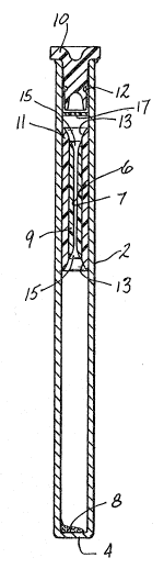
Referring now to the drawings, there is shown in FIGURE 1 a preferred embodiment of
the blood sampling paraphenalia formed in accordanc~ with this invention. The blood
sampling paraphenalia includes a transparent ~ube 2 formed preferably ot glass, and
having an in~egrally closed end 4. A plastic float member 6 is disposed in the tube 2,
as are the stain and red cell densifier reagents 8. An elastomeric plug 10 closes the
open end of the tube 2, and a supply of a thixotropic gel 12 is disposed inside of the
tube 2 around the plug 10 In placc of thP gel 12, a thin plastic disc or cup 17 can be
used. The tube is preferably about 75 mm long, the same length as a capillary tube,
and has a bore diameter of abou~ 40 mm. Its capacity for blood is about a.s ml. The
float wili be about B rnm in length and about 4 mm in diameter when static in the tube.
.
The float 6 is a compund structure which has a central through bore 7 into which the
white cells and platele~s layer out during centrifugation. The bore 7 is preJerably about
1.265 mm in diameter so as to achiev~ the necessary c~ll band elonga~ion to ailow
haNesting of the target cell band. The float 6 is formed with a core part 9 made from a
dimensionally stable transparent plastic, such as a rigid styrene plastic. A sleeve part
11 surrounds the core 9 and is bond~d thereto. The sleeve 11 is formed from a pliable
~ .i.~
2~$~7~
transparent plastic such as PVC. The ends o~ the sleeve 11 are flared, as at 13, ~nd
the ends of the bore 7 are also fiared as ag 15 to allow movernent of the ~lood in the
tube 2 during filling and centrifugation.
FIGURE 3 shows how the tube 2 can be filled with blood from a primary blood
collecting tube 14 by means of a transferring device 16 having a double piercingneedle or cannula 18. The transfer device 16 inciudes an outer shroud 20 with a
needle-carrying plug 22 telescoped thereinto. The needle 18 extends into a first weli
24 in the plug 22 sized to receive the stoppered end of the blood sarnpling tube 2. The
shroud 20 forms a second well 26 which is sized to receive the stoppered end of the
primary blood collecting tube 14. The transfer needle 18 pierces the plug 28 in the
tube 14 and also pierces the plug 10 in the sampling tube 2. The low pressure in the
tube 2 causes blood to be drawn from the tube 14 through the needle 18 into the tube
2, the flow of blood continuing until the tube 2 is substantially filled. Once filled, the
tube 2 is withdrawn from the well 24 and centri~uged. While trans~erring blood to the
testing tube 2 frorn a collection tube 14 is one way to fill the tube 2, it is readily
apparent that the sample could be taken directly from a patient using a needle and the
evacuated tube 2.
When the blood enters the tu~e 2, the reagents 8 wiil mix with ~he blood, and the tube 2
will be ready to centrifu~e. The tubes 2 are oriented in the centrffuge with the closed
end 4 out, so tha~ thP red cells will settle in the closed encl of the tube 2 and the plasma
wilt be adjacent to the stoppered end of the tube 2 after centrifugation. FIGURE 4
shows the condition of the tube 2 and blood after the cen~rifu~ation has been
completed. The red cells 30 collect in the closed end of the tube 2 and the float 6
becomes embedded in, and projects above the top of the red cell layer. The whitecells and plate~et layers which make up ~he bu~fy coat 32 set~le into the axial through
bore 7 in the float 10 and the plasma 34 is disposecl above the buffy coa~ and floa~ 10.
The thixotropic gel 12 (or plastic disc 17) covers and floats on the plasma layer 34
thereby holding the centrifuged blbod constituent layers in place in th~ bore 7 when
the tube 2 is handl~d af~er the centrifugatiorl step during harvesting ~f th~ target cell
band from the floa~ bore 7.
FIGURE 5 shows the manner in which the target cells can be harvested from the float
bore 7 with an aspirating needle 31. The needle 31 is inserted into the tube 2 ~hrough
the plug 10 so that its tip 23 may be positioned in ~he target cell ~and B, ~he other ~ell
bands being desiynated A, (:~, D and E. Suction is applied to the band B via theneedle 31 causing the cells to move in the directivn of the arrow 33 into the needle 31.
When the filled tube 2 is subjected to centrifugation forces of 10,000 G, which is the
force at which the prior art capillary tubes are centri~uged, the pliable sleeve part 11 of
the float 6 radially contracts whereby the effective diameter of ~he float 6 decreases.
Thus the float 6 is forced through the blood sample until the float encounters the
centrifuged red cell layer which, because of its specific gravity, resists fu~ther
movement of the float 6. Once this occurs, the float 6 will be stabilized and the sleeve
part 11 will expand back outwardly into snug engagement with the tube bore. The tube
bore wall may be coated with a siiicone lubricant to enhance the slidability of the float 6
in the tube 2.
It will be readily appreciat~d tha~ the tubes of this invention can be used to draw blo~
samples frorn pa~ients or from blcod collecting tubes, and the blood cell measurements
c~n then be made directly in ~he stoppered, closed tubes without exposing anyone to
the possibitity of contact with cont~minated blood. Thus the blood testing procedure
can even be used with patients who are known to have contaminated blood with no
danger to the person doing the testing. The dimensional tolerances observed in
producing the tubes and floa~s are relaxed, and the test assemblies have a longer shelf
l~e since the interior of the evaouated tubes is filled with an inert gas. Cell layer band
~rmation is preserved during handling of the tube ~ter centrifugation due to thepellicle formed on top of ~he plasma by the thixotropic material or by a piastic disc in
the tube during centrifugation. Target cells can be easily harvested from the readily
visible, elon~ated bands of cells in the float bore.
2 ~ 7 ~
Since many changes and variations of the disclosed embodinnen~ of the invention may
be rnade without ~eparting from the inven~ive concept, it is not intended to limit the
invention otherwise than as requirec by the appended claims.