Note: Descriptions are shown in the official language in which they were submitted.
2 0 ~
Patent
~0163 PROXIMAL CENTRALIZER WITH REMOVABLE HANDLE
Field of the Invention
This invention relates to a proximal centralizer for a femoral
implant and has specific relevance to a proximal centralizer having
a removable handle.
Summary of the Invention
This invention relates to a proximal centralizer for
positioning the proximal end of a femoral implant a predetermined
distance from the medial wall of the intramedullary canal. The
spacer is preferably formed from poly methyl methacrylate and
includes as integral components a handle portion and a centralizer
portion spaced apart from one another by breakable connectors. In
use the centralizer portion of the centralizer is carried by the
implant adjacent the collar in contact with the medial wall and
adjacent side walls of the implant. During insertion of the
implant into the cement filled intramedullary canal the handle
provides access to the surgeon to hold the centralizer portion
tightly against the implant. After the implant with the
centralizer attached is inserted into the intramedullary canal, the
surgeon pivots the handle portion abou-t the centralizer portion to
break the breakable connectors adjacent the centralizer portion for
removal of the handle portion. The handle portion is then
discarded.
In the preferred embodiment, the handle portion may be
substantially symmetrical with the centralizer portion in shape,
but formed having a different cross-sectional thickness. In
essence, the proximal centralizer of the preferred embodiment
consists of two centralizer portions of distinct cross sectional
thicknesses interconnected by a breakable connectors. During the
implant procedure, the surgeon may position either centralizer
portion adjacent the medial wall of the implant with the other
2~93~ -
surgeon. Since the two centralizer portions on the cen~ralizer
have distinct cross~sectional dimensions, the choice of which
centralizer portion to use is dictated by the desired spaciny
between the medial wall of the canal and the proximal medial wall
of the implant as determined by the surgeon.
Accordingly, it is an object of the invention to provide a
novel proximal centralizer for a femoral implant.
Another object of the invention is to provide a proximal
centralizer for a femoral implant having a centralizer portion and
a handle portion.
Yet another object of the invention is to provide a proximal
centralizer for a femoral implant having a pair of centralizer
portions connected by breakable connectors.
Still other objects of the invention will become apparent UpOIl
a reading of the following description taken with the accompanying
drawings.
Brief Description of the Fiqures
Figure 1 is a perspective view of the invention.
Figure 2 is a top down elevational view taken from Fig. 1
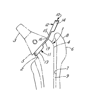
Figure 3 is a fragmented side elevational view of the
invention shown in use in its intended environment connected to a
hip stem implant which is partially inserted within a prepared
cement filled intramedullary canal. The implant and canal are
shown in broken lines.
Figure 4 is the fragmented side elevational view of Fig 3 with
the implant fully inserted and the unused portion of the
centralizer removed.
Figure 5 is a perspective view of an alternative embodiment.
Figure 6 is a top down elevational view of Fig. 5.
.
:-` 2 ~
Description of the P~referred ~ odiments
The preferred embodiments herein described are not intended to
be exhaustive or to limit the invention to the precise forms
dlsclosed. Rather, they are chosen and described in order to best
explain the invention so that others skilled in the art may utilize
the teachings.
For the purposes of illustration a prosthetic hip implant 1
and proximal end of a prepared femur 6 is illustrated in Figs. 3
and 4 in broken lines only. Implant l includes a stem 4 and a neck
3 integrally formed and defining a collar 5 at their junction.
Stem 4 includes a medial wall 2. The femur 6 includes a prepared
intramedullary canal 7 having a proximal end and a greater
trochanter 8. A quantity of poly methyl methacrylate 9 is placed
in canaI 7 in keeping with standard hip replacement techniques.
lS Proximal centralizer l~ as shown in Fi~s. 1-4 is illustrated
as being an integral unit and includes a centralizer portion 12 and
a centralizer portion ll. Centralizer portion 12 includes a
generally U-shaped spacer 14 with integral breakable connectors 15
extending longitudinally as shown. Spacer 14 is inclined slightly
relative to the vertical and has a cross-section of a predetermined
thickness. Similarly, centralizer portion ll includes a generally
U-shaped spacer 13 with integral breakable connectors 19 extending
longitudinally as shown. Spacer 14 is also incline~ slightly
relative to the vertical and is formed having a cross-section of a
predetermined thickness. In the preferred embodiment, the spacers
13 and 14 are inclined relative to the vertical (as mentioned) to
match the slope of the implant's proximal medial wall. Also in t~e
preferred embodiment the cross-sectional thickness of spacer 14 is
greater than the cross-section thickness of spacer 13. As
mentioned, centralizer portion 12 and centralizer portion ll are
interconnected by breakable connectors l~ and 19 at junction 16.
2~9~0
centralizer 10 is preferably relatively brittle when solid.
Therefore, connectors 15 and 19 are referred to as breakable since
bending the connectors causes them ko break adjacen~ their
respective spacer. The use of PMMA for spacers is well known and
possesses many advantages in use with PMMA cement.
In use, a surgeon determines the amount of spacing desired
between the medial wall of a prepared intramedullary canal and the
- proximal medial wall of a femoral implant 1. The centralizer 10 is
slid onto the distal end of the implant and along the stem toward
the proximal end. Either centralizer portion 12 or centralizer
portion 11 may be positioned to engage the underside of collar 5 of
the implant to orient the particular centralizer portion desired
adjacent the medial wall of the implant~ Which centralizer portion
(11,12) is positioned adjacent the medial wall is one of surgeon
choice dependent upon the desired spacing between the medial wall
of the implant and the adjacent bone structure. For example,
assume the thickness of spacer 13 is 2mm and the thickness of
spacer 14 is lmm. If a surgeon determines a 2mm spacing between
the proximal medial wall of the implant and the bone structure is
appropriate, centralizer 10 would be positioned as illustrated in
Fig. 3 with centralizer portion 11 in contact with the implant and
portion 12 extending rearwardly therefrom and slightly above the
greater trochanter. Centralizer portion 12 in assence becomes a
handle for centralizer 10 to allow the surgaon to hold the
centralizer portion 11 secure during insertion of the implant 1.
When the implant is fully inserted, centralizer portion 11 is
between the ~emoral bone and the proximal medial wall of the
implant thereby insuring a 2mm spacing between bone and implant.
If the surgeon requires only a lmm spacing between bone and implant
then centralizer portion 12 is positioned adjacent the implant
prior to insertion and portion 11 extends rearwardly to act as a
handle. It should be understood that the exact thickness of
spacers 13 and 14 is one of mere desi~n choice and should not ~e
2~3~
inserted into the intramedullary canal and the cement is allowed to
at leas-t partially cure and polymerize wi~h the centralizer, th~
surgeon pivots the rearwardly extendiny centralizer portion (12 in
Fig.~) upwardly toward implant neck 3. Pivoting the centralizer in
this manner causes legs 19 in Fig. 4 to break from spacer 13
preferably adjacent the spacer. The unused centralizer portion is
then discarded. It should be noted that centralizer portions 11
and 12 are angled slightly relative to one another to provide
clearance for the greater trochanter when the implant and
centralizer are insertion.
An alternative embodiment of the invention is illustrated in
Figs. 5 and 6. The proximal centralizer 20 of Figs. 5 and 6
includes a centralizer portion 21 and a handle portion 22
interconnected by temporary living hinge 26 formed by recesses 27.
As with centralizer 10 of the first embodiment, centralizer 20 is
preferably formed from PMMA. Handle portion 21 includes a spacer
23 having a predetermined cross sectional thickness and being
inclined slightly relative to vertical. Handle portion 22 includes
slightly arcuate finger tabs 24,25 extending transversely relative
to the plane of spacer 23. In use, centralizer 20 is slid upwardly
on the stem of an implant (not shown) until butting against the
collar of the implant. The implant is inserted into the
intramedullary canal while the surgeon maintains centralizer 20 in
position by grasping thè finger tabs 24, 25 and applying tension.
When the implant is fully seated and the cement is allowed to at
least partially cure, the surgeon pivots handle portion 22 about
hinge 26 to break off the handle from centralizer portion 210
Handle portion 22 may then be discarded.
It should be understood that the invention is not to be
limited to the precise forms disclosed but may be modified within
the keeping of the appended claims.
