Note: Descriptions are shown in the official language in which they were submitted.
7 ~
ELECTRONIC BIOP~,Y I~S~RUMENT
~1_LD OF T~IE INVENTION
This invention relates to an instrument for e~tracting
samples of tissue rom humans and other animals and more
particularly to an instrument for automatically perforrniny a
biopsy of a tissue mass in an accurate, expeditious manner
with a minimum of discomfort to the patient.
BA~_R lND OF T~1_INV~NTI~N
It is often desirable and frequently absolutely necessary
to sample or test a portion of tissue from humans and even
other animals, particularly in the diagnosis and treatment of
patients with cancerous tumors, pre-malignant conditions and
other diseases or disorders. Typically in the case of
cancer, when the physician establishes by means of procedures
such as palpitation, x-ray or ultra sound imaging that
suspicious circumstances exist, a very important process is
to establish whether the cells are cancerous by doing a
biopsy. Biopsy may be done hy an open or closed technique.
Open biopsy removes the entire mass ~excision biopsy) or a
part of the mass (incision biopsy). Closed biopsy on the
other hand is usually done with a needle-like instrument and
may be either an aspiration or a core biopsy. In needle
aspiration biopsy, individual cells or clusters of cells are
obtained for cytologic examination and may be prepared such
as in a Papanicolaou smear. In core biopsy, as the term
suggests, a core or Eragment of tissue is obtained for
histologic examination which may be done via a frozen section
or paraffin section.
-
.. , , ~ .; ,
.
2 ~ 7
The type of biopsy depends in large E)art in circurnstancespresent with respect to the patient and no single procedure
is ideal for all cases. However, core biopsy is extremely
useful in a number of conditions and is being used more
frequently by the medical profession.
A variety of biopsy needles and devices have been
described and used for obtaining specimens of tissue. For
example, reference is made to U.S. Patents 4,651,752;
4,702,260; and 4,243,048 which show biopsy needles of varyiny
types. ~dditionally, a number of very specialized devices
for extracting samples of tissue have been described such as
the biopsy device in U.S. Patent 4,461,305, which device is
designed specifically for removing a sample of tissue from
the female uterine cervix. Other devices have been disclosed
which relate to surgical cutting instruments. For example,
U.S. Patent 4,589,414 discloses an instrument which is
particularly designed to operate in the area of the knee to
withdraw tissue chips. Also available are so-called biopsy
guns for removing a core of tissue which customarily are
spring powered devices and must be cock0d with considerable
force. When actuated such guns produce a loud snapping
noise, combined with a jerking action. Such a biopsy gun may
employ a needle set consisting of an inner stylet and an
outer tube called a cannula. The stylet is a needle like
device with a notched cut-out at its distal end. The cannula
in effect is a hollow needle with an angled cutting surface
at its distal end which slides over the stylet. When the
stylet is forced into tissue, the tissue is pierced and
relaxes into the notched cut-out of the stylet. When the
cannula is then slid forward, the tissue in the notch of the
stylet is sliced off and retained in the notch until the
cannula is withdrawn. Examples of such devices are shown in
U.S. patents 4,600,014 and 4,699,154. Although such spring
powered biopsy guns will remove a core or sample of tissue,
they have rather serious disadvantages. For one, they must
:` , ~ .-
. . ~ , .
.: :
. ~ -
2 ~ 7 5
--3--
be manually cocked with a plunger bar. Such "cocking" of the
gun requires considerable force and the gun must be cocked
for each biopsy cut. A further disadvantage is that the
springs provided in the gun accelerate the needles until a
mechanical stop posi-tion is reached, creating a loud snapping
noise and jerkiny motion which is a problem both to the
physician and the patient. This noise and jerking action can
cause the patient to jump and in some cases even prevents the
physician from striking the intended tissue target. Another
disadvantage is that the force and velocity delivered to the
stylet and cannula rapidly dirninishes when traveling from a
retracted to a fully extended position resulting in tissue
samples of lower quality.
U.S. Patent No. 4,940,061 discloses a biopsy instrument
which represents a substantial improvement over the
aforementioned devices, substantially eliminating the loud
snapping noise and jerking motion associated with
spring-powered biopsy guns, for example. In the instrument
of Patent No. 4,940,061, an electric motor drives a rotary
cam assembly which converts rotary motion to linear motion to
sequentially extend and retract a stylet and cannula, and
employs electrically actuated stops to control the extension
and retraction of the stylet and cannula. Details of the
construction and operation of the biopsy instrument are
disclosed in U.S. Patent No. 4,940,061, which is hereby
incorporated by reference. Although well suited for many
applications, -that instrument suffers certain drawbacks, one
of them being mechanical wear associat~d with a limit switch
assembly and a toggle assembly which both include stationary
wiper plates and spring finger contacts which slide against
each other during normal operation.
Accordingly it is a principal object of this invention to
provide an instrument for obtaining samples of tissue from
tissue masses.
It is a fu:cther object of this invention to provide a
- - : :
2 ~ r~ ~3
-~ -
biopsy instrument which is able to provide a substantially
constant force and velocity to that portion of the instrument
which penetrates the tissue mass and severs a portion of
tissue for further examination.
It is another object of this invention to provide an
instrument for autornatically performing a biopsy of a tissue
mass in an accurate and expeditious manner with a maximurn of
accuracy and a minimum amount of discomfort to the patient.
It is a still further object of this invention to provide
an instrument for performing tissue rnass biopsies by removiny
a core or sample of tissue, which instrurnent eliminates the
need for springs and mechanical stops, which is silent in
operation and has the ability to effectively penetrate even
small tissue masses.
It is another object of this inventiorl to provide an
instrument for obtaining tissue samples from -tissue masses
which instrument requires no manual setting or cocking and
which may be "fired" multiple times without any abrupt starts
or stops.
It is still another object of this invention to provide a
biopsy instrument which includes means to convert rotary
motion to sequential, linear motion of substantially constant
force and velocity to the means for penetrating and severing
a tissue sample from a tissue mass.
These and other objects of the invention will be apparent
from the following description and claims.
; :,
~,
.,,:
; ~
2~'3~7~
SUMMAR~ OF THE INVENTION
Based on the prior art instruments for biopsy samples
from tissue masses, and the actual present state of this art,
there then exists a need for an instrument which is capable
of automatically removing a tissue sample or core sarnple of
predetermined size where the process is done very rapi~ly, is
easily repeated if required, is accurate, is relatively
simple for the physician to use, is virtually noiseles3, and
in use results in minimal discomfort to the patient.
Accordingly, we have invented an instrument for rernoving
tissue samples from a tissue mass which instrument
automatically penetrates, severs, an~ removss the tissue
portion for exarnination. The instrument is motor powered,
preferably by self-contained r~chargeable batteries, and
employs electrically actuated stops instead of mechanical
stops to control the action of penetration and retraction
from the tissue mass. The portion of the instrument which
penetrates the tissue mass and severs a portion thereof, the
tissue penetrating and severing means, includes an inner
stylet which penetrates the tissue rnass and a hollow outer
tube or cannula which surrounds the stylet and serves to
sever a sample of tissue. In a preferred form the tissue
penetrating end of the stylet is notched so that when the
stylet penetrates the tissue mass, a portion of the tissue
relaxes in the notched area. After tissue penetration by the
stylet, the cannula, having a cu~ting surface at its distal
end, penetrates the tissue and cuts off the tissue portion
residing in the notched area of the stylet. The tissue
penetrating and severing means are operably connected to a
special motor powered rotary cam assembly by means of cam
followers and lt is a Eeature of this invention that the
rotary motion of the cam is converted to sequential, linear
motion in the tissue penetrating and severing means, the
. ~ ..
.:
.
2 ~ ~ 9 ~
--6--
linear motion being of substantially constant force and
velocity.
In operation, the physician or technician actuates the
instrument by pressing a button causing the stylet to move
forward in a rapid, precise manner and penetrate the tissue
mass Eollowed with penetration of the mass by the cannula,
resulting in a portion or core of tissue being severed and
retained in the notched portion of the stylet. Further
actuation by the physician causes ths cannula to retract
exposing the tissue sample in the stylet for easy removal.
An additional actuation causes retraction of the stylet and a
resetting of the cannula/stylet assernbly for further use.
.:
,
2~g7~
--7--
BRIEF DESCRIPTION OF THE DRAWINGS
The above noted advantages and other characteristic
features of the present invention will be in part apparent
from the accompanying drawings, and in part pointed out in
the following detailed description of the preferred
embodiment of the invention in which reference will be made
to the accompanying draw.in~s wherein like reference numerals
designate corresponding parts and wherein:
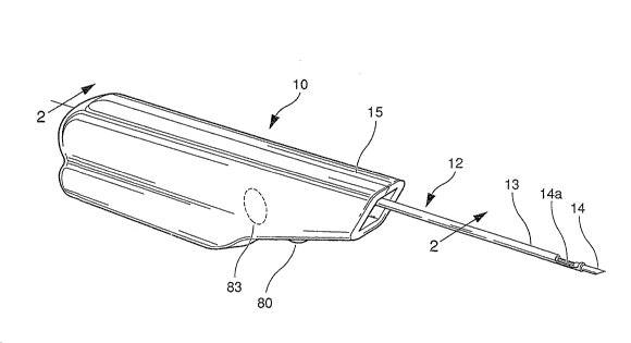
FIG. 1 is a perspective view of the biopsy instrurnent of
this invention.
FIG. 2 is a side elevational view talsen on the line 2-
~of FIG. 1.
FIG. 3 is an exploded view of the major component parts
of the instrument shown in FIG. 2.
FIG. 4 is an exploded perspective view of the biopsy
instrument further illustrating the major component parts
thereof.
FIG. 5 is a plan view of the outer surface of the rotary
cam illustrating the cam profile as a function of angular
position about the full circumference of the cam.
FIG. 6 is an electrical schematic of a control circuit
according to the preferred embodiment of this invention.
. ~ :
: .
DESCRIPTION OF THE PR:EFERRED EMBODIMENT
For the purposes of promoting an understanding of the
principles of the invention, reference will now be made to
the embodiment illustrated in the drawings and specific
language will be used to describe the same. It will
nevertheless be understood that no limitation of the scope of
the invention is thereby intended, such alterations and
further modifications in the illustrated device, and such
further applications of the principles of the invention as
illustrated therein being contemplated as would normally
occur to one sl~illed in -the art to which the invention
relates.
Considering now the drawings in detail, FIG. 1
illustrates a perspective view of one embodiment of the
inventive biopsy instrument which is shown generally at 10
with the tissue piercing and removing means shown generally
at 12. The tissue piercing and removing means comprises a
stylet 14 and cannula 13. R~ferring to FIG. 2 which is a
sectional view through the instrument shown in FIG. 1, and
FIGS. 3 and 4, which are exploded views of a number of the
components of the instrument, the ins-trument 10 is shown as
having an outer housing 15 provided with a motor 18 mounted
in one end thereof. Motor 18 is reversible and preferably of
the DC type and preferably powered by rechargeable batteries
16 contained within the housing. Suitable contacts 17 are
provided to recharge the batteries. Motor 18 is operably
engaged with planetary gear assembly 20 by means of shaft 19
which shaft engages central gear 21. Central gear 21 in turn
meshes with planetary gears 22 which in turn engage with
annulus gear 23. In a preferred embodiment the DC motor
operates at about 10,000 rpm with the gearing being about a
6:1 ratio. Drive shaft 25 is secured at its end 26 in the
D-shaped opening 35 of the planetary gear set by means of a
set screw or other suitable fastening means.
,` ' ' , : ~ .
,,
:
2 ~
- 9 -
The components o~ the instrument which guide the
stylet/cannula assembly 12 will now be detailed, A physician
or technician actuates the instrument causing the stylet 14
to move forward in a rapid and precise manner to penetrate
the tissue mass followed by penetration o~ the mass by the
cannula 13, resulting in a portion or core of tissue beiny
severed and retained in the notched portion of the stylet.
Further actuation causes the cannula to retract e~posing the
tissue sample in the notched portion at the distal end o~ the
stylet for easy removal. An additional actuation causes
retraction of the stylet and a resetting of the
cannula/stylet assembly for further use. The penetration and
retraction of the stylet and cannula assembly is controlled
in part by hollow rotary cam 55, one embodiment of which is
illustrated in FIG. 4. As will be described later, the cam
preferably has a cam profile as illustrated in FIG. 5. Cam
55 is provided with a continuous groove 56 which is made up
of three sections. A first groove section 56a is positioned
substantially parallel to one end of cam 55 and extends about
a portion of the circumference of the cam. A second ~roove
section 56b is positioned substantially parallel to the other
end of the cam and also extends about a portion of the
circurnference of the cam. Section 56c connects section 56a
and 56b in a generally diagonal manner in the embodiment of
FIG. 4. Cam 55 is rotated by means of drive shaft 25, which
is secured at its forward end 26 into the opening 63 of the
end wall 59 of cam 55. Thus rotation of shaft 25 in a
clockwise or counterclockwise direction causes identical
rotation of the cam.
As previously described, stylet 14 moves within and is
surrounded by cannula 13. The non-penetrating end of stylet
14 is mounted in stylet block 74. Correspondingly, the
non-penetrating end of cannula 13 is mounted into cannula
block 75. As shown in FIG. 4, stylet block 74 is provided
with extension 76 which is in alignment with and moves
.
7 ~
~o
through opening 77 of the cannula block 75 to aid in proper
alignment of the stylet and cannula blocks and therefore -the
stylet/cannula assembly. An alternative construction of the
stylet/cannula assembly is disclosed in copending U.S. patent
application Serial No. 07/583,597, which is hereby
incorporated by reference.
Mounted in the ends of each of the cannula and stylet
blocks ara drive rods 62 which are in turn secured to drive
arms 61. Each of drive arms 61 is provided with a cam
follower 60 which rides in the continuous groove 56 of carn
55. Thus, rotation of cam S5 will result in sequentia]
linear movement of the stylet and cannu]a.
Although a generally diagonal central groove section 56c
is useful with an optoelectronic sensor as shown in FIG. 4,
the cam profile is preferably as illustrated in FIG. 5, which
is a scale drawing of the outer surface of the cam, with the
front edge of the cam appearing at the top of the drawing.
The groove has a flat rear section 156a beginning at one end
point A and extending approximately 235 counterclockwise
(CCW) around the cam, as viewed from the rear, followed by a
an S-curve or sinusoidal section which includes sections
156c, 156d and 156e extending approximately 68, 9 and 68
CCW, respectively, around the cam, followed by a flat front
section 156b extending approximately 199 CCW around the cam,
to the other end point B.
Fixed within the housing is a printed circuit board 120
on which is mounted all of the motor driver and control
electronics for the instrument, as will be explained in
detail in connection with FIG. 6. The board is provided with
a central hole through which drive shaft 25 passes, as shown
in FIG. 2. A slotted disc 121 is affixed to the drive shaft,
preferably by means of a D-shaped opening mating with a
D-shaped section on the shaft as in the case of the
connection to the cam 55. The disc is positioned so as to
pass through the openings in two optoisolators referred to
.,
,
:
:: :
2~J~)7 ~
herein as sen~or 1 and sensor 2 and labeled #1 and #2,
respectively, in FIG. 3. The slot in the disc is 50 wide,
and the disc is fixedly mounted on the drive shaft such that
edge 122 of the slot is angularly offset 9-1~ with respect
to the endpoint A of the groove in the cam, as shown in
FIG. 4. It will be appreciated by those skilled in the art
that, although FIG. 4 shows cam followers 60 at the same
axial po~ition, this is Eor illustration purposes only in the
exploded view, and that, in operation, the angular pAosition
of the cam in FIG. 4 corresponds to the stylet in a partially
extended position.
Operation of the instrument begins with stylet 14 and
cannula 13 in a retracted position and with the exposed tip
of stylet 14 immediately adjacent the tissue mass 11.
Initial rotation of cam 55 results in forward movement of
stylet block 74 and its attached stylet to penetrate the
tissue mass where a portion of the tissue is caught in notch
14a. Continued rotation of the cam results in forward
movement of the cannula block 75 and its attached cannula
into the tissue mass severing the portion of the tissue
within notch 14 from the tissue mass. The instrument is then
withdrawn from the patient. Rotation of cam 55 is then
reversed thus causing retraction of the cannula e~posing the
tissue sample in notch 14a for easy removal by the
technician. Further rotation of cam 55 will result in
retraction of the stylet and, thus, a return to the initial
ready-to-fire condition.
Because of the need for precise movement of stylet and
cannula, guide means shown generally at 64 are used -to
further aid in pro~er alignment of the stylet/cannula
assembly. In the embodiment shown in FIG. 4, guide means 64
includes a generally cylindrical shaped housing 68 having a
rectangular opening 69 appro~imately si~ed to accommodate the
stylet and cannula blocks 74 and 75. Thus the stylet and
cannula blocks move laterally within the interior of housing
-. : -
,
r~
68 and bear on the interior walls of the hou.sing aiding
proper alignment. In addition, guide means 64 also includes
a cylindrical shaped guide 65 and bulkhead 70, the latter
separating guide 6~ and housing 68. Guide 65 is a solid
cylinder provided with vertical channels 66 through which
drive rods 62 opera-te. Guide 65 is constructed with a
separator between channels 66 to assist in maintaininy proper
spacing and alignment of the drive rods.
In the preferred ernbodiment, the instrument has three
actuators or buttons which set into motion the action of the
stylet/cannula assembly. As shown in FIG. 1, the instrument
includes retract button set 80 and a fire button 83, which
are preferably provided with a rubber seal. The retract
button set is located on the underside of the instrument and
mechanically connected to two pairs of contacts 81 in a
switch frame 89 which also includes a microswitch 85
mechanically connected to fire button 83. The retract button
set includes separate buttons for the cannula and stylet,
preferably positioned side by side. Thus, the instrument has
three separate pushbutton switches: (1) a fire switch, (2) a
cannula retract switch, and (3) a stylet retract switch.
Actuation of the fire button, when enabled, causes initial
penetration of the stylet into the tissue mass followed by
penetration of the cannula. Actuation of the cannula retract
button, when enabled, causes retraction of -the cannula
exposing the sample of tissue. Actuation of the stylet
retract button, when enabled, retracts the stylet whereupon
the instrument is ready for further use.
Referring now to FIG. 6, which is an electrical schematic
for the preferred embodiment of the motor driver and control
electronics according to this invention, the primary
components of the circuitry are three JK flip-flops, four AND
gates, two complementary pairs of MOSFETs, and two
optoisolators which return mechanical position data. U2 and
U3~B operate in conjunction with each other to dictate which
,
7 ~
-L3-
operator button is enabled via U3/A, Ul/C and Ul/D. Another
function of U3/B is to dictate which sensor output is enabled
via Ul/A and Ul/B. U3/A is primarily a latching circuit for
the fire button.
In the ready-to-fire position, which is the proper
initial position for operation of the device, the stylet and
cannula are both retracted and the circuit is in a reset or
standby mode. In this mode the outputs (Ql) of all three ~K
flip-flops are low (logic "0"), thus disabling sensor 1,
enabling sensor 2 and enabling the fire button and fire latch
U3/A.
Once the fire button is pressed, the output of U3/A goes
high (logic "1"), turning on the LEDs in sensors 1 and 2
through transistor Q5 and forward biasing MOSFET Q4, thereb~
connecting one terminal of the drive motor to ground. Ul/C
and Ul/D are both held low at this time by the low levels on
the Q outputs of JK flip-flops U2 and U3/A. Thus, MOSFET Q2
is on, enabling motor drive current to flow from VCC through
Q2, the motor, and Q4 to ground.
Therefore, in response to actuation of the fire button,
the drive motor begins turning and, through the planetary
gear box, causes the drive shaft to rotate clockwise (CW) as
viewed from the rear of the instrument. This causes the
slotted disc to rotate cloc~wise from its initial position,
in which the slot is adjacent to sensor 1 which is located
directly below the drive shaft. Sensor 2, located 120
counterclockwise from sensor 1 as viewed from the rear of the
instrumen-t, detects the passage of the slot after
approximately 230 of clockwise shaft rotation, and, in
response, generates an output pulse which passes through
Ul/B, enabled at this time by a high state output on the
output of U3/B, and clocks U3/B, thereby
disabling sensor 2 and enabling sensor 1. The motor
continues to drive the rotary cam and slotted disc clockwise
until the slot returns to a point adjacent sensor 1, which
, ' , ' ' ~ ."~
:
2 ~ 7 ~j
-14-
responds by clocking U2 through Ul/A. UZ goes high in
response, resetting U3/A and thereby stopping the motor. The
low output from U3/A not only turns o~f Q~ and thereby
deenergizes the motor, but also turns on Ql to connect both
terminals of the motor to VCC, thereby providing dynamic
braking. The motor stoys with the stylet and cannula in
their extended positions.
The high output from U2 is also supplied to one contact
of each of the stylet and cannula retract switches as an
enabling signal. With U3/B reset at this time, only the
cannu]a retract switch is actually enabled, because the low Q
output of U3/B prevents any pulse from the stylet retract
switch from passing through Ul/D. When the cannula retract
button is pressed, the output of Ul/C goes high, turning on
transistor Q3 and completing a circuit from VCC through ~1,
the motor and Q3 to ground, whereby the motor reverses
direction and causes the rotary cam and slottsd disc to
rotate counterclockwise. It should be noted, perhaps, that
both optoelectronic sensors are disabled whenever the motor
is deenergized. Once cannula retraction begins, howev~r,
both sensor LEDs are again turned on through Q5, although
only sensor 2 is enabled because Ul/~ is disabled at this
time. Thus, sensor 2 is the first sensor to respond to the
passage of the slot in the slotted disc, and it responds by
clocking U3/B, thereby disablin~ sensor 2 and the cannula
retract button and enabling sensor 1 and the stylet retract
button. The motor stops in response to the resulting low
state at the output of Ul/C.
The final step in the cycle is actuation of the stylet
retract button. Pressing this button with Ul/D enabled
re-enables the drive, causing the cam and slotted disc to
resume counterclockwise rotation and resulting in sensor 1
clocking U2 and resetting U3/B through Ul/A. This disables
the stylet retract button and re-enables the fire latch,
thereby completing the ~ull cycle.
' ' :
. ~
. ~.
2~$~
-15-
The instrument~s response time is short enough that
movement from any one of the predefined extended or retracted
positions is completed before any of the control buttons can
be released in normal operation.
The presently preferred components are specified as
follows:
Devicç ~_~ce TyPe
Ul CD 9081B
U2, U3 CD 9027B
10 Ql, Q2 FCG 23~2
Q3, Q4 ECG 2383
Sensors 1, 2 EE-SX 1067
The invention is described above in terms of
optoisolators and a slotted disc as the preferred position
sensor construction, primarily because of superior speed of
response. However, the present invention, more broadly,
contemplates the elimination of spring fingers or other wiper
elements and wiper plates such as disclosed in U.S. Patent
No. 4,940,061. The term "wiperless position sensor" is used
in this patent to mean any type of limit switch or other
position sensor which does not have such a wiper assembly,
and is intended to include optoelectronic devices,
electromagnetic devices, Hall effect devices, capacitive
devices, and microswitches, among others.
The present invention has a number of advantages over all
other forms of biopsy instruments including that disclosed in
U.S. Patent No. 4,940,061. In addition to greater
reliability as a result of a wiperless position sensor~ the
instrument has improved action because of the new cam
profile, as shown in FIG. 5. The curved section of the cam
extends less than 1~5 around the cam circumference, without
abrupt transitions, and begins approximately 55 from the
fully retracted position of the cam follower for the stylet.
One advantage of this construction is a large increase in the
~ 7~7
-16-
atnount of time the motor spends in a no-load condition upon
starting, thus allowing the motor to accelerate and reach a
motor speed above the loaded rating prior to hitting the rarnp
in the cam. This increase in speed directly results in
desired higher needle velocities. The reduction in ramp
length results in greater forward movetnent per degree of
rotation, further increasing the ma~imum needle velocity
during the stroke. Another desirable feature is a small
delay between the time the stylet finishes its stroke and the
time the cannula begins its stroke. This allows tnore tissue
to fall into the slotted stylet, and thereby results in
improved core samples.
While the invention has been illustrated and described in
detail in the drawings and foregoing description, the same is
to be considered as illustrative and not restrictive in
character, it being understood that only the preferred
embodiment has been shown and described and that all changes
and modifications that come within the spirit of the
invention are desired to be protected.
'