Note: Descriptions are shown in the official language in which they were submitted.
2O~c~2~
MODULAR GUIDE FOR SHAPING OF FEMUR TO ACCOMMODATE
INTERCONDYLAR STABILIZING HOUSING AND PATELLAR TRACK OF
IMPLANT
This invention generally relates to surgical
devices for use by orthopedic surgeons in the implantation of
distal femoral knee prostheses, particularly in cases where a
patellar prosthesis is also implanted and, more particularly,
where an intercondylar stabilizing housing is provided in the
implant.
Surgical instruments for various bone shaping or
resection operations, required in the implantation of
prosthetic devices, have been devised to assist orthopedic
surgeons in accurately performing the necessary cutting
steps.
One example of such instrumentation is U.S. Patent
No. 4,474,177 to Whiteside, disclosing a device and method
which utilize an intermedullary rod for aligning a shaping
tool(s) to make several angular resection cuts using a common
reference axis precisely established hy the rod.
Another example is the apparatus described U.S.
Patent No. 4,721,104 to Kaufman and Whiteside, which
describes a femoral shaping apparatus employing a template
having a straight slot therein for cutting a rela.tively deep
recess for an intercondylar stabilizing housing of a knee
implant. The patent, however, does not disclose a cutting
guide having a curved track useful for forming a groove to
accommodate a patellar track on such a prosthesis.
Heretofore it has been common practice for a
surgeon to form a patellar track groove in a resected distal
femur, if required, due to the nature of the particular knee
prosthesis being implanted, by use of various cutting or
abrading tools or instruments without aid of any type of
locating or guiding instrument. In design and seating of a
prosthesis it is important to retain the original
patella/femoral joint line to avoid placing undue stress on
the patella and its connective tissues.
U.S. Patent Application Serial No. 462,26~, filed
January 9, 1990 in the name of Coates et al., describes an
improved system for accurate placement and cutting of a
groove to accommodate a patellar track portion o~ a
prosthesis to insure accurate seating of the prosthesis for
long term wear and stability of a patellar prosthesis.
There still remains a need for using common
surgical instrumentation to form both a groove and deep
recess in the resected distal femur, respectively
accommodating a patellar track and intercondylar stabilizing
housing of the implant.
Modular surgical instrumentation and a method of
using same is provided, according to the invention. The
instrumentation comprises a first bracket defining a
generally U-shaped structure having an internal surface
adapted to be seated on the distal aspect of a resected femur
bone and an elongated central opening appointed to expose a
selected area of the resected femur, including a curved track
for guiding a first shaping tool along a predetermined path
for controlled shaping of a curved patellar groove in a
portion of the selected area exposed through the opening. A
second bracket defines a linear slotted bore extending
generally parallel to the long axis of the femur for guiding
a second shaping tool to form a relatively deep recess
accommodating an intercondylar stabilizing housing of a knee
implant. The second bracket has an internal surface defining
a shape adapted for releasable engagemen~ with the curved
track, including means for accurately aligning the slotted
bore relative to the opening while forming the recess.
2 (~ ~ 2 2 ~ r I
The method of the invention comprises the steps of
sea~ing the first bracket described above on the distal
aspect of the resected emur and moving the first shaping
tool along the curved track to form a patellar groove in a
selected area thereof. The first shaping tool is then
withdrawn and, leaving the first bracket in place, the second
bracket described above is seated on the curved track so that
the slotted bore of the second bracket is accurately aligned
with the opening of the first bracket, after which a second
shaping tool is introduced through the bore to form a recess
accommodating the intercondylar-stabilizing housing of the
implant.
An advantage of the invention is that a patellar
groove may be formed in the femur utilizing a cutting guide
which need not be removed in order to form a recess for an
intercondylar-stabilizing housing using a second cutting
guide, the two guides being cooperable with one another in a
modular structure.
Another advantage of the invention is that
alignment of the recess with the patellar groove is based on
the same cutting guide and, hence, is more accurate than the
use of subsequent and separate guides.
The invention wi.ll be urther explained with
reference to the following detailed description and drawings
wherein:
FIGURE 1 is a perspective view of a U-shaped
cutting guide of this invention adapted to fit over the
distal end of a resected femur;
FIGURE 2 is a side view illustrating the use of the
U-shaped guide of this invention in place on a femur in
conjunction with a cutting tool;
FIGURE 3 is a cross-sectional view of the U-shaped
cutting guide of FIGURE 1, talcen along line 3-3 of FIGURE 2;
FIGURE 4 is a cross-sectiona.l view of the cutting
tool shown in FIGURE 2, taken along line 4-4;
FIGURE 5 is a side view of an alternative cutting
tool for use with the U-shaped guide, which may be power
driven;
FIGURE 6 is a cross-sectional view of a further
embodiment of the U-shaped guide in conjunction with a
cutting tool of the invention, taken along a cross-section
~ similar to that of FIGURE 3;
; FIGURE 7 is a cross-sectional view of a still
further embodiment of the U-shaped bracket used with a
cutting tool according to ~he invention, also taken along a
cross-section similar to that of FIGURE 3;
FIGURE 8 is a side view of a further embodiment of
a power driven rotary cutter for use in combination with the
U-shaped bracket according to the invention, with parts shown
in cross-section;
FIGURE 9 is a perspective view of a shaping guide
: used with the U-shaped bracket of the invention, to fonn a
recess for accommodating the intercondylar stabilizing
~ housing of an implant; and
;~ FIGURE 10 is a top view of FIG. 9 showing another
~ tool for performing a further shapine opera.tion.
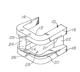
:~ Referring more pa.rticularly to the drawings, there
is seen in FIGURE l a cutting guide element 10 of generally
U-shaped configuration having a section 12 adapted to fit
over the distal end of a resected femur 11 as shown in FIGURE
2 End 14 is adapted to fit on the posterior side of the
resected femur and consists of 2 arms adapted to fit over the
medial and lateral condyles of the femur. The anterior side
of the cutting guide device consists of ends 16 and 18 which
are a continuation of the distal. sur:Ea.ce contacting portion
12 and are adapted to closely fit over the anterior side of
2~2~
--5--
the resected femur as indicated in EIGURE 2. Channels 20 and
21 are cut or sunk into the central edges of portion 16 and
18 of the device to form a cur~ed track in which the cutting
device can be moved during the course of cutting procedures.
The central portion of the cutting guide 10
consists of an elongated opening 23 which exposes at least
the distal and anterior aspects of the resected femur so that
a patellar groove may be cut therein. Connecting members 22
and 24 serve to connect the device together into an integral
structure. The distal surfaces of the connecting members may
also be curved as illustrated with element 24 to provide room
for the cutting tool during use.
Holes 2~ and 26 are preferably formed in the distal
surface portion of the guide member in order to provide a
means for securing the guide to the resected femur. These
holes can be used to accurately drill holes into the femur
for the purpose of installing anchoring pins to fix the guide
to the femur. These holes are preferably positioned so that
they will receive anchoring pegs on the actual implant.
As best seen in FIGURE 2 a hancl-operated cutting
tool 30 may be employed in conjunction with cutting guide 10.
Tool 30 is provided with a generally convex-shaped cutting
portion 32, the cross-section of which is seen in FIGURE 4. A
series of cutting teeth 34 are positioned along the length of
cutting surface 32. The edges of surface 32 are provided
with ledges 40 and 42 along the entire length thereof.
Ledges 40 and 42 act as track-engaging means so that the
cutting tool 10 can be moved along a desired central course
to cause resection of a patellar channel in the distal and
i anterior surfaces of femur 11. With the embodiment of FIGURE
2, the surgeon can by hand reciprocate cutting tool 30 in
guide 10 using handles 36 and 38.
- ~ ~ o ~
In the embodiment of FIGUR~ 5~ an alternative
cutting tool 60 is employed which is provided with an annular
convex-shaped cutting surface 61. Surface 61 is provided
with a series of cutting teeth 62 around the circumference
thereof for purposes of resection of femur 11. Shoulders 64
and 66 are provided on opposite ends of the cutting surface
so that the cutting tool 60 can be moved along cutting guide
tracks 20 and 21. Shaft 63 is provided so that tool 60 can
be driven by a conventional rotary power tool (not shown) so
that it rotates as indicated by arrow 68.
FIGURES 6 and 7 represent alternative embodiments
of track configurations with appropriately modified track
engaging surfaces on the cutting tool. Firstly, in FIGURE 6
there is seen a modified cutting guide element 110 having
anterior arms 114 and distal elements 116 and 11~, the
surfaces of which, adjoining the central opening
therebetween, are provided with raised rail track members 120
and 122. As also seen in FIGURE 6, the cutter 130 i5
provided with track engaging grooves 140 and 142 which are
adapted to engage the raised rail track e]e1nen$s and guide
cutter 130 in an appropriate path to form a resected patellar
groove.
FIGVRE 7 shows in analogous fashion a revised
embodi.ment in which channels 220 and 222 form the track means
in the distal surfaces 216 and 218 of guide element 210. In
this configuration, the track engaging portions 240 and 242
of cutting tool 230 comprised track engaging projections on
the flanges provided Oll each side of the cutting surface 232.
In other respects, the embodiments o~ FIGURE 6 and 7 have
other components analogous to those shown in Figs. 1 through
5.
FIGVRE 8 shows a preferred embodiment oE a rotary
cutter for use in connection with the combination of the
2~h~w
present invention. In that embodiment that cutting element
is power driven while the track engaging surfaces are free to
rotate separately therefrom. ~ handle 72 is rotatably
affixed to one end of power driven shaEt 74, for example, by
a screw 70. Handle 72 may be formed from polytetrafluoro-
ethylene or other low friction materials. A cutter 80
provided with cutting teeth 81 is affixed to shaft 74 by
means of set screw 78. On each side of the cutting element
80 is an indentation or race 77 containing ball bearings
which allow guide track engaging discs 75 and 76 to rotate
separately from shaft 74. Appropriate mechanical connectors
such as snap ring 79, which may fit into a groove on shaft
74, are utilized to hold the cutter in position on the shaft.
The power drive end shaft 74 is provided with an appropriate
connector to permit attachment to a rotary power driving
device. For example, end 82 may be a "Hudson~" end which
attaches to a power reamer drive.
The ball bearings as shown in races 77 may be
replaced, for example, by polytetrafluoroethylene washers or
other low friction washers such as ultra high molecular
weight polyethylene or nylon or the li.ke.
It has been found that due to the fact that washers
75 and 76 are free wheeling from power shaft 74, the cutter
80 will not jump out of the track if the shaft is
inadvertently twistedO Forcible pulling of the cutter alon~
the track and excessive wear of the track is also obviated by
this preferred embodiment.
In practicing the present invention, a surgeon
would follow normal procedures for resection of the distal
femur which would be resected to a size adapted to fit the
dimensions of the particular guide unit 10 being employed.
In general, the surgeon can observe a remaining part of the
original patellar track between the resected condyles and can
- ~ -
center the guide element 10 on such track. In the event that
the track is not sufficiently observable after resection, the
swrgeon would center the guide 10 on the posterior condyles
themselves. The guide element 10 would generally be provided
to the surgeon in a number of separate sizes which would
match the corresponding implant sizes provided to the
surgeon. Resection of a groove by the cutting tool to the
depth permitted by the guide will ensure a proper depth and
placement of the newly formed patellar track.
Modular surgical instrumentation and a method of
using same is further provided according to the invention,
and shown in FIG5. 9 and 10. The instrumentation comprises
the U-shaped bracket 10 (FIGS. 1-2) defining a generally
~-shaped structure which is seated on the distal aspect of
the resected femur 13 and the elongated central opening 23
appointed to e~pose a selected area of the femur, including
curved ledges 40, 42 for guiding the tool 30 along a
predetermined path for controlled shaping of a curved
patellar groove in a portion of the selected area exposed
through the opening. A second bracket generally indicated at
300 defines a linear bore 302 whicll guicles an end mill,
generally indicated at 304 rotating in the direction of arrow
306, in an axial direction shown by arrow 308 downwardly
toward the resected femur 13, i.e., essentially parallel to
the long axis of the intermedullary emoral (not shown). The
bore 302 has a pair of slots 310 which extend tangentially
from the bore 302 for guiding a U-shaped punch 312 downwardly
(arrow 308) through the bore 302 to form, together with the
end mill 304, a relatively deep recess accommodating an
intercondylar stabilizing housing of a knee implant (not
shown) a bracket 300 further comprises a top plate 314 which
is essentially perpendicular to the long axis of the femur
and through which the bore 302 is Eormed.
20~ ~h2.43
Extending perpendicularly from the top plate 314
are a pair of legs 316 each having seats 318 which extend in
an anterior-posterior direction and have a curved shape to
engage the ledges 40, 42 of the U-shaped bracket 10. The top
plate 314 of the bracket 300 has an extended arm 320 which is
aligned with respect to the U-shaped bracket 10 by means of a
positioning slot 322 which engages a peg 324 projecting
distally from the bracket 10. A tightening screw 326
pro~ects inward a medial-lateral direction through the arm
320 and engages the bracket 10 to securely lock the bracket
300 into proper position with respect to the U-shaped bracket
10 .
The end mill 304, shown in FIG. 9, has a shoulder
328 whi.ch bottoms-out in the stop 330 formed at the distal
end of the bore 302. After the end mill 304 is brought down
into the surface of the resected femur 13 and withdrawn, the
punch 312 is impacted with a mallet or the like to finish
forming the deep recess for the intercondylar stabilizing
housing~ until the tip 332 of the p-1nch 312 reaches the
bottom of the hole formed by the end mill 304.
The method of the invention comprises the steps of
seating the first bracket described above on the distal
aspect of the resected femur and moving the first shaping
tool along the curved track to form a patellar groove in a
selected area thereof. The first shaping tool is then
withdrawn and, leaving the first bracket in place, the second
bracket described above is seated on the curved track so that
the slotted bore of the second bracket is accurately aligned
with the opening of the first bracket, after which a second
shaping tool is introduced through the bore to form a recess
accommodating the intercondylar-stabilizing housing of the
implant. Preferably, the first shaping tool comprises end
mill 304 and the second shaping tool compr~ses punch 312
- 1 0 -
which, according to the method of the invention, together
form the recess (not shown~ accommodating the intercondylar
stabilizing housing of the implant.
While a number of embodiments of the invention have
been disclosed herein, further revisions a.nd alternative
embodiments falling within the scope and spirit of the
appendant claims will be apparent to those skilled in the
art.