Note: Descriptions are shown in the official language in which they were submitted.
2062741
27836/HAQU-l
HET~ODS, COMPO~ITIONS AND DEVICES
S ~OR GROWING CELLS
INTRODUCTION
Technical Field
The field of the invention is the growth of
no~mal mammalian cells in culture.
Backqround
There is significant interest in the ability
to use cells for a wide variety of therapeutic
purposes. The hematopoietic system exemplifies the
- extraordinary range of cells involved in protection of
mammalian hosts from pathogens, toxins, neoplastic -~
cells, and other diseases. The hematopoietic system is
believed to evolve from a single stem cell, from which
all the lineages of the hematopoietic system derive.
The particular manner in which the stem cell
proliferates and differentiates to become determined in
its lineage is not completely understood, nor are the
factors defined. However, once the ~tem cell has
become dedicated to a particular lineage, there appear
to be a number of factors, for example colony
stimulating factors, which allow, and may direct the
stem cell to a particular mature cell lineage.
There are many uses for blood cells.
Platelets find use in protection against hemorrhaging,
as well as a source of platelet derived growth factor.
Red blood cells can find use in tran~fusion~ to support
the transport of oxygen. Specific lymphocytes may find
application in the treatment of various diseases, where
the lymphocyte is specifically sensitized to an epitope
,, .. ,, , .. ... . . , . . . ,, ~
: ~ ', ! .
~ ' ' . ' ' . ''~'~ ' ' ',' . I ' ` ' ' , ' '
. . ,
2062741
of an antigen. These and many other purposes may be
contemplated.
In order to provide these cells, it will be
necessary to provide a means, whereby cells can be
grown in culture and result in the desired mature cell,
either prior to or after administration to a mammalian
host. The hematopoietic cells are known to grow and
mature to varying degrees in bone, as part of the bone
marrow. It therefore becomes of interest to recreate a
system which provides substantially the same
environment as is encountered in the bone marrow, as
well as being able to direct these cells which are
grown in culture to a specific lineage.
15 Relevant Literature : -
U.~. Patent No. 4,721,096 describes a 3-
dimensional system involving stromal cells for the
growth of hematopoietic cells. See also references -
cited therein. Glanville, et al., Nature 292:267-269,
(1981), describe the mouse metallothionein-I gene.
Wong, et al., Science 228:810-815, (1985), describe
human GM-CSF. Lemi~chka, et al., Cell 45:917-927,
(1986), describe retrovirus-mediated gene transfer as a
marker for hematopoietic stem cells and the tracking of
the fa.e of these cells after transplantation. Yang,
et al., Cell 47:3-10, (1986), describe human IL-3.
Chen and Okayama, Mol. Cell. Biol. 7:2745-2752, (1987),
describe transformation of mammalian cells by plasmid
DNA. Greaves, et al., Cell 56:979-986, (1989),
3~ describe the human CD2 ~ene.
SUMMARY O~ T~E INVENTION
Methods are provided employing reactors and
compositions which allow for the efficient prolifer-
ation o~ hematopoietic cells in culture, particularlycells at an early stage in maturation, including ~tem
cells. The methods employ transformed stromal cells
- ~ .
. .
.
'
`
3 20627~
which provide for constitutive or inducible production
of growth factors, which cells are physically separated
to allow for easy separation of hematopoietic cells.
By providing for continuous perfusion, hi3h densities
S and yields of viable hematopoietic cells may be
achieved. The reactor employs a protein surface for
the stromal cells and for maintaining separation of
stromal cells and hematopoietic cells.
BRIEF DESCRIPTION OF THE DRAWINGS -
Figure 1 is a schematic view of a perfusion
chamber: and
Figure 2 is a schematic representation and
flow diagram of the perfusion medium pathway.
DESCRIPTION OF THE SPECIFIC EMPODIMENTS
- Methods are provided for the growth of
hematopoietic cells in culture, employing transformed
fibroblast cells for providing growth factors,
proteinaceous components added to the mixtures of the
transformed cells and the hematopoietic cells and
substantially continuous perfusion to maintain an
effective growth environment. The description of the
method therefore may be divided into descriptions of
the perfusion eonditions, the reactor and its internal
structure, and ~he transformed fibroblasts.
The reactor comprises a vessel which may be of
any convenient shape which allows for the necessary
cell distribution, introduction of nutrients and
oxygen, permits removal of waste metabolic products,
and harvesting of cells. The reactor should provide
for conditions which substantially mimic bone
perfusion. In vivo, about 0.08 ml of serum per ml of
bone marrow per minute is perfused. This translate~
into about 3 ml of serum per 106 cells per day. The
media will therefore be changed on the average at least
50%, preferably at least 100%, in any 24 hour period,
4 20~2741
so as to maintain a level of metabolic products which
is not growth limiting. The rate of change will
generally be from about 5 to 10 ml of perfusion medium
per 106 cells per day, empirically mimicking in vivo
perfusion rates.
Various media may be employed for the growth
of hematopoietic and stromal cells. Illustrative media
include MEM, IMDM, RPMI, and may be supplemented by
combinations of 5-20% fetal calf serum, 5-20~ calf
serum, and 5-15% horse serum, or serum free media
supplemented with PDGF, EG~, FGF or other growth
factors to stimulate stromal cells or stem cells. To
supplement the growth factors provided by the
transformed fibroblasts, additional growth factors may
be included in the perfusion medium, particularly where
dedicated cells of a particular lineage are desired.
Among the growth factors which may be included in the
perfusion medium, either by stromal cell excretion or
addition, are GM-CS~, G-CSF, or M-CSF, interleukins 1-
20 7, particularly 1, 3, 6 and ~, TGF-~ or 3,
erythropoietin, or the like, particularly human
factors. It is understood that one or more, preferably
at least two of the growth factors will be provided by
secretion from transformed cells, which will be present
in an amount sufficient to maintain the desired level
of the growth factors in the perfusion medium.
Conveniently, in the reactor physiologic
temperature will be employed, namely 37C, although
lower temperatures may also be employed, including 33~,
usually not being below 25C. Humidity will generally
be about 100%, where the air will contain about 5%
carbon dioxide. The perfusion medium may be oxygenated
external to the reactor or internal to the reactor,
various means being provided for internal
oxygenation. Internal oxygenation may be achieved with
hollow fibers, porous sintered disks, silicone tubing
or other membranes of suitable porosity and
~ , . , . . , . .:
.
~ ' '
,
.
20527~1
hydrophobicity. The nutrient level and metabolic
product level will normally be maintained in a
relatively narrow range. Glucose level will usually be
in the range of about S to 20 mM, usually about 10 to
20 mM, lactate concentration will usually be maintained
below about 35 mM and may be allowed to be over 20
mM. Glutamine concentration will generally be
maintained in the range of about 1 to 3 mM, usually 1.5
to 2.5 mM, while ammonia concentration will usually be
maintained below about 2.5 mM, preferably below about
2.0 mM.
The flow of fluid may be by gravity, by a
pumpj or other means, where the flow may be in any
direction or a multiplicity of directions, depending
upon the nature of the packing in the reactor.
Desirably, laminar flow may be employed where the flow
may be substantially horizontal across the reactor or
vertical flow may be employed, where the flow is from
the bottom to the top of the reactor or visa-versa.
A variety of packings may be used in the
reactor to provide for adherent growth of the cells,
while maintaining some physical separation between the
stromal cells and the hematopoietic cells, and while
allowing for ~ome contact or close juxtaposition
between the stromal cells and the hematopoietic
cells. In thi~ way, the factors secreted by the
stromal cells may be readily taken up by the
he~atopoietic cells to encourage their proliferztion
and, as appropriate, differentiation and maturation.
The pcotein matrix to support the cells may
take the form of shredded collagen particles, e.g.,
sponges or porous collagen beads, sponge~ or beads
composed of extra-cellular bone matrix protein from
bone marrow, or protein coated membranes, where the
protein may be collagen, fibronectin, hemectin, RGDS
peptide, mixed bone marrow matrix protein, or the
like. Pore sizes of membranes will generally range
. . ~
.
. . .
.
20S2741
from about 1 to 5 ~ to allow for interaction between
the different cell types, while still retaining
physical separation.
Membranes may be employed, which will be
S protein coated. Various membrane materials may be
employed such as polypropylene, polyethylene,
polycarbonate, polysulfonate, etc. Various proteins
may be employed, particularly collagen or the other
proteins which were indicated previously. The membrane
should have sufficiently small pores, that the
transformed cells may not pass through the membranes,
but may grow and form a confluent layer on one side of
the membrane and extend portions of the cell membrane
into the pores. Generally the pores will be in the
range of about 1 to 5 ~. In this manner, the
hematopoietic stem cells may grow on the opposite side
of the membrane and interact with the transformed
cells, whereby factors may be transferred directly from
the transformed cells to the hematopoietic progenitor
cells. The progenitor cells, the stem cells, are able
to attach to the intruded cytoplasmic projections which
have passed into the pores. Hematopoietic
differentiation from the stem cells occurs on one side
of the membrane and differentiated progeny are unable
to squeeze back through the pores, which are already
largely occupied by cytopla~mic projections from the
fibroblasts. As hematopoietic cells mature and
differentiate, they will be released from the membrane
and into the nutrient medium.
The reactor may be packed with the various
particles in a central portion of the reactor to define
a central chamber, which will be separated from an
upper chamber and a lower chamber. Alternatively, one
or a plurality of membranes may be introduced, where
two membranes will define a region associated with
either the stromal cells or the hematopoietic cells,
where the regions will alternate between stromal and
20~27~ ~
hematopoietic cells. In this way, one may provide for
differential perfusion rates between the chambers of
the hematopoietic cellq and the stromal cells. The
medium exchange rate will generally fall within the
S ranges indicated above.
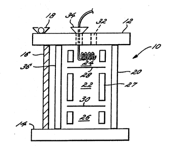
Figure 1 is a schematic view of a perfusion
chamber. Reactor 10 with cover plate 12 and floor
plate 14 are joined by bolts 16, held in position by
wing nuts 18. Three bolts are employed, so as to avoid
warping. The chamber 20 has three sections, the middle
section 22 containin~ the support matrix for the
stromal cells, the bed of stromal cells, and the bone
marrow cells. The central section 22 is sepa~ated from
the top section 24 and the bottom section 26 by
membranes or mesh 28 and 30 respectively.
Conveniently, polysulfonate membrane may be employed or
a stainless steel mesh, whose mesh size is small enough
so that cells are contained within the central section
of the chamber. The separating interphase may be
placed in the chamber using an inner cylinder 27 which
is sectioned to provide the separating membrane
mechanical support. The top section 24 and the bottom
section 26 need not be identical and will have tubing
or membranes across which liquid media and gases are
exchanged. The gases are exchanged acro~s a
hydrophobic, e.g., silicone, tube whose length (and
thereby gas/liquid contact area) may be varied to allow
for ~fficient gas fluxes to support the needs of the
cell population that is metabolizing in the central
section. The media can be pumped or withdrawn directly
from the top or bottom sections through port 32 and may
be fed through delivery tube 34.
If deYired, the top and bottom sections may be
eliminated by using an external oxygenator. In this
situation, the separating membrane is held in place
under the qlas3 cylinder 36 which fits into cylindrical
groove plates 12 and 14 and the area inside of the
.. :, -
,
.
. . .
8 2~27 ~ ~
cylindrical groove is indented to allow for good flow
distribution across the membrane. This geometry allows
the fluid from the finite number of inlet ports to mix
and for radial pressure to equilibrate, leading to a
uniform liquid flow across the separating membrane.
This setup is suitable for chambers which have
relatively few cells, so that oxygenation does not
become limiting.
In Figure 2 is depicted a schematic represen-
tation of the loop that connects the perfusion chamberto the side media reservoir, oxygenator, sensor
chamber, and sample/injection ports.
An external fresh media ~ource 50 is pumped by
means of pump 52 to a media reservoir through line 56
and spent media is withdrawn through line 58 from
reservoir 54 by means of pump 52 to the spent media
container 60 for further processing. A second pump 62
pumps media from the media reservoir 52 through line 64
through a hollow fiber oxygenator 66. The media is
directed through line 68 to the first chamber of
bioreactor 70. As appropriate, a means for injection
of media component 82 is provided, for introducing the
component into line 68 for transport by the media into
the first chamber of bioreactor 70. The component may
be test components, additional factors, or the like.
The media from bioreactor 70 is directed through
central chamber 72 into the second chamber 74 of the
bioreactor. From there the media is directed by line
76 to in-line sensors 78 for detecting the change in
composition of the media.
For example, it is desirable that the
glutamine:gluco~e ratio be in the range of about 1:5-8,
depending on the cell lines used; for instance,
preferably 1:8 for transfected 3T3 cells. Furthermore,
ammonium concentrations will preferably be below about
2.0 mM and lactate concentrations are preferably lecs
than about 40 m~. By mor.itoring the effluent from the
9 20627~1
bioreactor, the media introduced into the bioreactot
may be modified, oxygen partial pressu~e may be
changed, gas flow rate may be altered, various
components may be augmented, or the rate of perfusion
may be slowed or increased.
From the sensors 78, the media is directed
through line 80 by means of pump 62 to the reservoir
54.
By means of the flow path described above, the
media in the side reservoir is slowly exchanged using a
separate pump. This organization allows for separate
contzol of the media exchange rate (the outer pump) and
the flow rate through the oxygenator and perfusion
chamber. The former is used to control the longer term
lS change in the media composition and perfusion, while
the latter may be uced to control the dissolved oxygen
tension and flow patterns in the chamber. The use of a
small mesh biocompatible membrane allows for plug -
(piston) flow in the chamber and thus allows the
precise control of delivery of growth factors and other
special compounds that one may wish to introduce to the
hematopoietic cells and stromal cells in very precise
amounts.
After autoclaving the chamber and components
of the loop, the reactor i~ assembled in a sterile
environment. The media may be circulated through the
side $oop and chamber for a few days while signs of
contamination are monitored. If sterile assembly is
accomplished, the central section of the chamber is
inoculated with either the extra-oellular matrix alone
or a pre-inoculated extra-cellular matrix support that
contains the stromal cells. The stromal cells are then
either: 1) kept in the chamber for a period of a few
days while their metabolic performance and/or growth
factor responsiveness is monitored and if results are
satisfactory, the bone marrow is inoculated; or 2)
immediately seeded with bone marrow. In either case,
,~ . -
. . , , :
.
2052741
the cell layer is kept at the bottom of the central
section of the perfusion chamber. The cells lay down
additional extra-cellular matrix and the cell layer
adheres to the separating membrane. At this time, the
chamber may be inverted and the cell layer may then be
located at the ceiling of the central section. In this
configuration, the maturing cells will settle on the
bottom of the central chamber as they lose their
adherence to the stromal layer. This feature is
important to prevent the damage caused by mature cells
to the stromal layer and/or the less mature
hematopoietic cells. This feature also makes the
continuous removal of mature cells easier.
These cells are harvested by withdrawing the
cells by syringe, or by continuously allowing the cells
to flow out of the chamber, by the pressure of the
perfused medium, through the exit tubing.
The stromal cells will, for the most part, be
fibroblasts transformed with one or more genes
providing for desired hematopoietic growth factors.
~he same or different cells may be transfected with the
genes, depending upon the particular selection of host
cells, the same or different cells may be used for a
plurality of genes.
A wide variety of normal cells or stable lines
may be employed. However, it is found that not all
cell s~rains are permissible, since transformation of
some cell line~ may result in the overgrowth of the
cells. Desirably, the cells which are employed will
not be neoplastic~ but rather require adherence to a
support. The mammalian cells need not be human, nor
even primate. A variety of nontransformed cells may be
included in the adherent cell layer as well, including
normal human bone marrow adherent cells, normal human
spleen adherent cells, and normal human thymic
epithelium.
Methods for transforming mammalian cells,
.. - .. .. .. . . .
- .
.
.
11 2~27~
including ~ibroblasts, are well known and ~here is an
extensive literature of which only a few references
have been previously given. The constructs may employ
the naturally occurring transcriptional initiation
S regulatory region, comprising the promoter and, as
appropriate the enhancer, or a different transcrip-
tional initiation region may be involved, which may be
inducible or constitutive.
A large number of transcriptional initiation
regions are available which are inducible or
constitutive, may be associated with a naturally
occurring enhancer, or an enhancer may be provided, may
be induced only in a particular cell type, or may be
functional in a plurality or all cell types. The
lS transcriptional initiation region may be derived from a
virus, a naturally occurring gene, may be synthesized,
or combinations thereof.
Promoters which are available and have found
use include the chromosomal promoters, such as the
mouse or human metallothionein-I or II promoters, B-
actin promoter, etc., or viral promoters, such as SV40
early gene promoters, CMY promoter, adenoviruis
promoters, promoters associated with LT~s of retro-
viruses, etc. These promoters are available and may be
readily inserted into appropriate vectors which
comprise polylinkers for insertion of the transcrip-
tional initiation region as well as the gene of
interest. In other instances, expresision vectors are
available which provide for a polylinker between a
transcriptional initiation region and a transcriptional
termination region, also providing for the various
signals associated with the processing of the messenger
for translation, i.e., the cap site and the
polyadenylation signal. The construction of the
expression cassette compri ing the regulatory regions
and the structural gene may employ one or more of
restriction enzymes, adaptors, polylinkers, in vitro
. .
.~ .
12 20~27~1
mutagenesis, primer repair, resection, or the like.
The expression cassette will u~ually be part
of a vector which will include a ma~ker and one or more
replication ~ystems. The marker will allow fo~
detection and/or selection of cells into which the
expression cassette and marker have been introduced.
Various markers may be employed, particularly markers
which provide for resistance to a toxin, particularly
an antibiotic. Preferably, gentamycin re~istance is
employed, which provides resistance to G418 for a
mammalian cell host. The replication systems may
comprise a prokaryotic replication system, which will
allow for cloning during the various stages of bringing
together the individual component3 o~ the expression
cassette. The other replication system may be used for
maintenance of an episomal element in the host cell,
although for the most part the replication system will
be selected so as to allow for integration of the
expression cassette into a chromosome of the host.
The introduction of the expression cassette
into the host may employ any of the commonly employed
techniques, including tran~formation with calcium
precipitated DNA, transfection, infection, electro-
poration, ballistic particle~, or the like. Once the
host cells have been transformed, they may be amplified
in an appropriate nu~rient medium havins a selective
agent, to select for those cells which comprise the
marker. Surviving cells may then be amplified and
used.
Host cell~ which may be employed include
African green monkey cell line CVl, mouqe cells
NIH-3T3, normal human bone marrow fibroblasts, human
spleen fibrobla~ts, normal mouse bone marrow
fibrobla~ts, and normal mouse spleen fibroblasts. It
should be noted that in some instances, depending upon
the choice of vec~or and cell line, the cells may
become neoplastic. It is important that the resulting
-
` , : '
13 20&2~
transformed cells be capable of adherence, whereby the
tran~Formed cells maintain binding to a support, such
as protein sponges, protein coated membranes, or the
like.
Once the vector for expressing the appropriate
growth factors has been constructed, it may be used to
transform the cells by any convenient means. The
resulting transformed cells may then be used to seed
the supports, which have already been described. These
supports may be introduced into the reactor or may be
present at the time of seeding in the reactor. The
cells will be allowed to grow for sufficient time to
ensure that the cells are viable and are capable of
producing the desired growth factors.
The reactor may then be seeded as appropriate
with the hematopoietic cells. The hematopoietic cells
may include substantially pure stem cells, a mixture of
- hematopoietic cells substantially free of mature
hematopoietic cells of one or more lineages, or a
mixture comprising all or substantially all of the
various lineages of the hematopoietic system, at
various stages of their maturation.
The cells are allowed to grow with substan-
tially continuous perfusion through the reactor and
monitoring of the various nutrients and factors
involved. For the most part, the primary factors will
be provided by the stromal cells, so that a steady
state concentration of growth factors will normally be
achieved. Since conditioned supernatants are found to
be effective in the growth of the hematopoietic cells,
one can provide for a ratio of stromal cells to hemato-
poietic cells which will maintain the growth factor at
an appropriate concentration level in the reactor.
Transfected stroma can provide for the
introduction of genes into human stem cells. In mice,
retroviral mediated gene transfer into stem cells is
made possible by pretreating mice with S-FU and then
. ~ ' ' , ' ,
- . :
, ' . . .
.
. '-
14 2~S27~
growing the harvested bone marrow cells in ~EHI
conditioned media, which contains IL-3 and GM-CSF
(Lemischka, C _ 45:917, 1986). The artificial stroma,
grown with a retroviral packaging cell line secreting a
retroviral vector of interest, may be used to
efficiently introduce genes into human stem cells. For
example, human T-cells could be made resistant to HIV
infection by infecting stem cells with the retroviral
vector containing an HIV antisense sequence under
control of a CDC2 regulatory sequence (Greaves, Cell
56:979-986, 1939) which would allow for tissue specific
expression in T-cells. There would be a factor
provided by the retroviral packaging cell line
essential for replication of the retrovirus; this
factor would be absent in the hematopoietic target
cells. Once the virus was transferred to the
hematopoietic target cells, it would no longer be able
to replicate.
The following examples are offered by way of
illustration and not by way of limitation.
EXPERIMENTAL
I. Formation of Transformants
The growth factor human G~-CSF (Wong, Science,
228:810-815, (1985)) was inserted into a eukaryotic
expres~ion vector. The hGM-CSF cDNA (EcoRI to AhaIII,
approximately 700 bp fragment) was cloned into an EcoRI
to PstI fragment of pSP65. (Melton, Nucl. Acid~ ~es.
30 2:7035-7056 (l984)). The resulting plasmid was
pSP65GM-CSF. The mouse metallothionein promoter
(Glanville, Nature, 292:267-269, (1981)) wa~ digested
with EcoRI and 3~1II and the approximately 2 kb
fragment containing the promoter was inserted into the
35 EcoRI to 3amHI fragment o pSP65 to make p65MT. The
plasmid pMT GM-CSF wa~ then constructed by digesting
pSP65GM-CSF with EcoRI, filling in the overhang with
" : .
lS 20527~1
the Klenow fragment o~ DNA polymerase I and then
dige~ting the resulting linearized DNA with HindIII to
isolate the 700 bp fragment comprising the coding
region of GM-CSP. This fragment was subcloned into the
SalI filled/HindIII site of p65MT. The 2.7 kb fragment
comprising the metallothionein promoter and the GM-CSF
coding region was then isolated and placed into pSV2neo
(Southern and Berg, J. Mol. Appl. Genet 1:327 (1982))
from which the SV-40 promoter was removed. This
results in the SV-40 poly A signal downstream of the
GM-CSF coding sequence.
The neomycin resistant gene, which confers
resistance to the antibiotic gentamycin (G418) was
taken from pSV2neo by isolating the approximately 3 kb
PvuII to EcoRI fragment and placing EcoRI linkers onto
the PvuII site. The neo resistance gene with Eco~I
ends was subcloned into the EcoRI site of the G~-CSF
expression plasmid to create the plasmid MTGM-CSFneo.
The plasmid MTGM-CSFneo alone and as a
cotransfection with the plasmid (Yang, Cell 47:3-10,
1986) encoding the gibbon ape IL-3 gene under the
control of the SV-40 promoter and poly A site, were
transfected by electroporation of linearized DNA into
the African green monkey cell line CVl and the mouse
cell line NI~ 3T3 cells. Transformants were selected
by selection in media containing 500 mg/ml o G418,
isolated, and screened fsr production of GM-CSF or IL-3
by bioassay of supernatants using A~L-193 cells ~Adams,
et al., Leukemia 3:314 (1989)). Several of the
po~itive lines were then employed as stroma for human
bone marrow cells in Dexter culture.
In addition, normal mouse bone marrow cellc
were transfected with the above pla~mids using the
calcium/phosphate method of Okayama (Chen, Mol. Cell.
35 3iol. 7:2745-2752, 1987) and were found to efficiently
express the introduced gene~.
GM-CS~ and IL-3 secretion by the transfected
.. .. . . . . . .. .
~ - ; , ;
, .- ' .. ' ~
- .
.
16 20~27~
fibroblasts was investigated. Serum free 72 hour
culture supernatants were obtained from the NI~-3T3
cells and assayed for hGF secretion by 3H uptake on
target cells inhibitable by neutralizing rabbit anti-
GM-CSF or anti-IL-3 antibodies. Proliferation induced
by 20 mg/ml GM-CSF was set as 100 units GM-CSF and that
induced by 10 ng/ml IL-3 was set as 100 units IL-3.
The co-transfected cells produced about 35 units/ml of
GM-CSF and about 57 units/ml of IL-3.
II. Perfus_on Chamber
The perfusion chamber is a glass cylinder with
Delrin caps to allow for autoclaving without
deformation and biocompatability. The caps have
cylindrical groves into which the glass cylinder fits.
At the bottom of the grove an O-ring is placed to seal
the lumen of the chamber. The caps have several holes
into which Luer (Luer Lok) fittings are provided into
which media and gas delivery lines are put as well as
an extended tube into the central section of the
chamber to sample adherent and/or non-adherent cells.
The caps are attached with three long bolts, spaced
120, placed outside the glass cylinder; wing nut~ and
washers are used to tighten the assembly.
The chamber is hooked to a side reservoir.
The loop contains a pump, a chamber of on-line sensors,
oxygenator, and sample and injection ports in addi~ion
to the side media resDrvoir. The media in the side
reservoir is then qlowly exchanged using a separate
pump. This configuration allows for separate control
of the media exchange rate and the flow rate through
the oxygenator and perfusion chamber. The former is
used to control the longer term change in the media
composition and perfusion, while the latter may be used
to control the dissolved oxygen tension and flow
patterns in the chamber. The use of a small me h
polysulfonate membrane allows for plug flow in the
.
17 20~7~
chamber and the precise control of delivery of growth
factors and other special compounds which one may wish
to introduce into the bioreactor in very precise
amounts.
S The transfected stromal cells are seeded
either over a bed of shredded collagen sponge or the
stromal cells are placed on one side of a 5~ porous
polycarbonate filter precoated with collagen and the
stromal cells allowed to adhere to the filter over a
number of hours. The cells are allowed to grow in an
appropriate nutrient medium until the cells become
confluent on one side while sending cytoplasmic
projectionq through the pores. Bone marrow cells are
then ceeded on the other side of the membrane and the
lS stem cells attach to the intruded cytoplasmic
projections which have passed through the porec.
After autoclaving the chamber and components
of the loop, the reactor is assembled in a sterile
environment. The media is then circulated through the
side loop and chamber for a few days while signs of
contamination are monitored. The central section of
the bioreactor is then inoculated with either the
extra-cellular matrix alone or a pre-inoculated extra-
cellular matrix support that contains the stromal
cells. The stromal cells may then be kept in the
chamber for a period of a few days while their
metabolic performance and/or growth factor
respon~iveneg~ i3 monitored and if results are
satisfactory, the bone marrow is inoculated or
i D ediately seeded with bone marrow. In either case,
the cell layer is kept at the bottom of the central
section of the perfusion chamber.
The cells lay down additional extra-cellular
matrix and the cell layer adhere~ to the support.
Where the membrane i9 used, the chamber may be inverted
and the cell layer is then located at the ceiling of
the central section. In this configuration, the
- - ;- .
.
.
20S27~
maturing cells settle on the bottom of the central
chamber as they loose their adherence to the stromal
layer. The non-adherent cells are then harvested by
constant cell flow, driven by the medium perfusion
pressure, into the exit tubing.
In a typical run, the chamber was inoculated
with NIH-3T3 cells on day one on shredded collagen
sponge support. ~or the first 40 days perfusion rates
and other operating variables were adjusted. At day 40
a reasonable steady state was achieved which was
maintained for about 20 days. On day 64 the chamber
was seeded with 33 x 106 human bone marrow cells. For
the first 10 days the harvested cell count decreased
until it settled in a steady ctate of about 7-8 x 10⁵
cells produced every three days. Flow cytometric
analysis showed that a constant fraction, about 20~ of
the harvested cells were HLA-DR positive. On day 90 a
- pump failure was experienced and the pH dropped below
6.9 overnight. When the perfusion rate was restored
the production of non-adherent cells recovered and was
approaching the previous steady state production rate
when a bacterial contamination occurred. At this
point, the study was terminated.
The above results demonstrated that a
perfusion chamber i~ capable of performing ex vivo
hematopoiesis, hematopoiesis may be restored ex vivo
after a p~ drop, the glucose concentration data showed ~ `
that the hematopoietic cells grow primarily aerobically
on glucose, since the glucose concentration drops after
inoculation without increasing the lactate
concentration indicating that oxygenation is limiting.
The glucose/lactate (anaerobic) metabolism appears to
be primarily due to the NI~-3T3 stromal bed.
Similarly, the glutamine and ammonia concentrations
reach pre-inoculum levels once the hematopoietic cell
number levels off, implying that the glutamine
consumption by the bone marrow cells is much less than
~9
2~$~
that of the stromal bed.
III. Monitoring of Metabolic Products
The consumption and formation rates of glucose
S and lactate as well as glutamine and ammonia were
determined for transfected NIH-3T3 cells. (The medium
was IMDM plus 20~ FCS). Increased glucose consump~ion
was only observed for daily fed T-flasks, where as all
less frequently fed cultures follow the same slowly
diminishing glucose uptake rate pattern. Cultures that
were exchanged 50% daily were switched to the 100%
daily exchange schedule on day 18, which resulted in an
immediate increase in gluco~e consumption following the
same trend as that observed for cultures exchanged 100%
daily from day one. Lactate production rates follow a
similar pattern, as the lactate yield on glucose is
essentially a constant (0.9 lactate/glucose; indicating
a largely anaerobic stromal metabolism~.
The glutamine and ammonia concentrations show
a pattern analogous to the glucose/lactate metabolism.
Using values corrected for chemical decomposition of
glutamine at 37C, the glutamine consumption rate
versus the glucose con3umption rate ~hows relative
uptake rate~ are constant, about 1:8 glutamine:
glucose. The predicted optimum ratio varie~ with
oxygen uptake rate - the ratio drops with increasing
optimum uptake rate.
~nalogous conclusions were supported by ;!
glucose/lactate metabolic data derived from normal bone
marrow ~tromal fibroblasts~ Under conditions of
infrequent medium exchange the cultures were primarily ~ -
anaerobic, with high steady state levels of lactate
rapidly achieved and maintained. With more frequent
medium exchange, the cell metabolism became more rapid,
with increased glucose consumption and lactate
production. No detectable consumption of glutamine was
observed after correcting the data for spontaneous
,
~ , .
20~27~
chemical decomposition. For both 3T3 cells and normal
human bone marrow cells, the cells continue to divide
and crowd when the serum/media exchange rate wa~ above
what appears to be a critical replacement schedule.
To further ascertain the relative importance
of perfusion rate of serum versus that of nutrients,
the following experiments were performed: 1) one set
of T-flasks with 20% serum containing media exchanged
daily; 2) two sets o~ T-flasks, one with 20~ serum and
the media exchanged every other day and one with 10~
serum with the media exchanged daily; 3) two sets of
T-flasks, one with 10% serum and the media exchanged
every other day, one with 5% serum with the media
exchanged daily; 4) two sets of T-flasks, one with 5%
serum and the media exchanged every other day and one
with 2.5% serum with the media exchanged daily. The
serum exchange rate is the same within each group while
the exchange rate of the nutrient containing media
varies. The results from these experiments show that
it is the exchange rate of the serum that is critical.
While for the experiment 1) glucose consumption
increased and by day four had substantially flattened
out to a rate of about 9.5 mmole~/per day, in all of
the other cases, the glucose consumption started below
the original glucose consumption of Group I and dropped
off in a substantially linear manner regardless of
whether twice the amount of serum waR used and changed
every other day or half the amount of serum was used
- and the media changed every day. This supports the
need for a critical perfusion rate of serum or one or
more serum components that influence the metabolic
growth behavior of the stromal cells.
It is evident from the above results, that one
may grow hematopoietic cells in a bioreactor in an
efficient manner. Stromal cells can be provided from
homologous or heterologous sources, where the stromal
cells have been transfected with genes to provide for
.
.
,
the important growth factor~. In this manner, serum 2 ~ 5 2 7 4
need not be added to the media to support the growth of
the cells. ~y providing for stromal cells which adhere
to a support in a manner which allows for separation of
5 hematopoietic cells from the stromal cells, the
hematopoietic cells may be continuously harvested ~or
use. By appropriate choice of combinationR of growth
factors, specific lineages of hematopoietic cells may s
be grown. In addition, if desired, the stromal cells
10 may provide for a reservoir of transfecting viruses for
the introduction of genes into the hematopoietic cells.
All publicationc and patent applications cited
in this specification are herein incorporated by
15 reference as if each individual publication or patent
application were specifically and individually
indicated to be incorporated by reference.
Although the foregoing invention has been
described in some detail by way of illustration and
20 example for purposes of clarity of understanding, it
will be readily apparent to those of ordinary skill in
the art in light of the teachings of this invention -~ -
that certain changes and modifications may be made
thereto without departing from the spirit or scope of
25 the appended claims. ~ -
.