Note: Descriptions are shown in the official language in which they were submitted.
'~ 20~27~6
..,~
NEUROLOGICAL THERAPY SYSTEM
Back~round of the Invention
The technical field of this invention is the
treatment of neurological diseases and, in
particular, the treatment of neurotransmitter-
deficiency and dysfunction diseases.
Neurotransmitters are small molecules (less
than 1 kilodalton (kD) molecular weight) which act as
chemical means of communication between neurons.
They are synthesized by the presynaptic neuron and
released into the synaptic space where they then
effect receptors on postsynaptic neurons.
Neurotransmitter deficits have been
implicated in various neurological diseases. Lack of
neurotransmitter-mediated synaptic contact causes
neuropathological symptoms, and can also lead to the
ultimate destruction of the neurons involved.
Recently, it has been discovered and disclosed in
commonly-owned International Application No.
PCT/US88~04092 (W089/04655) that localized delivery
of the relevant neurotransmitter to the target tissue
may reverse the symptoms without the need for
specific synaptic contact.
u~E S~EE ~
~ -2- 20S2716
For example, paralysis agitans, more
commonly known as Parkinson's disease, is
characterized by a lack of the neurotransmitter,
5 dopamine, within the striatum of the brain, secondary
to the destruction of the dopamine secreting cells of
the substantia nigra. Affected subjects demonstrate
a stooped posture, stiffness and slowness of
movement, and rhythmic tremor of limbs, with dementia
10 being often encountered in very advanced stages of
the disease.
These clinical symptoms can be improved by
the systemic administration of dopamine precursors
15 such as levodopa (L-dopa)(Calne et al. (1969) Lancet
ii:973-976), or dopamine agonists such as
bromocriptine (Calne et al. (1974) Bri. Med. J.
4:442-444) and (+)-4-propyl-9-hydroxynapthoxacine (de
Yebenes et al. (1987) Movement Disorders 2:291-299),
20 both of which are able to cross the blood-brain
barrier, and which are converted into dopamine in the
brain. Dopamine, itself, cannot be administered
systemically because of its inability to cross the
blood-brain barrier.
However, a number of drawbacks are incurred
when using this type of chemical therapy. For
example, other neurological structures which
recognize dopamine as a neurotransmitter are also
30 affected. In addition, it becomes difficult to
administer the correct drug dosage with time because
the "therapeutic window" narrows (i.e., just after
administration, the patient is overdosed, exhibiting
- 3 -
excessive spontaneous movement; some time thereafter
the drug level may become insufficient, causing the
patient to again express Parkinsonian symptoms).
Furthermore, the limited potency and/or solubility of
most available dopamine agonists precludes continuous
in vivo infusions as means to reduce motor deficits
in Parkinson's disease. Therefore, what is needed is
a method of continuous or constitutive delivery of an
undegraded, active neurotransmitter to a localized
target region deficient in that neurotransmitter.
In an attempt to provide a continuous
supply of dopamine and related drugs to the brain at
a controlled rate, miniature osmotic pumps have been
used. However, limited solubility and stability of
dopamine and related drugs, as well as reservoir
limitations, have restricted the usefulness of this
technology. Controlled sustained release has also
been attempted by implanting dopamine encapsulated
within bioresorbable microcapsules ( "Implantable
microencapsulated dopamine (DA): a new approach for
slow-release DA delivery into brain tissue," McRae-
Degueurce et al. (1988) Neurosci. Lett. 92:303-309).
However, controlled sustained release of a drug from
a bioresorbable polymer relies on bulk surface
erosion, for example, due to various hydrolytic
events, increasing the likelihood of drug
degradation, and rendering predictable release rates
difficult.
The implantation of cells capable of
constitutively producing the needed neurotransmitter,
reportedly in response to environmental needs, has
also been attempted. Recently, remedial trans-
plantation of neurotransmitter-secreting tissue has
been accomplished using the patient's own tissue so
206274~
r _ 4
as not to elicit an immune response. For example,
dopamine-secreting tissue from the adrenal medulla of
patients suffering from Parkinson's disease has been
implanted in their striatum with some success.
5 However, this procedure is only used in patients less
than 60 years of age, as the adrenal gland of older
patients may not contain sufficient dopamine-
secreting cells. This restriction limits the
usefulness of the procedure as a remedy since the
10 disease most often affects older people.
Furthermore, abdominal surgery performed to
excise portions of the adrenal gland poses
substantial risks. Moreover, it is not actually
15 known whether it is the dopamine or other ~factors"
produced by the implanted cells, or the trauma of the
surgery, itself, which alleviates the clinical
symptoms. In fact, stereotaxic surgery, or the
placement of precisely localized lesions in the brain
20 has been practiced in younger, less affected patients
without transplantation and this procedure appears to
provide similar relief of Parkinsonian symptoms. The
procedure is risky, however, and opinions among
neurosurgeons still differ as to the best way of
25 making the lesion and what its ideal location should
be.
Alternatives to the transplantation of a
patient's brain tissue also include the
30 transplantation of either allograft (identical tissue
from another of the same species), or xenograft
(similar tissue from another of a different species)
dopamine-secreting tissue. However, recent studies
20627~
have shown that although the brain is considered
"immuno-privileged", rejection ultimately occurs with
both allografts and xenografts. This problem
5 necessitates the co-adminstration of immuno-
suppressors, the use of which renders their own set
of complications and deleterious side-effects.
Therefore, there exists a need for improved
10 therapies for neurotransmitter-deficiency diseases in
general, and in particular, a need for neurological
therapy devices which can augment or replace the
functions of dysfunctional neurotransmitter-producing
areas of the brain without causing excessive trauma.
15 More specifically, there exists a need for a method
of providing active, undegraded neurotransmitter to a
localized region of the nervous system of a subject
deficient in this neurotransmitter, the correct
dosage of which will be constitutively delivered over
20 time.
Accordingly, it is an object of the present
invention to provide an implantable neurological
therapy device useful for the sustained and
25 controlled delivery of a neurotransmitter to a
subject, and more particularly, to provide a device
which can deliver neurotransmitter to a localized
region in the brain of a subject.
~ -6- 2062746
It is another object to provide an
implantable device that contains and protects
neurotransmitter therein from in vivo degradation
5 such that it is delivered to the subject in an active
form. Yet another object of the present invention is
to provide an implantable device which can deliver an
amount of neurotransmitter responsive to in vivo
environmental needs. A further object is to provide
10 an implantable, protective cell culture device which
is retrievable, and whose contents are renewable with
new and/or additional source of neurotransmitter.
~ 2~S27~6
SummarY of the Invention
Neurological therapy devices are disclosed
5 for the local and controlled delivery of a
neurotransmitter to the brain of a subject suffering
from a neurotransmitter deficiency or dysfunction.
It has been discovered that various polymeric
materials have the ability to protect various
10 ~effector-type~ substances, such as neurotransmitters
and growth factors, from oxidation, hydrolysis, and
degradation when such substances are embedded
therein. In addition, these polymeric materials have
the capacity for sustained release of the embedded
15 substance at a controlled rate. Recently, it has
also been discovered that cells encapsulated within a
protective semipermeable membrane, and not in direct
contact with a target tissue, are capable of
producing neurotransmitter or growth factor in
20 response to the needs of the immediate environment.
These discoveries have been utilized to
develop the neurological therapy devices of the
present invention. One such device includes a
25 biocompatible, implantable, and retrievable polymeric
insert including a source of neurotransmitter. In
preferred embodiments of this device, the source
includes a neurotransmitter which has been embedded
within the insert. The polymeric insert protects the
30 neurotransmitter embedded therein from oxidation,
enzymatic degradation, and hydrolysis. It may take
2062~6
any shape which is conducive for supplying
neurotransmitter, and which can be accommodated by
the recipient. Preferred shapes include a fiber or
rod.
Useful neurotransmitters to be embedded
within the insert include gamma aminobutyric acid,
serotonin, acetylcholine, norepinephrine, endorphins,
enkephalins, and dopamine. Alternatively,
10 precursors, agonists, active analogs, and active
fragments of these neurotransmitters (e.g. dopamine
precursor L-dopa and dopamine agonist bromocriptine)
may be used.
The polymeric insert includes pores having a
molecular weight exclusion of from about 1 kD to
about 1,000 kD, but preferably from about 25 kD to
about 100 kD. In one preferred embodiment, the
polymeric insert includes a hydrophobic matrix such
20 as ethylene-vinyl acetate copolymer. In another
embodiment, the insert includes a hydrophilic matrix
such as a hydrogel. The insert may additionally
include an impermeable portion which is preferably
provided by an outer coating of a pure polymeric
25 material such as polyurethane or ethylene-vinyl
acetate. The impermeable portion can serve to make
the insert function as a conduit for neurotransmitter
or whatever substance is embedded therein, and can
also aid in supplying the substance to a specific
30 anatomical region of the subject.
'- -9- 20S27~16
An alternative neurological device includes
a retrievable source of neurotransmitter and a
retrievable source of growth factor in close
5 proximity. The source of neurotransmitter includes
at least one neurotransmitter-secreting cell
encapsulated within a semipermeable membrane allowing
the diffusion therethrough of the neurotransmitter.
The term "semipermeable~ is used herein to
describe biocompatible membranes that allow the
diffusion therethrough of molecules having a
relatively low molecular weight, while excluding the
passage of those having a relatively high molecular
15 weight.
In one embodiment of the invention, the
semipermeable membrane of the receptacle contains
pores having a molecular weight exclusion of from
20 about 50 kD to about 100 kD. This membrane excludes
the passage therethrough of large particles such as
those which are capable of degrading the
neurotransmitter or injuring the neurotransmitter-
producing cells (e.g. viruses, antibodies,
25 complement, and various proteases). The
semipermeable membrane can be made of various
polymeric compositions such as a polyvinylchloride,
polyacrylonitrile, polyvinylidene fluoride,
polystyrene, polymethylmethacrylate, polysulfone, and
30 acrylic copolymers.
-lo- 2~62746
" ~.
The neurotransmitter-secreting cell may
include any cell which is known, or has been
engineered to produce neurotransmitter, or agonists,
5 precursors, active analogs, or active fragments
thereof. For example, chromaffin cells of the
adrenal medulla, embryonic ventral mesencephalic
tissue, and various neuroblastic cell lines such as
PC12 function to supply dopamine, and therefore, are
10 preferred for incorporation into the device. In some
aspects of the invention, the cell is an allograft
(i.e., cells from another of the same species as the
subject in which it is to be implanted) or a
xenograft (i.e., cells from another of a different
15 species).
The source of growth factor is situated such
that it can easily come into contact with the
neurotransmitter-secreting cells encapsulated within
20 the semipermeable membrane. The growth factor
maintains cell differentiation and/or stimulates the
production and secretion of neurotransmitter. In one
preferred embodiment, the source of growth factor
includes a polymeric insert with growth factor
25 embedded therein. In another preferred embodiment,
the source is at least one growth factor-secreting
cell encapsulated within a semipermeable membrane
allowing the diffusion therethrough of the growth
factor. The growth factor is delivered to the
30 neurotransmitter-secreting cells as it leaches from
the insert or as it diffuses from the semipermeable
membrane after being secreted by the cells therein.
-11- 2C62~B
. .,~ .
The invention will next be described in
connection with certain illustrated embodiments.
However, it should be clear that various
S modifications, additions, and subtractions can be
made without departing from the spirit or scope of
the invention. For example, the present invention
should not be read to require, or be limited to, a
particular device shape, material, neurotransmitter,
10 growth factor, or cell line described by way of
example or illustration.
-12- 20627~ 6
."".,~
Brief Description of the Drawings
The invention itself can be more fully
understood from the following description when read
5 together with the accompanying drawings in which:
FIGS. lA-lC are schematic illustrations of
an implantable neurological therapy device according
to several aspects of the present invention;
FIG. 2 is a graphic representation of
cumulative in vitro release kinetics of eight
dopamine/ethylene-vinyl acetate inserts;
FIG. 3 is a graph demonstrating rotational
behavior of animals after the implantation of
dopamine-containing neurological therapy devices;
FIG. 4 is a graph showing extracellular
20 dopamine levels in the brain of animals having
acutely implanted dopamine-containing neurological
therapy devices;
FIG. 5 is a graph showing the level of
25 extracellular dopamine in the brain of 3 animals
having an implanted dopamine-containing neurological
therapy device 7 days post-implantation; and
FIG. 6 is a photographic representation of a
30 scanning electron micrograph of a dopamine/copolymer
EVAc insert revealing dopamine particles distributed
throughout the polymer matrix (A) before
implantation, and (B) after implantation.
-13- 20627~6
Detailed Description of the Invention
Neurological therapy devices are disclosed
for the constitutive and controlled delivery of a
5 neurotransmitter, a precursor, agonist, active
fragment, or active analog thereof to a target
region of a subject suffering from a neurological
dysfunction.
Exemplary embodiments of the neurological
therapy device of the present invention are shown in
FIGS. lA-lC in which like reference characters
indicate the same or essentially similar elements in
the various illustrations. FIG. lA is a device
15 including an implantable, biocompatible polymeric
insert containing the neurotransmitter embedded
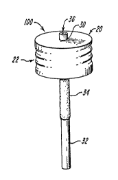
therein. The device 100 includes a cylindrical cap
20 having grooves 22 on the sides thereof, and a
polymeric insert 30 containing neurotransmitter
20 embedded therein. The insert has a permeable portion
32, an impermeable portion 34, and an end 36.
Neurotransmitter can diffuse through permeable
portion 32, but not through impermeable portion 34.
The insert of the neurotherapy device acts
as a conduit for the source of neurotransmitter or
growth factor as well as a directed passageway to the
anatomical region or specific tissue which requires
treatment. In FIG. lA the insert has the shape of a
30 rod. However, it should be appreciated that the
insert may have any shape which can accommodate the
source of neurotransmitter without causing undue
trauma in the subject upon its surgical implantation.
-14- 20G27~
The neurological therapy devices shown in
FIGs. lB and lC include a source of neurotransmitter
and a source of growth factor in close proximity.
The device 200 shown in FIG. lB includes insert 30
5 which contains growth factor embedded therein, and
further includes semipermeable membrane 50 containing
neurotransmitter-secreting cells 52 encapsulated
therein. Membrane 50 has the form of a U-shaped
tube. However, it should be appreciated that the
10 semipermeable membrane may have alternative shapes
that will accommodate the cells such as, for example,
a hollow rod, sack, or multiple fibers.
U-tube 50 may be loaded with cells through
15 end 26 or 28. Ends 26 and 28 may be reversibly
occluded with friction fitted caps 40, or
alternatively with an epoxy glue or sutures of a
biocompatible and nonresorbable material. In the
device 300 shown in FIG. lC, semipermeable membrane
20 50 containing neurotransmitter-secreting cells 52 is
accompanied by rod-shaped semipermeable membrane 54
containing growth factor-secreting cells 56
encapsulated therein.
The region targeted for implantation of the
neurological therapy device is preferably the brain,
as it is often the site of many neurological
deficiencies and dysfunctions. Devices 100, 200, and
300 can be surgically implanted into the brain such
30 that permeable portion 32 of insert 30, or
semipermeable membrane 50 and 54 are in immediate
contact with brain tissues and fluids. Once
implanted, the cylindrical cap 20 can be permanently
2062746
-15-
secured to the skull, for example, by screwing it in,
and further, by applying a glue or a cement such as
dental cement to the cap at the junction of the skull
and the cap.
S
In the event that the neurotransmitter or
growth factor supply in insert 30 is spent, the
insert can be removed and replaced. Retrieval of
implanted insert 30 can be accomplished by pulling it
10 out of cap 20, for example, using a pair of forceps
after exposing the device. Cap 20 may be located
directly under the patient's epithelial tissues.
Insert 30 may be replaced with a new insert in the
event that additional neurotransmitter therapy is
15 required. Cells encapsulated within semipermeable
membrane 54 (FIG. lC) can also be retrieved if the
cells cease to produce neurotransmitter or growth
factor, expire, or are no longer needed to correct
the neurological dysfunction. Cells in membrane 50
20 can be replenished by forcing the cells out of end 26
or 28 by pressure or suction using, for example, a
hypodermic syringe.
Permeable portion 32 of insert 30 is
25 implanted at or near the target region, while the
impermeable portion 34 confines the neurotransmitter
or growth factor to within the boundaries of the
insert. The permeable portion 32 includes a
polymeric material having pores of a particular size
30 (i.e., having a particular molecular weight cut-off)
that excludes some molecules from passage
therethrough, while permitting the passage of
others. In this way, the diffusion of
-16- 2~S274~
neurotransmitter from the insert to the target
region, or growth factor to the neurotransmitter-
producing cells, is allowed, while the passage of
larger deleterious elements such as viruses,
5 antibodies, complement, and various proteases is
effectively barred. For example, inserts with pores
having a molecular weight exclusion of from about 1
kD to about 1000 kD are useful, with those having
pores with a molecular weight cut off of from about
10 25 kD to about 100 kD being particularly preferred.
The insert may be composed of any
biocompatible material having the desired pore size
and being composed of materials which do not limit
15 the activity of the substance embedded therein.
Hydrophilic matrices such as hydrogels (e.g.,
hydroxyethyl methacrylate, polyvinyl alcohol, and
polyvinyl pyrrolidone) and hydrophobic matrices such
as ethylene vinyl acetate are particularly useful.
The neurological therapy device can provide
any neurotransmitter which will satisfy the
deficiency or remedy the dysfunction. These include
gamma aminobutyric acid, serotonin, acetylcholine,
25 epinephrine, norepinephrine, dopamine, enkephalins,
and endorphins. Alternatively, the device may
provide an active analog, active fragment, or active
derivative of the neurotransmitter, or may include a
precursor, which after processing, provides the same
30 activity as the neurotransmitter in the appropriate
in vivo location. The device may further include an
agonist of the neurotransmitter.
~ -17- 20S27~6
One way of providing the source of
neurotransmitter includes incorporating it into the
polymeric insert. The encapsulating material
5 provides a protective environment for substances such
as neurotransmitters or cell growth factors embedded
therein, while affording sustained release of the
substance at a controlled rate therefrom. For
instance, the use of polymeric insert composed of
10 hydrophobic matrices reduces neurotransmitter
degradation by inhibiting oxidation and hydrolysis of
the neurotransmitter encapsulated therein.
An exemplary method for incorporating the
15 effector substance (e.g., neurotransmitter or growth
factor) into the insert includes fabricating the
polymeric from a mixture or complex of the polymeric
material and the substance. For example, a
hydrophobic material such as ethylene-vinyl acetate
20 (EVAc) copolymer can be dissolved in a solvent to
which a lyophilate of a neurotransmitter such as
dopamine can be added. The mixture is agitated and
fabricated into the desired shape by extruding it
from a melt. Upon cooling of the mixture, a solid
25 polymeric matrix containing neurotransmitter embedded
throughout the matrix is formed (see FIG. 6).
Contact of the hydrophilic neurotransmitter with the
aqueous environment causes the slow leaching of
neurotransmitter therefrom, leading to the
30 development of micropores throughout the matrix of
the insert.
-18- 206274~
The concentration of neurotransmitter added
to the hydrophobic matrix material is one factor
controlling the rate of neurotransmitter release; the
5 more neurotransmitter incorporated, the more rapid
the release rate. The particulate size of the
neurotransmitter incorporated into the matrix
material is another variable; the larger the particle
size, the quicker the rate of release.
The release of growth factor from a
polymeric insert can be controlled by the amount of
carrier protein co-embedded therewith; the more
carrier protein incorporated, the higher the rate of
15 grown factor release. However, the ratio of growth
factor to carrier protein is dependent on the levels
of growth factor that are effective in the
physiologic environment within a therapeutic range.
A useful carrier protein is one having the ability to
20 readily dissolve while in the matrix, and having the
ability to leach from the matrix. Micropores through
which growth factor can leach are created in the
matrix when the carrier protein is dissolved by the
aqueous environment. Such a carrier protein is
25 bovine serum albumin (apparent molecular weight =
about 69 kD).
The release rate may also be controlled by
the amount of pure, impermeable polymeric material
30 coating the effector substance-embedded insert; the
more (or thicker the) coatings, the slower the
release rate. Materials such as polyurethane or
pure ethylene-vinyl acetate are particularly useful
for this purpose.
- 19 - ~ 7 ~ ~
Alternative methods of incorporating the
source of neurotransmitter include the provision of
neurotransmitter-producing cells accompanied by a
source of growth factor situated in close proximity
thereto. In this embodiment, a semipermeable
membrane functions as a protective cell culture
device for the neurotransmitter-secreting cells. The
pores of the membrane should be large enough to
enable the exchange of metabolites with body fluids,
and to permit the diffusion therethrough of
neurotransmitter produced by the cells therein, but
are small enough to bar the passage therethrough of
larger elements deleterious to the cells. Pores
having a molecular weight cut-off of from about 50 kD
to 100 kD are particularly useful for this purpose.
The semipermeable membrane may take any
useful form such as a U-tube, hollow fiber, cell sack
or container, or microcapsule. Likewise, if the
source of growth factor includes growth factor-
producing cells, they, too are encapsulated within amembrane allowing for the exchange of metabolites,
growth factor production and diffusion, cell
maintenance, and growth (limited by the boundary of
the membrane). For a further discussion of such
devices, see commonly-owned, copending Canadian
patent application no. 583,385, filed November 17,
1988, granted May 30, 1995 as Canadian Patent No.
1,335,715.
-20- 2062~4~
Any cells which secrete the desired
neurotransmitter or growth factor may be incorporated
into the device. For example, the cells may be any
5 which naturally produce the neurotransmitter, such as
neurons. Such cells are useful because they are able
to respond to the general environment by producing
neurotransmitter as it is needed. The cells can be
obtained from a number of sources such as a body
10 organ which normally secretes a particular
neurotransmitter in vivo. For example, tissues of
the embryonic ventral mesencephalon and adrenal
medulla (chromaffin cells) which normally produce
dopamine can be used. These tissues may be an
15 allograft, or they may be a xenograft.
Alternatively, the cell may be derived from various
cultured neuroblastoid cell lines, such as PC12.
In addition, any cell which secretes a
20 precursor, agonist, active analog, or active fragment
of a desired neurotransmitter or growth factor having
similar neurotransmitter activity can also be used,
including, for example, cells which elicit L-dopa, a
precursor of dopamine, or bromocriptine, a dopamine
25 agonist.
Further, any cells which have been
genetically engineered to express a neurotransmitter
or growth factor, or their agonists, precursors,
30 derivatives, analogs, or fragments thereof having
similar effector activities are also useful in
practicing this invention. Thus, in such an
approach, the gene which encodes the
!~
~ -21-
20527~6
neurotransmitter, or its analog or precursor is
either isolated from a cell line or constructed by
DNA manipulation. The gene can then be incorporated
into a plasmid, which, in turn, is transfected into a
5 cell such as a fibroblast for expression. (See,
e.g., Maniatis et al., Molecular Cloning (1982),
herein incorporated by reference for further
discussion of cloning vehicles and gene manipulation
procedures.) The cells which expresses the
10 neurotransmitter can be grown n vitro until a
suitable density is achieved.
Thereafter, the cells from this culture can
be loaded into the neurological therapy device by
15 seeding a portion of the already implanted
semipermeable membrane via an orifice located at the
skin surface. Alternatively, small tissue fragments
or culture aggregates may be preloaded into an
encapsulating semipermeable membrane which is then
20 implanted within the subject.
Various ~growth factors" having the ability
to stimulate cell growth, differentiation, and/or
neurotransmitter secretion may be co-implanted with
25 the neurotransmitter-secreting cells to insure
successful delivery of neurotransmitter to the
subject. These growth factors may be specific for a
cell type or have a generalized effect on a number of
different tissues. In the case of neurotransmitter-
30 producing cells such as neurons, growth factors canact to maintain neurotransmitter production, as well
as to promote cell maintenance and growth.
Alternatively, growth factors may maintain nerve
-22- 20S2~
cells in a differentiated state. Useful cell growth
factors include nerve growth factor (NGF), fibroblast
growth factor (FGF), platelet-derived growth factor
(PDGF), and epidermal growth factor (EGF), among
5 many. In addition, effectors of various membrane
receptors such as glutamate and nicotine may also be
useful.
The growth factor may be incorporated into
10 the device with the neurotransmitter-producing cells
by embedding it within the polymeric matrix of the
insert and placing it in the receptacle with the
cells. The embedded growth factor leaches slowly
from the insert into the receptacle, thereby acting,
15 for example, to maintain the differentiated state of
the cells therein such that they continue to produce
neurotransmitter. The encapsulating membrane of the
cells, if present, poses no hindrance as it is
permeable to the growth factor. This insert may be
20 retrieved from the receptacle and replaced as
described above.
Alternatively, growth factor-producing cells
such as hippocampal cells or fibroblasts engineered
25 to produce NGF (see e.g., Rosenberg et al. (1988)
Science 242:1575-1578) may be encapsulated and
implanted in proximity to the neurotransmitter-
secreting cells as described above.
The following non-limiting examples more
fully illustrate preferred features of the invention.
- 23 - ~ 7 ~ ~
_
EXAMPLE I
Experimental Model
Young adult (200-225 g) male Sprague-Dawley
rats (Charles River Laboratory, Wilmington, MA) were
anesthetized by intramuscular injection of an 87/13
mg/kg mixture of ketamine (Ketalar~)/ xylazine
(Rompun~). Stereotaxic injections of 6-OHDA
(12 mg 6-OHDA in 6 ~1 of 0.9% saline with 0.05 mg/ml
of ascorbic acid) were performed into the
anteriomedial region of the substantia nigra
(coordinates: -2.9 mm bregma, 2.3 mm lateral and 8.1
mm deep to the dura with the incisor bar set at 5.0
above the intraaural line). Two weeks after the
lesion, rotational behavior was assessed under
apomorphine (APO) (0.05 mg/kg sc) challenge.
Behavior was characterized both in an open field and
modified Ungerstedt rotometer essentially as
described by Ungerstedt et al. (Brain Res. (1970)
24:485-493). Animals exhibiting more than eight
turns/minute over a 40 minute test period were
selected for the study. Groups of 3 animals were
housed in plastic cages on a 12 hour on-off light
cycle, with food and water available ad libitum.
EXAMPLE 2
Intracranial Cannulation
Sixteen animals received intrastriatal
devices made of a 9.5 mm long ~0.85 mm ID)
semipermeable polyvinylchloride-acrylic copolymer
(AC) tubular inserts with a molecular weight
exclusion of 50 kD. The distal end of the insert was
- 24 - ~ 0
occluded with a solution of the same acrylic
copolymer. The first 6 mm from the open end of the
insert were coated with a polyurethane solution,
rendering this portion impermeable, and therefore,
limiting fluid exchange to the striatum.
Sterilized therapy devices were inserted
stereotaxically into the striatum (+0.3 mm bregma,
2.7 mm lateral to the midline and 8.0 mm deep to the
dura). Once implanted, the therapy devices were
secured by equidistant placement of 2 bone screws
into the skull, providing anchorage for dental
cement. The proximal port was closed with an AC
glue. The therapy device remained in vivo for the
duration of the study.
The implantation of empty striatal therapy
devices did not induce any new neurological deficits
in any of the animals. At the time of implantation
of the acrylic copolymer inserts, the proximal cap of
the therapy device was hermetically fused to the
tube. After cap removal, the device lumen was filled
with an acellular clear liquid.
EXAMPLE 3
Dopamine-Releasing Insert Fabrication
Ethylene-vinyl acetate copolymer (EVAc)
resin (40% by weight vinyl acetate, Elvax~ 40w,
DuPont, Inc., Wilmington, DE) was washed 20 times in
distilled water and 95% ethanol to remove impurities.
Purified EVAc was subsequently dissolved in methylene
chloride to make a 10% (w/v) solution. Dopamine
(Sigma, St. Louis, MO) was ground in a
-25- 20~27 ~
mortar to a fine powder, sieved to 50 ~m, and added
to the EVAc solution to a final concentration of 20%
dopamine to EVAc (w/w). The dopamine/EVAc solution
was ultrasonicated for 5 minutes, agitated in a
5 vortex mixer for 15 minutes, and rapidly cooled in
liquid nitrogen in order to form a solid matrix with
fixed dopamine particles. The methylene chloride was
removed by lyophilization.
Strings with a diameter of 0.5 mm were
pressure extruded at a temperature of 55~C and
sectioned into rods 8 mm long. To retard dopamine
release, three coats of 10% EVAc were applied to each
rod by repeated immersion resulting in rods with a
15 final diameter of 0.7 mm. The distribution of
dopamine particles in the EVAc was analyzed by
scanning electron microscopy (AMRay-lOOOA, Lico,
Inc., Bedford, MA).
EXAMPLE 4
In Vitro Release Kinetics
In vitro dopamine release kinetics were
studied by placing a 0.7 x 8 mm long rod in 1 ml of
25 0.9% physiologic saline with 0.05 mg/ml of ascorbic
acid (+) or (-) 20% dopamine incubated in individual
wells at 37~C. At daily time-points, the fluid was
collected and its concentration measured by high
pressure liquid chromatography (HPLC) with an
30 electrochemical detector. The system used included a
Model 5700 solvent delivery module and a model 5100A
Coulochem multi-electrode electrochemical detector
(ESA, Bedford, MA). A 20 ~1 aliquot of each sample
was injected onto the column (CA-HR 80; ESA) with no
-26- 20~274S
sample pretreatment. The mobile phase contained 0.05
M NaPO4, 0.2 M EDTA, 212 mq/L heptane sulfonic acid,
and 3% methanol, at a pH of 2.6. Total run time was
approximately 8 min. The concentration of each
5 compound was determined by comparison with the peak
height of serial diluted standards run with each
assay. The dopamine detection limit of the
chromatographic system used was S0 pg.
The wells were replenished with fresh
saline/ascorbate solution after each measurement.
Dopamine release was calculated as cumulative percent
release.
FIG. 2 shows the cumulative dopamine release
of eight 0.7 x 8 mm 20% dopamine/EVAc rods in 0.9%
physiologic saline with 0.05 mg/ml ascorbic acid at
37~C over 17 days (FIG. 2). Total dopamine content
prior to the release studies amounted to 340 +/- 25
20 mg per rod.
EXAMPLE 5
In Vivo Studies
Successfully lesioned animals with striatal
therapy devices were anesthetized and placed in a
stereotaxic apparatus. Following midline incision,
the proximal cap on the therapy device was located
and excised, and a 20% dopamine/EVAc insert rod was
30 placed in the cap of the device. The proximal end of
the device was again sealed with the AC glue. Skin
closure was achieved with 6-0 monofilament nylon
(Ethicon, Inc., Somerville, NJ).
-27- 2~27~6
",",.
Rotation behavior was evaluated under
apomorphine challenge (0.05 mg/kg) at 7 and 14 days
post-dopamine/EVAc loading. The dopamine/EVAc rod
5 was subsequently removed from the receptacle under
methoxyfluorane anesthesia at day 14. Behavior was
analyzed two and four weeks later.
During the first few hours after
10 implantation, the animals receiving dopamine/EVAc
inserts spontaneously rotated contralateral to the
implant side, whereas animals receiving control
inserts did not exhibit such behavior. FIG. 3
summarizes the effect of the APO challenge before,
15 during, and after the implantation of a 20%
dopamine/EVAc inserts as compared to control animals,
who had received inserts of EVAc alone. Controls
showed a slight improvement in rotational behavior 7
days post-implantation with return to pre-
20 implantation values at all subsequent time points.Experimental animals displayed a statistically
significant decrease in rotational behavior at both 7
and 14 days post-implantation (30.1% at 7 days and
82.6% at 14 days). Two weeks after the removal of
25 the dopamine-release insert, rotational behavior
increased again, leaving no statistical difference
between the control and the experimental group at 42
days.
-28-
20627~S
EXAMPLE 6
Dopamine Determination
The microdialysis probes used in these
5 experiments were composed of AC semipermeable tubes
(600 ~m ID, 8 mm long, 50 kD molecular weight
exclusion) fabricated by a phase-inversion, dry-jet
wet spinning technique (de Yebenes et al. (1987)
Movement Disorders 2:291-299). A few hours prior to
10 the experiment, the dialysis probe recovery was
determined by placing the probe in a beaker of
artificial cerebrospinal fluid (CSF) (150 mmol Na+,
1.4 mmol Ca++, 0.8 mmol Mg++, 1.0 mmol PO4, 155 mmol
Cl-, pH 7.4) containing known concentrations of
15 dopamine, DOPAC, and DHBA at 800 pg/20 ml with 1 mg
ascorbic acid in a 100 ml solution. Dopamine
concentration was determined by HPLC with an
electrochemical detector (EC). The system used
included a Model 5700 solvent delivery module
20 (ESA, Bedford, MA) and a model 5100A Coulochem
multi-electrode electro-chemical detector (ESA,
Bedford, MA). The relative recovery of the dialysate
probes was 24-29% at room temperature. Dialysate
values are reported as pg per 20 minute collection
25 period.
For in vivo analyses, a dialysis probe was
stereotaxically placed in proximity to the previously
implanted receptacle in the rat striatum. The animal
30 was anesthetized as previously described. Artificial
CSF was pumped through the probe at a flow rate of
2.5 ml/minute throughout the experiment. The
dialysate was collected over 20 minute intervals into
20~274~
tubes containing 5 ml 1.1 N perchloric acid. The
sample was analyzed immediately by HPLC-EC.
After collecting a number of samples to
5 determine baseline extracellular fluid (ECF) dopamine
overflow, a 20% dopamine/EVAc insert was placed in
the therapy device. Dialysis samples were collected
for 20 minute intervals post-implantation to
determine if ECF dopamine levels were affected by the
10 dopamine-releasing insert. Dopamine levels were
determined acutely following the implantation of the
dopamine-EVAc inserts in 3 animals and 7 days post-
implantation in the 3 remaining animals.
A 20 ml aliquot of each sample was injected
onto the column (CA-HR 80, ESA) with no sample
pretreatment. The mobile phase contained 0.05 M
NaPO4, 0.2 M EDTA, 212 mg/L heptane sulfonic acid,
and 3% methanol, at pH 2.6. Total run time with
20 resolution of dopamine and DOPAC was approximately 11
minutes. The concentration of each compound was
determined by comparison with the peak height of
serial diluted standards run with each assay.
As shown in FIG. 4, dopamine levels in the
extracellular fluid of lesioned striata were
consistently undetectable by microdialysis. Twenty
minutes after the implantation of a 20% dopamine-
releasing EVAc polymeric insert, high levels of
30 dopamine were recovered. The dopamine levels
remained elevated throughout the next 80 minutes. In
a lesioned striatum studied seven days post-
implantation of dopamine/EVAc, the extracellular
20~2~
striatal dopamine concentration in the dialysate was
comparable to the levels observed in the acute
experiment shown in FIG. 5. Histologically, the
microdialysis probes were found to be located 300-500
5 ~m from the devices.
EXAMPLE 7
Histoloay
Upon completion of the study, deeply
anesthetized animals were perfused transcardially.
The brain was removed and sections 25 ~m thick were
cut on a freezing sliding microtome (AO Reichert
Model 976 C, Austria), and either picked-up directly
15 on glass slides coated with 3-amino-propyltriethoxy-
silane, or immersed directly in Tris buffer.
Selected sections were stained for Nissl substance
with cresyl violet. Such histological analysis
revealed consistent placement of the receptacle
20 within the striatum.
Other sections were processed for
immunocytochemical localization of tyrosine
hydroxylase (TH) utilizing the avidin-biotin
25 procedure. Brain sections were incubated 2 days at
4~C in primary antisera to TH (Eugene Tech,
Allendale, NJ). Incubations in the secondary
antisera and the Avidin-Biotin complex (Vectors Labs,
Burlingame, CA) were carried out at room temperature
30 and the peroxidase reaction was developed essentially
as described by Winn et al. (J. Biomed. Mater. Res.
(1989) 23:31-44). Mounted slides were analyzed with
a Zeiss IM 35 interfaced with a morphometric system
(CUE-2, Olympus Corp., Lake Success, NY).
~ -31- 20S274~
At the conclusion of the study,
immunohistochemical localization of TH on the
substantia nigra and the striatum confirmed greater
5 than 90% destruction of the nigrostratial pathway.
No evidence of sprouting surrounding the device was
observed.
The 20% dopamine/EVAc insert were e~amined
10 by scanning electron microscopy (SEM) using an AMRay
lOOOA machine (Lico, Inc., Bedford, MA) prior to and
2 weeks after implantation (FIG. 6). Cross-sectional
scanning electron microscopy revealed an even
distribution of dopamine particles suspended
15 throughout the polymer matrix (FIG. 6A). Two weeks
after incubation in physiologic saline and in vivo,
the polymer inserts showed disseminated pits and
holes, indicative of dopamine particle dissolution
(FIG. 6B).
EXAMPLE 8
ImPlantation of Dopamine-Producing Cells and
Nerve Growth Factor-Releasing Inserts
EVAc inserts containing 0.01 - 0.2% nerve
growth factor (NFG) were prepared as described in
EXAMPLE 3 except that dopamine is replaced with NGF
NGF/EVAc inserts were implanted within the striatal
neurotherapy devices of successfully lesioned animals
30 as described in EXAMPLE 5. A suspension of adrenal
medulla chromaffin cells was prepared by enzymatic
dissociation. The suspension was seeded within the
semipermeable membrane by injection of cells in
suspension. The proximal end of the device was
~ -32- 2~74~
sealed with AC glue, and skin closure was achieved
with 6-0 monofilament nylon (Ethicon, Inc.,
Somerville, NJ). Rotational behavior was evaluated
as described in EXAMPLE 5 at 7, 14, 21, and 28 days
5 post-NGF/EVAc and cell loading.
Behavioral modification tests and
histological analysis after implantation were
performed as described in EXAMPLES 5-7, revealing
10 essentially similar results.
What is claimed is: