Note: Descriptions are shown in the official language in which they were submitted.
' - 1 --
;,
. TITLE OF THE INVENTI01~
ENDOSCOPTC MEDICAL DEVICE
BACKGROUND OF THE INVENTION
As medical and hospital casts continue to increase,
surgeons are constantly striving to develop advanced
surgical techniques. Advances in the surgical field are
often related to the development of operative techniques
which involve less invasive surgical procedures and reduce
overall patient trauma. In this manner, the length of
hospital stays can be significantly reduced, and therefore
the hospital and medical costs can be reduced as well.
One of the truly great advances in recent years to
reduce the invasiveness of surgical procedures is
endoscopic surgery. Endoscopic surgery involves the use of
an endoscape, which is an instrument permitting the visual
inspection and magnification of any cavity of the body.
The endoscope is inserted through a cannula after puncture
through the wall of the,body cavity with a trocar, which
is a sharp-pointed instrument. The surgeon can then
perform diagnostic and therapeutic procedures at the
surgical site with the aid of specialized instrumentation
designed to fit through additional cannulas providing
openings into the desired body cavity as may be required.
In many surgical procedures, including those involved
in endoscopic surgery, it is often necessary to remove
bodily tissue or damaged bodily organs. This is
especially challenging during endoscopic surgery because
of the small openings through which such tissue or organs
must be removed. Under these circumstances at is
necessary to fragment. or morcellate, the bodily tissue so
ETH 803
2 - 2~04~~~
that it can be readily removed through the small
endoscopic openings.
In response to the need to morcellate and remove
bodily tissue during endoscopic surgery, devices have been
developed to aid the surgeon. For example, Icon et al.,
Journal of Medical Engineering and Technology, Vol. 13,
No. 6 (November/December 1989). pages 285-289, discloses
an endoscopic or laparoscopic instrument for removing
tissue, referred to in the art as a tissue morcellator,
through a small cross--section. An article in General
Surgery News, 11 (10) 1990 illustrates the feasibility of
laparoscopic nephrectomy which is an endoscopic procedure
for excising a kidney, by first enclosing the desired
kidney in a nylon drawstring entrapment sack and then
using a tissue morcellator to fragment and aspirate the
kidney from the sack.
In other endoscopic surgical procedures, it is often
necessary or desired to enclose a fractured organ during
surgical repair to aid the surgeon in maintaining the
integrity of the bodily organ. A device for accomplishing
this task is disclosed in U.S. Patent 4,428,375. This
patent describes a drawstring mesh or net bag for
encapsulating a fractured organ during surgical repair.
The bag is intended to conform to the organ shape and
compress the organ sufficiently to close any organ
fractures and provide hemostasis. Similar type devices
for enclosing fractured or damaged organs are described in
U.S. Patent Nos. 4,878,890; 2,143,910 and 3,983,863.
Unfortunately, none of the devices described in these
patents are particularly suitable for endoscopic surgery.
ETH 803
- 3 - 2~~~~'~3
In view of the advances made to date in the field of
endoscopic surgery, it would be desirable to fabricate an
endoscopic instrument which can perform a variety of
functions to enable the surgeon to carry out surgical
procedures endoscopically. More specifically, it would be
desirable to fabricate an endoscopic device capable of
morcellating bodily tissue or organs and to remove such
tissue or organs, and to facilitate the surgical repair of
fractured organs with an endoscopic device capable of
ZO enclosing such fractured organs.
SUMMARY OF THE INVENTION
The invention is a medical device for enclosing an
internal bodily organ or tissue during surgery. The
- device comprises a continuous, filamentary strand having a
distal noose portion and a proximal free end portion, said
free end portion enclosed within a generally rigid
longitudinal tubular sleeve so as to facilitate handling
of said device; a surgical bag having an open end therein,
said bag fixedly attached along substantially the entire
perimeter of said open end thereof to said noose portion
of said filamentary strand along substantially the entire
circumferential length of said noose portion thereof; and
means fox pulling said free end portion of said
filamentary strand proximally so as to continually reduce
the diameter of said noose portion thereby continually
closing said open end of said surgical bag.
The medical device of this invention is particularly
adapted fox use during endoscopic surgical techniques.
The device can be used during any operative procedure
requiring the enclosure of bodily tissue ar bodily organs.
ETH 803
CA 02064673 2000-11-21
- 3a -
According to a further broad aspect of the present
invention there is provided a medical device for enclosing
an internal bodily organ or tissue during surgery. The
device comprises a continuous, filamentary strand having a
s distal noose portion secured with a knot, and a proximal
free end portion. The free end portion is enclosed within a
generally rigid, longitudinal tubular sleeve so as to
facilitate handling of the device. The knot is configured
in a manner so as to allow movement of the free end portion
~o proximally and to prevent the noose portion from loosening
when engaged about the bodily organ or tissue. A surgical
bag having an open end therein is fixedly attached along
substantially the entire perimeter of the open end thereof
to the noose portion of the filamentary strand along
~5 substantially the entire circumferential length of the noose
portion. Means is provided for pulling the free end portion
of the filamentary strand proximally so as to continually
reduce the diameter of the noose portion thereby continually
closing the open end of the surgical bag. The tubular
zo sleeve is tapered at its distal end. The knot abuts the
tapered distal end when the free end portion of the
filamentary strand is pulled proximally, and the tapered
distal end has a cross-sectional diameter effective to
prevent the knot from passing through the tubular sleeve
is when the free end portion of the filamentary strand is
pulled proximally.
~o~~~~~~
_~_
BRIEF DE~~RTPTION OF THE DRAWINGS
Figure 1 is a perspective view of the medical device
of this invention.
Figure 2 is a perspective view of the medical device
on a reduced scale after the device is used to enclose a
bodily organ.
Figure 3 is an enlarged perspective view of the
medical device with a portion of said device broken away.
Figure 9 is an exploded perspective view of the
medical device of this invention in combination with
conventional endoscopic instruments to facilitate the use
of the device during endoscopic surgery.
Figure 5 is a perspective view illustrating the
operation of the medical device of this invention.
Figures 6 and 7 are perspective views illustrating the
operation of the medical device of this invention within
bodily tissue.
DETAIZaED DESCRIPTION OF THE PREFERRED EMBODIMENTS
As defined in this application, the word "distal' is
used to describe that portion of the device which extends
away from the user during use. and the word "proximal" is
used to describe that portion of the device that extends
toward the user during use.
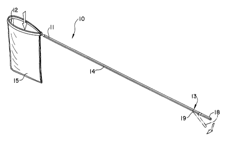
Referring to the Figures, Figure 1 illustrates a
medical device at 10 representing a preferred embodiment
of this invention. The device includes a continuous
ETH $03
-~ 5 -
filamentary strand 11 having a distal noose portion 12 and
a proximal free end portion 13. Strand 11 can be prepared
from any conventional surgical suture material, e.g.
nylon, silk, steel, catgut, and conventional bioabsorbable
suture materials such as polymers and copolymers of
lactide, glycolide, para-dioganone and trimethylene
carbonate. Surgical bag 15 having an opening therein for
placement of bodily tissue is attached to distal noose
portion 12 along substantially the entire perimeter of the
apen end of the bag by engagement with substantially the
entire length of distal noose portion 12. The free end
portion 13 of continuous filamentary strand 11 is enclased
within a generally rigid, longitudinal tubular sleeve 14,
often referred to in this art as a cannula. Tubular
sleeve 14 has a frangible portion 18 at its proximal end
and a score line 19 disposed at the distal end of the
frangible portion 18. Continuous filamentary strand 11 is
adhesively attached within the frangible portion 18 of the
tubular sleeve 14 with a conventional adhesive, e.g. epoxy.
As illustrated in more detail at Figure 3, the noose
portion 12 of continuous filamentary strand 11 is
sealingly engaged within a channel 20 running along the
circumferential length of the open end of the surgical bag
15. rn this embodiment, channel 20 is formed by folding
the top portion of the bag over about itself and sealed
along a lower portion of bag 15 to create a top flap
portion 15a. In like manner, bag 15 can also have a
peripheral edge seal 24 having a closed bottom portion 15b
if a hermetic seal is reduired for the particular surgical
application. Still referring to Figure 3, the noose
portion 12 of continuous filamentary strand 11 is secured
with a slip knot 17, which allows movement of the free end
portion 13 of the continuous filamentary strand 11
proximally and prevents the noose portion 12 from
ETH 803
- 6 -
loosening when engaged about bodily tissue. hongitudinal
tubular sleeve 19 has a tapered distal end 16 which
prevents slip knot 17 from passing through tubular sleeve
19 during use.
Surgical bag 15 can be constructed of a wide variety
of materials, but generally the bag used should be
biocompatible and non-toxic to bodily tissue, and should
exhibit the requisite conformability so that it can
readily fit down a trocar. If the medical device is to be
used to morcellate and remove bodily tissue, then the bag
is desirably waterproof to prevent fragmented tissue from
escaping the bag. Additionally, for this application, the
bag should have a high tsar and burst resistance, a low
modulus and moderate elongation. Although a variety of
materials can be used for this purpose, the preferred
material of construction for the surgical bag for this
application is PEBAXT" block copolyetheramide.
Alternatively, if the bag is to be used for encapsulating
a fractured organ during surgical repair, then it may be
desirable to employ a bag which has a mesh network. See,
for example, U.S. Patent 4,928,375, which describes a
variety of pliable surgical materials well known in the
art for this application that can be fabricated into a
desired mesh structure.
Referring now to Figures 1 and 2 in combination, one
can see generally how the device is used to enclose bodily
tissue. In order to encapsulate bodily organ 24, as seen
in Figure 2, the user would first grip frangible portion
18 of tubular sleeve 14 with one hand and the remaining
portion of tubular sleeve 19 with the other hand. and then
snap apart the two pieces about score line 19. This
allows fox the continuous filamentary strand 11 to be
retracted through the longitudinal tubular sleeve 19 as
ETH 803
- 7
shown in Figure 2. Following this simple procedure, the
user could then place the surgical bag 15 about a desired
bodily organ 24, positioning the bag at the appropriate
location about bodily organ 24. To complete the
procedure, continuous filamentary strand 11 is pulled
proximally as shown by the arrow at Figure 2, causing the
distal noose portion 12 of strand 11 to close the open end
of surgical bag 15. As shown more clearly at Figure 3.
knot 17 is restrained by tapered end 16 of the
longitudinal tubular sleeve 14 while the user is pulling
strand 11 proximally and allows strand 11 to pass through
the tubular sleeve 14 so that the distal noose portion 12
may be closed about bodily organ 24. The knot
configuration must be such that once the distal noose
portion 12 is closed about bodily organ 24, it remains
closed and doss not loosen.
Referring now to Figure 4, the medical device of this
invention can be used in combination with introduces 23
and trocar 22 to facilitate its use during endoscopic
surgery. First. the surgical bag 15 is folded about the
axis of the proximal free end portion 13 of continuous
filamentary strand 11 so as to facilitate the insertion of
the medical device into introduces 21. After the medical
device is inserted into introduces 21, the introduces can
then be placed within an appropriately sized trocar 22 for
insertion into the desired bodily cavity. As shown in
Figures 5-7, the trocar 22 is introduced into a desired
bodily cavity until penetration of the desired bodily
tissue 23. As shown in Figure 7, once the trocar is
appropriately placed, the medical device of this invention
can be moved distally through introduces 21 and trocar 22
so as to cause surgical bag 15 to protrude from introduces
21 and into bodily tissue 23. Once bag 15 is placed
within desired bodily tissue 23 free of the confines of
FTH 803
-8-
the reducer and trocar, it can unfold as shown by the
arrows at Figures 5 and 7. After surgical bag 15 unfolds,
the user can then manipulate the device so as to place
unfolded surgical bag 15 about a desired bodily tissue,
and then the user can perform the procedure outlined above
to carry out the required operation.
Following the surgical operation, the bag 15 can
either be removed from the surgical site or be left intact
at the site, depending on the operative procedure
performed. For example, if bodily tissue is morcellated
within the bag 15, and therefore it becomes necessary to
remove the fragmented tissue from the body. then the bag
can be readily removed by pulling the tubular sleeve 14
15 proximally through introduces 21 and trocar 22 until the
entire medical device 10. including bag 15, has been
removed from the body. Alternatively. if bag 15 is
composed of a bioabsorbable surgical mesh, and the bag 15
is used to facilitate the repair of a damaged organ over
an extended period of time, it may be desirable to leave
bag 15 intact at the surgical site. This can be
accomplished simply by first cutting strand 11 at or near
the junction of distal noose portion 12 and free end
portion 13, and then pulling tubular sleeve 14 proximally
so as to remove the free end portion 13 of strand 11 from
introduces 21 and trocar 22 while leaving bag 15 intact
within the body.
Although only the most preferred surgical device of
this invention is described herein, numerous additional
embodiments will become apparent to those skilled in this
art, all of which are well within the scope and spirit of
the claimed invention.
ETH 803