Note: Descriptions are shown in the official language in which they were submitted.
2065~97
INTRAOCULAR LENS INJECTOR
The present invention relates to an intraocular lens
injector, and more particularly to an injector head and an
injector assembly for an intraocular lens injector, and a
method of so using the assembly.
In eye surgery for treating conditions such as natural eye
lens cataracts, a common procedure is to remove the cataracted
lens through an incision in the cornea of the eyeball, and
replace it by an artificial intraocular lens. The intraocular
lens, typically 6 mm in diameter, is usually temporarily
resiliently deformable, i.e. foldable into generally
cylindrical shape, by curling, etc., to reduce its girth, and
while kept folded is inserted through a corneal incision,
typically 3.5-4 mm long, to minimize patient trauma. The lens
has a lens body or optic, e.g. of ~oft material such as sili-
cone, with normally stiffer resiliently deformable position
fixation mean~ or haptics, e.g. of polypropylene, extending
therefrom to seat the lens in ~he eye.
2 ~ e~ 7
The haptics are kept in stable relation to the folded lens
body during insertion into the eye so as to pass without diffi-
culty through the incision. Once inserted, unfolding the lens
body and haptics in the confined space at the implantation site
must be controlled to avoid patient trauma from contact of
these expanding mechanical elements with the inner wall of the
cornea or other eye parts. Inserting the folded lens into an
eye so as to minimize patient trauma is very difficult. A tool
is needed to hold and insert the folded lens, requiring an
incision large enough to accommodate both. Often, a separate
retainer keeps the lens folded, so that the inserted retainer
and tool clutter the eye interior during lens unfolding, after
which the tool and retainer must be retrieved via the incision.
U.S. Pat. No. 4,573,998 to Mazzocco shows various tools to
insert a deformable lens through an incision into the eye by
pushing, stretching, ejecting or compressing technique. The
lens and/or tool grossly contact the incision during insertion
and the lens must be released carefully in the eye to keep the
expanding lens elements from injuring internal eye parts.
U.S. Pat. No. 4,906,247 to Fritch shows a deformable lens
held folded by forceps inserted in a stretchable plastic tube
so as to stretch the tube diameter and also squeeze the tube
around the folded lens like a mitten. Upon inserting the lens
into the eye, the forceps must release it with great care to
keep the expanding lens elements from injuring internal eye
parts.
U.S. Pat. No. 4, 911,714 to Poley shows a deformable lens
held folded by sutures connecting apertures on opposed edges of
the lens, or by integral lock means or adhesive on such edges,
for insertion by a first tool through a first incision into the
eye. A second tool inserted through a second incision is used
to cut and remove the sutures, unlock the lock means or break
the adhesive bond while the first tool holds the lens to keep
the expanding lens elements from injuring internal eye parts.
U.S. Pat. No. 4,917,680 to Poley shows a deformable lens
h ~
held folded by a severable retainer or band for insertion by a
first tool through a first incision into the eye. A second
tool inserted through a second incision is used to sever and
remove the band while the first tool holds the lens to keep the
expanding lens elements from injuring internal eye parts.
U.S. Pat. No. 4,976,716 to Cumming shows an insertion
device with telescoping lnner and outer tubes, and a plunger in
the inner tube, for a deformable lens having leading and
trailing haptics. Two opposed flexible, widely diverging,
inner fingers integral with the inner tube front are notched
near their free ends to form bendable tips. Two opposed rigid
outer fingers pivoted to the outer tube front are lockable to
hold the inner fingers in narrower diverging state when the
inner tube is retracted in the outer tube so that only its tips
protrude. Jaws on a lens loading tray squeeze and fold the
lens between the inner fingers so that the leading haptic
protrudes from their narrower diverging tips, and then the
outer fingers are locked.
The leading haptic and diverging finger tips are inserted
into the eye incision, and a lever and ratchet drive on the
device is used to move the inner tube and plunger forwardly of
the outer tube until the inner finger notches clear the
incision, enabling the stored force of the folded lens to bend
the inner finger tips outwardly of their widely diverging
position for partial lens unfolding. Further use of the drive
causes the plunger to push the lens beyond the inner finger
tips for complete lens unfolding. A spring return of the drive
is used to retract the plunger and inner tube from the
incision. The device is complex, costly and cumbersome to
operate, and its diverging fingers stress the incision or
require use of a larger incision. As the fingers do not
surround the folded lens, but only hold its diametrically
opposed girth portions, the outwardly projecting girth portions
therebetween can contact and stress the incision and inner wall
of the cornea during the insertion procedure.
206~97
Also, a device manufactured by Allergen Inc., known as
"The Prodigy," includes a lens injector having a straight,
rigid tubular portion whose tip is inserted into the incision
and through which the folded lens is injected into the eye.
The tip is not tapered and has no means to control unfolding of
the lens.
It would be desirable to insert a temporarily folded
intraocular lens through a minimum size incision into the eye
by an instrument that does not unduly stress or traumatize the
incision, permitting controlled gradual lens injection and
unfolding in the confined space within the eye, while avoiding
incision trauma from such stress or contact of the unfolding
lens with the inner wall of the cornea or other eye parts.
It is an object of the invention to overcome prior art
drawbacks, and to provide an intraocular lens injector having a
head for partial insertion into a minimum size eye incision to
inject a temporarily folded, i.e. deformable, intraocular lene
into the eye, and a device to push the folded lens through the
head, and a method of using the injector for controlled gradual
injection of the lens into the eye and simultaneous controlled
gradual unfolding thereof within the eye, 80 as to avoid
patient trauma from stress on the incision or contact of the
unfolding lens with the inner wall of the cornea or other eye
parts.
It is another object of the invention to provide auch an
injector of structurally simple parts, readily fabricated at
relatively low cost so a~ to be economically feasible for it to
be disposable.
According to the invention, an injector head is provided
for partial insertion into a minimum size eye incision to
inject a temporarily folded intraocular lens into the eye. The
head comprises a hollow cone having a base end and a generally
pointed end, and defines a plurality of circumferentially
2~7~7
distributed cantilever leaves convergingly tapering from the
base end to the generally pointed end. The leaves are
resiliently flexible for outward displacement from a normally
closed position in which they are adjacent each other to an
open position in which they are spread apart to allow the
folded lens to be injected through the cone into the eye.
The base end has a mounting formation adapted to mount the
head on an injector device adapted for controlled gradual
injection of the folded lens through the cone and into the eye.
The leaves in closed position are in generally side by side
abutting contact, and are sufficiently flexible for dis-
placement to open position under the urging contact force of
the folded lens being injected through the cone. The head may
be provided in sterile packaged form.
The invention also concerns an injector assembly
comprising the injector head and the injector device. The
device comprises a holder and a plunger. The holder has a
plunger end and a delivery end interconnected by a longitudinal
through bore adapted to hold the folded lens, and a
longitudinal slot generally parallel to the bore and
communicating the bore with the exterior of the holder. The
delivery end is connected to the base end of the cone for
connecting the bore with the cone. The plunger is insertable
into the bore ~rom the plunger end for controlled gradual
delivery of the lens from the delivery end into and through the
cone for controlled gradual injection of the lens into the eye
and simultaneous controlled gradual unfolding of the lens
within the eye.
- The holder slot preferably has a V-shaped entrance.
Typically, a lens inserter is included having a longitudinal
contact edge for forcing the lens in unfolded state at the
exterior of the holder, against and inwardly through the slot
into the bore, to provide the lens in folded state in the bore.
The inserter may include a plate portion having a bulbous
longitudinal contact edge, or a pin portion defining a round
2065~7
longitudinal contact edge. The assembly may be provided as a
kit containing the head, holder and plunger, and optionally the
inserter, in sterile packaged form.
According to the invention, a method of using the assembly
is also provided to inject the intraocular lens into the eye.
The method comprises placing a lens atop the longitudinal
slot, forcing the lens through the slot by applying a
longitudinal edge, e.g. of the inserter, thereto for folding
the lens in the holder bore, moving the plunger into the bore
from the plunger end, partially inserting the cone into a
minimum size eye incision, and gradually pushing the plunger
against the lens in the bore to deliver it into the cone and
urge it against the leaves to spread them apart without unduly
stressing the incision, for controlled gradual lens injection
into the eye and simultaneous controlled gradual lens unfolding
within the eye preparatory to implanting the lens therein.
Generally, the lens has a pair of opposed haptics, and is
provided in the bore with one haptic ahead of the lens as a
leading haptic and the other behind the lens as a trailing
haptic, so that the leading haptic is injected into the eye
ahead of the lens for movement away from the incision as the
lens enters and unfolds in the eye, followed by insertion of
the trailing haptic.
Other objects of the invention will become apparent from
the within specification and accompanying drawings, in which:
Fig. 1 is a perspective view of an assembly of an injector
head and an injector device therefor, together with a lens
inserter to insert the lens in the device, according to a first
embodiment of the invention;
Fig. 2 is a cross sectional view showing the loading of
the lens into the device via the inserter;
Fig. 3 is a side view, partially sectioned and broken
away, of the head and device, assembled for use with the lens
2065~g7
loaded therein in folded state;
Fig. 4 i9 a longitudinal sectional view of the head;
Fig. 5 is a view of the head, device and inserter as a
combined assembly in a sterile package;
Fig. 6 is a schematic view showing the lens as it unfolds
during injection via the head and device into the eye; and
Fig. 7 is a schematic view of an assembly of the head with
an injector device and a lens inserter according to a second
embodiment of the invention.
:' 10
Referring to the drawings, and initially to Figs. 1-6, an
assembly 1 is shown of an injector head 2 for partial insertion
into a minimum size eye incision to inject a temporarily folded
intraocular lens 11 into the eye, and an injector device 21 for
use with head 2, plus a lens inserter 32 for inserting lens 11
into device 21, according to a first embodiment of the
invention.
Lens 11 is conventional, having a temporarily foldable,
i.e. resiliently deformable, optic or lens body 12 and a pair
of opposed resiliently deformable position fixation means or
: haptics for seating the lens in the eye, including a leading
haptic 13 and a trailing haptic 14. Lens 11 is capable of
being temporarily folded under compression force into a
compact, e.g. cylindrical, shape of reduced girth for
facilitated insertion through a minimum size incision into the
eye, and of unfolding to original undeformed state upon release
of such compression force.
Head 2 is formed of a hollow split cone 3 with a fixed
base end 4 and a generally pointed, openable tip end 5. Cone 3
defines a plurality of, e.g. four, circumferentially
distributed, cantilever leaves 6 convergingly tapering from
base end 4 to tip end 5, formed by slits 7. Leaves 6 are
resiliently flexible for outward displacement from a normally
closed position adjacent each other (Figs. 3 and 4), e.g. in
,:
2 ~ 7
side by side abutting contact, to an open position in which
they are spread apart to allow lens 11 to be injected through
cone 3 into the eye (Fig. 6). Leaves 6 are sufficiently
flexible for displacement to open position under the urging
contact force of lens 11 as it is being injected through cone
3.
Device 21 includes a holder 22 and a plunger 23. Holder
22 has a plunger end 24 and a delivery end 25 interconnected by
a longitudinal through bore 26 to hold folded lens 11.
Base end 4 has a mounting formation, e.g. a collar 8 with
internal threads 9 in the interior 10 of cone 3. Delivery end
25 has a counterpart mounting formation engageable with such
mounting formation, e.g. a neck 27 with external threads 28
mating with threads 9 on collar 8 (Fig. 3), to mount head 2 on
device 21 and connect bore 26 with interior 10. The mounting
formation and counterpart formation may be of any other
suitable form, such as a rib on one of these parts and a groove
on the other, sized for snap fit releasable interlocking to
mount cone 3 on holder 22 and communicate interior 10 with bore
26. Also, base end 4 may be integrally connected to delivery
end 25.
Plunger 23 is inserted in bore 26 via plunger end 24 for
controlled gradual delivery of lens 11 from delivery end 25
into and through interior 10 for controlled gradual injection
into the eye and simultaneous controlled gradual unfolding
therein. Bore 26 may be of circular or other suitable, e.g.
oval or polygonal, cross sectional shape for holding and
delivering lens 11 in folded state, with plunger 23 being of
conforming shape.
To load lens 11, holder 22 has a longitudinal slot 29
generally parallel to bore 26 and which communicates bore 26
with the exterior of holder 22. Slot 29 may extend from just
behind delivery end 25 to the extreme edge of plunger end 24.
As shown in Fig. 1, a lens inserter 32, e.g. formed as a
T-shaped member by a horizontal press block 33 and a vertical
.
2~r~rl
plate 34 attached to its underside, is included to force lens
11 in unfolded state at the exterior of holder 22, against and
inwardly through slot 29 into bore 26 to load lens 11 in folded
state therein. Lens 11 is protected from damage during such
loading by providing slot 29 with a V-shaped entrance 30 (Fig.
2), and the bottom of plate 34 with a bulbous longitudinal
contact edge 35. Plate 34 and edge 35 are desirably
coextensive with slot 29.
Lens 11 may be loaded into holder 22 by pressing inserter
32 with one hand in the direction of arrow 36 against unfolded
lens 11 resting across slot 29, while palming holder 22 in the
other hand. By providing holder 22 with a polygonal external
profile, e.g. a rectangular block shape 31 as shown in phantom
in Fig. 2, holder 22 may be placed on a table or other support
and lens 11 loaded by pressing inserter 32 with only one hand
to fold lens 11 into bore 26 via slot 29. Slot 29 is sized to
receive the composite thickness of contact edge 35, plate 34
and the folded thickness of lens 11, for inserting lens 11 into
bore 26 without hindrance or damage.
Once lens 11 is loaded in holder 22 and inserter 32 is
removed, the plunger shank 37 is inserted in bore 26 via
plunger end 24 and pushed by the plunger handle 38 in the
direction of arrow 39 until plunger 23 forces trailing haptic
14 forward and abuts lens body 12. Head 2 is then mounted on
25 holder 22 to form assembly 1 (Fig. 3). Head 2 is partially
inserted into the incision 89 in the cornea 81 (shown in
phantom in Fig. 3). Plunger 23 is pushed in the direction of
arrow 39 to force lens 11 from bore 26 into interior 10, and
against surrounding leaves 6 to spread them apart for
controlled gradual injecting of lens 11 into the eye and simul-
taneous controlled gradual unfolding as it exits from head 2
(Fig. 6).
Head 2 may be provided alone, or with the components of
device 21, optionally including inserter 32, in kit form, in a
sterile package 41, e.g. with a separation 42 providing head 2
2 ~ 7
in a sub-package 43 distinct from holder 22 and plunger 23, and
optionally inserter 32 (Fig. 5).
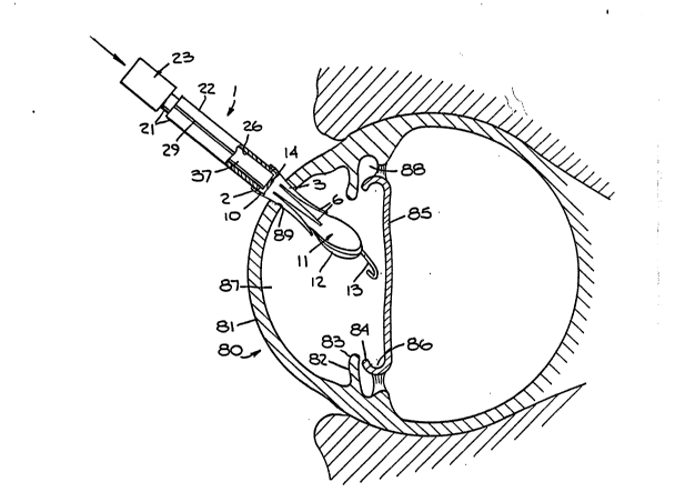
As shown in Fig. 6, the pertinent parts of the human
eyeball 80 include the cornea 81, the iris 82 with its central
opening or pupil 83, the remainder of the anterior lens capsule
84 after extracapsulary removal of a cataracted natural lens,
and the posterior lens capsule 85, such that posterior capsule
85 defines a cul-de-sac 86 at its peripheral margins that is
formed between anterior and posterior capsules 84,85. The
aqueous humor zone between cornea 81 and posterior capsule 85
- is divided by iris 82 into an anterior chamber 87 and a
posterior chamber 88.
Haptics 13,14 may seat in cul-de-sac 86, between anterior
and posterior capsules 84,55, to position lens 11 so that lens
body 12 performs its light focusing function.
Typically, lens body 12 has a diameter of about 5-6 mm and
a thickness of about 0.4 mm, and haptics 13,14 have a thickness
of about 0.2 mm and a width of about 1.2 mm. Haptics 13,14 may
have a composite expanded length in undeformed state of about
13 mm from the outer edge of one haptic to the diametrically
opposite outer edge of the other. As folded, lens body 12
typically has a girth diameter of about 3 mm, permitting its
facile insertion through an eye incision at most about 3.5 mm
long.
Since folded lens 11 is of bowed shape as loaded in bore
26 (shown in phantom in Fig. 2), the diameter of bore 26 may
i roughly equal the, e.g. about 3 mm, girth diameter of folded
lens body 12, enabling lens 11 to be pushed by plunger 23 along
bore 26 and into cone interior 10 easily and without hindrance
or damage.
Lens body 12 may be of any suitable temporarily
resiliently deformable light focusing optic serving material
that is sufficiently soft for the desired folding and also non-
toxic and eye fluid compatible such as silicone, with haptics
13,14 typically being of slightly stiffer resiliently
2~7~ 7
11
deformable material such as polypropylene. Lens body 12 must
have a memory, so that when folded to reduce its girth, it will
return readily to unfolded state, so long as promptly inserted
through the incision for unfolding in the eye, thus insuring
against loss of such memory. Of course, the lens folding and
injection procedures are effected under sterile conditions.
Such folding must be done during or just prior to surgery
to avoid the loss of "memory" of the lens which would take
place if "folded" by the manufacturer days or weeks before
surgery.
As used herein, "folding" means doubling, rolling,
curling, gathering, crinkling, and like type compressing of
lens body 12 onto itself one or more times to reduce its com-
posite girth.
As shown in Fig. 6, to inject lens 11, a minimum size
corneal incision 89 is made by the surgeon, e.g. about 3.5 mm
long, just sufficient to accommodate the reduced girth of
folded lens body 12. Then, holding assembly 1 with head 2
adjacent incision 89, cone 3 is inserted partially thereinto so
as not to stress unduly incision 89, and plunger 23 is
gradually pushed in bore 26 to force lens 11 into interior 10
and against surrounding leaves 6 to cause them to spread apart
as lens 11 starts to unfold in the eye. As the leading end 100
of cone 3 is located inwardly of incision 89, the spreading of
cantilever leaves 6 occurs without stressing incision 89.
Continued gradual pushing of plunger 23 results in injec-
tion of lens 11 into the eye and its gradual unfolding to
normal state. During this unfolding, the free ends of leaves 6
readily distend outwardly beyond the normal confines of cone 3
without stressing incision 89 as leaves 6 inwardly clear
incision 89. Trailing haptic 14 may remain outside incision 89
and assembly 1 simply removed to release cone 3 therefrom,
whereupon forceps are used to l'snakell haptic 14 through
incision 89 and seat lens 11 in the eye in the usual way.
Assembly 1 thus injects lens 11 into the eye without
2 ~ r~
12
inserting head 2 fully into incision 89. Forceps are only used
to insert trailing haptic 14 fully and seat lens 11 in the eye
after head 2 is removed from incision 89, thus avoiding stress
on the incision. As lens 11 is controlled by the gradual
movement of plunger 23, it is gradually injected and gradually
unfolds in controlled manner by the surrounding leaves 6, thus
preventing the lens from bursting open, in uncontrolled manner,
from its closed state, and in turn avoiding contact of the
entering and unfolding lens 11 with the inner wall of cornea 81
or other eye parts.
~eretofore, when a tool was used to insert a folded lens,
it fully occupied the incision along with the lens, causing
patient trauma by stressing the incision or by increasing its
size.
In using assembly 1, preliminarily lens 11 is provided in
folded state in bore 26, formation 8 is engaged with
counterpart formation 27 to connect cone 3 to bore 26, and
plunger 23 is inserted in bore 26 via plunger end 24. Then,
cone 3 is partially inserted into incision 89 so as to avoid
stressing it, and plunger 23 gradually pushed against lens 11
in bore 26 to deliver it to cone 3 and urge it against leaves 6
to spread them apart without stressing the incision, for
controlled gradual injection of lens 11 into the eye and simul-
` taneous controlled gradual unfolding of it therein.
Typically, leading haptic 13 is injected ahead of lens
body 12 and moves away from incision 89 as lens 11 enters and
unfolds in the eye, followed by trailing haptic 14 insertion
and lens 11 implanting by seating haptics 13,14 against
adjacent eye tissue.
Assembly 1 facilitates exploitation of the minimum size
`~ incision used for extracapsular removal of the natural lens, as
head 2 may be partially inserted in that same incision to
inject lens 11 into the eye. This is significant as the
smaller the incision size the less the patient trauma, includ-
ing pain and discomfort then and later, not only due to the
.; .
13
incision itself but also to the number and/or size oE any
needed sutures.
Referring to Fig. 7, a second embodiment of an assembly 50
is shown having the same head 2 and plunger 23 as in the
embodiment of Figs. 1-6, but a holder 52 of different type
forming an injector device 51 with plunger 23. Holder 52 has a
plunger end 53 and a delivery end 54 interconnected by a longi-
tudinal through bore 55 to hold folded lens 11. Delivery end
54 has a counterpart mounting formation engageable with
mounting formation 8, e.g. a neck 56 with external threads (not
shown), mating with threads 9, to mount head 2 on device 51 and
connect bore 55 with interior 10. Plunger 23 is inserted in
bore 55 via plunger end 53 for delivery of lens 11 from
delivery end 54 into and through interior 10, for use of device
50 in the same way as device 21.
To load lens 11, holder 52 has a longitudinal slot 57
generally parallel to bore 55 and which communicates bore 55
with the exterior of holder 52. Holder 52 is of rectangular
external profile or shape and longer than holder 22 to accommo-
date a suitably shaped, e.g. rectangular, recess 59 at the rear
end of slot 57 and communicating therewith. Slot 57 may extend
from just behind delivery end 54 to recess 59. Recess 59
communicates with the rear face of plunger end 53 via a bore
extension 55a.
A lens inserter 60, e.g. formed by a rear press block 61
and a horizontally extending front pin 62 attached thereto, is
included to force unfolded lens 11 at the exterior of holder
52, against and inwardly through slot 57 into bore 55 to load
lens 11 in folded state therein. Lens 11 is protected from
damage during such loading by providing slot 57 with a V-shaped
entrance 58 and pin 62 with a round cross section forming a
longitudinal contact edge 63. Pin 62 is desirably coextensive
with slot 57. Press block 61 is sized and shaped for play-
free, sliding fit in recess 59 for downward movement therein to
move pin 62 via slot 57 into bore 55. Bore extension 55a is
2 ~
14
sized to accommodate press block 61 for rearward removal of
inserter 60 therethrough after lens 11 is loaded in bore 55.
Holder 52 has a rectangular external profile to facilitate
loading of lens 11, by resting holder 52 on a table or other
support, and using one hand to force press block 61 downwardly
in a manner similar to the loading of holder 22. Slot 57 is
sized to receive the composite thickness of pin 62 and the
folded thickness of lens 11 to insert lens 11 into bore 55
without hindrance or damage. Lens 11 is loaded in holder 52 in
folded or otherwise compacted, e.g. cylindrically rolled, state
to reduce its composite girth, and is delivered without
hindrance or damage in that state from holder bore 55 to
interior 10 for insertion via head 2 through incision 89 into
the eye.
Once lens 11 is loaded in holder 52, inserter 60 is pulled
out rearwardly. Then, plunger shank 37 is inserted in bore 55
via plunger end 24 for the above described purposes, and head 2
is mounted on holder 52 to form assembly 50.
Head 2 may also be mounted on the tubular tip of a lens
insertion instrument, like the aforesaid Allergen Inc. device
known as "The Prodigy," suitably modified for connecting head 2
thereon, for use with plunger 23, to achieve the purposes of
the present invention.
Head 2 may be of any suitable non-toxic material, e.g.
rigid or resiliently flexible plastic, with at least leaves 6
being of resiliently flexible material, e.g. resiliently
flexible plastic. Holders 22 and 52, plunger 23 and inserters
32 and 60, may be of suitable rigid material, e.g. metal or
plastic such as Teflon. Head 2 may be a single use disposable
30 part, and holders 22 and 52, plunger 23 and inserters 32 and 60
may be reusable parts.
Clearly, the injector head, injector device and lens
inserter of the invention are structurally simple parts that
are readily fabricated at relatively low cost.
The specification and drawings are set forth by way of
2 ~ 7
illustration and not limitation, and various modifications may
be made therein without departing from the spirit of the
invention which is to be limited solely by the scope of the
claims.