Note: Descriptions are shown in the official language in which they were submitted.
-1-
MUI,'rIPLF, ELECTRODE S'~IP
This application is a continuatioll-in-part of United States Patent
Application Serial No. 07/687,302, the benefit of the filing date of which is hereby
claimed under 35 U.S.C. 120. United States Patent Application Serial
No. 07/687,302 is hereby incorporated by reference.
Field of the Invention
The invention relates to monitoring systems and, in particular, to an
electrode strip ~for use in monitoring electrical activities of a living body.
Back~round of the Invention
Conventional electrocardiography is concerned with the measurement and
analysis of voltage potential readings taken from a limited number of anatomically
defined locations. The voltages between various locations are cornbined to form
electrocardiograph (ECG) leads that are represented as waveforms and are
compared to clinically developed criteria to diagnose or classify the state of a1~ person's heart. One type of conventional electrocardiographic system has focused
on the application of ten electrodes to a person's skin; six across the precordial or
chest area of the person and one on each of the arms and legs. This type of system
is typically called the standard l~-lead ECG system. The electrodes are commonlyattached to the body by a conductive gel with;n an adhesive structure, or by a gel
which is both conductive and adhesive.
More recently, electrocardiologists have been experimenting with a body
surface potential mapping technique as a tool in scientific investigations and in
improving clinical diagnosis of heart disease. In body surface potential mapping, a
large number of electrodes are applied to a person's torso to obtain an estimate of
the total body surface distribution of cardiac-generated potentials. This distribution
is commonly displayed as a series of isopotential contours plotted on a map thatrepresents the person's torso. The resultant isopotential map is then evaluated for
~ PHYS\61)n~P.DOC
.
,
. : -
-2- 2~13~;
the presence of features representing the particular cardiac characteristic of
interest.
Proper electrode placement is a major concern in electrocardiography.
More particularly, to allow a person's ECG data to be meaningfully compared to
S clinical data obtained from known populations, the electrode readings must be
made at uniformly defined, anatomical locations. Proper place~lent poses
difflCUltieS, in part, because the electrodes must be positioned on people of
different sizes. In body surface mapping, the desired electrode sites are arrange~
in a number of columns and rows, with sorne mapping systems utili~ing as many
10 as 2a,0 body surface electrodes. Th~ls, proper electrode placement rnay be further
complicated by the large number of electrodes to be attached.
In an attempt to alleviate electrode placement problems, a number of
electrocarcliograph electrode systems have been developed One type of system
simply uses individual electrodes whose relative positions are unconstra~ned by the
15 separate and distinct conductive wires that couple the electrodes to a cable that is
connected to monitoring equipment. Thus, this system allows individual
positioning of the electrodes upon the subject person. A second type of system
provides a number of electrodes directly attached to a cable, with differently
proportioned electrode-cable sets used with different-sized bodies. Other systems
20 implement a cable or harness whereby individual electrodes attached thereto can be
selectively positioned along the cable or harness structure. In one device, the
electrodes are connected with spring clips to the harness allowing individual
electrodes to be slidingly positioned along the harness.
The electrode arrangements described above are generally cumbersome to
25 use and are often relatively expensive. The time required for proper placement
with the more cumbersome prior art systems can be particularly important in
emergency situations or when a large number of electrodes are re~uired, for
example, to perform body surface mapping. Care must be exercised with a system
utilizing a separat& lead for each individual electrode so that individual electrodes
30 do not become entangled, a problem that can increase the chance that any given
electrode will be placed in the wrong position, particularly in emergency
situations. If differently-sized electrode-cable sets are to be used to compensate for
differences in body sizes, an electrocardiologist must have electrode-cable sets of
several sizes at his or her fingertips. More important, the person charged with
35 placement of electrodes is also required to select the proper size and accurately
place the electrodes onto the body in a minimum amount of time. Even then, the
PlrYS\60nAP.DOC
,
'
2 ~ 3 ~;
-3 -
electrode-cable set selected may not ~llow accurate electrode placement on persons
between two sizes or at each end of the spectrum of average-sized bodies. Devices
utilizing a scheme whereby the individual electrodes can be slidably positioned
along an electrode cable or harness are disadvantageous in their bulk and
S complexity, and again, are not particularly well suited for body surface potential
mapping because of the large number of electrodes required.
As can be seen, there is a continuing need to provide an electrode device
which allows accurate and timely placement of individual electrodes on the body of
a person, whether it be conventional electrocardiography or a technique utilizing
10 body surface potential mapping, while reducing the complexity ancl cost of the
device.
Summarv of the Invention
An electrode strip in accordance with the present invention is a unitary strip
for measuring the electrical activities of the heart or other bioelectrical events of a
lS body while still providing a degree of flexibility in the positioning of individual
electrodes. A plurality of regions of extensibility in the strip provide adaptive
spacing between electrodes. The electrode strip is a potentially disposable
alternative to costlier and less manageable cables known in the art. In addition, a
number of electrode strips can be configured to be simultaneously and conveniently
20 placed on a patient for use in applications such as body surface potential mapping.
The electrode strip includes a substantially inextendible substrate, a
plurality of electrode sites defined on the substrate, and a region of extensibility
defined in the substrate between a pair of adjacent electrode sites to allow selective
positioning of the adjacent electrode sites on the body. The electrode strip further
25 includes conductive elements for providing an electrical path to each electrode site.
In a preferred embodiment of the invention, the electrode strip includes a
layer of malleable material attached to the substrate. The malleable material
provides nonelastic extensibility out of the inextendible substrate. The electrode
strip includes a plurality of spaced-apart conductors, each one of which extends30 from the connector area to an individual conductive element at each of the
electrode sites. The electrode strip further includes a plurality of dielectric cover
layers~ a different one of which extends over each conductor.
In one disclosed embodiment of the invention, each region of extensibility
is formed by a plurality of transverse folds in the electrode strip to allow adaptive
35 spacing between adjacent apertures. Each fold, in cross section, defines a
~ PHYS~P.DOC
-4 -
U-shaped section in the substrate Other illustrative configurations lhat can be
employed as the regions of extensibility are disclosed.
In currently preferred embodiments of the invention, the conductors are
formed on a substrate of polyester resin. Each electrode site is connected to a
5 conductive gel pad which has an adhesive surtace to contact a body. A protective
release liner is included to protect the adhesive surface of each gel pad prior to
attaching the strip to the body.
Brief Description of the Drawin~s
The various ~eatures and advantages of the invention will be understood in
10 view of the following detailed description taken in conjunction with the following
drawings in which:
FIGURE 1 is a perspective view of a ~Irst exemplary embodirnent of an
electrode strip of the present invention shown in an operative position on the chest
area of a person;
FIGURE 2 is a lower, patient-side view of the electrode strip of FIGURE 1
prior to the formation of regions of extensibility between adjacent apertures;
FIGU:RE 3 is an enlarged view of an electrode region (substrate) of the
electrode strip shown in FIGURE 2;
FIGURE 4 is a partial perspective view of the electrode strip depicted in
FIGURE 2 after the electrode strip has been formed to provide regions of
extensibility between adjacent apertures;
FIGURE 5 is a partial perspective view of a second embodiment of an
electrode strip of the present invention;
FIGURE 6 is a partial perspective view of a third embodiment of an
electrode strip of the present invention;
FIGURE 7A is a partial perspective view of a fourth embodiment of an
electrode strip of the present invention;
FIGURE 7B is a partial perspective view of the electrode strip of
FIGURE 7A after the regions of extensibility are partially expanded;
FIGURE 8A is a partial perspective view of a fifth embodiment of an
electrode strip of the present invention;
FIGURE 813 is a partial perspective view of ihe electrode strip of
FIGURE 8A after the regions of extensibility are partially expanded;
FIGURE 9A is a partial perspective view of a sixth embodiment of an
electrode strip of the present invention;
PHYs~nA~
1 8 ~
FIGURE 9B is a partial perspective view of the electrode strip of
FIGURE 9A after the regions of extensibility are partially expanded;
FI(3URE 10 is a partial perspective view of the electrode strip of
FIGURE 1 illustrating the use of an adhesive conductive gel pad to interface the5 electrode strip with the body of a person;
FIGURE 11 is a partial perspective view of the electrode strip of
FIGURE 10 where the ends of each pad opposite the electrode contact with the
strip are allowed to remain free of attachment to the strip;
FIGURE 12 is a patient side view of an electrode strip substrate which
10 includes an alternative pattern of apertllres, and hence, electrodes;
FIGURE 13 is a perspective view of a number of the electrocle strips of
FIGURE 1 shown in an operative position on the chest area of a person;
FIGURE 14 is a perspective view of a second exemplary embodiment of an
electrode strip of the present invention shown in an operative position on a person,
15 with electrodes located at a plurality of precordial and limb sites;
FIGURE 15 is a top elevation view of the electrode strip of FIGUE~E 14
prior to the formation of regions of extensibility between adjacent electrode sites;
FIGURE 16 is an elevation view of the electrode strip of FIGURE 14;
FIGURE 17 is an exploded view of a section of the electrode strip of
20 FIGURE 14;
FIGIJRE 1~ illustrates an alternative means for connecting the electrode
sites of the electrode strip of FIGURE 1 or FI(3URE 14 to monitoring equipment;
FIGURE 19 is a perspective view of a body surface potential mapping
assembly utilizing a number of electrode strips in accordance with the invention;
FIGURE 20 is a schematic view of the body surface potential mapping
assembly shown in FIGURE 19 and illustrating the placement of the electrode sites
along a plane superimposed on a person;
FIGURE 21 is a perspective view of a connector used to couple the
electrode strip to medical and diagnostic equipment; and
FIGURE 22 is an exploded perspective view of the connector of
PIGURE 21.
~:~ Detai ed Description
In accordance with the present invention, the electrode strip provides a
relatively inexpensive and potentially disposable device for measuring the activities
35 of the heart and other muscles and organs of a body while including electrodes
which can be selectively positioned to accommodate different-sized bodies.
~:~
PHYS\6072~P. UOC
2 ~3 ~
-6-
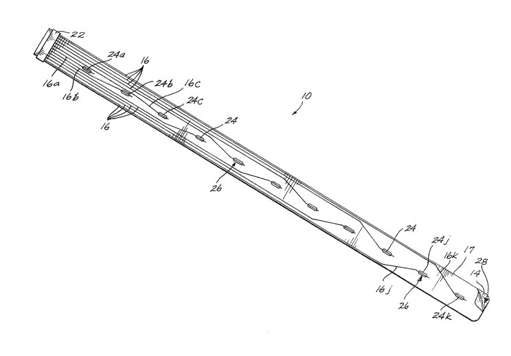
FIGURES 1-2 ill~lstrate a ~Irst exemplary embodiment in accordance with the
invention. An electrode strip 10 includes ~n elongate substrate 14, a plurality of
spaced apart conductors 16 that extend along one surface of substrate 14, and aninsulating cover layer 17 that insulates all but a portion of each conductor 16.5 With reference to FIGURE 1, the conductors 16 couple electrical signals between a
person lg and various medical and therapeutic equipment, such as a monitoring
device 20, which is generally known in the art. ~s is shown, one end of electrode
strip 10 includes a connector 22 which is configured to mate with a cable extending
to the monitoring device 20 or other such equipment.
With reference again to FIGURE 2, the cover layer 17 includes a plurality
of apertures 24 witll each apertllre being positionecl over a portion of a different
one of the conductors 16 to allow electrical contact with the body of a person or
other living being. In the arrangement of FIC~URE 3, the depicted aperture 24 isof elongate oval geometry to form an electrode site 26 of corresponding shape.
Various other aperture shapes can be employed as long as the associated conductor
16 passes partially or entirely across the aperture.
The arrangement of conductors 16 as they extend from connector22 to
different ones of the electrode sites 26 may be varied as long as the conductors do
not make ele trical contact with one another. In the particular embodiment shownin FIGURE 2, the conductors are substantially parallel to one another and extendfrom connector 22 to apertures 24 which are longitudinally spaced apart along the
center width of substrate 14. As the conductors 16 approach individual
apertures 24, they generally taper toward the center width of the substrate to
intersect with the apertures. In that regard, the centermost conductor 16a
terminates at the aperture (24a) nearest the connector 22. Conductors 16b and 16c,
which lie acljacent the centermost conductor 16a, terminate at the two apertures(24b and 24c) which are second and third nearest the connector 22. The pairing of
conductors 16 with apertu~es 24 continues in the arrangement of FIGURE 2, such
that the outermost conductors 16j and 16k terminate at the apertures (24j and 24k)
furthest from connector 22.
The electrode strip 1~ is constructed by depositing or otherwise forming the
conductors 16 on a first surface 28 of the substrate 14. In this regard various
known processes such as painting, screen printing, vacuum coating or sputtering
can be used. The cover layer 17 in which the apertures 24 have been previously
cut is then affixed to the first surface28 of the substrate 14 by means of an
adhesive material. As an alternative method of forming the conductors 16, the
PHfS~P.DOC
2 ~ ~
-7-
substrate material may be clad with a layer of cond~lctive material in which theconductors are formed by conventional photolithographic and chemical etching
techniques. Preferably, the substrate 14 and cover layer 17 are formed of a
polyester resin such as that commercially available under the trade name Myla~,
5 each being on the order of 3 mils thick.
Substrate 14, including the conductors 16 and cover layer 17, is flexible but
substantially inextendible along its length. A plurality of sections or regions of
extensibility 30 are subsequently formed into the substrate between pairs of
adjacent electrode sites 26. Illustrative embodiments of the regions of
extensibility 30 are shown in FIGURES 4-9E3 The formation of materials of the
type employed in substrate 14 and cover layer 17 is generally understood by those
skilled in their use.
The regions of extensibility 30 as clepicted in FIGURES 4-6 are formed into
the strip 10 by (1) preshaping the strip using a jig, mandrel or other device; (2)
15 heating the strip while in the preshaped position; and ~3) cooling the strip
An illustrative formation process for the strip lO involves clamping the
strip 10 to a mandrel having a plurality of triangular-shaped sections similar to
those illustrated in FIGURE 4; immersing the strip in hot water at a temperaturebetween, for example, 180-212F; immersing the strip in cool water, for
20 example, between 45-60~; and, removing the strip from the mandrel. With
regard to FIGURE 5, each region can be formed by the same process using a jig inplace of the mandrel. The jig includes four posts wherein each post causes a 180
turn in the strip 10 when the strip is wrapped around the jig. As can be
appreciated, for this process to be used, the substrate 14 (and cover layer 17) must
25 be therrnally formable. The optimal parameters of the formation process are
dependent upon the particular material used in substrate 14 and cover layer 17.
The regions of extensibility 30 as depicted in FIGIJRES 7A-9B demonstrate
alternate methods of creating extensible regions. FIGIJRES 7A, 8A and 9A
illustrate alternate regions of extensibility 30 prior to extending the regions to
30 accommodate electrode placement on a body. FIGURES 7B, 8B and 9B illustrate
the regions of extensibility 30 in FIGIJRES 7A, 8A and 9A, respectively, after the
regions have been somewhat extended, as if they were positioned on a person.
FIGURES 7A-7B demonstrate the use of a band 37 of material attached to
the lower, patient-side surface of the electrode strip 10, between adjacent pairs of
35 the regions of extensibility 30. The bands 37 may be comprised of an elastic
ma$erial or of an inelastic material that will deform as the regions of extensibility
P~5~72AP.DO~
2 ~
-8-
are separated. FIGlJRES 8A-8B illustrate the llse of layers 41 of a material, such
as Yelcro~9, that separates from itself as the regions of extensibility 30 are
separated. In FIC;UR~S 9A-9E3, each region of extensibility 30 is formed by
doubling a section of the electrode strip lO over onto itself and holding the section
5 in place using a band 47 of material.
The regions of extensibility 30 as depicted in FIGURES 4-6 are resilient
and extendible sections between adjacent electrode sites 26, on the otherwise
inextendible substrate 14. The regions of extensibility 30 as depicted in
FIGURE~S 7A-7B can be both resilient and nonresilient extendible sections between
10 adjacent electrode sites 26 on the otherwise inextenclible substrate 14. Thus,
although the electrode sites 26 are normally separated by predefined distances, the
application of longitudinal force to the substrate 14 and regions of extensibility 30
allows the electrode site separation to be altered. As a result, the strip 10 is easily
adapted to individual body shapes and sizes while remaining an integral unit.
With reference to FlGUREi 4, in a first embodiment, each region of
extensibility 30 is a tri-fold, triangular-shaped section of substrate 14 formed by
three transverse curves or folds 32a, 32b, and 32c. When in the unextended,
quiescent state, the geometry of one of the regions 30 roughly parallels that of an
isosceles triangle. The angle cY of the fold forming the peak of the arc 32b (i.e.,
20 the angle opposite the base of the isosceles triangle) is less than 60 with the
angles ,B and ~ of the adjacent folds ~32a and 32c) being less than 120 each. The
angle a of fold 32b will increase (i.e., approach or exceed 60) when the electrode
strip is in effect stretched to increase the distance between acljacent electrode
sites 26; conversely, the angle cY will decrease to less than 60 when the adjacent
25 electrode sites 26 are moved toward one another. The angles of each fold 32
described offer a desirable degree of extendibility for each region of
extensibility 30.
With reference to FIGURE5, in a second embodiment, each region of
extensibility 30 includes four transverse folds 34 which collectively form a stubby
30 T-shaped section of substrate 14. More particularly, each fold 34 represents
a 180 turn in the substrate. The first fold 34a and fourth fold 34d collectively
form the vertical portion of the T, respectively. The folds 34b and 34c form thehorizontal portion of the T.
With reference to FIGURE 6, in a third embodiment, each region of
35 extensibility 30 includes two opposing spiral wound regions36. Each spiral
wound region 36 is formed by doubling a section of substrate 14 over onto itself
P~S~nUP.DOC
f~
and coiling the doubled-over region into a spiral having at least a half turn. In the
embodiment of FIGURE 6, the spirals forming each region of extensibility have onthe order of two and one-half turns. The spiral wound regions 36 of a particularregion of extensibility 30 will tend to unwind as the electrode strip lO is in effect
S stretched to increase the distance between adjacent electrode sites 26.
With reference to FIGURES 7A and 7B, in a fourth embodiment, each
region of extensibility 30 includes a fold in the electrode strip 10 that is maintained
by a band 37 of material. Lateral force applied to the electrode strip 10 causes the
band 37 to stretch between the electrode sites 26. The bands 37 are attached to the
10 electrode strip by, for example, applying an adhesive, or using heat to bond the
materials together. Band 37 stretches resiliently if a material such as natural
rubber is used or inelastically if a thin plastic sheet of polyethylene is used. The
bands 37 are preferably attached to the lower, patient-side surface of the electrode
strip lO as depicted. Alternatively, a pair of bands may be coupled to the sides of
15 each fold.
With reference to FIGURES 8A and 8B, in a fifth embodiment, each region
of extensibility 30 includes a fold in the electrode strip 10 that is maintained by
layers of material 41. Layers 41 are peeled apart when lateral force is applied to
electrode strip 10, as depicted in FIGURE 8B. Suitable materials for use as the
20 layers 41 include Velcro~D, an adhesive with poor self-stick properties, a
nonadhesive material with cohesive properties, or a fragile material which tearsapart when lateral force is applied to the strip. The layers are preferably
adhesively attached to the electrode strip 10.
With reference to FIGURES 9A and 9B, in a sixth embodiment, each
25 region of extensibility 30 is a section of the electrode striy that is doubled over
onto itself and held in place by a band 47. The band 47 is constructed from a part
of the substrate 14 itself, or may be a separate strip of material that is looped
around the electrode strip. In FIGURES 9A and 9B, each band comprises two
strips of the substrate 14 that are coupled together by extending one of the strips
30 through a slot 49 in the other strip. As an alternative, the two strips may be
coupled together by an adhesive bond.
The degree of adjustment that can be made to the spacing between adjacent
electrode sites 26 is dependent, first upon the distance between adjacent
apertures 24, and second by the shape, size, and complexity of the regions of
35 extensibility 30. In general, these factors can be varied during both the
manufactu~ing and forming processes of the strip 10 to tailor the strip to any
PHYS~P,~
2i~3.l~
-10-
desired application. For example, the embodiment of FIGURE 6 may be ~Iseful in
some situations because the relatively large amount of substrate 14 within each
spiral wound region 36 allows a great deal of latitude (in separation) when
positioning the electrode sites 26 on a body.
S The strip 10 can also be tailored by selecting the number of electrocle sites
to accommodate specific needs. As another example, a precordial strip requires
six electrode sites. To allow strip 10 to properly adjust to the general population in
a precordial application, tlle regions of extensibility 30 are con~lg~lred to provide a
longitudinal adjustment on the order of several centimeters between adjacent
electrode sites 26. Further, the nonlinear nature of the anatomically deflned
electrode locations for precordial rnonitoring reqllires that the regions of
extensibility 30 be formed to allow both longitudinal and curvilinear adjustment of
the electrode strip 10. A strip could be constructed in accordance with the
invention to meet these constraints by, for example, employing multiple folds
which when bent will conform in a curvilinear fashion similar to the spreading of a
fan
With reference to -FIGURE 10, the conductors 16 must be electrically
coupled to the body of a person or other being (i.e., in signal communication with
the body). One method of establishing this electrical contact is through a plurality
of eor~ductive gel pads 38 associated with the various electrode sites 26. Thus, the
electrode sites 26 do not directly contact the skin. Rather, the conductors 16
terminating at each electrode site 26 are coupled to the skin via the gel pads 38.
Each pad has adhesive properties on both oppositely disposed surfaces -- an upper
surface40 to attach the gel pad 3~ to the strip 10 and a lower surface 42 to
detachably mount the pad to the body of a person (not shown). Adhesive, ionically
conductis~e gels in this form are generally known in the art.
Once mounted to a person, the entire lower surface 42 of the gel pads 38
will provide ionic conductivity between the person's skin and the conductors 16.The impedance of the electrode/patient interface is determined primarily by the
area of the lower surface 42 and not the size of the electrode site 26. This property
allows the apertures ~4 and the conductors 16 to have a relatively small size
without affecting the strength of the signals monitored by the end equipment, for
example, the monitoring device 20 shown in FIGURE 1.
A desirable skin surface 3rea to obtain electrode readings is on the order of
one square inch (i.e., 6.45 square centimeters). Preferably~ the strip 10 is
provided with one-inch-square gel pads 38 which are preattached to the area
P~n,s~n,~P.Doc
f;
I I
surrounding each electrode site 26. An outer liner 44 protecting the adhesive onthe lower surface 42 of the pads 38 can then be removed just prior to attaching the
strip to the body. In one ernbodiment, shown in FIGURE lO, the protective outer
liner 44 is a single strip which covers the lower surfaces 42 of all of the gel
5 pads 38. In this embodisnent, removal of the single liner 44 will expose the lower
surface 42 of each gel pad 38 for placement on the body.
With reference to FIGURE 11, a second method of attaching the gel
pads 38 to the electrode strip 10 is illustrated. When applied to a person's body
(not shown), longitudinal extension of the strip 10 tends ts) cause the regions of the
10 substratel4 around the electrode sites26 to bend away from the body, i.e.,
presenting a concave surface to the person's body. To accommodate such bending
while maintaining electrical contact between the body and the electrode sites 26,
only a portion of the conductive gel pad 38 is attached to the electrode site 26.
More specifically, one end of the upper surface 40 of the gel pad 38is adhesively
15 attached to the substrate 14 at the aperture 24. 'I he remaining portion of the upper
surface 40 of gel pad 38, adjacent the electrode site 26, includes a strip 45 of paper
or other suitable material that prevents it from adhering to the electrode strip. The
entire lower surface 42 of the gel pad 38 is still attached to the person's body.
When the gel pad 38is adhesively joined to the person's ~ody it effectively couples
20 electrical signals to and from the body while still allowing the electrode site 26 to
bend away from the body. As previously noted, the protective outer liner 44
applied to the adhesive regions on the lower surface 42 must be removed prior toattaching the pad 38. Where adhesion of the pads 38 to sites 26 is limit~d by the
strips 45, it will be noted that removal of the outer liner 44 must proceecl starting
25 from the end of strip 10 that is opposite the limited adhesion end of pads 38.
As is discussed above, the arrangement of the conductors 16 as they extend
to the apertures 24 is not of critical importance. With reference to FIGURE 12, in
an alternative arrangement, each conductor 16 extends substantially the entire
length of the substrate 14, rather than having the individual conductors terminate
30 after reaching an aperture as in FIGURE 2. In this arrangement, the conductors 16
are substantially parallel to one another ancl to the length of the strip 10. The
conductors 16 are equally spaced apart across the width of the strip. The
corresponding apertures 24 associated with the conductors 16 are located at varying
widths along the strip 10 and thus are not in longitudinal alignment with the strip
35 as are the apertures of FIGURE 2. However, it should be noted that this
arrangement of apertures 24, and hence electrode sites 26, will not cause electrode
PN'fS\6~n~P.DOC
2 ~3 ~
-12-
placement problems with respect to the person's skin because the gel pads 38 arelongitudinally aligned with the strip 10 and it is the gel pads and not the actual
electrode sites 26, which contact the skin (as describecl above).
The alternative arrangement of conductors and apertures in FIC;URE 12
allows the substrate 14 and conductors 16 to be manufactured in a continuous
process with adjustments of the apertures' locations in the cover layer facilitating
particular applications of the strip. For exannple, some applications may require
the apertures to be spaced further apart or, conversely, closer to one another. This
is accomplished with the arrangement of ~I(iURE 12 simply by a(ljllsting the
longitudincll spacing between the apertures. In contrast, the embodiment shown in
FIGURE 2 may require an adjustment in leacl layout, as well as aperture positions,
to alter the substrate 14 for different applications.
Body surface potential mapping techniques often require the placement of a
large nllmber of electrodes on a person in an arrangement comprising any number
of columns and rows. With reference to FIGIJ~E 13, an exemplary arrangement
including a number of columns of electrodes is shown. Each column includes an
electrode strip. In that regard, a first electrode strip 50a is illustrated on the right
side of the person 18 and a sPcond electrode strip SOb is illustrated on the person's
left side. An electrode strip 50n (shown in phantom) representing the nth strip
attached to the person is also illustrated. The electrode strips 50 are substantially
similar to the electrode strip 10 of FIGURE 1. In that regard, each electrode
strip 50 includes a plurality s)f spaced-apart conductors 52 and a connector SD~ at
one end thereof.
The electrode strips 50 are joined through their connectors 54 to a plurality
of connectors 55 in a connector strip 56. The connectors 54 of the electrode
strips 50 cooperatively interact with the connectors 55 of the connector strip 56 to
couple electrical signals therebetween. Further, the connector strip 56 includes a
plurality of spaced-apart conductors (not shown) which couple electrical signalsbetween the conductors 5~ (and, hence, the person 18) and the monitoring
device 20. As is shown, the connector strip 56 includes a cable connector 58,
which is configured to mate with a cable extending to the monitoring device 20 or
other such equipment.
The connector strip 56 utilizes the technology of the present invention to
allow flexibility in the placement of the electrode strips 50. To this end, the
connector strip 56 includes a number of extensible regions 60 similar to the regions
of extensibility 30 of th electrode strip 10. Preferably, the extensible regions 60
~HYS~nAP.DOC
3 ~ 8
-13-
each include two opposing spiral wound regions 62 similar to the spiral wound
regions 36 of FIGURE 6. As will be appreciated by those skilled in the art, the
number of strips S5) depicted in FIGURE 13 can be increased to provide a larger
number of readings. Further, the length, width and shape of the connector strip 56
S can be adjusted to accommodate the number and arrangement of strips employed.
FIGURES 14-16 illustrate a second exemplary electrode strip 100
constructed in accordance with the invention. Electrode strip lO0 is tailore(l for
use with a standard 12-lead E~CG system, wherein six electrodes are placed acros.s
the chest of a person at precordial placement positions Vl-V6 and four electrodes
are placed on the arms and legs (or torso~ at right arm RA, left arrn LA, left
leg LL, and right leg RL placement positions. As is shown most clearly in
FIGURE 16, the electrode strip includes a plurality of regions of extensibility 102
that allow selective and adjustable spacing of the electrodes on the body of a
person 18. In contrast to the regions of extensibility 30 of electrode strip lO
(shown in FIGURE 1), the regions of extensibility 102 are better suited to provide
curvilinear bending, as well as longitudinal extension and contraction of the strip.
Thus, the required curvature of strip 100 between the precordial placement
positions is readily achieved.
A top, elevation view of the electrode strip lO0, prior to formation of the
regions of extensibility 102, is illustrated in FIGURE 15. The electrode strip 100
includes a substantially elongate substrate 104 that is separable into five
substantially elongate sections 104a-104e along cut-lines 106. It is assumed in
FIGURE 15 that the substrate 104 is transparent to allow the conductors and
electrode sites on the lower, patient-side of the strip to be clearly shown. Theentire substrate 104 may be formed of a single material and then cut along the cut-
lines 106. In a preferred use, the cut-lines 106 are only perforated and the
electrode strip 100 is packaged and sent to the end-user as a single, elongate piece,
with each substrate section 104a-104e inclucling the regions of extensibility 102
(not shown in FIGURl3 15). During placement of the device, the substrate
sections 104b-104e are then partially separated from each other at a first end 108
along the perforated cut-lines 106 and stretched to contact the limb placement
positions RA, LA, LL and RL, respectively.
In a preferred configuration, the substrate 104 includes an end segment 110
that extends perpendicularly from a second end 112 of the electrode strip. A
plurality of spaced-apart conductors 114 begin at the end segment 110~ extend
along the lower (patient-side) surface of the substrate 104, and terminate at spaced-
PHYS\60n.~P.DOC '
8 3';
-14-
apart locations along the electrode strip. Each conductor includes a circular-shaped
area at the end segment 110, providing a sufficient surface area to allow contact by
a connector clamp, described below. Opposite the connector end, i.e., at the
terminating end, each conductor includes an elongate oval-shaped area o~
5 conductive material. Preferably, a second elongate oval-shaped layer 116 of
conducti~e material is placed in electrical contact with the terminating end of each
conductor 114 to form electrode sites, i.e., precordial sites (Vl-V6) and limb sites
(RA, LA, LL and RL). Each oval-shaped layer 116 is then coupled to the person's
skin, pre~erably throu~h a conductive interface such as the gel pads 38, depicted
and described in reference to E~IGURES 10 and 11. The conductors 114, including
the circular-shaped beginning end and oval-shaped terminating end, are preferably
formed of silver ink tracings. F,xcluding the ends, a sllitable wiclth for the
conductors is a width on the order of 60 mils (1.5 mm).
With reference again to E~IGURE 14, the conductors (not shown) couple
electrical signals between the person 18 and meclical and therapcutic equipment,for example, monitoring device 20 via a cable 118. The cable 118 includes at least
ten conductive leads to couple each of the conductors of the electrode strip to the
monitonng device 20. The cable 118 is releasably coupled to the electrode strip by
a spring-loaded clamp 120 that is configured to mate with the end segment 110 ofthe strip. In one embodiment, the clamp includes at least ten conductive teeth, one
for each conductor, with each tooth being coupled to a di~ferent one of the
conductive leads in cable 118. When coupled to the end segment 110, the teeth
engage their respective conductors in the circular-shaped area of each conductor(shown in FIGU~E 15~ and are held against the conductors by the spring housed
within the clamp. The clamp 120 is described and illustrated in greater detail in
FIGURES 21 and 22 and accompanying text.
The particular arrangement of conductors 114, as they extend frorn end
segment 110 to the precordial and limb electrode sites, is not of substantial
importance as long as the conductors remain insulated from one another. In thP
particular embodiment shown in FIGURE 15, the conductors generally extend in a
parallel fashion frorn the end segment toward the electrode sites.
The conductors on substrate section 104a extend from the end segment 110
along the substrate until reaching individual precordial electrode sites V1-V6. In
that regard, the outermost conductors 114a and 114b (on substrate section 104a)
extend to sites V6 ~u~d V5, respectively. The conductors 114c and 114d, adjacent
PHYS~.DOC
2 ~ 8 ~i
,5
the outermost conductors, extend to sites Vl arld V3, respectively. The innermost
conductors, 114e and 114f, extend to sites V~, and V2, respectively.
The conductors on substrate sections 104b-10~e extend from the end
segment 110 to the limb sites RA, LA, LL and RI, respectively. The substrate
S sections are relatively narrow as they extend frorn the end segment 110 to each
limb site. At each limb site, however, the associated substrate section becomes
wider and has a subst~mtially square-shaped segment to accommodate the limb
sites, i.e., including the oval-shaped layers 116. As the conductors extend fromthe end segment and reach the square-shaped segments, they form a right-angle out
and away from the center of substrate 104a to intersect the limb sites RA, LA, LL
and RL.
Each of the conductors 114 is insulate(l trom the person by a separate
dielectric cover layer 122. The cover layers 122 are preferably formed of a UV
curable clielectric coating such as those manufactllred by Acheson Colloids
Company. The cover layers 122 are preferably deposited onto the electrode strip
through a single silk screening process whereby only those areas needing insulation
are covered. This method of dielectric placement is advantageous as it conservesdielectric material. As will be appreciated, the entire lower patient-side surface of
the electrode strip, with the exception of the electrode sites and the end of each
conductor, may be covered with a single dielectric layer if desired.
An exploded, sectional view of the first end 108 of the substrate 104a is
shown in FIGURE 17, including the conductors 114, oval-shaped layers 116 and
cover layers 122. Only three of the cover layers 122 are shown in FIGIJRE 17.
The cover layers are slightly wider than the conductors they insulate and extendfrom the end segment 110 to the oval-shaped layers 116. For example, if the
conductors are 60 mils (1.5 mm) wide, the cover layers 122 are preferably on theorder of 100 mils (2.5 mm) wide. As will be appreciated, the conductors may
alternatively be insulated by a continuous layer of dielectric having a plurality of
apertures extending orthogonally therethrough, and spaced along the dielectric at
locations corresponding to the oval-shaped layers 116, in a manner similar to
electrode strip 10 of FIGURE 1.
The electrocle strip 100 is constructed by depositing or othe~wise forming
the conductors 114 on a lower (patient-side) surface 124 of the substra~e 104. In
this regard, various known processes such as painting~ screen printing, vacuum
coating or sputtering can be used. As an alternative method of forming the
conductors 114, the substrate may be clad with a layer of conductiv~ material in
PllYS\amAP.DOC
3 ~
,~
which the conductors are formed by conventional photolithographic and chemical
etching techniques. The cover layers may also be formed using painting, screen
printing, vacuum coating or sputtering techniques.
The substrate 104 itself is preferably formed of a polyester resin, such as
S that commercially available under the tra(le name Mylar~, being on the order
of 3 mils (0.076 mm) thick. The substrate may also be formed of Kapton~D, or
other suitable material. The preferred length of the substrate section 104a is
23.75" (60 cm), with a distance of 3.75" (9.5 cm) between each precordial
electrode site Vl-V6. A suitable width for the substrate section 104a is 1.2"
(3 cm), excluding end segment 110. A suitable length for the substrate
sections 104b and 104e is 19.25" (49 cm). A suitable width for the substrate
sections 104b-104e is on the order of 0.3" (7.6 mm).
Once the conductors 114 have been formed on the substrate 1()4, the oval-
shaped layers 116 are deposited or otherwise placed onto the lower surface 124 of
the substrate, with a different layer 116 being in electrical contact with each
conductor 114. Although not depicted in FIGURE 15, the precordial and limb
sites may be ionically coupled to a person's skin through a conductive interface,
such as the conductive gel pads 38 shown in FIGURES 10 and 11. The oval-
shaped layers 116 provide better contact with a conductive interface such as the gel
pads 38, as opposed to simply having the end of the conductors 114 contact the
interface. The oval-shaped layers 116 may also be of other shapes, for example,
circular or ,of various thicknesses. The oval-shaped layers 116 are preferably of a
silver/silver chloride compound. The silver/silver chloride layer converts the ionic
current flow of the body into electron flow that the monitor can amplify in a
chemically reversible manner which is well known to those skilled in the art. The
conductors 114 could be formed of silver/silver chloride but are generally formed
of silver which is more conductive and does not represent a source of chloride ions
which would corrocle the connector. The silver by itself does not bidirectionally
transform ionic flow to electron flow. After deposition of the oval-shaped
layers 116, the cover layers 122 are deposited or otherwise formed on the
conductors 114 to insulate the portions of the conductors between the end
segment 110 and each oval-shaped layer 116.
After forrnation of the conductors 114, oval-shaped layers 116 and cover
layer 122, a band 126 of malleable meta~ or other material is attached to the upper
surface 128 of each substrate section 104a-104e, i.e., opposite the conductors 114
(only substrate sectilon 104a is shown in FIGUR 17). Each band 126 is somewhat
P~IYS~P.DOC
2 1~ ~ , 3 J 2 . j
-17-
narrower than the substrate section on which it is placeci. The band attachecl to the
substrate section 104a is significantly wider than the bands attached to substrate
sections 104b-104e, although the bands have not been illustrated in FIGURE 16 for
purposes of clarity in the illustration. In an actllal embodiment, the bands 126 are
S formed of a dead-soft alurninum on the order of 6 mils (0.15 mm) thick. Other
malleable metals or plastics may also be used.
The bands 126 are attached, for example, by inclu(ling a pressure-sensitive
adhesive layer on one side thereof and firmly pressing the adhesive layer against
the upper surface 128 of the clectrode strip. The band 126 neeci not be a
10 continuous band that extends the entire length of the substrate sections 104a-104e.
Rather, it is useful in some applications to have sections of the bcmd attached only
to the areas upon which regions of extensibility are to be formed. Thus, for
example, substrate section 104a would include five separate malleable bands, each
separated by one of the electrode sites Vl-V6.
The substrate 104, including the conductors 114, cover layers 122 and
bands 126, is flexible but substantially inextendible along its length prior to
formation of the regions of extensibility 102. The regions of extensibility 102 are
formed into the substrate section 104a between pairs of adjacent precordial
electrode sites Vl-V6. Preferably, the regions of extensibility 102 are also formed
into the substrate sections 104b-104e during the same process. However, it is
noted that the regions of ex~ensibility do not have to be formed into the substrate
sections 104b-104e, but formation therein is often a manufacturing convenience.
As will be appreciated, if regions of extensibility are not formed in the substrate
sections 104b-104e, the band 126 may be omitted from these sections, if desired.With reference again to FIGURE 17, the electrode strip 100 preferably
includes gel pads 38, similar to those depicted in FIGURE 10, that are preattached
to the area surrounding each electrode site. The gel pads 38 each have a lower
surface42 that provides ionic conductivity between the conductorslla. and a
person's skin. An outer liner 44 protects the adhesive lower surface of the gel
pads 38 and is removed just prior to attaching the strip to a body.
A preferred arrangement of the regions of extensibility 102 is shown in
FIGURE 16, wherein each region includes four transverse, upwardly extending
folds 13û in the substrate/band assembly. In cross section, each fold 130 defines
an inverted, U-shaped section of substrate. The bands 126 retain the initially
formed four-fold configuration of each region of extensibility until acted upon
during placement of the electrode strip. Upon adjusting the regions of extensibility
P~IYS\~P.DOC
~ .
e~ C3 L ~ ~;
-18-
during placement of the strip, the regions will retain the adjusted form (as a result
of the bands 126), and thus the electrode sites will have a tendency to remain
where they are positionecl, even before coupling each site to the body. As will be
appreciated, other configurations of the regions of extensibility 102 m;~y be used.
For example, fewer or additional transverse folds 130 may be induced into the
substrate. Moreover, various shapes and sizes of folds may also be implemented to
form the regions of extensibility 102.
The regions of extensibility 102 are formed into the electrode strip 100
through mechanical processes. One process involves (1) aligning the strip alonK
the top of a toothed surface, (2) extending an arm downwar(lly between a pair ofadjacent teeth, thereby forcing the electrode strip into the groove formed by the
adjacent teeth and creating one of the transverse folds, (3) positioning the armabove the next pair of teeth, ancl (4) repeating steps (2) and (3) until each region of
extensibility ;s forrned. Other processes known and used for the general formation
of malleable material may also be used to form the regions of extensibility 102.The process of forming the regions of extensibility 102 can be compared to
formation of the regions of extensibility 30 shown in FIGIJRES 4-6. Formation ofthe regions of extensibility 30 requires the step of (1) preshaping the electrode
strip, (2) heating the strip while in the preshaped position, and (3) cooling the
strip. In contrast, heating is not required in the formation of the region~ of
extensibility 102; a desired shape is mechanically induced into the electrode strip,
wherein the shape is held by the malleable layer attached to the substrate. The
latter process has manufacturing advantages including that it is generally quicker
and does not require heating and cooling of the electrode strip.
As will be appreciated, formation of each fold 130 decreases the
longitudinal length of the strip, bringing the precordial electrode sites on either
side of the fold closer together. In the application of the electrode strip 10a to
person 181 the longitudinal distance between the electrode sites can then be
increased by deforming the regions of extensibility 102, i.e., stretching the
folds 130. Further, the strip may be bent laterally at the regions of extensibility by
holding one side of the strip and pulling the free end of the strip, on the opposite
side, in the direction of the desired bend. Thus, the resultant regions of
extensibility 102 allow extension and curvilinear motion between adjacent electrode
sites on the otherwise inextendible substrate 104.
There are a number of differences between the electrode strip 100 of
FIGURES 14-16 and the electrode strip 10 of FIGURE 1. The most significant
P~IYS~P.DOC
~ ~3 ~ 3 ~;
-19-
difference is with respect to the regions of extensibility that are formed in each
strip. The regions of extensibility 30 in strip 10 are resilient and have a tendency
to return back to their formed shape once longitudinal or c~lrvilinear pressure is
removed from the strip. In contrast, the regions of extensibility 102 in strip 100,
5 as a result of the bands 126, tend to conform to and retain any shape induced upon
them, for example, as the electrode strip 100 is manipulated to properly position
the precordial and limb sites on the body of a person. In effect, each region ofextensibility 102 has a memory-like feature that allows the strip 100 to be shaped
prior to actually connecting the electrode si~es to the person. This provides an10 opportunity for greater accuracy in electrode placement. Fllrther, as is illllstrated
most clearly in FIGURF, 16, the mllltifolded str~lcture of each region of
extensibility 102 provides for an accordion-like bending of the regions of
extensibility, whereby curves in the electrode strip are readily achieved along with
thc desired electrode site separations.
It is noted that the bands 126 may also act as a shielding layer to shield the
conductors 114 from electromagnetic waves if the bands 126 are connected to
ground. As is known in the art, highly sensitive medical instruments will typically
have shielded cables to block at least a portion of the spurious currents induced by
ambient electromagnetic waves. In a somewhat similar fashion, the bancls 126
could shield the conductors 11~ from electromagnetic interference, thereby
enhancing the accuracy of the ECG readings. The amount of shielding provided
by each band will generally depend upon the composition of the material used to
form the band, the dimension thereof, and mainly where it is connected to the
electrical ground reference.
FIG~JRE 18 is a partial illustration of a third exemplary embodiment of an
electrode strip in accordance with the invention. In the embodiment of
FIGURE 18, a variation of strip 100 is shown, including the substrate section 104a
and barld 126. lIowever, the conductors 114, oval-shaped layers 116 and cover
layers 12~ are not manufactured as part of the strip. Rather, the conductors andcover layers are in effect replaced by a plurality of electrode pads 140 and separate
and distinct cables 142 that carry electrical signals from the electrode pads 140 to
therapeutic and monitoring equipment. Although a variation of the electrode
strip 100 is depicted in FI(:;URE 18 and described below, the method shown and
described herein will work equally well with a variation of the electrode strip 10 of
FIGURE 1, i.e., one that does not include the conductors or cover layer but doesinclude an electrode element electrically connected to a tabular region.
PHYS~I'.DOC
2 ~ ~ ~3 .~
-~0-
An alternative embodiment to that depicted in F~GURE 18 would be an
extendible substrate that did not require the use of a plurality of transverse
folds 130. Examples of such a material might be Coban~ manufactured by 3M, a
natural rubber, or an inelastic plastically deformable material s~lch as thin
polyethylene sheet.
Each electrode pad 140 has adhesive layers on oppositely disposed surfaces:
an upper adhesive layer 1~4 to attach the electrode pad 140 to the electrode strip,
and a lower conductive adhesive layer 146 to detachably mount the pad to the body
of a person (not shown). The lower adhesive layer 146 is exposed by removing a
protective liner 148. Each electrode pad 1~0 further inclu(les a conductive tabular
region 150 that projects from the pad. An alligator clip 152, coupled to the
cable 14~, is then used to provide an electrical connection to the cable. A
conductive element within the electrode pad couples electrical signals from the
lower adhesive layer 146 (i.e., which provides a conductive interface with the
person's skin) to the tabular region 150.
In the embodiment of FIGURE 18, the electrocle sites are t;rst positioned
on the body of the person, and then each site is coupled to medical equipment
through a different one of the cables 142. As will be appreciated, the conductive
gel pads 38 of FIGURE 11 may be used as an alternative to the electrode pads 140if they are provided with a conductive tabular region to allow electrical connection
between the pad and cables 142. Use of the adhesive electrode pads 140 and
cables 142 is advantageous from a manufactllring standpoint in that the electrode
strip does not need conductors or the dielectric cover layers. However, use of the
pad/cable combination in lieu of the conductors is disadvantageous because care
must be exercised to ensure that the right cable is connected to the corresponding
electrode. ~rrors in the connection scheme can lead to incorrect computer analysis
of the results.
PIGURES 19 and 20 illustrate an exemplary body surface potential
mapping assembly 160 constructed in accordance with the present invention. As
will be appreciated, any number of other body surface potential mapping patternsmay also be realized.
The mapping assembly 160 includes29 electrode sites provided on four
separate electrode strips 162, 164, 166 and 168, each employing the basic
constructional techniques described in connection with the electrode strip 100 of
FI~iURE 14. Although not explicitly illustrated in FIC~URES 19 and 20, each
e1ectrode strip preferably includes (a) a substrate, (b) a plurality of conductors that
PHYS\60nAP.DOC
,: :
. .
~ 13~
extend frorn a connector segment 17û of each electrode strip, along a lower surface
of the substrate, to individual electrode sites, (c) a malleable band attached to an
upper surface of the substrate, (d) a plurality of cover layers that extend over the
conductors to insulate all but a portion of the conductors from the body of a
S person, (e) a silver/silver chloride layer over each electrode site, and (t) an
adhesive layer positioned over each silver/silver chloride layer to attach the strip to
a person. Other methods of constructing the electrode strips, such as those
discussed above in regard to the electrode strip 10 of FIGURE 1, may also be
employed.
As is shown in FIGURE 19, a number of clamps 120 cooperate with the
connector segments 170 to releasably couple the conductors of each electrode strip
to individual cables 172. The connector segments 170 are constructed similar to
the end segment 110 of the electrode strip 100, shl)wn in FIGURB 15. The
cables 172, in turn, couple the conductors to medical and therapeutic equipment
(not shown) through a main cable connector 174 including four sets of receptacles
and a main cable 176. The cables 172 each include a connector at the end thereof,
opposite clamp 120. The connectors are releasably coupleable to the receptacles in
the main cable connector 174, which has at least as many conductors as the four
strips 162, 164, 166 and 16~ combined. Each conductor in the main cable
connector 174 is coupled to medical equipment through the cable 176, which also
includes at least as many conductors as the number contained in the strips.
A plurality of regions of extensibility 102 are provided between at least
some of the electrode sites on the strips to provide electrode site separation and
strip curvature, thereby allowing the electrode sites to be properly positioned on
the body of a person 18. The regions of extensibility 102 have in some instancesbeen labeled with different reference numerals for clarity in the description of the
electrode strips. With reference to FIGURE 19, the electrode sites positioned onthe person's sides are not shown because of the view presented. FIGURE 20 is
provided to better visualize the location of all of the 29 electrodes, particularly
those not illustrated in FIGURE 19. In FIGURE 2~ the electrode strips and
electrode sites are illustrated on a plane superimposed on the body of a person 18.
A pair of mid-lines 178 and 180, located along the right and left sides of the
person 18, respectively, represent imaginary lines that would extend along the
person's sides, separating the body into front and back portions, in the event the
strips were applied to the person's body.
~r. s~P.DOc
-~2-
Reviewing each electrode strip in greater (letail, electrode strip 162 includes
left and right columns 162L and 162R of electrode sites 182a-18~h that are couple(J
together at their respective upper and lower ends by regions of extensibility l84a
and 184b, respectively. Each column inc:ludes four electrode sites. When
S strip 162 is properly applied to the person's chest, columns 162L and 162R are
positioned on opposite sides of the person's sternum. It is noted that regions of
extensibility are not included between the electrode sites within a column because
the preferred locations and separations of these electrode sites along the sternum
are generally the same for a large percentage of the population.
~,lectrode strip 164 is positioned on the left, rnid-level chest area of the
person and includes two rows 164a and 164b of electrocle sites that are separable
from one another at an end of the strip opposite the connector segment 170. The
upper electrode strip 16qa includes four electrode sites 186a-186d; the lower
electrode strip 164b includes three electrode sites 186e-186g. With reference toFIGURE20, the electrode sites 186c and 186g (third from the connector
segment 170) are positioned on the person's left side, in front of mid-line 180.The electrode site 186d, fourth from the connector segment 170, is also positioned
on the person's left side, but behind mid-line 180.
The electrode strips 166 and 168 are positioned above and below,
respectively, the electrode strips 162 and 164. The electrode strips 166 and 168are identical, with strip 166 including seven electrode sites 188a-188g and strip 168
including sites 190a-19Og. The right outermost electrode sites, 188a and l90a, are
positioned on the person's right side, behind mid-line 178. The electrode sites
adjacent the right outermost electrode sites, 188b and l90b, are positioned on the
person's right side, in front of mid-line 178. The left outermost electrode sites,
188g and 190g, are positioned on the person's left side, behind mid-line 180. The
electrodes adjacent the left outermost electrode sites, 188f and 190f, are positioned
on the person's left side, in front of mid-line 180.
The remaining electrode sites on the top electrode strip 166 are positioned
across the chest area of the person, one on the person's right, 188c, and two on the
left 188c and 188d. The remaining electrode sites on the bottom electrode
strip 168 are positioned across the abdominal area, with electrode site 190c being
on the right side and electrode sites l90d and l90e being on the person's left side.
The electrode assembly 160 utilizes the technology described in the present
invention to providle a ~9 electrode site body surface potential mapping scheme. It
will be appreciated by those skilled in the art that virtually any number of electrode
PHYs\~OnAP.WC
.
.
-23 -
sites may be implernented llsing this technology. Moreover, various other
conductor-electrode site arrangements may be employed in accordance with the
present invention.
FIGURES 21 and 22 illustrate in greater detail the clamp 120 used to
S connect the electrode strip 100 of FIGUR~S 14-17 to medical and cliagnostic
equipment (not shown) through cable 118. A similar clamp confi~uration would
be used to couple the electrode strips of FIGURES 19 and 20 to medical and
diagnostic equipment. Clamp 120 includes an upper and lower housing 7,ûO
and 202, respectively, that are coupled together by a pin 204. A pair of
springs 206 are captured between the upper an(l lower housing near a ~ack side 208
of the clamp. The lower housing 202 includes a plurality of cond~lctive teeth 210
that are located in a pattern such that they intersect the terminating ends 212 of the
conductors 11~, when the electrode strip lO0 is positioned within the clamp 120.~s is shown, each terminating end 212 includes a circular conductive region that is
slightly larger than the s~lrface area of the conductive teeth 210. The arrangement
of the conductive teeth 210is based on the configuration of the terrninating ends of
the electrode strip.
In the embodiment of FIGtJRES 21 and 22, twelve conductive teeth are
included in the lower housing 202, ten of which correspond to the
conductors 114a-114 j of FI~URE 15, and two that may be connected to ground or
used for other purposes. ~or example, additional conductive teeth may be used todetermine whether the electrode strip has been inserted into the connector properly
or whether the appropriate connector/electrode strip pairing has been implemented.
Each of the conductive teeth 210 are coupled to a separate conductor within the
cable 118. A plurality of resistors 213 may be coupled between at least sorne ofthe conductive teeth and cable 118. The resistors213 are used to limit
defibrillation current through the cable and monitor. In the ernbodiment of
FIGURE 22, nine resistors 213 are depicted, although one of the resistors is
partially hidden by the lower housing 202.
The upper housing 200 includes a pad 214 having a plurality of
apertures 216 extending therethrough. The apertures 216 are positioned directly
above the conductive teeth 210 when the upper housing is seated on the lower
housing such that the pad 21~ abuts the electrode strip 100, which in turn is
pressed against the conductive teeth 210. Apertures allow elastic deformation ofthe compliant electrode strip connector end segment 110 between the conductive
teeth 210 and the aperture perimeter which is also resiliently deformable. This
PUYS~nAP.DO~
-24 -
secondary spring action helps assure electrical contact without the added
manufacturing expense of spring loaded contacts and or tighter tolerances.
To releasably connect the clamp 120 to the electrode strip 100, the upper
and lower housings are pressecl together at the back 208 of the clamp, causing the
upper housing 200 to rotate, relative to the lower housing 202, around the pin 2~.
Rotation of the upper housing 200 provides an opening for insertion of the end
segment 110 of the electrode strip 100 into the clamp. The lower housing ~02
includes a back wall 218 that prevents the encl segment 110 from being inserted too
far into the clarnp 120, and left and right side walls 220 and 222 that ensure the
electrode strip is properly seated as it is positioned into the clamp. After the end
segment has been inserted, pressure is released from the back 218 of the clamp.
The springs206 cause the upper housing 200 to rotate about the pin 2û4,
pressuring the pad 214 into abutment with the electrode strip 100 and the
terminating ends212 of the conductors 114 against the conductive teeth 210,
thereby making electrical contact.
A suitable material for constructing the upper and lower housings is a
plastic that has been molded into the depicted shapes. The pad is preferably
comprised of a compliant material such as silicone rubber. The conductive teeth
are preferably noble metal plated electrical contacts.
It should be recognized by those skilled in the art that various modifications
and changes can be made in the disclosed embodiments of the invention without
departing from the spirit and scope of the invention. Therefore, the scope of the
invention should be determined solely by reference to the following claims.
PtNS\6072AP.DOC
.