Note: Descriptions are shown in the official language in which they were submitted.
~f~~'~(~'~~a
SENSOR FOR INTRAUTERINE USE
BACKGROUND
This invention relates generally to instruments used to measure or detect
a condition of the fetus in utero and, in particular, to pulse oximeter
sensors used
to measure the blood oxygen saturation of the fetus during labor and delivery.
Pulse oximeters are typically used to measure various blood
characteristics, including arterial blood oxygen saturation and pulse rate.
Pulse
oximetry sensors pass light through a portion of the patient's tissue and
photoelectrically detect
pulsatile changes in the absorption of the light by the tissue. The detected
light is
then used to determine the characteristic of interest.
Pulse oximetry sensors generally fall into two categories. 'rransmissive
pulse oximetry sensors shine light through opposed blood perfused tissue
surfaces,
such as a finger or an ear, by disposing the light emitters and photodetectors
on
opposite sides of the tissue. Transflectance sensors, on the other hand, emit
light
into and detect light from the same side of the tissue.
The quality of the optical signal generated by the pulse oximeter sensor
depends on the quality of optical coupling between the sensor and the patient.
Optical coupling refers to a relationship betsrveen two objects permitting
light to be
transmitted from one object to the other. In the context of a pulse oximeter
sensor
and a patient, optical coupling refers to a relationship between the sensor
and the
patient permitting the sensor to transmit light into the patient's blood-
perfused tissue
and permitting a portion of the light to return to the sensor after passing
through
the tissue. The quality of the optical coupling is related to the amount of
light
enutted by the sensor that actually enters the patient's tissue and to the
portion of
the light received by the sensor that actually passed through the patient's
blood-
perfused tissue.
CA 02066088 2002-11-27
2
Tissue characteristics at the sensor site can affect the quality of the
optical coupling between the sensor and the patient. For example, the presence
of hair or mucous on the skin will attenuate the light transmitted into the
tissue by
the sensor.
In addition, the physical position and orientation of a sensor with respect
to the patient's skin will affect the optical coupling of the sensor with the
patient.
An improperly applied sensor may permit some of the light from the emitters to
shunt directly to the photodetector without passing through the patient's
tissue.
This latter problem is more prevalent with transflectance sensors than with
transmissive sensors.
Pulse oximeters may be used to measure fetal blood oxygen saturation
during labor and delivery. Since the accessible part of the fetus (usually the
top
of the head) does not offer opposed tissue surfaces for transmissive pulse
oximetry, transflectance sensors are used. The use of transflectance sensors
in
the fetal environment presents some unique optical coupling problems, both as
to tissue characteristics at the sensor site and as to retention of the sensor
at the
chosen site.
Prior art fetal pulse oximetry sensors were placed on the portion of the
fetus showing through the dilated cervix (the "presenting part") or on the
portion
of the fetus within the uterus and adjacent to the cervix (the "transcervical
region"). Sensors placed on the presenting part were typically attached by
hooks
inserted through the fetus' skin or by suction to retain the sensor in place.
Sensors placed on the transcervical region were held in place by the pressure
of
the cervical wall against the fetus. While neither fetal tissue region could
be seen
by the user, both regions could be reached by the user's fingers to ensure
that
the sensor was firmly in place on the fetus to provide adequate optical
coupling
between the sensor and the tissue.
SUMMARY OF THE INVENTION
As discussed in U.S.patent 5,228,440, cervicalpressure
No.
on the presentingpartof the fetus creates edema (caput)
local
which can suppress the fetalpulse and make pulse oximetryreadings
a .
f.. f (~ i ,i
~~ ~~ '~.r SJ ~.~ .,)
3
unreliable. In addition, the amplitude of the prise in the presenting part and
in the
transcervical region can diminish as the cervix dilates.
During the periodic contractions o:f the uterine wall, additional local
forces on the presenting part and the transcervic:al region are exerted
actively by the
cervix and passively by the mother's pelvic bones. These transient local
forces may
further affect pulse amplitude. Thus, obtaining strong and consistent pulses
throughout labor and delivery may be difficult"
The readings rnay also be affected by fetal hair at the sensor site.
Depending on its color and amount, hair attenuates the light to various extent
s.
Hair also rnay cause light to be shunted from the light source to the light
detector.
The light attenuation and light shunting diminislx the quality of the optical
coupling
between the sensor and the Fetus.
To overcome some of these drawbacks to 'the placement of fetal sensors
on the presenting part and the transcervical region, a transflectance sensor
may be
placed on a portion of the fetus beyond the transcervical region on fetal
tissue that
provides better oximetry signal characteristics (the "preferred region").
Because this
pt-eferred region is beyond the user's reach, however, the user cannot confirm
that
the sensor has been properly placed against the fetal tissue surface. In
addition,
prior art hook and suction sensor retention mechanisms cannot be used for fear
that
the sensor might be placed on sensitive areas, such as the fetus' eyes.
This invention is a sensor placement and retention mechanism for use
with fetal sensor sites beyond the user's reach. The sensor of this invention
provides
adequate optical coupling while minittuzing the potential for damage to both
fetus
and mother. In addition, the pulse oximeter sensor of this invention has a
contact
signal for indicating when the face of the sensor is properly placed against
the fetus'
skin.
The preferred embodiment of this invention is a fetal pulse oximetry
sensor having an active face through which a light souree and a light detector
operate. The sensor :includes a handle that facilitates placement of the
active face
at a sensor site in a preferred region beyond the transcervical region and
beyond the
reach of the user. t~ self-inflating bladder presses the active face of the
sensor
~~~b~;~~~~i(~~
4
against the fetus' skin to optically couple the sensor with the tissue at the
sensor
site. A pair of electrodes--one disposed against the fetus' skin and one
exposed to
the amniotic fluid--are used to confirm that vthe sensor is firmly in place on
the ,
fetus.
The invention is discussed in greater detail below with reference to the
drawings.
BRIEF DESCRIPTION OF TI-iE DRAWINGS
Fig. 1 is a block diagram of a fetal pulse aximetry system according to
the preferred embodiment of this invention.
Fig. 2 is a schematic diagram of the current generator and voltage
measuring unit of the sensor contact indicating system of this invention.
Fig. 3 is a crass-sectional view of a sensor according to the preferred
embodiment of this invention.
x5 Fig. 4 is a front elevational view of the sensor shown in Fig. 3.
Fig. 5 is a diagram showing the user inserting a sensor into the vagina.
Fig. 6 is a diagram showing the user's hand guiding the sensor to the
preferred region.
Fig. 7 is a diagram showing the sensor in place in the preferred region.
Fig. 8 is a flaw chart showing the preferred method of using the eontact
indication signal.
Fig. 9 is a crass-sectional view of an alternative embodiment of the
sensor of this invention,
DETAILED DESCRIPTION OF THE PREFERRED E11IIBODIMENT
Fig. 1 is a block diagram of a fetal pulse oximeter system according to
the preferred embodiment of this invention. The system includes a pulse
oximeter
sensor 200 comprising an optical signal unit 202 and a contact signal unit
20~.
Optical signal unit 202 may include a light source and a light detector in a
manner
known in the pulse oximetry art,
~~~~3ri~iu.7
Contact signal unit 204 comprises a first electrode 210 adapted to be
placed firmly against the surface 211 of the fetal skin when optical signal
unit 202
of puss oximeter sensor 200 is in place at the: sensor site 213 in a manner
that is
likely to minimize the likelihood that light will shunt from the sensoz's
light source
5 to the sensor's light detector i.e. in a manner likely to maximize the
quality of the
optical coupling). Contact signal unit also comprises a second electrode 212
adapted
to be exposed to the amniotic fluid 215 withun the uterus when optical signal
unit
202 is in place at the sensor site 213.
Optical signal unit 202 communicates via bus 206 with an oxygen
saturation caltu1acion unit 102 in a pulse oximeter monitor 100. Oxygen
saturation
calculation unit 102 may be configured in any manner known in the art,
preferably
as in the N-200 oximeter sold by Nellcor Incorporated.
Contact signal unit 204 communicates via buses 208 and 209 with a
contact indicating unit 104 in oximeter monitor 100. Contact indicating unit
104
comprises a current generator 106 coupled to fixst electrode 210 via bus 208
and
coupled to second electrode 212 via bus 209. Current generator 106 generates a
current between first electrode 210 and second electrode 212.
Contact indicating unit 104 also comprises a voltage measuring unit 108
which (1) measures a voltage corresponding to the current flowing between
first
electrode 210 and second electrode 212 and (2) produces a measured voltage
signal
on a bus 110. The voltage received over bus 110 is dixectly proportional to
the
impedance of thA electrical path between electrodes 210 and 212.
A comparator 112 receives the measured voltage signal from bus 110
and compares the received voltage to a threshold voltage value T received over
a
bus 114. Comparator 112 may be implemented entirely in hardware, or it may
include an analog to digital converter and associated software.
Contact signal unit 204 and contact indicating unit 104 may be used to
indicate whether senior 200 is in proper contact with the fetal skin surface.
As
stated above, when optical signal unit 202 is properly in place against the
fetal skin
211 at the sensor site 213, electrode 210 will be against the fetal skin 211,
and
electrode 212 will be exposed to the amniotic fluid 215. If, however, optical
signal
a) ~l i.~ ri
6
unit 202 is not firmly against the fetal skin (and is therefore not properly
optically
coupled with the fetus' tissue), both electrodes 210 and 212 will be exposed
to the
amniotic fluid surrounding the sensor. Since l:he impedatxce of the amniotic
fluid
215 surrounding sensor 200 is lower than the .impedance of the surface 21 I of
the
fetal skin on which sensor 200 is placed, the voltage measured by voltage
measuring
unit 108 will be relatively high when electrode 210 is placed firmly agair~t
the
fetal skin i.e when the electrical path between the electrodes crosses the
fetal skin
surface before reaching the amniotic fluid) and relatively low when both
electrodes
are exposed to the amniotic fluid i.e. when the electrical path between the
electrodes is an uninterrupted path through the amniotic fluid).
If threshold value T is chosen to be betweetx the expected high and low
voltage values, comparator 112 will provide a contact signal on a bus 116 to a
contact indicator 118 indicating proper contact between first electrode 210
and the
surface 211 of the fetal skin (and, hence, proper contact between optical
signal unit
204 and the surface 211 of the fetal skin) when the voltage received over bus
110
is greater than the threshold value T received over bus 114. In addition, the
contact
signal may be sent over bus 120 to oxygen saturation calculation unit 102 to
be
used in the saturation calculation as discussed below with reference to Fig.
8.
Fig. 2 is a schematic diagram of current generator 106 and voltage
measuring unit 108 shown in Fig. 1. Curxent generator 106 comprises a clock
generator 302 and a low pass filter 304. In this embodiment, clock generator
302
generates a G~ 5 volt, 50 kHz square wave signal on a line 306 which is
connected
to one terminal of a capacitor 308 in low pass filter 304. The other terminal
of
capacitor 308 is coupled to a node 310 between a resistor R1 and a resistor
R2.
The other terminal of resistor R1 is coupled to a ground potential. The other
terminal of resistor R2 is coupled to a node 312 between a resistor R3 and a
capacitor C2. The other terminal of resistor R3 is coupled to a nods 314
between
the non-inverting input terminal of an operational amplifier (OP AMP) 316 and
a
capacitor C3. The other terminal of capacitor C3 is coupled to a ground
potential.
The output terminal of c~P AMP 316 is coupled to a node 318 between an AC
coupling capacitor C4, a resistor R4, and the other terminal of capacitor C2.
The
' r~;~~y
~; ~ ~i ~:~ ,.,
' C.~ tj
other terminal of resistor R4 is coupled to a node 320 between a resistor RS
and
the inverting input terminal of OP AMP 316. The other terminal of resistor R5
is
coupled to a ground potential. The other terminal of capacitor C4 is coupled
to a
node 322 between a current limiting resistor Rfi, the cathode of a diode D1
and the
anode of a diode D2. 'rhe anode of diode D1 is coupled to a -15 volt power
supply,
and the cathode of diode D2 is coupled to a +1S volt power supply. The other
terminal of resistor R6 is coupled to a node 324. The function of low pass
filter
304 is to produce an approximately sinusoidal S0 kH~e, signal with a peak
amplitude
of approximately S-6 volts at node 324.
Fade 324 is coupled between one terminal of a resistor R7, one terminal
of a high frequency filtering capacitor CS, and a first primary input
tetxninal 326 of
a transformer 328. The other terminal of capacitor CS is coupled to a node 330
between a second primary input terminal 332 of transformer 328 and a ground
potential. The signal at node 324 is thus applied across the primary input
terminals
326, 332 of transformer 328. If current limiting resistor R6 has a value of
approximately 100 kOhms, than the maximum current through the secondary side
of transformer 328 is approximately 60 microamps.
A first secondary output terminal 334 of transformer 328 is coupled to
bus 208 (and hence to first sensor 210) through a coupling capacitor C6 and a
further current limiting resistor R8. Similarly, a second secondary output
terminal
336 is coupled to bus 209 through a coupling capacitor C7 and a current
limiting
resistor R9.
Since the impedance across sensors 210 and 212 causes a voltage to be
developed in response to the cuxxent flowing through secondary terminals 334
and
2S 336 of transformer 328, and since this voltage is reflected across
transformer 328
to primary input terminals 326 and 332 (and to node 324), then transformer 328
and node 324 may be considered a part of voltage measuring unit 108 as well as
current generator 106.
Voltage measuring unit 108 comprises an amplifier 338, a peak detector
340, and a buffer 342. Amplifier 338 amplifies the voltage at node 324 by a
factor
s ~ ~~ ~a rb -7 ~~
of 3, and peak detectcor 340 senses and holds the peak voltage (positive or
negative)
output by amplifier 338 before communicating this voltage to buffer 342.
As mentioned previously, node 324 is coupled to one terminal of a
resistor R7 which resides within amplifier 338. The other terminal of resistor
R7
S is coupled to a node 344 between the non-inverting input terminal of an OP
AMP
346, the cathode of a diode D3, and the anode of a diode D4. The anode of
diode
D3 is coupled to a -15 volt power supply, and the cathode of diode D4 is
coupled
to a +15 volt power supply. The output terminal of OP AMP 346 is coupled to a
node 348 between one input terminal of a resistor R10 and the anode of a diode
D5 in peak detector circuit 340. The other terzrunal of resistor R1U is
coupled to
a node 352 between the inverting input terminal of OP AMP 346 and a resistor
R11. The other terminal of resistor Rll is coupled to a ground potential. Node
348 provides the amplified voltage to peak detector 340.
The cathode of diode D5 is coupled to one terminal of a resistor R12.
The other terminal of resistor R12 is coupled to a node 354 between the non
inverting input terminal of an OP AMP 3S6 in buffer 342, one terminal of a
capacitor C8, and one input terminal of a resistor R13. 1"he other terminals
of
capacitor C8 and resistor R13 are coupled to a ground potential. The values of
the foregoing components are chosen so that peak detector 340 may follow
changing
inputs on the order of several hertz.
OP AMP 3S6 forms buffer 342. The output terminal of OP AMP 356 is
coupled to a node 358 between bus 110 and the non-inverting input terminal of
OP
AMP 355. Node 3S8 provides the voltage to be compared with the threshold
voltage T in comparator 112.
Various modifications to the preferred circuit may be employed. For
example, transformer 328 and its associated protection circuitry Le.g.-, the
current
limiting resistors) may be eliminated and the remaining current generating
circuitry
coupled directly to first sensor 210 and second sensor 212. Comparator 112 may
be a window comparator which provides the contact signal only if the measured
voltage is within a selected range, since a very high voltage may indicate
that one
~ ~ -~ ~) ~~; i~
9
of the sensors is disposed in air or against some other high impedance medium
which is not of interest.
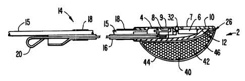
Fig. 3 shows a side cross-sectional ~~iew of a sensor apparatus according
to a preferred embodiment of this invention, pulse oximetry sensor 2 comprises
a
two-piece resilient housing 4. Cover piece 10 :is preferably formed of black
silicone
rubber and body piece 12 is formed from conductive black silicone rubber so
that
the sensor is able to bend a small amount longitudinally to conform to the
shape
of the site.
An electromagnetic radiation directing unit 6 and an electromagnetic
radiation detecting unit 8 are disposed in the cover 10 of housing 4 to form
the
active face of the sensor. Radiation directing unit 6 comprises a first light
emitting
diode (LED) and a second LED (not shown). The first LED emits red light having
a wavelength of approximately 660 manometers (a red 1.ED), and the second LED
emits infrared light having a wavelength of approximately 900 manometers (an
infrared LED). Electromagnetic detecting unit 8 is a standard photodetector
which
may be shielded by a faraday shield to prevent electromagnetic interference.
Radiation directing unit 6 and radiation detecting unit 8 are coupled to wires
(not
shown) which form a bus (corresponding to bus 206 shown in Figs. 1 and 2)
communicating with oxygen saturation calculating unit in a remote oximeter
monitor
(not shown). Any exposed parts of the wires may also be shielded by a grounded
faraday shield. Clear silicone lenses 7 and 9 cover units 6 and 8,
respectively.
An electrode 32 (corresponding to electrode 210 in Fig. 1) is disposed
between units 6 and 8 in cover 10 of sensor housing 4. Electrode 32 is
preferably
formed from sterling silver, although other conductive materials may be used.
Conductive silicone housing body 12 (which corresponds to electrode 212 of
Figs.
1 and 2) and electrode 32 are coupled to wires within cable 15 which form a
bus
(corresponding to buses 208 and 209 shown in Figs. 1 and 2) communicating with
a contact indicating unit in a remote oximeter monitor (not shown).
tn an alternative configuration, the sensor body may be formed from a
molded, opaque, flexible P'VC or thermoplastic elastomer (such as
polyurethane).
~~ ~a~~~r~
Since these materials are not conductive, both electrodes would have to be
separate
conductive members disposed in the sensor body.
In addition, the optical components and the electrode on the active face
of the sensor may be disposed on a standard fiberglass circuit board with
conductive
5 traces forming (in part) the buses communicating with the electrodes and
optical
components. In this embodiment, the electrode corresponding to electrode 212
of
Figs. 1 and 2 may be a conductive portion of the circuit board instead of a
button
electrode. The sensor body may be molded around the circuit board, with
appropriate openings formed in the body for the electrodes and optical
components.
10 Affixed to housing 4 of the preferred embodiment shown in Fig. 3 is a
handle 14 which functions as an insertion and placement aid. Handle 14
comprises
a substantially flat guide tube 16 which, together with the cable 1S
containing the
wires coupled to units 6 and 8, is enclosed by a tube 18 which may comprise
heat
shrink tubing. A removable stiffener 20 is disposed within guide tube 16
before
shrinking tube 18. Stiffener 20 ensures that handle 14 has the desired
property of
allowing bending along the fetal head and the curve of the mother's pelvis
toward
the region to be probed while resisting lateral bending.
In the preferred embodiment, handle 14 has a series of regularly spaced
markings 22, as shown in Fig. 4. These markings provide a visual indication of
the insertion depth of the sensor in the mother's vagina. Markings 22 may also
be
used to gauge the descent of the fetus as labor progresses.
In addition, a ridge 24 may be formed on the handle at a predetermined
distance from the leading edge 26 of the housing 4. The spacing between ridge
24
and the housing's leading edge 26 is such that, for the average fetus carried
to term,
housing 4 is in the preferred monitoring region within the mother's uterus
when
ridge 24 is at the sagittal suture of the fetal head.
A biasing bladder 40 partially covers conductive housing body 12 to
provide an optional sensor retention feature. Bladder 40 is made, e.~., of a
resilient,
open~celled polyurethane foam 42 surrounded by a silicone skin 44 in which a
small
opening 46 has been formed. Opening 46 allows the bladder to be flattened
during
insertion of the sensor, as discussed further below, by permitting the air or
other
i /
l ~~:~~93~':~t
11
fluid within foam 42 and skin 44 to escape when a force is applied to the
exterior
of bladder 40. The resilient foam 42 will re-expand as the exterior force
decreases,
thereby drawing fluid back into bladder 40 through opening 46. In tl-ais
embodiment, opening 46 is approximately .051) inches, although the size of the
opening may be varied to produce the desired rate of re-expansion of bladder
40.
The function of bladder 40 is to press the active face of sensor housing 4
firmly
against the fetus at the sensor site and to keep the sensor in place during
the
contractions associated with labor.
Alternatively, foam 42 may be replaced with a spring, a diaphragm, or
other biasing mechanism. Also, a self-skinning foam may be used in place of
the
foam 42 and skin 44.
In another alternative embodiment, self-ia~lating bladder 40 may be
replaced with a hollow, sealed bladder that may be selectively inflated with
saline
solution or another suitable fluid after the sensor has been inserted into the
uterus.
1 S The selectively inflatable bladder may be provided with a mechanism for
maintaining
a predetermined bladder fluid pressure, such as a pressure regulating valve.
The preferred method of using the apparatus of Fig. 3 is as follows.
The user determines the location of the fetal back and the height and
orientation
of the fetal head by abdominal examination of the mother. The user then makes
a vaginal assessment of cervical status using the Bishop score. This score
grades the
cervix on five elements: dilatation, effacement, position, station (of the
fetal head,
i.e.. above, below, or at the ischial spines), and consistency (firm, soft,
etc.) The
vaginal examination also may precisely confirm the position of the fetal head.
The preferred method of insertion and application of sensor 2 is shown
in Figs. S-7. With the examizliing fingers 401 already in the vagina 402 and
at the
posterior cervix 405, the user grasps the apparatus by handle 14 witlx the
other
hand 400. Sensor housing 4 is inserted into the vagina with housing cover 10
faced
toward the fetus. Sensor housing 4 is then threaded up between the index and
middle fingers of the examining hand 404.
The fingers 401 of the examining hand stretch the posterior cervix 403
to make room for sensor housing 4. Pressure exerted by the cervix against the
~~i7~~i.~~~a
12
sensor compresses bladder 40. The user further advances sensor housing 4 into
the
uterus past the presenting part 405 and past the transcerv~ical region, as
shown in
Fig. 6. Sensor housing 4 is then in the preferred region 406. For a fetus at
term,
ridge 24 on handle 14 will be flush with the vertex of the fetal head when the
sensor housing 4 is in the preferred region 406, as shown in Fig. 7.
Bladder 40 will re-expand to hll the space between the uterine wall 410
and the fetus. The resilient force of bladder 40 will press sensor housing
cover 10
i.e the active face of sensor 2 containing lenses 7 and 9 and electrode :32)
firmly
against the surface of the fetus' skin. This action by the bladder maxirrii~es
the
quality of the optical coupling between the sensor and the, tissue at the
sensor site
and helps retain the sensor at the sensor site in the preferred region 406
during
labor and delivery.
In the preferred embodiment, a Nellcor Incorporated model N-200 pulse
oximeter is modified to include the contact indicating unit 104 i.e., the
current
generator, voltage measuring unit and comparator) of Figs. 1 and 2. Electrode
32
and conductive sensor housing body 12 of sensor 2 connect to contact
indicating
unit 104 via wires disposed in sensor cable 15. Cable 15 connects to the N-200
pulse oximeter via a 9-pin connector.
In addition to retaining the sensor housing 4 at the chosen sensor site,
the force between housing cover 10 and the resilient fetal skin created by the
action
of bladder 40 isolates electrode 32 from the amniotic fluid. Thus, the current
generated by the current generator and flowing between electrodes 12 and 32
traverses the fetal skin as well as the amniotic fluid. In other words, the
skin and
amniotic fluid are resistors in series. The voltage measured by the oximeter's
voltage measuring unit i.e. element 108 in Figs. 1 and 2) is therefore higher
than
the voltage that would have been measured if both electrodes were exposed to
the
relatively conductive amniotic fluid, in which case the amniotic fluid and
skin would
act as resistors in parallel (if the oxientation of sensor 2 is such that
electrode 32
is touching the skin and is also exposed to the amniotic fluid) or the
amniotic fluid
would act as a singles resistor (if electrode 32 does not touch the skin at
all). As
discussed below, the voltage measuring unit will then send a signal to give a
visual
~ r
~~~9~3~~ri;~
13
inclication that the sensor is firmly in place. If the visual indicator fails
to light,
the user may move the sensor to an adjacent site in the preferred region in
order
to improve the contact between sensor and ski .
In an alternative embodiment, a plurality of interconnected electrodes
may be used in place of electrode 32. For example, a pair of electrodes may be
disposed laterally on the sensor's active face between the LEDs and
photodetector.
In this embodiment, the electrodes are electrically interconnected so that if
either
electrode is exposed to the amniotic fluid, the contact indicating unit will
indicate
that the sensor is not in place. Disposing the electrodes laterally helps
ensure that
the contact indicating unit will not indicate proper placement when the active
face
of the sensor is not flat against the fetus' skin but is rotated about the
long axis of
the sensor housing. Other electrode configurations are also possible.
The flow chart of Fig. 8 shows the preferred manner in which the
measured voltage is used. Block 500 represents the measurement of the voltage
between electrodes 12 and 32. Diamond 502 represents the comparison of the
measured voltage with a predetermined threshold T. Threshold T is selected to
be
a value between the expected voltage when electrode 32 is in contact with the
fetus'
skin and the expected voltage when electrode 32 is away from the skin, i,~.,
when
both electrodes are exposed to the amniotic fluid. Diamond 502 is a gate: if
the
measured voltage is not greater than the threshold 'I', oxygen saturation is
not
calculated.
If, however, the measured voltage is greater than T, an indicator lamp
on the oximeter monitor is lit as represented by block 504, and the oxirneter
examines the most recent optical pulse data sent to its oxygen saturation
calculation
unit i.e. element 102 of Fig. 1) by the sensoras photodetector 8 as shown by
block
506. The oximeter's review of the optical pulse data preferably includes
qualification of the most recent pulse with parameters such as (l) comparison
with
historical pulse amplitude, (ii) comparison with historical pulse frequency
and (iii)
correlation with an independent EGG signal.
As represented by diamond 508, if the pulse does not qualify, it is
rejected. The algorithm then returns to block 500 and looks to see if the
measured
? ~) ~~ ~~ ~ 3 y~ ;~
14
voltage still exceeds the threshold 'T. If the pulse meets the
qualif°mation criteria,
however, 'the optical pulse is further processed and the saturation value is
displayed
in the manner of the prior art N-200 oximeter.
As an alternative to the "sensor contact" signal represented by block 50~
in Fig. $, the voltage measured by the voltage measuring unit can be used to
generate a "no sensor contact" signal when the voltage is, below the
threshold. Also,
the desired threshold value 'I" may be coded into the ser~or itself in a
manner
known in the art. The oximeter monitor would then be provided with means to
read the threshold value from the sensor far use by the contact indicating
unit.
Fig. 9 shows an alternative embodiment of the pulse oximeter sensor
according to this invention. This embodiment omits the biasing bladder 40 of
the
embodiment of Fig. 3. Instead, sensor 2 has a curved handle 50. Handle 50
comprises a fixed curved sleeve 52 and an oppositely curved removable
stiffener 5~,
both enclosed by a tube 56. Curved sleeve S2 and removable stiffener 54 are
preferably formed from .020 inch and .025 inch stainless steel, respectively.
Tube
56 has the same markings and ridge as the embodiment shown in Figs. 3 and 4.
The spring constants of curved sleeve 52 and removable stiffener 54 are
chosen so that the handle is substantially straight when stiffener 54 is in
place.
With stiffener 54 removed, i.e., in the sensor's relaxed state as shown in
Fig. 9, the
radius of handle 50 corresponds to one-half the radius of the head of a fetus
at
term. Inserting stiffener S4 straightens handle 50 (as shown in phantom
outline 60
in Fig. 9) to facilitate insertion of the sensor housing into the uterus.
After insertion of the sensor housing the required depth into the uterus,
straightener 54 is withdrawn. Since the radius of curvature of the fetus' head
is
greater than the radius of curvature of the sensor in the relaxed state, the
sensor
will be in the position shown in phantom outline 62, and the spring ford of
curved
sleeve 52 will press cover 10 of sensor housing 4 i.e. the acrive face of the
sensor
containing lenses 7 and 9 and electrode 32) against the surface of the fetus'
skin.
Because the handle's continuous curve helps the sensor housing conform to the
shape of the fetus' head, this action optically couples the sensor's LEDs and
~ f'' ;) f'o
~~~~~i~)!.i!)
1S
photodetector with the tissue at the sensor site and helps keep the sensor in
place
at the sensor site during labor and delivery.
In an alternative embodiment, the shape and/or spring characteristics of
the removable stiffener can be chosen so that the handle is slightly curved
even with
the stiffener in place within the handle. The initial curve could help
insertion of the
sensor by approximating the pelvic curve of the mother. Upon withdxawal of the
stiffener in this embodiment, the sensor would assume the position shown in
phantom outline 62 in Fig. 9.
Other modifications may be made to the disclosed apparatus and method
without departing from the scope of the invention. For example, other sensor
retention mechanisms, such as natural or applied suction, may be used in place
of,
or together with, the bladder or curved handle described above.
In addition, other uses may be made of the contact signal fxom the
comparator of the contact signal unit. For example, the contact signal may be
used
as a gate for the oximeter's audible beep tone. Alternatively, the absence of
a
contact signal (or presence of a "no contact" signal) may sound an audible
alarm.
Also, the sensor may be provided with a movable articulated handle
whose position and shape may be controlled from a remote location outside the
uterus.
In other embodiments of tMs invention, the sensor handle may be
provided with a channel to provide access for a tool used to rupture the
amniotic
membranes and for the introduction of other transducers, such as an
intrauterine
pressure transducer.
Other modifications of the invention will 'be apparent to those skilled in
the art.
F~ '
t