Note: Descriptions are shown in the official language in which they were submitted.
~~~'~~
1
2307U-324 / UC 90-236-1
I~~R~~l~~ 1»~~~5~ M~°d"~i~1D~
~'~R ~X"fEItNA~ C~iEt3'1' (:C~IPRESSIO~T
~~at°.3C~CR~~iD Off' '.~°~&.' ~~T~'f~02$
1. F'ieloi of the ~Lav~atioa
The present invention relates generally to
devices and methods for performing external chest
compression as a part of cardiopulmonary resuscitation
procedures. In particular, the present invention relates
to the use of devices which provide for alternately
compressing and actively expanding a patient's chest to
induce both ventilation and blood circulation.
sudden cardiac arrest is a major cause of death
worldwide and can arise from a variety of circumstances,
including heart disease and trauma such as electrical
shock and suffocation. To improve a patient's chance of
survival (and diminish the likelihood of brain and heart
damage resulting from oxygen deprivation), it is
essential that measures be taken as soon as possible to
at least partially restore the patient's respiration arid
blood circulation. Approximately thirty years ago,
techniques for external chest compression, generally
referred to as cardiopulmonary resuscitation (CPR), were
developed and have enjoyed great success in reducing
mortality resulting from sudden cardiac arrest. such
techniques, however, have remained largely unchanged over
the past two decades.
External chest compression relies on actively
applying pressure to the patient's chest in order to
increase intrathoracic pressure. Such pressure increase
will induce blood movement from the region of the heart
and lungs through the peripheral arteries, thus partially
restcaz ing the patie~at's circulation. Phase 1 of
traditional CPR is referred to as the '°active compression
phase°° where the chest is compressed by the direct
application of external pressure. Phase 2, referred to
as the '°relaxation phase," occurs when pressure is
2
withdrawn and the natural elasticity of the patient's
chest wall causes expansian. While such expansion is
generally sufficient to refill the cardiac chambers with
some blood, it is insufficient to ventilate the patien'c,
i.e., fill the lungs with sufficient air to oxygenate the
blood. Thus, conventional CPR further requires periodic
ventilation of the patient, e.g., mouth-to-mouth
ventilation, in order to provide the air necessary for
blood oxygenation.
l0 Manual CPR procedures generally require
performers to kneel over the patient and to apply
pressure using the palms of their hands to the patient°s
sternum as the patient lies supine on a flat surface. Tf
no one else is available, the performer must periodically
shift position to vexatila~te the patient through a mouth-
to-mouth procedure. Such manual procedures are thus very
tiring to the performer and furthermore have been found
to result in only limited restoration of respiration and
circulation.
2o Manual CPR procedures can also result in injury
to the patient. For example, pressure applied by the
palm of the hand can fracture the patient°s sternum
and/or ribs and cause other traumatic injury, especially
if the performer's hand position is inadvertently shifted
laterally to an improper location on the patient's chest. ,
The performance and safety of CP1~ procedures
can be enhanced through the use of various mechanical and
automatic machines for applying external chest
compression and optionally ventilating the patient by
providing supplemental oxygen or air. The machines may
be as simple as a "cardiac press" which is a manually
operated lever which provides a mechanical advantage in
perfo~ninr chest compbe:.:,ion. More sophi~; icated
machines can provide chest compression and/or ventilation
through a variety of ether mechanisms, including the use
of pressurized chambers fox compressing the chest cavity.
While such machines can be very effective, their bulk,
3
weight, and cost limit their availabz.lity. In
particular, such machines are not widely available
outside of medical facilities and their size is a
deterrent to providing such equipment in emergency
vehicles.
CPR is often administered in conjunction with
other procedures which, taken together, are referred to
as advanced cardiac life support (ACLS). Most commonly,
CPR is administered while the patient undergoes both
electrocardiographic monitoring (ECM) and electrical
defibrillation. Both ECM and defibrillation require the
attachment of electrodes to the patient's chest. The
need to attach electrodes can interfere with the ability
to administer CPR, particularly the ability to administer
manual CPR. In the case of manual CPR, the performer can
also be exposed to electrical shock when current is
applied to perform defibrillation.
It would therefore be desirable to provide
improved devices and methods for performing external
chest compression in conjunction with CPR and ACLS
procedures, Tt would be particularly desirable if such
methods and devices provided enhanced ventilation and
blood circulation in the patient undergoing treatment,
preferably reducing or eliminating the need to separately
ventilate the patient. Desirably, the methods and
devices should be simple and easily stored so that they
can be maintained in emergency vehicles, non--medical
facilities, and even the home. The devices should be
suitable for performing enhanced manual CPR, in
particular by converting Phase 2 chest expansion from a
passive event to an active process to improve venous
blood return from the heart and enhance airflow into the
lungs (facilitated ventilat~~n). The devaua5 should
further facilitate the simultaneous performance of
electrocardiographic monitoring and/or electrical
defibrillation, preferably reducing the perfor'mer's
CA 02066297 2000-02-03
4
exposure to electrical shock from the electrode
attachment.
2. Description of the Hackaround Art
U.S. Patent No. 4,881,527, describes a chamber
which may be placed over a patient's chest to alternately
apply pressure and vacuum to compress and expand the
chest. U.S. Patent Nos. 4,429,688 and 4,196,722,
describe hand-held vacuum cups which are intended for
applying percussive therapy to the lungs (chest
physiotherapy). The devices are intended for repeatedly
striking a patient's chest, not for applying a continuous
compression and expansion. A variety of vacuum cup
designs have been proposed as body massage devices. See,
for example, U.S. Patent Nos. 2,879,765; 2,742,251;
1,460,927; and 728,003, and British Patent Specification
274,306. German Patentschrift 468358 may also be
pertinent.
A device for applying pressure and vacuum to a
patient's abdomen to assist in breathing was described by
Dr. Rudolf Eisenmenger in Wiener Medizinische
Wochenschrift, page 807, August 5, 1939. The device is
further described in a brochure of the Biomotor Company,
Munich, Germany, undated.
Anecdotal reports of the use of toilet plungers
for performing CPR have been made by one of the inventors
herein. See, Lurie et al. (1990), Journal of the
American Medical Association, October 3, 1990, page 1661;
and San Francisco Examiner, article entitled "Toilet
Plunger Successful in CPR," October 1990.
The use of mechanical devices for performing
chest compression and CPR is described in Textbook of
Advanced Cardiac Life Support, c''hapter 4, American Heart
Association, Second Edition, 1987.
CA 02066297 2000-02-03
S
SUMMARY OF THE INVENTION
According to the present invention, devices are
provided for the enhanced performance of cardiopulmonary
resuscitation (CPR) and advanced cardiac life support
(ACLS) procedures. The devices comprise an applicator
which facilitates the application of pressure to compress
the chest of a patient suffering from cardiac arrest.
The applicator is intended to distribute the applied
force substantially evenly over a portion of a patient's
chest, thus reducing the risk of injury to the patient.
The applicator further includes an adherent lower surface
which allows active positive expansion of the patient's
chest by lifting of the applicator between successive
compression strokes. In this way, significant
improvement in patient respiration and circulation can be
achieved when compared to conventional CPR where the
chest is passively, not actively, expanded.
Accordingly, the present invention provides a device
for performing cardiopulmonary resuscitation of a
patient, said device comprising:
an applicator body having an upper surface and a
lower surface;
a handle secured to and extending upwardly from the
upper surface of the applicator;
means on the applicator body for adhering the lower
surface to the patient's chest, whereby the performer can
alternately compress and actively expand the patient's
chest by pressing and pulling on the applicator using the
handle; and
a pressure gauge on the handle, said pressure gauge
measuring compression force applied by the applicator.
In a particular embodiment of the present invention,
the device facilitates the performance of manual CPR
where a performer applies pressure directly to an upper
surface of the applicator, typically using the open palms
of both hands. In such cases, the applicator will
CA 02066297 2000-02-03
5a
comprise an applicator body having upper and lower
surfaces, where the upper surface includes means for
securing at least one hand thereto. In this particular
embodiment, the performer can continuously press and lift
on the upper surface without the need to grasp any
portion of the applicator. Thus, the performer can carry
out CPR using his or her hands in a generally
conventional manner, with the additional benefit that the
ventilation and blood circulation is enhanced while the
risk-of injury to the patient is reduced.
The manual applicator of the present invention will
usually be in the form of a flexible vacuum cup, where
the vacuum or suction provides at least a portion of the
adherence and the resilient nature of the cup provides
the desired cushion. Optionally, a lower lip of the cup
will be coated with an adhesive to further promote
adherence. Alternatively, the applicator can be in the
form of a resilient pad with an adhesive material or
layer present on a lower surface thereof. In the latter
case, the pad distributes the applied pressure
substantially uniformly over the contact area with the
patient's chest while the adhesive surface provides for
expansion of the chest as the performer lifts up on the
pad. In some cases, it may be desirable to combine the
two approaches with an adhesive present on the lower
surface of the vacuum cup. In this way, once the
applicator is properly positioned on the patient's chest,
the physician will shift only minimally if at all. Thus,
traumatic sternal and rib injuries resulting from the
mislocated application of compression force are reduced.
The manual applicator can further include one or
more electrodes present on the lower surface of the
applicator body. The electrode will be disposed so that
it contacts the patient chest when the applicator is in
place and will be useful in performing
electrocardiographic or other monitoring procedures
and/or electrical defibrillation when connected to
CA 02066297 2000-02-03
6
appropriate external systems. When the applicator
includes such an electrode, it may be desirable to
provide a glove or other protective barrier as part of
the hand securing means. In this way, the risk of
accidental electrical injury to the performer is reduced.
In a further aspect, the present invention provides
a device for performing cardiopulmonary resuscitation of
a patient, said device comprising:
a flexible cup having an open hollow interior a
lower lip formed about the open interior;
a handle secured to an extending upwardly from the
flexible cup, and
a pressure gauge on the handle, said pressure gauge
measuring compression force applied by the vacuum cup.
The present invention also provides a device for
performing cardiopulmonary resuscitation of a patient,
said device comprising:
a flexible cup having a hollow interior and a lower
lip formed about the open interior;
a handle secured to and extending upwardly from the
flexible cup, said handle having means for manually
grasping spaced-upwardly from the flexible cup;
an axially compressible spring in the handle,
wherein said spring acts as a shock absorber between the
handle and the flexible cup; and
a gauge on the handle which measures compression of
the handle as the device is pressed against the patient's
chest.
In another particular embodiment of the present
invention, an applicator body similar to that described
above can be connected to a mechanical drive member. The
mechanical drive member can be a simple handle, a powered
drive system, or any other mechanical link which is used
in place of direct manual manipulation of the applicator
as described above. When the applicator is other than for
manual use, the applicator body will include an electrode
disposed on its lower surface to facilitate
7
electrocardiographic monitoring and/or electrical
defibrillation. In the case of manual devices with a
handle, it will be particularly useful. to include a
handle--mounted display which provides patient status and
feedback information to the performer.
_HRZ'~~ ~~~C~~~'~~9P1 oF' ~'~~ %~Fi~'~~t~T~s
Fig. 1 is a perspective view of an applicator
device constructed in accordance with the principles of
the present invention.
Fig. 2 is a cross-sectional view of the
applicator device of Fig. 1.
Fig. 3 is a cross~sectional view of a first
alternate embodiment of an applicator device constructed
in accordance with the principles of the present
invention.
Fig. 4 is a top plan view of a second alternate
embodiment of an applicator device constructed in
accordance with the principles of the present invention.
Fig. 5 is a cross-sectional view taken along
line 5°5 of Fig. 4.
Fig. 5A is a side view of a third alternate
embodiment of an applicator constructed in accordance
with the principles of the present invention.
Fig. 6 is an elevational view, with portions
broken away, of a third alternate embodiment of the -
applicator device of the present invention.
Fig. 7 is a schematic illustration of an
applicator device constructed in accordance with the
principles of the present invention employed in a powered
actuation system.
Fig. 8 illustrates the applicator device of
Fig. 1 being used to perfo~a manual cardiopulmonary
resuscitati.un.
Fig. 9 illustrated the proper placement of the
applicator on a patient for resuscitation as illustrated
in Fig. 8.
~~~~~~~~~o~ ~~ (.~~~ ~~~~~~~~ ~~~~~~
According to the present invention, methods and
devices are provided for performing manual and automated
cardiopulmonary resuscitation (CPR), optionally in
combination with electrocardiogxaphic monitoring (ECM)
and/or electrical defibrillation as part of advanced
cardiac life support (ACLS) procedures. The device
comprises an applicator body having an upper surface and
a lower surface. The lower surface is adapted to adhere
to a patient°s chest during the performance of CPR so
that the intrathoxacic region of the chest can be both
compressed by pressing on the applicator body and
actively expanded by lifting upward on the applicator
body.
Tn a particular embodiment intended for manual
CPR, the upper surface of the applicator body will
include a strap or other:means for securing at least one
hand of the person performing the CPR procedure. In this
way, the performer can alternately apply active
compression and active expansion by pushing and pulling
with the strapped hands) without the need to grasp the
applicator body in any way. ''this is a great advantage
when the CPR is being performed over extended periods
since the need to periodically grasp the applicator to
expand the patientes chest would be very tiring to the -
performer. In addition, time wasted in relocating the
performer's hands to the proper chest position would be
reduced since the applicator would remain secured to the
proper location on the chest by vacuum and/or other
adhesive means.
In another specifis embodiment, the upper
surface of the applicator body can be attached to a
mechanical urine element, sur:rA as a handle or a
mechanical link which is part of a powered automatic
drive system. In this way, active automatic compression
arid eatpansion of the patient's chest can be performed.
9
In both manual and powered systems, the active
expansion of the chest which occurs when the applicator
body is lifted causes a negative pressure within the
intrathoracic region, drawing air into the lungs to
ventilate the patient. This is a particular advantage
since it reduces ar eliminates the need to otherwise
ventilate the patient, such as through mouth-to-mouth
resuscitation. In addition, such active expansion causes
peripheral blood to move more rapidly back into the right
side of the heart and lungs, resulting in increased left
heart blood flow during the next compression phase.
Optionally, the applicator body will include an
electrode in its lower surface which can facilitate
perfarmance of ACM and/or electrical defibrillation.
The applicator body acts as an interface
element between a force-applying source, e.g., the
performer°s hands or the mechanical drive element, and
the sternum region on the patient°s chest to which the
force is applied. The applicator is designed to both
uniformly distribute the applied force over a
predetermined area, i.e., the contact area between the
applicator and the chest, as well as t~ provide a cushion
to decrease the likelihood of injury resulting from the
applied compressive force. Usually, the applicator body
will be resilient to provide the desired cushion and may
further have the ability to distribute the force
uniformly by conforming to the contour of the patient°s
chest. In addition, the applicator is designed to remain
fixed to the chest wall at the desired location for
applying compression and expansion, thus eliminating the
need to relocate the proper location each time
compression is resumed, as is necessary with traditional
CpR.
A variety of specific designs for the
applicator body can fulfill these objectives. The
applicator bady can be formed as a solid pad from a
resilient material, such as a natural or synthetic
10
elastomer. Alternatively, the applicator body may be
formed as an open or partially open structure, optionally
containing an enclosed gas, gel, or the like, to enhance
the shock absorbing and distributing capability of the
body. In the case of pad~like applicator bodies, it will
be necessary to provide additional means for adhering the
lower surface of the body to the patient°s chest.
Typically, an adhesive material can be formed over all or
part of the lower surface.
l0 suitable adhesive materials include pressure-
sensitive adhesives such as those which axe commonly used
on medical bandages, transdermal patches, and other
medical applications. The most useful adhesives will be
natural and synthetic rubber based formulations,
particularly polyisobutylenes. other suitable adhesives
include acrylic and silicon-base materials. When used in
conjunction with electrodes, as described hereinafter,
swollen hydrogels, such as polyvinyl pyrrolidone), may
find use.
The preferred embodiment of the applicator body
will comprise a resilient vacuum cup having a hollow
interior, where the hollow interior is placed against the
patients chest sty that a Vac11l8m or "suGtiOn'o is created
when the applicator body is compressed thereagainst.
Thus, when the vacuum cup structure is subsequently
lifted according to the method of the present invention,
the patient's chest will be actively expanded. The
vacuum cup design is advantageous both because of its
inherent adherent characteristics as well as its natural
resilience which provides a cushion to protect the
patient and promote the even distribution of pressure
(force) over, the interface region with the patient°s
chest. Even with the vacuum cup design, it will
frequently be desirable to provide an adhesive layer
(using the materials described above) over at least a'
portion of the lip of the vacuum cup which contacts the
patient's chest. Adhesive helps hold the vacuum cup
r ."
s
~~~'~?~
11
applicator body in place and helps assure that the
desired vacuuan is maintained,
It will frequently be desirable to form the
applicator body as a laminated or layered structure,
usually having one or morn upper layers which are rigid
relative to the lower layer(s). The relatively rigid
upper layers) will act to receive a localized
compressive force, either Pram the performer's hand or
from a mechanical driver, and to evenly distribute the
l0 force over the lower, more resilient layers. The ability
to distribute the farce aver the resilient lower layers
is particularly important with solid applicatar body
structures which are subject to localized compression,
possibly causing a '°punch°through°' effect.
For manual applicator designs, means for
securing at least one hand will be provided on the upper
surface of the applicator body. The means for securing
can be a strap, mitten, glove, or the like, which permits
the performer to both press down on the applicator body
and lift upward on the body without the need to grasp
the applicator body in any way. The securing means
should be attached to the upper surface so that the
upward force applied by the performer's hand will be
relatively evenly distributed over the applicator body.
The use of a relatively rigid upper surface on the
applicatar body will help provide such even force
distribution.
The dimensions of the applicator body will be
chosen to provide a desired interface area between the
applicator and the patient's chest. Typically, for adult
patients, the applicator will have a circular periphery
with a diameter in the range from about 8 to 25 cm,
preferably being in the range from about 10 to 20 cm. For
children, the dimensions may be as small as 3 cm. Other,
non°circular geometries may also find use, and it is
necessary only that the applicator body be shaped to
provide for a desired force distribution over the
12
patient's sternum as well as to provide for sufficient
adherence to allow the patient's chest to be expanded
when the applicator body is raised upward.
The thickness of the applicator. body is not
critical and will depend on the particular body design.
For solid, pad~like applicator bodies, the thickness will
typically be in the range from about 1 to 10 cm, more
typically in the range from about 2 to S cm, depending on
the resiliency of the material employed. For vacuum cup
designs, the maximum thickness, i.e., the maximum air
gap, will be in the range from about 1. to 15 cm, more
usually from about 5 to 12 cm. F°or manual applications,
it will be desirable to provide a flat upper surface so
that the user can press down evenly over the surface with
one or both hands in a manner similar to conventional
CPR. In this way, the performer will experience the same
"feel" as conventional CPR with the advantages of the
present invention of patient protection and improved
ventilation and circulation. In some cases, it may be
2o desirable to shape the lower surface of the applicator
body to conform to the general contours of the human
chest. In addition, it may be desirable to provide a
plurality of sizes-of the applicator in a single kit so
that a particular applicator may be selected for the
individual patient. Such kits would have applicators as .
small as about 3 cm i.n diameter for children to as large
as 25 cm, usua11y.20 cm, in diameter for adults.
It will frequently be desirable to provide one
or more electrodes in the lower surface of the appliaa~tor
body. The electrodes will be exposed on the surface so
that they will contact the patients chest when the
applicator body is in use. The electrode will be
internally connected to an electrical conyiector or plug,
typically located on the side of upper surface of the
applicator body. The connector or plug will be selected
to allow interconnection with conventional ~C~t and/or
electrical defibrillation equipment. Combination
13
ECM/defibrillation equipment is commercially available
from suppliers such as Hewlett-Packard Co>, Palo Alto,
California, and Physio Control, Seattle, Washington.
When used with such systems, the applicator of the
present invention can act as one of the two (or more)
°'paddle°° connectors which are secured to the
patient°s
chest for monitoring and/or defibrillation.
Referring now to Figs. 1 and 2, a first
embodiment of the applicator device of the present
l0 invention, intended for manual CF'R, is illustrated. The
device 10 comprises a vacuum cup body 12 having a concave
interior 14 which opens into a lower surface defined by a
peripheral lip 16. The vacuum cup 12 has a substantially
flat upper surface 18 having a strap 20 extending
thereacross. The strap 20 is firmly secured to the upper
surface so that a user can place one or both hands
beneath the strap with the hands) being open to press
directly against the upper surface.
The vacuum cup body 12 is relatively thick
across its flat upper surface and tapers down to form a
skirt 22 terminating at the periphery of lip 16. The
thick upper surface region provides sufficient rigidity
so that the applicator body 12 will not involute or
°'cave in" as the user presses against the upper surface.
Instead, the lip 16 and skirt 22 will tend to spread -
outward reducing the volume of air in the concave
interior 14 and providing the desired vacuum. The vacuum
cup 12 will transmit sufficient force to compress the
patient's chest by a desired amount, typically 3.5 to
5 cm. After the desired compression of the chest is
completed, the user will lift on the applicator body 12
by raising one or both hands which in turn lift through
the strap 20. The reduced pressure within the concave
interior 14 will cause a vacuum or suction which acts to
raise.the patient's chest and actively expand the
intrathoracic cavity.
Using the device 10, the performer is able to
perform CPR in a manner similar to conventional manual
CPR, with reduced exposure to injury since the
application of force is localized to the intended region
on the patient's chest with the position being "anchored°°
by the device itself. Additionally, the ability to
actively raise the patient's chest and expand the
intrathoracic cavity provides for improved ventilation
and circulation of the patient.
l0 Referring now to Pig. 3, an alternate
embodiment of a manual applicator device 30 constructed
in accordance with the principles of the present
invention is illustrated. The device 30 comprises a
solid resilient pad structure 32 laminated to a
relatively rigid upper plate 34. The resilient pad 32
can be formed from a wide variety of natural and
synthetic polymers having sufficient resiliency to
conform to the contours of the patient°s chest while
retaining sufficient compressive strength t~ permit the
transmission of the desired force to the chest. Suitable
polymers include neoprenes, low density polyethylene,
soft polyvinylchloride (PVC) compounds, natural rubbers,
synthetic rubbers, and. the like. Suiaable polymeric
structure include open cell and closed cell foams. The
solid pad may also comprise fluid-filled bags and .
structures, such as gel-filled bags and air-filled
structures, which can transmit the desired force while
providing desired resilience and conformity.
The upper plate 34 will be rigid relative to
the resilient pad 32, typically being a rigid plastic
material. A strap 36 is secured to the upper plate 34
and allows the user to place one or both hands therein in
a manner similar ~co that described for device :t0.
Applicator 30 further includes an electrode 38
which is in the form of a ring extending about the
periphery of the lower surface of the pad 32. The
electrode 38 will be formed from a suitable material,
~5
such as electrically conductive metals, and will be
interconnected with an electrical connector cord 40 wha.ch
is suitable for interconnection with an ECP3 system, an
electrical defibrillator, or a combination
ECM/defibrillator unit. Such electrode applicators will
frequently be used in combination with an electrically-
conductive gel, such as those commonly used with
defibrillator electrodes, which can further enhance the
adhesive characteristics of the applicator.
To provide the necessary adherence, the lower
surface of pad 32 is covered with an adhesive which is
suitable for detachably adhering to the patients chest.
Suitable adhesives are described above.
A second alternate embodiment 50 of the
applicator device of the present invention is illustrated
in Figs. 4 and 5. The applicator device 50 includes a
relatively rigid upper plate 52 and a depending vacuum
cup structure 5~ formed from a relatively resilient
material, i.e., a material which is able to spring back.
A mitten 54 is secured to the upper surface of plate 52
and is shaped to receive a performer°s hand. An
electrode 58 is disposed in the lip 60 of the vacuum
cup 54. The electrode is interconnected to a cord ~0
intended for hook-up to conventional ECM and/or
defibrillation equipment.
The device 50 combines certain of the
advantages of each of the previous embodiments. The use
of the rigid upper plate 52 helps assure the even
application of force to the patient's chest. Use of the
vacuum cup structure 54 provides for an entrapped cushion
of air in its concave interior, further assuring
substantially uniform distribution of pressure to the
patient's chest. Finally,wthe use of the mitten 56,
rather than a strap that has previously been described,
helps isolate the performer's hand from the other
electrodes being used f~r ECM and/or defibrillation.
f~ f~ ~ 7a
~~~.7~~7~
16
Fig. 5A illustrates an applicator 150 having a
bellows or accordian configuration. In particular,
applicator 150 comprises an applicator body 152 having a
handle 154 secured on an upper surface 156. The
applicator body 152 includes an upper pleated section 158
and a lower skirt section 1.60 which together define the
desired bellows construction. It will be appreciated
that the bellows structure may include additional pleated
sections, although usually the structure illustrated will
be sufficient. The applicator body ~.5~ will usually be
composed of a resilient elastic material, such as a
natural rubber or synthetic rubber, and may be formed by
conventional molding techniques. The applicator body 156
provides a vacuum cup which permits significant air
intrusion or leakage to take place before the desired
vacuum is lost.
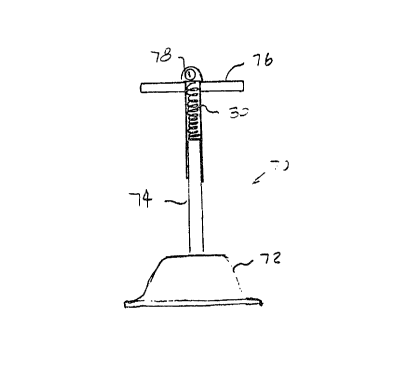
An applicator device 70 comprising an
applicator body 72 and a handle 74 attached to the upper
surface of the applicator body 72 is illustrated in
Fig. 6. The applicator body 72 is illustrated as a
vacuum cup, similar to that illustrated in Figs. 1 and 2,
but could be any of the other applicator body structures
described herein. The use of an elongate handle 74 with
the applicator body 72 is desirable for patient's lying
on the floor or ground. A T-bar 76 on the handle 74 -
allows the performer to stand over the patient caith one -
leg straddled on each side of the patient°s chest,
applying successive downward and upward strokes to
compress and actively expand the chest.
Use of the handle interferes with the
performer's ability to feel and regulate the pressure and
compression being applied to the chest. For that reason,
it is desirable to provide feedback a.ni~ormation, sucix as
a pressure gauge 78, on the handled de~rice 70. .As
illustrated, the pressure gauge employs a spring 80 which
is disposed between the T-bar 76 and the fixed portion of
handle 74. The spring 80 also acts as a shock-absorber
17
which helps limit excessive force applied to the patient.
Other pressure measuring devices and transducers would
also be suitable.
In embodiments w~.th a handle, it may be
s desirable to provide a more advanced monitoring panel or
readout on the handle (not illustrated) which can display
a variety of patient status information and/or feedback
to the person performing the CPR. Patient status
information includes minute ventilation, temperature,
blood pressure, heart .rate, respiratory rate, and other
vital signs. Such status information will often require
separate monitoring devices (not illustrated) attached to
the patient, and the display on the handle makes the
information immediately available to the person
performing the CPR. Feedback information includes
pressure or force applied to the patient, depth of
compression, compression rate (i.e., cycles per minute),
duty cycle (i.e., portion of each cycle in which the
patient is compressed), and the like. Such feedback
information can be provided as discrete values, e.g.,
with gauges or digital readouts, or may be provided with
a light or sound system which indicates when certain
threshold values have been met or exceeded. It may be
further desirable to prbvide a pacing signal, e.g.,
either a sound or flashing light, to facilitate
maintaining a desired compression rate.
The applicator device of the present invention
may also be employed in a powered system 90 as
illustrated schematically in Fig. 7. Applicator body 92
is secured to a vertical drive element 94 which is
attached to a reciprocating lever arm 96. The lever arni
96 may be driven in a wide variety of ways. As
illustrated, a fixed fulcrum point 9S is provided by
post 100 and the lever is reciprocated up and down by a
piston and cylinder 102 to provide the desired
compression and expansion of the chest.
18
The applicator 92 is again illustrated as a
vacuum cup structure, similar to that illustrated in
Figs. 1 and 2. The applicator 92 could employ any of the
other applicator body structures illustrated herein, and
will be particularly useful with those structures which
include integral electrodes which permit ECM and
defibrillation.
Referring now to Figs. 8 and 9, a method
according to the present invention for applying manual
CPR using the applicator device 10 is illustrated. A
patient P suffering from cardiac arrest and apnea is laid
on his back on a flat surface and the shirt and collar
loosened to provide access to the chest. After the
patient's airway is cleared and the chin lifted to tilt
the head, the device 10 is placed over the 1~wer portion.
of the patient's sternum in the region where conventional
CPR is applied (Fig. 9).
The performer then places one or both hands
under the strap 20 of device 10 and begins external chest
compressions at a rate of from 80 to 100 per minute.
Optionally, the performer will periodically apply mouth-
to~mouth resuscitation or other ventilation in order to
ventilate the patient. Tt is an advantage of the present
method, however, that the number of ventilations which
must be performed is reduced.
Each chest compression should achieve a
compression in the range from about 3.5 to 5 cm, and will
be followed by a positive lifting on the chest by the
performer by lifting on the applicator device 10. The
chest will be lifted and allowed to re~nai.n ventilated
until the next compression step. Typically, the
compression portion of the cycle will last from about 0.2
to 0.7 seconds, while the e~cpansion portion of the cycle
will last from about 0.2 to 0.7 seconds, with the
compression and expansion portions usually being equal.
1~
The method, as'described above, will be
continued until heartbeat and respiration are restored or
until medical support arrives.
Although the foregoing invention has been
described in detail for purposes of clarity of
understanding, it will be obvious that certain
modifications may be practiced within the scope of the
appended claims.