Note: Descriptions are shown in the official language in which they were submitted.
2066354
Cannula
Pield of the Invention
The~ present ~nvention relates to the field of surgical devices.
Background of the Invention
Cannulas are hollow tube instruments used to deliver fluids or
remove fluids from ~lood vessels, duct6 or other hollow organ~
of animal~. While ~any sizes of cannulas are available
co~mercially, microcannulas suitable for use in ~urgery on 6mall
animals are of limited design and utility. The smalle6t
available cannulas are generally flat tipped and are large
enough to accommodate a 16 to 24 gauge nsedle in the lumen of
the cannula, which needle is used a~ a trochar~ Small
cannulas are generally made of 6mall bore polyethylene tubing
and are supplied with a hypodermic needle which is used to block
a cannula to prevent fluid contained in the vessel of other
hollow organ from draining until the cannula is in place in a
pre-made incision in the vessel or ot~er hollow organ.
As cannulas decrease in size, they are more flexible and easily
bent and therefor difficult to manipulate. The flexibility of
6mall cannulas occurs because of the decreasing absolute wall
thickness of the cannula as they get smaller in diameter and
concomitant loss of rigidity of the cannula wall.
Conventional cannulas are 6upplied with trochars that ~ove
~reely in the lumen of the cannula ~ince lt i8 conventlonally
desirable to be able to guickly remove t~e trochar once ~ vessel
is cannulated.
Conventional small cannula~ appropriate for use i~ cannulation
of small blood vessels in microsurgery are notorîously difficult
2~63~
to use. The smallest cannulas available frequently reguire many
minutes of patient and skilled manipulation to prepare B micro-
incision in a blood ves6el and properly place the cannula in
small blood vessels.
Sun~ary ~nd Objects of the Invention
The present invention comprises a very s~all bore cannula
having a beveled tip with a s~arp pointed trochar tightly, but
removably placed in the lumen of the cannula. ~his cannula or
m~crocannula and associated trochar have sufficient rigidity to
be relatively ea6ily manipulated. Further~orc by ~ans of using
a sharp pointed trochar and bevel tipped cannula, it is possible
to cannulate a s~all blood vessel with out the necessity of
incising the blood vessel wall before inserting the cannula and
trochar.
It is ~n object of the ~nvention to provide an easily
~anipulated cannula which can be used to cannulate small hollow
organ~ and blood vessels in a short period o~ time.
It is another object of the invention to provide a microcannula
and trochar that function together ~s a unit to provide rap~d
cannulation with a minimum of blood loss.
It is yet another object of the invention to provide a
microcannula and trochar that can be u6ed to cannulate a blood
vessel without making a preparatory incision before lnserting
the trochar and cannula into the blood vessel.0
arief Description of the Figures
Figure 1 i6 a side view of the trochar according to the
invention.
Figure 2 is a cross-section of the ~icrocannula according to ~he
2~63~
invention.
Figure 3 6hows the trochar and microcannula in use as a unit
ju~t prior to penetration of a blood vessel wall or other hollow
tube by the point of the trochar.
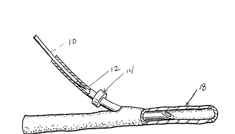
Figure 4 shows the ~icrocannula in place in n blood ves6el or
other hollow tube with t~e trochar in the process of being w~th
drawn.
Figure 5 shows the ~icrocannul~ in place in a blood vessel or
other hollow tu~e with the blood ve6sel tied off around the
proximal end of the microcannula and the ~icrocannula 6ecured
in place.
~etalled Description of the InYention
In greater detail the microcannula according to the invention
comprises a hollow tube 12 having a cross sectional size smAller
than a 24 gauge needle. More precisely the outside diameter of
said tube which co~prises the body of the microcannula ~6 ~bout
0.016 inch. The outside diameter of the tubing will vary
slightly but in general the outside diameter of he tube will be
between 0.018 ~nd 0.014 inch. Usually the out6ide diameter of
the tubing will be 0.016" +/- 0.001 inch.
The inside diameter of the tube which co~pri6es the body o~ the
microcannula i6 ~bout 0.008 inch. The in6ide diameter of the
tubing will vary slightly but in general the inside diameter of
the tube will be between O.Q10 and 0Ø006 inch. U6ually the
inside diameter of ~he tubing will be 0.008 l/- 0.001 inch.
The ~icrocannul~ according to the invention will h~ve a 6imple
beveled tip. $he angle of said bevel i8 about 25. ~ngle of the
2~6`~3~
bevel may vary between 23 and 27 degree6, but the best
performance of the microcannula i6 achieved when the bevel is
25+/- 0.5.
The microcannula of the invention will generally be ~ade of
bio-compatible polymer tubing. It is preferred that the bio-
compatible poly~er be perfluorocarbon material.
The ~icrocannula described above is highly flexible and delicate
and is difficult to insert into the lumen of a blood vessel 18
or other hollow organ requiring cannulation. In order to
facilitatQ manipulation of the microcannula, a trochar 10 is
provided for use with the microcannula. The trochar fits in t~e
lumen of the microcannula tube and may be remo~ed therefrom.
It i6 preferred that the trochar be of a size that fits tightly
in the lumen of the microcannula tube and does not move freely
in the lumen; however, the trochar must also be small enough to
be remove from the lumen of the microcannula when the side of
the microcannula is gra6ped and held and the trochar i6 pushed
or pulled from the lumen of the tube. The preferred performance
of the trochar i6 best obtained when the outside diameter of the
trochar i6 61ightly smaller than the inside diameter of the
tube.
The trochar used with the cannula is pointed and the point forms
an angle of about 8. The angle of tbe point of 6aid trochar
may vary between about 11- ~nd 5. An angle of 8 i8 preferred.
The length of the point of the trochar is about 8iX times the
di~meter of said trochar. Lengths substantially greater than
about 6iX time~ the d~ameter of the trochar lead to undesirable
delicate points that can flex an breaX. Therefore it is
preferred that the trochar point is about six times the diameter
of the trochar or less.
The trochar may be ~ade of any strong wire stock. It is
20663~
preferred that the wire is not of a ductile ~etal since the
trochar confers rigidity on t~e microcannula when it iB lnserted
into the lumen thereof. Furthermore ductile wires cannot be
easily inserted into the lumen of the ~icrocannula with the
required tight fit without bending or breaking. It i8 preferred
th~lt the trochar i~ made of stainle6s steel.
The microcannula of the invention further compri~es a segment
of the ~icrocannula tube located distal to the tip of the
~icrocannula that has an expanded outside diametsr. The
expanded out~ide diameter or shoulder 14 ~ay be 1n the form of
a ring of tubing or an "0" ring adhered to the outside w~ll of
the microcannula tube, dried plastic glue or a thickening in the
wall of the microcannula itself.
When in use the 6houlder is used to ~ecure the di6tal end of the
microcannula using a ligature 20 one end of which is tied
around the distal end of the ~icrocannula and the other end of
which is tied around the blood vessel 18 surrounding the end of
the microcannula proximal to the beveled tip, a~ depicted in
Figure 5.
The distal end of the microcannula, which i6 the end of the
microcannula away from the beveled tip may be 6ecured optionally
within the lumen of a larger tube which ~ay in turn be secured
to the tip of a needle or 6till larger piece of tubing. The
distal end of the microcannula can in this fashion be
conveniently connected to conventional fittings for tubing or
~yringes ~uch as luer lock fittings and the like.
Tubing of the type used to make the microcannula accordlng to
the in~ention can be obtained from supplier of laboratory wares
6uch ~5 Cole-Parmer, Chicago, Illinoi~, U.S.A.. Wire suitable
for fabrication into the trochar described here in can be
obtained from National Standard Company, Santa Fe Springs,
2066~4
Ca:Lifornia, U.S.A.
The present invention further comprises the microcannula
described hereinabove with the trochar described herein above
placed in the lumen of the tube forming the ~icrocannula.
When in use to cannulate a small blood vessel or hollow organ
the trochar 10 and microcannula 12 are used as a unit. The
trochar 10 is placed tightly fitting in the microcannula 12 with
the trochar tip 11 protruding beyond the microcannula bevel's
leading edge 16. The trochar tip 11 i6 used to pierce the wall
of the blood vessel 18. Tbe microcannula bevel t6 leading edge
comes into contact with the out6ide of the blood veesel 18 outer
wall sightly displacing the wall of the blood vessel and
stretching the hole punctured in the blood vessel wall. By
further advancing the microcannula and trochar together, the
~icrocannula i6 easily threaded through the hole into the lumen
of the blood vessel.
As a result of the tight fit of the trochar and microcannula the
cannulation of small blood vessels can be accomplished with out
the necessity of first incising ~ blood vessel followed by
insertion of a cannula and trochar. By using the trochar and
microcannula as a unit, bleeding can be minimized and the
possibility of damaging the blood vessel with an incision that
is too large is eliminated.
