Note: Descriptions are shown in the official language in which they were submitted.
W092/03989 PCT/US91/0~14
~- -1- 2~72~7
~ TITLE
VARIABLE POWER INTRAOCULAR LENS
WITH ASTIGMATISM CORRECTION
BACXGROUND OF THE INVENTION
The present invention relates generally to an
intraocular lens and, in particular, to an apparatus for
varying the power of and providing astigmatism correction
in an intraocular lens.
The lens of the human eye is located centrally behind
the pupil and is protected by the cornea. In the normal
eye, the lens is clear and is substant~ally symmetrical,
with opposed convex surfaces defining generally spherical
sections. The lens and the cornea cooperate to focus
- light on the retina. The retina in turn cooperates with
the nerves and the brain, so that light impinging on the
retina is perceived as an image.
The light refraction which takes place in the cornea
and the lens translates into an optical correction of
i about 60 diopters, with the cornea accounting for about 40
diopters and the lens accounting for about 20 diopters.
~ Other refracting structures also are present in the eye,
,~ but are disregarded to simply the subject explanation.
,
A cataract is a condition where the normally clear
lens of the eye becomes progressively opaque. This
opacification typically occurs over an extended period of
time, and the amount of light which passes through the
lens decreases with increasing degrees of opacity. As the
~ . ability of the cataract lens to transmit light decreases,
,~ the ability of the eye to perceive images also decreases.
"~ .
.,
. .
,............................................. .
,,
. . . ..
- ; . . . . .
,-.; . , , ., ~ . . . .
W092/0398s PcT/US9lto~l4
~`0;~2~ 2
Blindness ultimately can result. Since there are no known
methods for eliminating the opacity of a cataract lens, it
generally is necessary to surgically remove the opaque
lens to permit the unobstructed passage of light through
the pupil to the retina. The cataract lens is removed
through a generally horizontal incision made at the
superior part of the juncture where the cornea and sclera
meet.
Once the lens has been surgically removed, light can
be readily transmitted through the pupil and toward the
retina. As noted above, the lens of the eye performs a
significant light focusing function. Consequently, with
the lens removed, the optical system of the eye is left
about 20 diopters "short", and light is no longer properly
focused on the retina. Eyeglasses, contact lenses and
intraocular lenses are the three types of optical aids
that commonly may be employed after cataract surgery to
refocus the light on the retina.
Eyeglasses include lenses which are spaced from the
cornea of the eye. The air space between the lens and the
cornea causes an image magnification of more than 7%.
Unfortunately, the brain cannot assimilate this
magnification in one eye, and as a result an object
appears double. This is a particular problem if the
individual had only one cataract eye. Eyeglasses also
substantially limit peripheral vision.
Contact lenses rest directly on the cornea of the
; eye, thus eliminating the air space. As a result, there
,:i
,, .
`''
. ~ ~
.~ .
.. . .
.
,
.
. ..
W092/03989 PCT/US91/0~14
~ ~ 3 2~`72~7
is a much smaller image magnification with contact lenses
than there is with eyeglasses, and the brain typically can
fuse the images perceived by an eye with a contact lens
and one without. Contact lenses, however, are less than
perfect. For example, contact lenses are quite fragile
and can be easily displaced from their proper position on
the cornea. Additionally, the lenses must be periodically
replaced because of protein build-up on the surface of the
lens which can cause conjunctivitis. Furthermore, many of
the elderly people who require cataract operations do not
have the required hand coordination to properly remove or
insert the lens.
- Intraocular lenses first because available as optical
aids to replace removed cataract lenses in about 1955.
These lenses are placed in the eye, and thus closely
simulate the optics of the natural lens which they are
replacing. Unlike eyeglasses, there is virtually no image
j
distortion with a properly made and placed intraocular
lens. Also, unlike contact lenses, there is no protein
build-up on the intraocular lenses and the lenses require
no care by the patient.
To place the lens in the eye, the surgeon ordinarily
~; makes an incision or opening in the sclera and cornea to
allow the insertion of the }ens into the eye. Normally,
the stabilizing loops of the attachment members of the
lens are flexi~le and can be bent, if necessary, to pass
through the opening. Accordingly, the minimum length of
opening which must be made and is ordinarily determined by
: .
"~
~ , .
. ~ ., . ~ . , . , , ., , , . . ., ... . ~, . . . .. ... . . .. . . . ..
~,". .. . . .. , ., . , . .,, . ,. ,:.. , ., . . .. . , ~ .
W092/03989 PCT/US91/0~14
. ., ~
2~72~7 4
the diameter of the substantially rigid lens body, or
optic, usually having a circular periphery. It is, of
course, desirable to make the opening into the eye as
small as possible to minimize the risk of damage to the
5 eye. In the past few years, some lenses have been made of ,
flexible material like silicone that can be folded so as
to go into the eye through a smaller opening.
The current practice in the implantation of
intraocular lenses is to replace a normal crystalline
human lens of the eye removed at the time of surgery, such
as in cataract surgery, with an intraocular lens such as
an anterior chamber lens or posterior chamber lens formed
of appropriate biocompatible material such as PMMA
(polymethyl methacrylate) material. However, one of the
present problems with intraocular lenses is that it is
necessary to decide on the power of the lens
preoperatively. This can be accomplished, for example, by
performing an ultrasound scan and/or evaluating the
patient's refraction preoperatively and then making a
clinical estimate of the proper power of the lens in order
to determine proper refraction of the eye. However, even
with the best medical techniques and sophisticated optical
instruments available, ophthalmologists have never been
able to correct for accommodation which is the ability to
change the focus of vision from distance to near vision
and there is no lens system that can be adjusted after
implantation for even minor changes in spherical or
;'
` astigmatic power. Thus, most patients, following routine
. .
.. :
~,
.~ ,. . . . ,: . . .. ...... . . .. . . . . . . .
. . .. , ... ., ., .. . . . , ~. - . . ..
- .. .. . . . . ~ . .
W092/03989 PCT/US91/0~14
f--~ ' ' 2~72~7
lens implantation, require the use of glasses for
precisely focused distance and near vision.
The prior art intraocular lens typically is either of
plano-convex construction or double convex construction,
with each curved surface defining a spherical section.
The lens is placed in the eye through the same incision
which is made to remove the cataract lens. As noted
above, this incision typically is made along the superior
part of the eye near the juncture of the cornea and the
sclera. About one third of all postoperative patients
will have significant astigmatism and, approximately one
third will need a spherical adjustment in their
postoperative glasses to see clearly. In virtually all
instances, the surgery itself induces astigmatism which
fluctuates significantly during the first few weeks, or
even months, after the surgery.
Postoperative induced astigmatism is attributable to
the healing characteristics of the eye ad;acent the
incision through which the cataract lens is removed and
the intraocular lens is inserted. More particularly, the
incision in the eye tends to heal slowly. The incision in
the eye may take eight weeks to a year to properly heal.
During the period when the eye is healing, the wound area
tends to spread and thus a cornea that may have been
spherical before surgery is made other than spherical.
Since the incision is generally horizontally aligned, the
;' spreading is generally along the vertical meridian.
; Initially, after the surgery, the cornea is relatively
, . .
, ,.;. , . , , ~ - . . . , .............. ., . . : . ,.
: , . , . .. : ,.......... ... . ., . , , :
- , : . .. .~ , :
W092t03989 PCT/US91/0~4
` 2~72~7 6 ~-
.
steep in the vertical meridian. As the eye heals, the
cornea becomes relatively flat in the vertical meridian.
Consequently, the optical system of the eye, which may
previously have been spherical, becomes "toric" with the
vertical meridian of the optical system providing a
different optical power than the horizontal meridian.
This non-spherical configuration of the optic system is
generally referred to as "astigmatism".
The degree of this induced astigmatism varies
lo according to the type of incision made, the presence or
absence of sutures or the number and type of sutures used,
the technical skill and care employed by the surgeon, and
the physical attributes of the eye. For example, the use
; of a fine nylon suturing material typically results in a
smaller deviation from sphericity than the use of silk,or
absorbable sutures. Generally, the induced astigmatism
varies from 0.5 to 5 diopters. The initial postoperative
astigmatism is generally caused by the steepening of the
vertical meridian. Late astigmatism is caused by the
flattening of the vertical meridian of the cornea. The
orientation and amount of postoperative astigmatism are,
in most cases, not accurately predictable. Postoperative
astigmatism typically is corrected by prescription
eyeglasses which need to be changed periodically as the
; 25 eye heals.
In some cases, despite the best efforts of the
' ophthalmologist, the lens surgically placed in the
patient's eye does not provide good distance visual acuity
... . .
, ~
.
, , .,.. ~. ~ . . . ., . : , : .: ~ , . .,. : :
. . . ,. . . . - . - .: : : :
- . . :,
W092/03989 PCT/US91/0~14
~ 2 ~ ~ 7 2 ~ i
due to spherical miscalculations and due to the changing
astigmatic requirements. Since the surgery itself can
cause significant change in the amount and axis of the
astigmatism present after cataract surgeryr the exact
amount and axis of astigmatism can not be accurately
determined until sometime, usually several weeks or
months, after the surgery. Since the old intraocular lens
can not be readily removed and a new intraocular lens with
a different power surgically installed without unduly
jeopardizing the patient's vision, the patient must rely
on spectacles to provide accurately focused visual acuity.
In other words, although the need to wear heavy, bulky,
higher power spectacles is eliminated, the patient
nevertheless usually must wear spectacles for best focused
vision.
Several attempts have been made to provide an
intraocular lens which corrects for the astigmatism
expected after surgery or can be varied in power after
implantation. U.S. Patent No. 4,575,373 discloses a laser
adjustable intraocular lens which utilizes a laser to
alter, in situ, the power of an implanted intraocular
lens. The outer ring of the lens is manufactured of a
non-toxic heat shrinkable colored plastic material to
permit selective absorption of laser energy, thereby
causing the shape of the lens to change increasing the
,,
power non-reversibly.
U.S. Patent No. 4,816,031 discloses an intraocular
lens system including a PMMA lens implant, a second soft
.
i:
.i .
....... . . . . . . . . . .
, . .
. . -: . . . . . .
: ~, . - .
,.. . .. , . . :
. ~
.
~ .
W092/03989 PCT/US91/0~14
2~72~7. 8 `'
and pliable lens positioned thereover, and
electromechanical circuitry for regulating the distance
between the two lenses, thereby providing for adjustment
of the focal point of the lens system.
U.S. Patent No. 4,601,722 discloses an intraocular
lens having a lens body formed of a plurality of lens body
portions and magnet means for the assembly of the portions
into the lens body within the eye after the portions are
individually inserted through an incision in the eye.
U.S. Patent No. 4,512,039 discloses an intraocular
lens for offsetting postoperative astigmatism having the
finally placed vertical meridian optically weaker than the
horizontal meridian. Proper placement is ensured by
disposing the haptics along the vertical meridian.
U.S. Patent No. 4,298,996 discloses a magnetic
retention system for an intraocular lens having one or
more supports extending from the lens body. Each support
carries a pair of magnetic fixation members positioned on
; opposite sides of the iris, whereby a trans-iris magnetic
force secures the lens in place without sutures or
incisions in the iris.
;~ U.S. Patent No. 4,277,852 discloses an intraocular
lens with astigmatism correction combined with a
supporting mount or haptic structure to assure correct
optical orientation of the implant.
Several attempts have been made to provide a variable
power intraocular lens, which power varies according to an
application of a force external to the lens, for
,
~I .
.. . . . ... . .:: . .. :: ,, , , . ,
W092/03989 PCT/US91/0~14
2 0 ~ 72 ~ 7~
- ~-. . . g ~ . . ; .
correcting the astigmatism expected after surgery. U.S.
Patent No. 4,787,903 discloses an intraocular lens
including an annular Fresnel (prism) lens, made of a high
index of refraction material such as
5 polymethylmethacrylate. A composite material overlays the
Fresnel elements to provide a smooth external surface and
is made of a suitable material, for example, crystalline
lattice or liquid crystal material, which changes the
index of refraction when excited with electrical power or
10 radiant energy. The lens carries a complementary loop or
other energy pick-up device, for receiving the power from
an electric field generated by an external power source
feeding a coupling loop. The coupling loop can be carried
in an eyeglass frame, implanted about the eye socket or
15 positioned by the lens wearer or an ophthalmologist. It
is stated in the patent specification that some overlay
materials can be switchable between more than two states,
each with a different index of refraction, while other
materials will provide a continuously variable index of
, 20 refraction which may be stable or may return to an initial
value when the energy is removed. However, such materials
are not identified in the patent.
~ U.5. Patent No. 4,601,545 discloses a variable power
r lens system including an optically active molecular
25 matexial such as liquid crystals. A variable gradient
index of refraction is achieved by applying a controlled
stimulus field, such as a geometrically configured matrix
i of electrical voltages, to the lens. A corresponding
:.
" ~ .
,'~
;- - ^^ . .. .
. .
, ,' '.: . ,~ ' ~ . . ,: .-
. .
. ~ - . .
:,
W092/03989 PCT/US91/O~lq
2~67%47 lo
matrix of horizontal and vertical conductors applies the
electrostatic field produced by the applied voltage to be
selectively controlled at discrete points so that a
gradient index of refraction is produced.
U.S. Patent No. 4,564,267 discloses a variable focal
length lens which can be electrically controlled by
applying an electric field to a compound lens including at
least one lens formed of electrooptic crystals. The
electrooptic crystals are juxtaposed between first and
second transparent electrode plates each comprising a
plurality of concentric annular transparent electrodes. A
power source connected to the electrodes generates an
electric field across the crystals creating a refracting
index distribution having a lens action. The electric
field effectuates a change in the focal length of the lens
which varies according to the potential imparted.
U.S. Patent No. 4,373,218 discloses a variable power
i;, intraocular lens including a fluid expandable sac for
containing a liquid crystal material that is uced in
combination with an electrode and a microprocessor for
changing the index of refraction of the lens. An
electrode is located in a ciliary body to provide an input
' signal that is proportional to a desired acco~modation to
a microprocessor which can be implanted into a sclera of a
human eye. The microprocessor produces a potential across
the liquid crystal material to control the index of
j refraction to obtain the desired accommodation based upon
; the relative position of the eyes. The voltage output of
;: ' :.
~ . .
, : . ~.. . , ~ : - -
;~ . . - .
. . : . . .,... , , , . : ~ .,
.. . . .. . ... . .
.
W092/03989 PCT/US91/~
- 11 2~72'~7
the microprocessor is applied to electrodes which can be a
thin transparent material forming a coating on the
interior of the fluid expandable sac.
SUMMARY OF THE INVENTION
In recent years, exploratory surgery and radiography
have been replaced by magnetic resonance imag~ng (MRI) as
a method of seeing inside the human body. The body is
subjected to a powerful magnetic field which aligns the
atoms of the body in a north-south orientation. An FM
radio signal is transmitted through the body vibrating the
molecules until they flip upside down. When the radio
signal is terminated, the molecules flip back turning each
atom into a tiny FM radio station whose signals are
detected by an MRI scanner. It is an object of the
present invention to change the focal power and
astigmatism correction of a lens in an eye, in much the
same manner as MRI, by applying an external force field
which aligns actuating means in the lens.
The present invention concerns an intraocular lens
having a flexible lens body center portion formed from an
optically clear material surrounded by an outer ring which
is sensitive to an external force field, such as a
magnetic force. Utilizing the external force field, the
; 25 shape of the outer ring can be changed to elongate the
lens body along a predetermined axis for correcting
astigmatism. The outer ring can also be used to change
; .
. .
.. . .
....... ~.. ... . . , ~ :
,
.
W092/03989 PCT/US91/0~14
. .
2067-2~7 12
the power of the lens by altering the spherical shape of
the lens.
In general, an intraocular lens apparatus, according
to the present invention, for implantation into an eye
- 5 includes an optically clear, flexible, generally circular
lens body having a periphery; a relatively rigid ring
having an inner periphery attached to the periphery of the
lens body; and actuating means attached to one of the lens
body and the ring for selectively and reversibly altering
a shape of the lens body and maintaining an altered shape
to adjust one or more characteristics of the lens body
including the power and astigmatism correction, the
actuating means being responsive to a presence of an
external force field for altering the shape of the lens
body.
The present invention concerns an adjustable focus
lens which can be formed as an intraocular lens implanted
,' in the human eye. The lens apparatus includes a
transparent lens body having a periphery; a mounting ring
extending about at least a portion of the periphery of the
lens body; and a plurality of micromotor means spaced
equally about and coupled between the ring and the
periphery of the lens body. Each of the micromotor means
is responsive to an external control signal for selective
action to change position and/or the diameter and/or
circumference of an associated portion of the lens body
periphery for power and astigmatism correction. Power to
~ operate the micromotors can be supplied from an external
.. :
' ' ' ' ' ,:: ' - : :
,,...................... ~ , , ., ~ : .
-: ~
` . - ' ' ' ' ' ' ~ ' " ', ' - ' , ' '
.
.
W092/03989 PCT/US9l/0~14
2~72~7
- . 13
source and/or stored when the lens apparatus is implanted
for later use.
In one embodiment, the lens apparatus includes an
expandable and contractible inner ring and a relatively
rigid outer ring, the micromotor means being attached to
the outer ring and adjustably engaging the inner ring.
The micromotor means can be formed as a tuning fork having
a pair of generally parallel prongs extending on either
side of the inner ring and connected to a base attached to
the outer ring. The inner ring has a pair of flanges
formed thereon and facing surfaces of the prongs have
grooves formed therein for releasably retaining the
flanges. The micromotor means also can include a linear
positioning device connected between the base of the
tuning fork and the inner ring. Power for the micromotor
means can be provided from an external source which can be
ultrasound, static electricity, magnetic field, laser
- beam, etc. Power for the micromotor means also can be
t' stored, as potential energy for example, in the micromotor
, .
' 20 means before implantation for later use.
In another embodiment, the mounting ring is formed of
a plurality of segments and the micromotor means controls
overlapping portions of the segments wherein facing
surfaces of the overlapping portions have cooperating
grooves formed therein. The lens body has a hollow edge
portion formed at the periphery thereof and the
overlapping portions extend through the hollow edge
: portion. The micromotor means acts to change the
':
.
:... . . . ,.................... . . , , . :
,
. , . :
.
.
, . :. , '
W092/03989 ~ PCT/US91/O~J4
. : .. .
2~7247 14 ~`
circumference of the ring thereby changing the
configuration of the lens body.
In other embodiments, the micromotor means can
include a fluid powered piston and cylinder system, a
helical groove or thread and cooperating nut system, or a
; track and motive means system coupled between inner and
outer~rings. These systems permit relative axial and/or
radial movement between adjacent portions of the two rings
thereby changing the position or configuration of the lens
body in the eye.
In an intraocular lens application, the postoperative
vision of the lens implant recipient may be repeatably
corrected or adjusted to perfect or near perfect vision.
The changed power and/or astigmatism correction of the
lens remains stable until such time the implant recipient
needs to have the external force field applied to correct
a deviation from perfect vision caused by other sources
(such as the changes in astigmatism common in the healing
process) thus eliminating the need for changes in glasses
to keep the eye in good focus. Furthermore, due to the
passive restraint system in place, the lens according to
the present invention is stable, retaining the focus
and/or astigmatism correction after the external force
field has been removed. Such Iens does not require a
continuous power source, nor a power source being coupled
to the lens material by circuitry and a matrix of
electrodes, nor power coupling loops to supply continuous
power to the lens. The lens can be easily adjustable:
.
"'
'' ' , : -. ' ' ~' .': - - :
~ . . . . : '
~ . :
, . .
- . . ,., . , ~. . . . ..
:
W092/03989 2~ U7gl/o~
,. .. . . .
.. . .
adding or subtracting spherical lens power or adding or
subtracting astigmatic lens power thus fine tuning the
lens focus as needed as often as necessary over the life
of the patient.
The present invention concerns an adjustable focus
lens which can be formed as an intraocular lens implanted
in the human eye. The lens apparatus includes a
transparent and flexible lens body having a periphery;
means for mounting the lens body in an eye such as legs, a
loop or a ring; and a selective position and orientation
control device in the form of a plurality of micromotor
means spaced equally about and connected between the
periphery of the lens body and the mounting means, each of
the micromotor means being responsive to a predetermined
lS external source of energy (such as ultrasound) for
selectively changing the position of the lens body or a
portion thereof in the eye for power and astigmatism
, modification.
In one embodiment, the means for mounting includes an
, 20 expandable and contractible inner ring formed at the
periphery of the lens body and a relatively rigid outer
ring, the micromotor means being connected between the
outer ring and the inner ring. In another embodiment, the
means for mounting can be a pair of loops having ends
connected to a periphery of the lens body by the
, micromotor means. In yet another embodiment, the means
for mounting is a pair of hooks having ends attached to
the periphery of the lens body by the micromotor means.
i~ .
. .i .
, A
,
1"'
'' ', , ' , . '
.,
. ~ '
W092/03989 PCT/US91/0~14
;. - . . . .
2~72~ 7 16
The micromotor means can be a linear positioning
device having a base attached to the outer ring, loop or
hook and an extendable rod attached to the lens body.
Power for the micromotor means can be provided from an
external source which can be ultrasoundl static
electricity, magnetic field, laser beam, etc. In
addition, potential energy can be stored, for example, in
the outer ring or the linear positioning device for use
after implantation in response to an external triggering
device.
In an intraocular lens application, the postoperative
vision of the lens implant recipient may be repeatably
; corrected or adjusted to near perfectly focused vision.
The changed power and/or astigmatism correction of the
lens remains stable until such time the implant recipient
needs to have the external force field applied to correct
a deviation from perfect vision caused by other sources
(such as the changes in astigmatism common in the healing
process) thus eliminating the need for changes in glasses
to keep the eye in good focus. Furthermore, due to the
passive restraint system in place, the lens according to
the present invention is stable, retaining the focus
and/or astigmatism correction after the external force
field has been removed. Such lens does not require a
Z5 continuous power source, nor a power source being coupled
; to the lens material by circuitry and a matrix of
electrodes, nor power coupling loops to supply continuous
power to the lens. The lens can be easily adjustable:
. ! .
., .
. . .
.,, , '
.~ . , . ,, ~ . . . . . . .
'' ' ' ' , ' ' ~ ' ' ,.. ~ ' .
: . , ' ' ' , ' ' , ~ . ' ' ' ' '
', '
. ' '' ' ', ' ' ' ' , ' '': ' .' ' . 1~. : . .
': '' ' :. ' ' ~ . ' .' ' ' ' ' . . ' ~ '
W092/03989 PCT/VS91/0~14
2~72 ~
.
17
adding or subtracting spherical lens power or adding or
subtracting astigmatic lens power thus fine tuning the
lens focus as needed as often as necessary over the life
of the patient.
- 5
` BRIEF DESCRIPTION OF THE DRAWINGS
The above, as well as other advantages of the present
invention, will become readily apparent to those skilled
in the art from the following detailed description of a
preferred embodiment when considered in the light of the
accompanying drawings in which:
Fig. 1 is a cross-sectional side elevational view of
a normal human eye prior to removal of the natural lens;
' Fig. 2 is a front elevational view of a typical prior
~ 15 art intraocular lens;
,', Fig. 3 is a cross-sectional view of the lens shown in
the Fig. 2 taken along the line 3-3 on the vertical
meridian;
Fig. 4 is a cross-sectional view of the lens shown in
the Fig. 2 taken along the line 4-4 on the horizontal
meridian;
Fig. 5 is a cross-sectional side elevational view of
the human eye shown in the Fig. 1 after the insertion of
the intraocular lens shown in the Fig. 2;
Fig. 6 is a front elevational view of an intraocular
~' lens apparatus in accordance with the present invention;
;,~ Fig. 7 is a cross-sectional view of the lens
,~i apparatus shown in the Fig. 6 taken along the line 7-7;
.. . .
;
., .
,
.
. ~ .. :
. ,
.
W092~03989 PCTtUS91/0~4
20~7~7 18
Fig. 8 is a front elevational view of an intraocular
lens apparatus in accordance with a first alternate
embodiment of the present invention;
Fig. 9 is a cross-sectional view of the lens
apparatus shown in the Fig. 8 taken along the line 9-9;
Fig. 10 is a front elevational view of an intraocular
lens apparatus in accordance with a second alternate
embodiment of the present invention;
Fig. 11 is a cross-sectional view of the lens
apparatus shown in the Fig. lO taken along the line 11-11;
Fig. 12 is a cross-sectional view of the lens
apparatus shown in the Fig. 10 taken along the line 12-12;
Fig. 13 is a front elevational view of an intraocular
lens apparatus in accordance with a third alternate
embodiment of the present invention;
Fig. 14 is a cross-sectional view of the periphery of
the lens apparatus shown in the Fig. 13 taken along the
line 14-14;
Fig. 15 is an enlarged fragmentary view of a portion
of the ring of the lens apparatus shown in the Fig. 14;
Fig. 16 is an enlarged fragmentary rear elevational
view of the flexible lens body of the lens apparatus shown
in the Fig. 14;
Fig. 17 is a front elevational view of an intraocular
lens apparatus in accordance with a fourth alternate
~ embodiment of the present invention;
:~
i;~ , . . .
.
,. . . ~. ~. . . ,........ : .
'- ' ' ' ' , ' ' '~ '' ' '1 .. . . .
.
W092/03989 PCT/US91/0~14
~: - 19 20672~7
Fig. 18 is a fragmentary front elevational view of
the lens apparatus shown in the Fig. 17 set for increased
power;
Fig. 19 is a fragmentary front elevational view of an
intraocular lens apparatus in accordance with a fifth
alternate embodiment of the present invention;
Fig. 20 is a front elevational view of an intraocular
lens apparatus in accordance with a sixth alternate
embodiment of the present invention;
Fig. 21 is an enlarged fragmentary view of the
selectively adjustable shape retainer included in the lens
apparatus shown in the Fig. 20;
Fig. 22 is a cross-sec5ional view of a portion of the
periphery of the lens apparatus shown in the Fig. 21 taken
along the line 22-22;
Fig. 23 is a fragmentary front elevational view of an
intraocular lens apparatus in accordance with a seventh
alternate embodiment of the present invention;
Fig. 24 is a schematic block diagram of a basic
control apparatus for operating the actuator bodies in
', each of the lens assemblies according to the present
invention;
Fig. 25 is a front elevational view of an instrument
used with the control apparatus shown in the Fig. 24; and
Fig. 26 is a schematic block diagram of an automated
control apparatus for operating the instrument shown in
the Fig. 25 and the actuator bodies in each of the lens
assemblies according to the present invention.
..
.
.
.
.~
.
W092/03989 PCTtUS91/0~14
2~72~7 20 i-
Fig. 27 is a front elevation view of an intraocular
lens apparatus in accordance with the present invention;
Fig. 28 is an enlarged cross-sectional view of a
portion of the lens apparatus shown in the Fig. 27 taken
along the line 7-7;
Fig. 29 is an enlarged cross-sectional view of a
portion of the lens apparatus shown in the Fig. 27 taken
along the line 8-8;
Fig. 30 is a front elevation view of an alternate
embodiment of the intraocular lens apparatus according to
the present invention;
Fig. 31 is an enlarged cross-sectional view of a
portion of the lens apparatus shown in the Fig. 30 taken
along the line 10-10 ;
Fig. 32 is an enlarged cross-sectional view of a
portion of the lens apparatus shown in the Fig. 30 taken
along the line 11-11;
Fig. 33 is an enlarged cross-sectional view of a
second alternate embodiment of a micromotor for the lens
,' 20 apparatus shown in the Fig. 28;
, Fig. 34 is an enlarged cross-sectional view of a
` third alternate embodiment of a micromotor for the lens
apparatus shown in the Fig. 28; and
Fig. 35 is an enlarged cross-sectional view of a
fourth alternate embodiment of a micromotor for the lens
apparatus shown in the Fig. 28.
ii
~, .
~,
.
~
.
- ~
- . . .
W092/03989 PCT/U591/0~14
~'`, .
21 2~7~ ~rl
Fig. 36 is a front elevation view of an intraocular
lens apparatus having a ring type attachment device in
accordance with the present invention;
Fig. 37 is a front elevation view of an alternate
embodiment of the intraocular lens apparatus according to
the present invention having a pair Qf loop type
attachment devices;
Fig. 38 is an enlarged cross-sectional view of a
portion of a second alternate embodiment of the
intraocular lens apparatus according to the present
invention having a leg type attachment device;
Fig. 39 is an enlarged cross-sectional view of a
portion of a third alternate embodiment of the intraocular
lens apparatus according to the present invention having a
leg type attachment device;
Fig. 40 is a block diagram of a system for testing
, and storing data to be used to selectively position and
; orient the intraocular lens apparatus according to the
present invention; and
Fig. 41 is a block diagram of a system for
selectively positioning and orienting the intraocular lens
apparatus according to the present invention after
implantation in the eye.
.1 .
DESCRIPTION OF THE PREFERRED EMBODIMENT
Referring to the Fig. 1, there is illustrated a
normal human eye generally indicated by the reference
numeral 10. The eye 10 includes a cornea 12 covering an
,
;;
. , ' : ' :
"
. ::
.
W092/03989 PCT/US91/0~14
2~7%~7 ~ :
~ - 22
opening in a generally spherical sclera 14. Positioned
interiorly of the cornea 12 in the opening in the sclera
14 is an iris 16 having a pupil 18. Positioned behind the
pupil 18 is a lens 20 which focuses entering light onto a
retina 22 on the interior surface of the eye, the retina
being connected to the brain (not shown) by an optic nerve
24. The lens 20 is located centrally behind the pupil 18
and is protected by the cornea 12. In the normal eye 10,
the lens 20 is clear and is substantially symmetrical,
with opposed convex surfaces defining generally spherical
sections. The lens 20 and the cornea 12 cooperate to
focus incoming light on the retina 22. The retina 22 in
turn cooperates with the optic nerve 24 and
the brain, so that light impinging on the retina 22 is
; 15 perceived as an image.
The light refraction which takes place in the cornea
12 and the lens 20 translates into an optical correction
of about sixty diopters, with the cornea 12 accounting for
about forty diopters and the lens 20 accounting for about
20 twenty diopters. Other refracting structures also are
present in the eye 10, but are disregarded here to
simplify the explanation.
A cataract is a condition where the normally clear
natural lens 20 of the eye 10 becomes progressively
A 25 opaque. This opacification typically occurs over an
extended period of time, and the amount of light which
; i passes through the lens 20 decreases with increasing
degrees of opacity. As the ability of the cataract lens
,'' .
:'
.
. . . . . . .
:. . . : - -
:, ,. . ~ - , : :
WO92/03s8s PCT/US91/0~4
, ,,
, 23 2~72d7
20 to transmit light decreases, the ability of the eye 10
to perceive to images also decreases. Ultimately,
blindness can result. Since there are no known methods
for eliminating the opacity of a cataract lens 20, it
generally is necessary to surgically remove the opaque
lens 20 to permit the unobstructed passage of light
through the pupil 18 to the retina 22. The cataract lens
20 is removed through a generally horizontal incision made
at the superior part of a juncture 26 where the cornea 12
and ~he sclera 14 meet.
Once the cataractous lens 20 has been surgically
removed, light can be readily transmitted through the
pupil 18 and toward the retina 22. However, the lens 20
performs a significant light focusing function.
Consequently, with the lens 20 removed, the optical system
; of the eye is left about twenty diopters "short", and
ii light is no longer properly focused on the retina 22.
When a lens 20 iS removed to eliminate cataracts, it must
be replaced by an artificial lens. An intraocular lens,
such as a prior art intraocular lens 28 shown in the Fig.
'`,! 2, is commonly employed after cataract surgery to refocus
the light on the retina 22.
The intraocular lens 28 can be constructed of any
.,
biologically inert, transparent material suitable for
optical correction such as, for example, silicone. The
lens 28 iS a section of a sphere, generally circular as
viewed from the front with a diameter of approximately six
, millimeters. A pair of haptics 30 function as legs or
. .
:
:
. , : -
. . ~
'
'
' . , ' , '~ "~ ..
W092~03989 PCT/US91/0~14
~, ,:
20~72~7. 24 '`~
stabilizing loops which support the lens 28 in the proper
position in the posterior chamber of the eye 10 (Fig. 5).
Each haptic 30 extends approximately four millimeters from
a straight end attached to a periphery of the lens z8 to a
curved end to be attached to the eye. Thus, the total
width of the lens 28 and the haptics 30 is approximately
fourteen millimeters.
The intraocular lens 28 is inserted behind the iris
16 as illustrated in the Fig. 5. This type of lens is
referred to as a posterior chamber lens, the latest and
most popular of the many designs of intraocular lenses.
It should be understood that the prior art lens 28
can be manufactured for positions in the eye other than
the posterior chamber. For example, the lens 28 can be
placed in the anterior chamber, the area between the
cornea 12 and the iris 16. However, such positioning is
sometimes considered undesirable because positioning the
;i lens very close to the cornea may result in traumatization
of the endothelium of the cornea.
A problem associated with the proper implantation of
an intraocular lens is the accurate postoperative
determination of the exact prescriptive or refracting
power of the lens to be placed in the eye of the patient.
The ophthalmologlst or optometrist can, for example,
attempt to estimate the prescriptive power of the natural
lens 20 of the patient and, through the use of various
measuring devices, e.g. ultrasound, measure the depth and
diameter of the eye 10. These measurements in conjunction
,, .
,
W092/03989 PCT/US91/0~14
25 2 ~ ~2l~7
with clinical experience permit thé ophthalmologist or
optometrist to relatively accurately determine the proper
refraction or power of the intraocular lens 28 to be
implanted.
In some cases however, despite the best efforts of
the ophthalmologist or optometrist, the lens surgically
placed in the eye is not the correct dioptric power and
the patient does not obtain good unaided visual acuity.
During the postoperative healing period, the patient has a
variable amount of astigmatism, a refracting defect which
prevents focusing of sharp distinct images. Some
astigmatism present after cataract surgery is due to the
surgical incision and changes in corneal curvature as a
consequence of the healing of the incision.
The curvature in the lens 28 can be formed
;, asymmetricly such that a vertical meridian, along a cross
section line 3-3 as illustrated in the Fig. 3, is
optically weaker (longer diameter for less curvature) than
an horizontal meridian along a cross section line 4-4 as
,. ,
illustrated in the Fig. 4. The thickness of the lens 28
at a center 28a remains constant. Thus, the difference in
~i the respective optical strengths of vertical and
`~ horizontal meridians is created by different structural
contours (such as different radii of curvature), 28b and
,,.! 25 28c, in the vertical and hor ontal meridians respectively
resulting in different light refracting characteristics.
~;~Thus, the lens 28 defines a section of a sphere. In order
to properly align the lens 28 at the time of insertion in
. .
:'!
:'
'; ' ' ' ' . :
:' . ' : ' ''
,`. ' , ' ' ' ' .
W092/03989 PCT/~S91/~4
2~S7'~47 ~
- 26
the eye, the haptics 30 are offset from and extend
generally parallel to the vertical meridian. Thus, as
explained above, the prior art intraocular iens 28 has a
fixed correction and angle for astigmatic power as well as
a fixed spherical power.
Thus, the prior art intraocular lens 28 has a fixed
correction for aætigmatism and a fixed power. In the
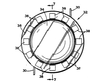
Figs. 6 and 7, there is shown an intraocular lens
apparatus 32 which, according to the present invention; is
provided with means for selectively changing the power of
the lens and means for selectively providing correction
for astigmatism. The lens apparatus 32 includes a central
lens body 3~ formed of a transparent flexible material and
attached about a periphery thereof to an inner periphery
of a ring 36 formed of a more rigid material. A pair of
the haptics 30 can be attached to the ring 3C.
Actuating means, in the form of a plurality of
actuator bodies 38, are equally spaced about the
, circumference of the ring 36. The bodies 38 can be
attached to a surface of the ring 36 or embedded when the
ring is formed. The bodies 38 are formed of a
magnetizable material such that each individual body
functions as a permanent magnet having a north pole and a
south pole. If adjacent ones of the bodies 38 are
magnetized to repel, the ring 36 will be expanded and
increase in circumference causing the lens body 34 to
; become less convex and, therefore, weaker in power. If
; adjacent ones of the bodies 38 are magnetized to attract,
.: .
, ' ' .
.
. : ~: , . - . ..
.. .. . .. . . .. .
.
WO92/0398s PCT/US91/0~14
27 2 ~ 72'~
the ring 36 will be contracted and decrease in
circumference causing the lens body 34 to become more
convex and, therefore, stronger in power. Thus, the
actuator bodies 38 can be utilized to selectively control
the power of the lens apparatus 32 after installation in
the eye and will retain the selected shape of the lens
body 36 until reset.
The degree of the astigmatism is readily determinable
through the use of conventional methods. An external
magnetic force can then be applied to predetermined ones
of the bodies 38 to expand or contract a portion of the
ring 36 aligned with the astigmatism to create the
necessæ.-y toric shape.
The magnetic force required to magnetize the bodies
38 should be sufficient to prevent exposure to normal
level everyday magnetic forces from resetting the lens 32.
In the Figs. 8 and 9 there is shown an alternate
` embodiment of the present invention. A lens apparatus 40
has a flexible central lens body 42 attached at a
periphery thereof to an inner periphery of a more rigid
ring 44. A plurality of magnetizable actuator bodies 46
are equally s~aced about the ring 44 for selectively
changing the power and the astigmatism correction as
discussed with respect to the lens apparatus 32. However,
25 portions 48 of the ring 44 positioned between the bodies
46 are formed with a reduced thickness as compared with
ad;acent portions which enclose the bodies. Thus,
additional flexibility of the ring is achieved requiring
.' ' .
~, .
. I .
' ' ' .: ' . ' .:
W092/03989 PCT/US91/0~14
2~672~7 28 ~
less force to compress or expand the ring and change the
shape of the lens body. A pair of the haptics 30 can be
attached to the ring ~.
The lens bodies 34 and 42 of the lens assemblies
shown in the Figs. 6-9 have a convex surface facing toward
the pupil 18 and a concave rearwardly facing surface.
Alternatively, a second alternate embodiment is shown in
the Figs. 31-33 as a lens apparatus 50 having convex front
and rear surfaces. A lens body 52 is formed from a
transparent, somewhat flexible material with a circular
shape in plan view and an ellipsoid shape in edge view.
An annular groove 54 extends about the periphery of the
body S2. Positioned in the groove 5~ is a ring 56 having
, a plurality of magnetizable actuator bodies 58 embedded
j 15 therein. If all adjacent actuator bodies are magnetized
to attract or repel, the ring 56 will contract or expand
respectively equally in all portions to selectively change
~! the shape of the lens thereby changing the power of the
lens. Also, two or three adjacent magnets can be
magnetized to produce an astigmatism correction, either at
opposite ends of a diameter to correct for regular
astigmatism as illustrated in the Fig. ll, or at one end
of the diameter to correct for irregular astigmatism as
illustrated in the Fig. 12. A pair of adjacent bodies 58a
, 25 and 58b have been magnetized to attract thereby changing
.1 .
the shape of the upper portion of the lens body 52 along
~, the line 12-12 in the Fig. 10. A pair of the haptics 30
can be attached to the ring 56.
. s
.`'
~ .
:~
.. . :
... : ,. ... ... :
- . ..
': :
.
, : . : . .
; ~,. ~ .
W092/03989 PCT/US91/0~14
~. 2~7~7
29
A third alternate embodiment is shown in the Figs.
13-16 as a lens apparatus 60 having a flexible or
malleable material (such as silicone) lens body 62
releasably attached to a rigid material (such as PMMA)
ring 64. A pair of the haptics 30 can be attached to the
ring 64. A plurality of spaced apart different diameter
circumferential grooves 66 are formed in a forwardly
facing surface 68 of the ring 6~. The lens body 62 has a
circumferential tongue 70 formed on an rearwardly facing
surface 72 thereof. The grooves 66 and the tongue 70 can
be formed with any suitable cross-sectional shape, such as
the trapezoidal shape illustrated, provided that the
grooves firmly retain the tongue to prevent separation of
the lens body 62 from the ring 64. The power of the
, 15 flexible or malleable lens body 62 can be changed
j selectively by moving the tongue 70 to an appropriate one
of the grooves 66, the outermost one of the grooves
resulting in the lowest power and the innermost one of the
grooves resulting in the highest power. Movement between
the grooves is accomplished by forming the grooves 66 with
a plurality of interruptions or adjustment spaces 74. In
a similar manner, the tongue 70 is segmented with each
segment corresponding in position to and being of no
,
greater length than one of the adjustment spaces 74.
The lens apparatus 60 can be preset for any desired
power prior to insertion in the eye by aligning the tongue
70 with a selected one of the grooves 66 and rotating lens
body 62 with respect to the ring 64 to insert the tongue
.
-
- ~:".
''
W092/03989 PCT/US91/0~14
2~72~7 30
into that groove. If a change in power is required, the
lens body 62 is rotated to align the segments of the
tongue 70 with the adjustment spaces 74, the tongue is
moved to the selected one of the grooves 66 for the power
desired and the tongue is rotated into the newly selected
groove. The entire tongue 70 can be formed of a
magnetically responsive material or an actuator body 76
(Fig. 16) formed of magnetically responsive material can
be embedded in each segment of the tongue. An external
magnetic field can be utilized to move each of the
actuator bodies 76.
In the Figs. 17 and 18, there is shown a fourth
alternate embodiment of the present invention. A lens
apparatus 78 includes a central lens body 80 attached at a
periphery thereof to an inner periphery of a more rigid
ring 82. A plurality of magnetizable actuator bodies 84
; are equally spaced about the ring 82 for selectively
changing the power and the astigmatism correction. The
bodies 84 can be embedded in the ring 82 together with a
means for retaining the shape of the lens apparatus 78
such as a shape retainer 86 formed of wire. The wire 86
extends circumferentially through the ring 82 between the
bodies 8~ and an outer periphery of the ring. A portion
of the wire 86 between each of the adjacent pairs of
bodies 8~ extends inwardly toward the center of the lens
apparatus 78 and turns sharply outwardly in a V-shape. If
an external electromagnetic force is applied to a pair of
adjacent ones of the bodies 84 tending to move the bodies
; 1". . ., ' ' .
. . .. , , ' ~. . . ' .
,,' ' '' ': ; '~ : , ' " `i ., . .. :
W092/03989 PCT/US91/0~14
,,~.. ~,............................................ . .
31 2B~72/~
together contracting the ring 82, at the same time, the V-
shaped pOrtion in the wire 86 between the adjacent bodies
is made narrower. When the external force is removed, the
wire 86 holds its new shape until the adjacent bodies 84
are mo~ed again even if the bodies are not permanently
magnetized. Thus, all of the bodies 86 can be moved to
compress or expand the ring 82 thereby increasing and
decreasing respectively the power of the lens apparatus
78. Furthermore, selected ones of the bodies 84 can be
acted upon to produce astigmatism correction in the
desired area of the lens apparatus 78. An example of the
ring 82 contracted from the shape shown in the Fig. 17 is
shown in the Fig. 18.
In Fig. 19, there is shown a fifth alternate
embodiment of the present invention. A lens apparatus 88
includes the lens body 80 from the previous embodiment
attached at a periphery thereof to an inner periphery of a
ring 90. The ring 90 has embedded therein a plurality of
the actuator bodies 8~ and the shape retainer 86. An
outer periphery 92 of the ring 90 has a plurality of V-
shaped notches 94 formed therein adjacent the V-shaped
portions of the shape retainer 86 in order to render the
ring 90 more responsive to compression and stretching.
Thus, the ring 90 is more flexible, requiring less force
to contract or expand than the previously described ring
82.
There is shown in Figs. 20-22, a sixth alternate
embodiment of the present invention. A lens apparatus 96
,'
:
. ... . . .
.
.
. : , . .
, . ., . : : ~ : ..
' ' ..
,, . . . ~ .
W092/03989 PCT/US91/0~14
~.
20$72~ 7 32
has a fleXible central lens body 98 attached at a
periphery thereof to an inner periphery of an inner ring
100. A plurality of magnetizable actuator bodies 102 are
e~ually spaced about the ring loo for selectively changing
the power and the astigmatism correction as discussed with
respect to the other lens assemblies above. However, an
outer periphery of the inner ring 100 is attached to an
inner periphery of an outer ring 106 having a shape
retainer lC4 embedded therein. The shape retainer 104, as
more clearly seen in Fig. 21, is of tubular shape and acts
in an accordion fashion to lengthen and shorten. When two
adjacent ones of the actuator bodies 102 are moved toward
; one another or away from one another, the inner ring lOo
and the outer ring 106 tend to contract and expand
respectively. The shape retainer 104 is also contracted
or expanded and is formed of a material which retains its
position until it is again forced to move by the bodies
102. Thus, the power and the astigmatism correction for
the lens 96 can be controlled through the movement of the
actuator bodies 102 and the shape retention capabilities
of the shape retainer 10~.
There is shown in Fig. 23, a seventh alternate
; embodiment of the present invention based upon the lens
apparatus 96 shown in Fig. 20. A lens apparatus 108
utilizes the lens body 98, the inner ring 100, the
actuator bodies 102 and the outer ring 104 as described
above. However, the shape retainer 104 has been replaced
with a helically formed wire shape retainer 110 which
~.
~ .. . .
b . '
~'
. ~ ~_.. .. . . '
' ' . . ,,, ."' '' ' ' '' ""' ' '
;' ',' " ' ~ ' ' , ' ' ' ' , :., '' " ' .''"'' '~ ' ' ' , ' '
,' ' ' .'' ' : : ' ' ' ' '
~ ,,, ' : , ' . ' ' ' ' ' ' ' -
' ' ' . ' . '
"";' ' ' . .. ' '~ ~ ' ' ''
W092/03989 PCT/US91/0~14
~ 2~72~
retains its shape once set by the movement of~the actuator
bodies 102.
There is shown in the Fig. 24 a block diagram of a
control system for operating the actuator bodies in each
of the above-described lens assemblies. A power supply
112 has an output connected to an input of a field
strength control 11~. An output of the field strength
control 11~ is connected to a first coil 116. A second
output of the field strength control 114 is connected to a
second coil 118. Each of the coils 116 and 118 can be
mounted on a holder 120. The holder is any suitable
device for positioning the coils 116 and 118 adjacent
associated ones of the actuator bodies in any of the
above-described lens assemblies. The field strength
control 114 is selectively adjustable for applying a wide
range of electrical power to each of the coils 116 and 118
individually in order to either permanently magnetize or
simply move the associated body in accordance with the
method of operation of one of the lens assemblies
according to the present invention. ~he coils 116 and 118
, are representative of either a single coil which can be
separately aligned with each of the actuator bodies in
turn or any other number of such coils including a
separate coil for each of the actuator bodies such that
all of the actuator bodies in a lens apparatus can be
operated at the same ti~e.
As shown in the Fig. 25, the holder 120 can form a
portion of an instrument 122 for operating the actuator
., .
... . .
W092/03989 PCT/US91/0~il4
. -;, j . .
2 ~ ~ 7 2 4 7 34 ~
bodies of a lens apparatus according to the present
invention. The holder 120 can be ring-shaped having a
center aperture i2~ which can be utilized to align the
instrument 122 with one of the lens assemblies according
to the present invention ins~alled in the human eye.
Mounted on the ring-shaped holder 120 are the coils 116
and 118, each of the coils being positioned in alignment
with an associated one of the actuator bodies in the lens
apparatus to be operated. Additional coils 126 mounted on
the holder 120 are shown schematically and represent any
.- .
deslrable number of such coils. The coil 116 is connected
by a pair of lead wires 128 to any suitable control such
, ~ as the field strength control 11~ shown in Fig. 24.
i Similarly, the coil 118 is connected by a pair of lead
wires 130 to a suitable control.
The polarity of the magnetic field generated by the
coils 116 and 118 can be reversed by simply reversing the
current ~low through the associatéd wires lZ8 and 130.
The coils 116 and 118 can also be provided with mechanical
means for orienting them to selectively operate the
actuator bodies in a desired manner. For example, the
coil 116 can be mounted on a carrier 132 slidably retained
~ in a circumferentially extending slot 13~ formed in a face
h~; of the holder 12~; The carrier 132 can be moved in either
i25 direction along the slot 13~ to accurately position the
coil 116 with respect to an associated actuator body.
Alternatively, the coil 118 is shown mounted on a circular
carrier 136 which is rotatably mounted on the holder 120.
, ~
j :
W092/03989 PCT/US91/0~14
!-~ 2~672~7
Thus, the angular orientation of the coil 118 with respect
to a radius of the holder 120 can be selectively changed
as desired.
The instrument 122 can be incorporated into a device
which can be utilized by the patient to change the shape
of the lens body as the situation requires. For example,
the instrument 122 can be built into a pair of eyeglasses
or formed as a handheld control and operated by the
patient to change the lens focus between near vision and
far vision
There is shown in the Fig. 26 a control for
automatically operating the actuator bodies of any of the
above-described lens assemblies according to the present
invention. The previously described power supply 112 is
connected to an input of a control unit 138. A pair of
.
outputs of the control unit 138 are connected to the coils
;;116 and 118 which are mounted on the holder 120. The
control unit 138 can include a general purpose, programmed
microprocessor having a standard operating software system
and a program for receiving instructions through a
keyboard 1~0 connected to an input of a control unit 138
as to the strength and duration of the electrical power to
be applied from the power supply 112 to the coils 116 and
118 in order to operate the actuator bodies as desired.
'25 In addition, a position sensor 142 can be connected to an
input of the control unit 138 for generating a signal
representing the position of the holder 120 and the coils
116 and 118 with respect to the lens apparatus to be
.`,'
~:,
....
.... .... . .. . . . , . ~ , . , ~ :: . . :-
W092/03989 PCT/US~1/0~14
2 ~ $ 7 ~ a r6~
36
operated. The position sensor 142 can be any suitable
device, typically light sensitive, for detecting any of
the physical features on the lens apparatus. For example,
the position sensor 142 could detect the actuator bodies,
a periphery of the lens body, or a periphery of the ring
shown in any of the preceding lens apparatus embodiments.
The actuator bodies can be formed of a ferromagnetic
element or one of a variety of alloys of ferromagnetic and
other elements which respond to a nearby magnetic field.
In some cases, the actuator elements can be "permanently"
magnetized, magnetized even though the external magnetic
field is removed, or simply aligned in response to the
alignment of the electromagnetic field where a shape
retainer is employed.
In summary, an improved intraocular lens is provided
to eliminate or reduce the post-operative regular and
irregular astigmatism. The invention utilizes an
intraocular lens having a flexible center lens body
; surrounded by an outer ring having actuator bodies
sensitive to an external force such as a magnetic field.
Through the implementation of an external magnetic force,
the shape of the lens can be changed to elongate the lens
along a predetermined axis for correcting astigmatism. If
the axis of the astigmatism changes, the shape of the lens
can be changed by reapplying the force along a different
~; aXis. The ring can also be utilized to change the power
of the ].ens, by changing the spherical shape. The
utilization of such an intraocular lens may eliminate the
s
, .
,~.
- ' ~ . . . : - . ,
: : .
W092/03989 PCT/US91/0~14
.~-;; 2~72~7
need of the recovering cataract patient to wear eye
glasses or contact lenses necessary. The elimination of
the glasses or contact lenses amounts to an immense
benefit to the recovering cataract patient, many of whom
are elderly and have enough hardships without being
burdened with wearing glasses or contact lenses.
Furthermore, a source of the external force can be
incorporated into a pair of eyeglasses, if needed, or a
handheld device to be selectively operated by the patient
for the accommodation of different focal lengths.
Although the actuator bodies have been described as
formed of ferromagnetic material responsive to a magnetic
field, such bodies could be formed of any suitable
material responsive to electromagnetic or mechanical
energy waves which cause the ring to compress and expand
circumferentialiy~
The various lens apparatuses discussed above can be
categorized as "active" or "passive" systems. The
"active" systems (32, ~6 and 50) require the application
of a force field to the actuating means to selectively and
reversibly alter the shape of the lens body. The
; actuating means then actively generates its own force
field to maintain the selected shape. The "passive"
systems ~60, 78, 88, 96 and 108) also require the
application of a force field to the actuating means to
` selectively and reversibly alter the shape of the lens
; body. However, the actuating means does not require any
force field to maintain the selected shape. Any of the
, I .
';' -
. . .: . , . , . - . . .
: , .. .. : - .. :. : .
W092/03989 PCTtUS91/0~14
2~72~7 38 ~
actuating means of the "active" systems and any of the
actuating means of the "passive" systems in the
embodiments shown and described can be substituted for
each other as desired. Furthermore, any of the actuator
segments shown can be mounted for rotation in the
associated ring similar to the coil 116 and the carrier
- 136 shown in the Fig. 25. Such an actuator segment would
remain magnetized and could be rotated by the instrument
; 122 to attract or repel an adjacent actuator segment or be
oriented in a neutral position.
In the Figs. 27-29, there is shown a micromotor
actuated variable focus intraocular lens apparatus
according to the present invention generally indicated by
a reference numeral 32, which lens is provided with means
for selectively changing the spherical power of the lens
and means for selectively providing correction for
astigmatism. The lens apparatus 32 includes a central
lens body 3~ formed of a transparent flexible material,
;' such as a silicone or the like. The lens body 34 is
generally disc-shaped and has an anterior convex surface
' adapted to be centered in the pupil of an eye and planar
rear surface. However, the anterior and rear surfaces can
, be any desired combination of concave, planar and convex.
An inner ring 36 is attached about a periphery of the lens
body 3~ by any suitable means, such as being molded
integral therewith as shown in the Fig. 28. The inner
ring or rings 36 can be formed of any suitable elastomeric
..~
, material to provide for appropriate expansion and
,
: i
, ~
: . . . - . , . - ,
,.': . " ,, ~ ,. ~ .' . ,
:
.: . .. .
-,
. . . , .
.
wo92/o3s89 PCT/US91/0~4
2~72~7
39
contraction of its periphery with ~he abilities to become
oval, segmented or wave like and the peripheral
circumference depending upon the orientation of the
micromotor.
The ring 36 is retained by a plurality of spaced
micromotors 38 extending radially inwardly toward the
center of the lens body 3~. The micromotors 38 each have
an inner end for retaining the inner ring 36 and an outer
end attached to an outer ring 40 which extends
concentrically about the inner ring 36. The outer
supporting or mounting ring ~0 is made from a rigid
plastic or other material and provides a fixed support for
the micromotors 38 and the lens body 3~. A pair of
haptics 30 can be attached to the ring ~0 to support the
lens apparatus 32 in the proper position in the eye.
As shown in the Fig. 28, each of the micromotors 38
can be formed as a tuning fork having a pair of spaced
apart generally parallel prongs or legs 42 and ~
branching from a base or handle ~6 attached to the outer
ring ~0. Facing surfaces of the prongs ~2 and ~4 have
grooves or teeth ~8 and 50 respectively formed therein.
The grooves ~8 and 50 cooperate with a pair of opposed
flanges 52 and 5~ respectively formed on the inner ring 36
to retain the adjacent portion of the lens body 34 and the
inner ring 36 a selected distance from the outer ring 40.
If the inner ring 36 is expanded and increased in
diameter, the lens body 34 will tend to become less curved
and the power of the lens assembly 32 will be reduced. If
i, :
.: ~. . . - . . . .~ ~. .. . ..
W092/03989 PCT/US91/0~14
. . , ,~
2~7 40
the inne~ ring 36 is csntracted and reduced in diameter,
the lens body 3~ will tend to become more curved and the
power of the lens assembly 32 will be increased. The
micromotors 38 cooperate with the inner ring 36 to provide
selective adjustment of the power of the lens from outside
the eye. Each of the micromotors 38 can be powered by a
control signal from an external energy source, not shown,
such as a source of ultrasonic energy at a predetermined
controlled frequency and/or amplitude which tends to
vibrate the prongs 42 and 44, oscillating them in the
direction of the double headed arrows 58 and 60
respectively. This produces a wave action that can cause
selective movement of the flanges 52 and 54 in the
direction of the double headed arrows 58 and 60
respectively. As each of the prongs 42 and 44 moves
, horizontally, the associated flanges S2 and 54 will
disengage from the grooves 48 and 50 respectively and then
to reengage in a different groove further away or closer
to the outer ring ~0 depending on the externally
`20 controlled frequency and thus the propelling wave action
generated
longitudinally along the prongs 42 and ~. The micromotor
38 can be responsive to different amplitudes of ultrasonic
energy. it can be responsive to ultrasonic energy at a
j 25 first predetermined frequency for contracting and a second
; predetermined frequency for expanding the inner ring 36.
There is illustrated in the Fig. 29 an alternate
embodiment of the micromotor 38. A micromotor 38~ is
'''
~i .
.,
`,~ .
. . .
. -: . .1 , . : , .
.
WO92/03s89 PCT/US91/0~14
,, ............................................ . :; .
41 2`~72l.~7
formed similarly to the micromotor 38, but includes a
linear positioning device 64 located between the prongs 42
and ~4. The positioning device 64 has a body 66 attached
at one end to the base ~ 6 . A rod 68 extends from the
opposite end of the body 66 and has a free end attached to
the inner ring 36. The linear positioning device is
responsive to a control signal from an external source of
energy (not shown) for extending and retracting the rod 68
thereby moving the ring 36 in the direction of the arrow
62 to. change the shape of the lens body 3~. The source of
energy can be ultrasonic as discussed above. The source
of energy can be electromagnetic (laser beam, radio waves,
etc.). In either case, conventional devices are known for
converting such energy into the linear motion 62.
If all of the micromotors 38 and 38~ are operated to
maintain the inner ring 36 in a circular configuration,
only the power of the lens assembly 32 will be changed.
If individual ones of the micromotors are operated to
change the shape of associated segments of the lens body
34, a selective correction for astigmatism can be made.
one micromotor can be actuated to correct for irregular
astigmatism and two opposing micromotors can be actuated
to correct for regular astigmatism. Each of the
micromotors 38 and 38~ can be responsive to a different
frequency for selective actuation or each micromotor can
, be selectively activated by external selective
stimulation.
.'
, . . .: .
. ~ ,, . , . . . , , .. . , ~ . .
. . .~. . ~' . ':.
W092/03989 PCT/US91/0~14
2U672~7 42 ~ `
In the Figs. 30-33 there is illustrated an alternat~e ~-
embodiment of the present invention. A lens apparatus 70
has a flexible central lens body 72 attached at a
periphery thereof to a relatively rigid mounting or
supporting ring 7~ formed of a plurality of overlapping
curved segments. Although only three segments 76, 78 and
80 are shown, the ring 7~ can be formed of any suitable
number of segments. The ring 7~ can be attached to the
periphery of the lens body 72, as discussed below with
respect to the Fig. 31, or extend through a hollow edge
portion of the lens body 72, as discussed below with
respect to the Fig. 32. A pair of haptics 30 can be
attached to the ring 74, one haptic 30 being attached to
the segment 78 and the other haptic 30 being attached to
lS the haptic 80.
Referring to the Fi~. 31, there is shown an enlarged
cross-sectional view of the overlapping segments 76, 78
and 80 which form a micromotor. on the facing sur~aces of
the overlapping portions of the segments 76 and 80 are
formed cooperating grooves 82. On the facing surfaces of
the overlapping portions of the segments 78 and 80 are
formed cooperating grooves 8~. The grooves 82 and 8~
, selectively permit relative motion between the associated
i segments thereby fixing the power and astigmatism
!
correction of the lens apparatus 70 at selected values as
explained below.
Referring to Fig. 32, there is shown a portion of the
I lens apparatus 70 wherein the segments 76 and 78 overlap
. .
,
.. ~
,: ,:
.: .
.. . . .. . . .
,, ~ . ~ . , , . :
W092/03989 PCT~USgl/0~14
43 2 ~72~7
to form a micromotor. On the facing surfaces of the
overlapping portions of the segments 76 and 78 are
formed cooperating grooves 86 which selectively permit
relative motion between the segments as explained ~elow.
The overlapping portions of the segments 76 and 78
slidably extend through a hollow edge portion 88 of the
lens body 72.
The overlapping portions of the segments and the
grooves shown in the Figs. lO and 11 form micromotors 90
and 92 respectively. These micromotors are representative
of a plurality o such elements which can be spaced about
the periphery of the lens body 72 in a manner similar to
the micromotors 38 and 38~ shown in the Fig. 27. The more
; micromotors that are used, the more uniform will be the
curvature of the lens body 72 and the more precise will be
the ability to adjust spherical and astigmatism
correction.
Relativ~ly little sliding movement between
overlapping segments is required to change the shape of
the lens body 72. One means for achieving such movement
would be to form the grooves 82, 8~ and 86 such that
vibration of a segment at a first frequency would cause
; relative movement in one direction between the segments
and vibration at a second frequency would cause relative
movement in the opposite direction. The vibration could
be induced by externally applied ultrasonic energy.
Another method of achieving such movement would be to
induce magnetic poles in the segments which poles would be
.- ' ,
: , , ~ ' , ' ' ~.
, , : . , . , :
.. , ., :, , ~ .
., : , , :: , . , :
- ,~
.
W092/03989 PCT/US91tO~14
20~72~7 44 ~
paired to attract or repel as required. In any case, the
grooves selectively permit relative motion between the
associated segments thereby fixing the power and
astigmatism correction of the lens apparatus 70 at
selected values. External mechanical pressure on the
haptics 30 could be used to trigger a micromotor to cause
circumferential movement of the overlapping ridges. If
; the micromotor has the capability to store potential
energy, then external mechanical pressure on the haptics
could be utilized to release such energy or to restore
such energy. For example, a compressed spring located in
- the positioning device 64 could be released or
recompressed.
There is shown in the Fig. 33 a micromotor 94
lS connected between the inner ring 36 and the outer ring 40.
The micromotor 94 has a central body 96 which can be
generally cylindrical in shape and capped at opposite ends
by a pair of end walls 96a and 96b. One end wall 96b of
the body can be attached to the outer ring ~0. Extending
from an opposite end of the body is a rod 98a having an
exposed end attached to the inner ring 36. The rod 98a
!~, extends through the end wall 96a into a cylinder chamber
; lOOa formed in the body 96. A piston 98b is slidably
retained in the chamber lOOa and is attached at an upper
surface to an abutting end of the rod 98a. The cylinder
lOOa is filled with a fluid under pressure such as a gas,
the gas in an upper portion of the cylinder being at a
, .
:. ~
.
; ~ .
.~..
W092/03989 PCT/US91/0~14
~i~s 2~72~7
.; . . . .
higher pressure which forces the piston ssb toward the
lower end of the cylinder lOOa.
Formed concentrically about the chamber lOOa is a
reservoir lOOb filled with a compressible fluid under
pressure. The upper end of the cylinder 100~ is connected
to an upper end of the reservoir lOOb by a two-way valve
: 102a located in the end wall 96b. A lower end of the
cylinder 100~ is connected to a lower end of the reservoir
lOOb by a two-way valve 102b located in the body 96 and a
radially extending passageway 104 formed in the end wall
96b. The reservoir lOOb is divided into upper and lower
portions by an annular piston 106. The piston 106 can be
responsive to a control signal such as an
external source of power for actuation to move in a
; 15 downward direction decreasing the pressure on the fluid in
. the upper portion of the reservoir lOOb and increasing the
l pressure on the fluid in the lower portion of the
; reservoir lOOb. Fluid will flow from the upper portion of
the chamber lOOa through the valve 102a into the upper
portion of the reservoir lOOb and fluid will flow from the
..
lower portion of the reservoir lOOb through the passageway
10~ and the valve 102b into the lower portion of the
chamber lOOa thereby forcing the piston 98b and the rod
98a in an upward direction as indicated by an arrow 108.
Movement of the piston 106 in an upward direction will
cause opposite movement of the piston 98b. Such movement
of the piston 98b will cause relative displacement between
the inner ring 36 and the outer ring ~0 thereby causing a
. . .
., ~ .
..
~ , . : . ~ .. : .. -
... ~ .. .. . . . . . . ..
. ~ - , . . ~ .. .. : . ... , : :
W092/03989 - PCT/US9l/0~14
, . ~ . .. ~ ~ ,
2 ~ ~ 7 7 ~ 7 46 l~ ,F~` '
change of shape in the lens body 3i. Other forms of
reservoirs could be utilized such as flexi~le membranes
responsive to external mechanical pressure for actuating
the micromotor or recharging the fluid in the reservoir
thereby storing energy for later use. External mechanical
,i pressure or external triggers such as ultrasound or laser
~, . .
could be utilized to actuate a micromotor or the valves
102a and 102b, or recharge a reservoir, for example by
making the fluid or gas in the chamber 100a expand thereby
storing energy for later use.
There is shown in the Fig. 34 another embodiment of
the present invention. A micromotor 110 has body 112
which can be generally cylindrical and have one end
~ attached to the outer ring ~0. A rod 11~ extends through
.~ .
an end wall of the body 112 opposite the outer ring ~0.
An outer end of the rod 11~ is attached to the inner ring
36. An inner end of the rod 11~ extends into a central
cavity 116 which can be internally grooved or threaded in
a helical pattern. Rotatably mounted on the rod 11~ is an
externally threaded nut 118 which threadably engages the
wall of the cavity 116. In response to a source of
~' external power, the nut 118 can be caused to rotate. If
., .', the nut 118 is fixed in position on the rod 11~, then as
the nut rotates and travels along the longitudinal axis of
~ 25 the cavity 116, the rod will be moved in the direction of
,~`l a double headed arrow 120 to cause relative movement
between the inner ring 3C and the outer ring 40 thereby
changing the shape of the lens body 3~.
~ '
~;, .
'~' ' ' .; .~.. . .
`' '...... . :: : . , ,
,.: ,' . . '. ~ ,.
, . .~
W092/03989 PCT/US91/0~14
~ 47 2 ~ 72~ 7
There is shown in the Fig, 35 yet another embodiment
of the present invention. A micromotor 122 has body 124
which can be of any suitable shape with an inner end
attached to the inner ring 36. The micromotor 122 also
includes a motive means 126 mounted on an outer end of the
body 12~ adjacent the outer ring ~0. An annular track 128
is attached to the inner circumference of the outer ring
~o and has an inwardly facing helical groove 130 formed
therein. The motive means 126 can be any suitable device
such as a wheel or endless belt which can be moved along
the groove 130 to rotate the lens body 3~ about its
center. The motive means 126 can be driven in a
conventional manner in response to a source of external
power and/or control signal to rotate the lens body 34 and
move the lens body in the direction of a double headed
arrow 132 thereby changing the axial position and
functional power of the lens body 34 when installed in an
, eye. The micromotor 122 is representative of a plurality
of such devices which can be spaced about the inner ring
36 in a manner similar to the micromotors 38 and 38~ shown
; in the Fig. 27.
If the groove 130 shown in the Fig. 35 is formed as a
plurality of annular grooves rather than a single helical
groove and the lens body 34 and the inner ring 36 are
formed of a flexible material, then astigmatism correction
can be made. Openings (not shown) can be formed through
the walls between adjacent grooves to permit the motive
, means 12~ to travel between the grooves. If the grooves
,~' ,
., .
. . , -.,
... . .
,. . .. ~ - ., ., . - . . .
: :.. -. -: ,, -
.
: . . - ., , : . . . .
; W092/03989 PCT/US91/0~14
~ ~ ~ 7 ~
48
are formed with different depths, éach of the motive means
12~ can be moved individually to select the desired groove
thereby changing the configuration of the associated
portion of the inner ring 36 and the lens body 3~ to
provide astigmatism correction.
The utilization of such an intraocular lens in
accordance with the present invention may eliminate the
need of the recovering cataract patient to wear eye
glasses or contact lenses. The elimination of the glasses
or contact lenses amounts to an immense benefit to the
i recovering cataract patient, many of whom are elderly,
sometimes forgetful, and many have financial and physical
hardships. Furthermore, a source of the external force
` can be incorporated into a pair of eyeglasses, if needed,
or a hand held device to be selectively operated by the
patient, or the micromotor could be responsive to pressure
applied from outside the eye to produce a change in the
focal length (focus) so the lens would have accommodation
(the ability to change focus from distance to near).
present invention has the advantage over prior art devices
of not requiring a physical or electrical connection
between the source of the power and the lens in order to
change the lens.
The variable focus lens of the present invention has
a variety of applications, in addition to the application
as an intraocular lens. For example, the variable focus
lens can be used as a camera lens. The lens could be used
' as an alternative to or in conjunction with cameras having
,
'
,
.~ .
. ,
., ~ . .
: . :
W092/03989 PCT/US91/0~14
29~72~7
- 49
either a fixed lens, an adjustable lens, or a plurality of
interchangeable lenses.
In the Figs. 36-39, there are shown various
embodiments of a micromotor actuated adjustable focus
intraocular lens apparatus according to the present
invention. In the Fig. 36, the appa~atus is generally
indicated by a reference numeral 32, which lens is
provided with means for selectively changing the
functional spherical power of the lens and for selectively
providing correction for astigmatism. The lens apparatus
! 32 includes a central lens body 34 formed of a transparent
flexible material, such as a silicone or the like. The
; lens body 34 is generally disc-shaped and has a convex
surface adapted to be centered in the pupil of an eye and
may have a concave, planar or convex rear surface. A
per~phery 36 of the lens body 34 can be formed as an inner
ring of any suitable material to provide a stable mounting
means for actuators. For example, the periphery 36 could
be molded integral with the lens body 34, but thicker in
cross section.
A plurality of spaced micromotors 38 extend radially
inwardly toward the center of the lens body 34. The
micromotors 38 each have an inner end attached to the
inner ring 36 and an outer end attached to an outer ring
40 which extends concentrically about the lens body 34.
The outer ring ~0 is made from a relatively rigid material
and provides a fixed support for the micromotors 38 and
~l ', . .
';
.
,
1.",. ': - ' :' '
.
.. , . . . ~ . : , . .
W092/03989 PCT/US91/0~14
.', . ~,
~7~7 ~ 50
the lens body 34. The outer ring io supports the lens
apparatus 32 in the proper position in the eye.
In the Fig. 36, if it is assumed that the lens
assembly 32 is being viewed from the front, outside the
cornea of the eye, then movement of the lens body 34
toward the viewer, the anterior direction, will increase
the functional power of the lens based upon well known
optical principles. Conversely, movement the lens body 34
toward the optic nerve, the posterior direction, will
decrease the functional power of the lens assembly 32.
The micromotors 38 cooperate with the inner ring 36 and
the outer ring ~0 to provide selective adjustment of the
power of the lens from outside the eye. Each of the
micromotors 38 can be powered by an external energy
lS source, not shown, such as a source of ultrasonic energy
at a frequency which causes extension action by the
micromotors 38 equally moving the lens body 34 forward in
a horizontal direction while maintaining the lens body 34
i in a generally vertical plane. The application of energy
, 20 at a different frequency will cause retraction action by
the micromotors 38 equally moving the lens body 34
posteriorly in the horizontal direction while maintaining
the lens body in the generally vertical plane.
There is shown in the Fig. 36 a radius 42 of the lens
apparatus 32 extending outwardly from a center point 44 of
the lens body 34 through one of the micromotors 38, a
i~ micromotor 38a. Any rotation of the lens body 34 about an
axis in the plane normal to the path of light rays between
i
" ' : ' ' .. ~
.
,: .,
.
.
WO92/03s8s PCT/US91/0~14 ~ ~
~ 51 2Q~72d7
the cornea 12 and the optiç nerve 24 (Fig. 5) will cause
an induced astigmatic effect for modifying astigmatism.
If an irregular astigmatism which by its nature is
segmental were located along the radius 42 and the lens
5 body was made of flexible material, the micromotor 38a
could be actuated to bend a segment of the lens body 34
relative to the remaining portion of the lens body. The
more common variety of astigmatism is regular astigmatism
which extends completely across the optical axis.
correcting regular astigmatism oriented along an axis ~6
can be adjusted by activating the micromotors 38a in an
anterior direction and 38c in a posterior direction, or
visa versa.
Prior to implantation in the eye, the optical
properties of the lens could be measured and stored in a
computer, for example, with reference to the various
combinations of actuation of the micromotors. After
implantation in the eye, the data stored in the computer
can be utilized along with postoperative information to
guide the actuation of the micromotors to produce the
desired dioptic power and astigmatic modifications. To
! correct for irregular and regular astigmatism and for
dioptic power adjustments, micromotor manipulation may be
aided by a computer program that calculates the amount of
activation to be used on each micromotor by analyzing
information from the following sources: corneal
topography, corneal curvature radii, the refraction, axial
length of the eye, and other ocular and lens data.
'
.- . ~. : . . ................... ~ - :
.. .
- , , ~ ,: , . . : . . . , : . : . ,
W092/03989 PCT/US91/0~14
52
~ ~T~e~ illustrated in the Fig. 37 an alternate
embodiment of the lens apparatus 32. A lens apparatus 48
includes the lens body 34 having the inner ring 36
attached to a plurality of micromotors 38. An attachment
means in the form of a pair of loops 50 is attached to the
lens body with an end of each of the loops 50 attached to
an associated one of the micromotors 38. The lens
apparatus ~8 operates in a manner similar to the lens
apparatus 32.
In the Fig. 38 there is illustrated an alternate
embodiment of the present invention. A lens apparatus 52
includes the lens body 34 having the periphery 36 thereof
attached to one end of a generally horizontally extending
one of the micromotors 38. The opposite end of the
micromotor 38 is attached to one end of one of the legs
30. Thus, the micromotor 38 can be actuated in the
, direction of an arrow 54 thus changing the focal power
'' and/or the astigmatic power of the lens body 3~.
Referring to the Fig. 39, there is shown an enlarged
; 20 cross-sectional view of another alternate embodiment of
1 the present invention. A lens apparatus 56 includes the
^, lens body 34 having the periphery 36 thereof attached to
one end of a generally vertically extending one of the
micromotors 38. The opposite end of the micromotor 38 is
attached to one end of one of the legs 30. Thus, the
micromotor 38 can be actuated in the direction of an arrow
58 to change the focal power and/or the astigmatic power
of the lens body 34. If one end of the micromotor 38 is
.
.
~, . "'
:.
W092t03989 PCT/US91/0~14
~ 2~72~7
53
attached to the leg 30 by a pivot means 60 and the other
end of the micromotor 38 is attached to the ring 36 by a
pivot means 62, then the micromotor 38 can be actuated to
extend, as shown in phantom, and mov~ along a curved path
as illustrated by an arrow 64.
The Fig. 40 is a block diagram of a system 66 for
testing and storing data to be used to selectively
position and orient the intraocular lens apparatus 32. A
micromotor control device 68 individually controls the
actuation of each of the micromotors provided in the lens
apparatus 32. The micromotor control device 68 generates
control signals and/or the power necessary to actuate the
micromotors. At the same time, the control device 68
generates information signals to a computer 70 identifying
which micromotors have been actuated. An optical sensing
device 72 is provided for sensing the optical properties
of the lens apparatus 32 and providing such information to
the computer 70. The computer 70 stores the optical
information from the sensing device in association with
the control information from the control device 68 for
later use.
The Fig. 41 is a block diagram of a system 74 for
selectively positioning and orientating the intraocular
lens apparatus 32 after implantation in the eye. When it
is desired to change the functional power of the lens
apparatus 32 and/or provide an astigmatism correction, the
. ~ .
, computer 70 will provide the necessary output signals to
the micromotor device 68. The micromotor device 68
.
:.- . . ., : . : .:
W092/03989 PCT/US91/0~4
,; . .
2~72~7
54
responds to the information from the computer 70 to
generate the appropriate control signals and/or power
necessary to actuate a predetermined combination of the
micromotors in the,lens apparatus 32 to produce the
desired results.
Not only can the adjustment process described with
respect to the Figs. lo and 11 be performed in a doctor's
office or medical facility, for example, but the system
for selectively positioning and orientating the
intraocular lens apparatus could be provided for use by
the patient. ~he computer 70 and the micromotor control
device 68 could be located in a pair of eye glass frames
with controls for use by the patient., When the patient
i sensed a need to change the functional power, the patient
would put on the glass frames, push an appropriate button,
, and the intraocular lens apparatus would be automatically
t changed. One example of such use could be when the
patient wished to switch from distance vision to vision
; for close work such as reading or watch repair. The
system according to the present invention could be
, provided so that it could be manipulated by the patient to
self adjust the intraocular lens apparatus even providing
adjustments for good vision as close as six inches from
the work.
, 25 ,The utilization of such an intraocular lens in
,~, accordance with the present invention may eliminate the
need of the recovering cataract patient to wear eye
' glasses or contact lenses. The elimination of the glasses
J
~'
.
.
~ ~ .
. . ~ .
` ' ' ': ' -
W092/03989 PCT/US91/0~14
,': 55
or contact lenses amounts,to an immense benefit to the
recovering cataract patient, many of whom are elderly,
sometimes forgetful, and many have financial and physical
hardships.
The adjustable focus lens of the present invention
has a variety of applications, in addition to the
application as an intraocular lens. For example, the
adjustable focus lens can be used as a camera lens. The
lens could be used as an alternative to or in conjunction
with cameras having either a fixed lens, an adjustable
lens, or a plurality of interchangeable lenses.
In accordance with the provisions of the patent
statutes, the present invention has been described in what
is considered to represent its preferred embodiment.
15 However, it should be noted that the invention can be ''
practiced otherwise than as specifically illustrated and
described without departing from its spirit or scope.
:~
.
. .
~ '
.
. . .
,
. '' , .' ': ' -'' ':.' ' '."' ~' ~ "
: ' ~: ~; . -. . ' ' ' : , '
. .