Note: Descriptions are shown in the official language in which they were submitted.
i
CA 02067412 2003-10-17
35614/JP11/RTP
1IY4CARDI71L R89A8cvIJ~tI871TIO~f THROVaB
T8E ENDOClIRDIIIh ByR~'11CE UBI~ia 11 ?~lIBBR
HACICaRO~IND Ol~' THE INti8I~1'1'IO~T
The present invention relates to a myocardial
revascularization device and method for making channels
in the inside of the heart ventricle to perfuse the
myocardium.
Within this application several publications are
references by arabic numerals within parentheses. Full
citations for these and other references may be found at
the end of the specification immediately preceding the
claims.
It is well known that coronary artery disease is a
pervasive disease afflicting many people in this country.
Many of these people are treatable by coronary artery
bypass surgery. However, alternative methods of
myocardial revascularization are required for patients
with coronary artery disease not amenable to coronary
artery bypass. Investigators have used the COZ laser in
arrested hearts to create transmural channels from the
epicardial surface. The channels increase cardiac
perfusion by shunting blood from the ventricle to
myocardial sinusoids, and can endothelialize and remain
patent indefinitely. In this approach, the energy is
delivered from outside the ventricle, and the channels
formed by the laser energy penetrate the lull thickness
through the ventricular wall.
The method may include the steps of positioning an aiming
beam energy emitter inside the ventricle of the heart,
said aiming beam energy emitter having an emitting beam
which identifies the location of the emitted energy from
the channel forming energy emitter, locating an aiming
beam energy detector outside the heart at a position
adjacent a desired channel forming site, and wherein the
step of directing energy from the channel forming energy
emitter is performed after the aiming beam energy
detector detects aiming beam energy to thereby indicate
that the channel forming energy emitter is directed to
the desired channel forming site. The desired channel
forming site may be based on familiar epicardial anatomic
landmarl~s, such as the epicardial branches of the
coronary arteries.
According to another aspect, the present invention
provides a method for myocardial revascularization of the
heart in a patient, comprising entering the ventricle of
the heart with a catheter having a lumen which houses a
(fiber which emits energy at a (fiber end, locating the
fiber end proximate ~o the ventricular wall, and emitting
energy from said fiber end in an amount sufficient to
form a channel in the ventricular wall into the
myocardium to thereby increase myocardial blood flow from
the endocardium to the myocardium.
A myocardial revascularization devise is also provided in
aCCOrdance with the invention, comprising a handpiece
having at least one lumen, and having an insertable end
and a handling end, a fiber for carrying energy from an
energy source to a fiber end from which the energy is
emitted, said fiber being received in one of said lumens,
means for moving the fiber within the lumen to different
stop positions, whereby the fiber end extends from the
handpiece insertable end at different sites of a
ventricular wall corresponding to said stop positions,
and means for transmitting energy to said fiber end in an
amount sufficient to form a channel in the ventricular
wall into the myocardium at each of said sites, to
thereby increase myocardial blood flow from the
endocardium to the myocardium.
The present invention also provides a myocardial
revascularization device, comprising a channel forming
energy emitter means, for insertion into the ventricle
cavity of a heart, for emitting energy to form at least
one channel in the ventricular wall into the myocardium,
an aiming beam energy emitter means for emitting an
aiming beam which .identifies the location of energy
emitted from the channel forming energy emitter means, an
a~,ming beam energy detector, for placing against the
exterior of the heart, for detecting an aiming beam from
the aiming beam energy emitter means, and means for
energizing the channel forming energy emitter means in
response to detection of an aiming beam by the aiming
beam energy emitter. The aiming beam energy detector
location may be selected on the basis of familiar
epicardial anatomic landmarks, those being the epicardial
branches of the coronary arteries.
These and other advantages will become apparent from the
detailed description accompanying claims and attached
drawing figures.
',~~'~~ ~~_
~~~c~i~r~~a~ ~~ xxa n~,xxr~~
Fig. 1 is a cross~sectional view of a ventricular wall of
a heart, showing the epicardium, myocardium, endocardium
and a channel formed by a laser energy source according
to the present invention:
Fig. 2A is a myocardial revascularization device
according to the invention:
l0 Fig. 2B shows in more detail gripping means such as
suction cups on the insertable end of the catheter;
Fi,g. ~ shows an aiming grid to focus a transatrial laser
at specigic sites based on visible epicardial landmarks
with the heart surgically exposed; and
Fig. 4 shows a transthoracic aiming thorascope according
to the invention.
r~~~~~1~.~
E R OId R F' MROD PiE
According to one aspect of the invention, a method for
myocardial revascularization of the heart in a patient is
provided, comprising positioning a channel forming energy
emitter inside the ventricle of the heart, and directing
energy from the channel forming energy emitter toward the
ventricular wall in an amount sufficient to form at least
one channel in the ventricular wall into the myocardium
to thereby increase blood flow from the endocardium to
the myocardium. The energy emitter may be a laser. The
steps of positioning and directing are preferably
repeated to form channels at different sites in the
ventricular 'wall.
The method preferably includes the steps of positioning
an aiming beam energy emitter inside the ventricle of the
heart, said aiming beam energy emitter having an emitting
beam which identifies the location of the emitted energy
from the channel forming energy emitter, locating an
aiming beam energy detector outside the heart at a
position adjacent a desired channel forming site. The
channel forming site may be selected based on familiar
epicardial anatomic landmarks, those being the epicardial
branches of the coronary arteries. Further, the step of
directing energy from the channel forming energy emitter
is preferably performed after the aiming beam energy
detector detects aiming beam energy to thereby indicate
that the channel forming energy emitter is directed to
the desired channel forging site.
The present invention also provides, in a patient, a
method fox myocardial revascularization of the heart,
comprising entering the ventricle of the heart with a
catheter having a lumen which houses a fiber which emits
energy at a fiber end, locating the fiber end proximate
to the ventricular wall, and emitting energy from said
0
fiber end in an amount sufficient to form a channel in
the ventricular wall into the myocardium to thereby
increase myocardial blood flow from the endocardium to
the myocardium. The fiber is preferably connected to a
laser, so that the fiber end emits laser energy.
The steps of locating and emitting are preferably
repeated to form channels at different sites in the
ventricular wall. The step of locating preferably
comprises advancing the fiber end relative to the
catheter a selected distance, whereby channels are formed
in the ventricular wall at said selected distances.
according to another aspect of the invention, a
myocardial revascularization device is provided,
comprising a handpiece having at least one lumen, and
having an insertable end and a handling end, a fiber for
carrying energy from an energy source to a fiber end from
which the energy is emitted, said fiber being received in
one of said lumens, means for moving the fiber within the
lumen to different stop positions, whereby the fiber end
extends from the handpiece insertable end at different
sites of a ventricular wall corresponding to said stop
positions, and means for transmitting energy to said
fiber end in an amount sufficient to form a channel in
the ventricular wall into the myocardium at each of said
sites, to thereby increase myocardial blood flow from the
~ndocard.ium to the myocardium.
The means for moving the fiber preferably comprises means
for moving the fiber within the lumen to different stop
positions each a selected distance apart. The means for
transmitting energy preferably comprises a laser. The
handpiece may include means for supplying medicinal
fluid, and may have means for supplying the medicinal
~~"~~s~
fluid under pressure. The medicinal fluid may be
heparin, for examr.~s.
The means for moving the fiber may comprise servomotor
means for moving the fiber a selected distance, and may
comprise a foot switch to activate the servomotor means.
The handpiece insertable end may include gripping means
extending therefrom to grip a ventricular wall. The
gripping means may comprise three suction cups.
According to another aspect of the invention, a
myocardial revascularization device is provided,
comprising a channel forming energy emitter means, for
insertion into the ventricle cavity of a heart, for
emitting energy to f~rm at least one channel in the
ventricular wall into the myocardium, an aiming beam
energy emitter means fox emitting an aiming beam which
identifies the location of energy emitted from the
channel forming energy emitter means, an aiming beam
energy detector, for placing against the exterior of the
heart, for detecting an aiming beam from the aiming beam
energy emitter means, and means for energizing the
channel forming energy emitter means in response to
detection of an aiming beam by the aiming beam energy
emitter. The aiming beam energy detector location may be
selected on the basis of familiar epicardial anatomic
S,andmarks, those being the epicardial branches of the
c~ronary arteries: The aiming beam energy detector may
comprise an army of detector elements.
~0
The means for energizing may c~mprise control means far
receiving an EKG signal from a patient. The channel
forming energy emitter means preferably is energized in
response to detection of an aiming beam by the aiming
beam ea~ergy emitter and at a suitable time in the EKG
cycle.
_9_
The myocardial revascularization device may comprise a
magnetic element on ';xe ,hannel forming energy emitter
means, and an electromagnet on the aiming beam energy
detector, for electromagnetically coupling the channel
forming energy emitter and detector, for stabilizing the
channel forming energy emitter.
Referring now to the Figs., as shown in Fig. 1, a
ventricle wall 10 has an epicardium 12, myocardium Z4 and
endocardium 16. A laser channel 18 is also shown, which
extends into the ventricle wall 10 sufficiently to
communicate with the myocardium layer but which does not
extend entirely through the ventricle wall to and through
the epicardium. The laser channel 18 was formed using
the method and apparatus according to the present
invention.
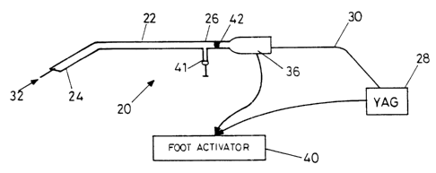
Fig. 2A shows a myocardial revascularization device
according to the present invention, which can be used to
perform the method according to the present invention.
The device 20 comprises a catheter 22 having at least ane
lumen, and having an insertable end 24, and an operating
enc~ 26 to be held by a physician. An energy source, such
as a laser 28, which may be a TfIC:YA~ laser, has
connected to it a fiber optic 30, which may be one or
more cyuartz fibers. The fiber optic 30 is received
through the catheter lumen and is shown having an end 32
extending from the insertable end 24 of the catheter. A
servomotor 36 serves to advance the fiber end 32 to stop
positions spaced certain distances from each other. The
spacing may be 1 to to mm. for example. The servomotor
36 is connected to and controlled by a foot activator 40.
The foot activator is also connected to and controls the
firing of the laser 28 when the fiber end is at the stop
positions.
-5.. J
e~~m
The device of Fig. 2A also has means for introducing
medicinal fluid to the site, in the preferred form of
heparin. The heparin is introduced under pressure as
shoran at 41 in Fig. 2~. A diaphragza 42 inside the
catheter prevents the pressurized heparin from flowing
out of the operating end 2~ of the catheter.
As shown in Fig. 2B the insertable end 24 of the device
has gripping means extending therefrom in the form of
three suction cups 44. These cups 44 provide a means to
removably mount and stabilize the insertable end 24 to
the inner ventricular wall, and serve as a tripod for the
end 24, and the fiber end 32.
Fig. 3 shows an aiming grid and aiming beam arrangement
useful for locating the desired target positions to fire
the laser which creates the channels. This arrangement
could be used in open chest surgery and can be used in a
procedure as an adjunct t~ coronary bypass or other
procedures.
This arrangement comprises an aiming grid 50 having
sensors in the form of ph~todiodes 52 located in an array
on a suitable sheet material. The grid 50 is adapted to
be positsoned inside of the thoracic cavity adjacent the
heart outside the ventricular wall in which laser
channels are desired. The grid 50 is connected to a
controller 54 by cable 56.
A handpiece 60, having a shell of suitable plastic
material, for example, houses an aiming bean source 62.
The aiming beam source may be an 80~ nm diode laser, but
could also be other sources of electromagnetic,
ultrasonic or magnetic energy. The aiming beam grid 50
has sensors compatible with and adapted to detect the
~~~~"~~.~~' '3
.2 N
m~~m
energy source. The handpiece f>0 may actually be a
catheter having two lumens. Also disposed in the
handpiece is a fiber optic end 64 for projecting a laser
beam, similar to that of Figs. 1 and 2. The fiber optic
end 64 is connected, by a fiber optic s7 within the
handpiece, to a laser 56 outside the handpiece. Control
means to fire the laser 66 is provided in controller s6.
The controller, by way of cable 68, also provides a means
t~ control the servomotor 70, located in the base of the
handpiece 60, for advancing the fiber optic end ~64 to
selected stop positions similarly as in Figs. 1 and 2.
The controller 54 is also connected to receive signals
fx'Om a surface EKG by way of cable 72.
It is usually desired that only specific regions of the
myocardium will be targets. The targets are based on the
watershed areas of each of the coronary branches, such
that a region poorly perfused by an occluded coronary
branch would be a target, while an adjacent area might
not. There are virtually no visual landmarks to provide
a roadmap of the coronary branches when the ventricle is
viewed from the inside and even if there were, an optical
system w~uld be necessary to visually guide such a
system. The coronary arteries are largely epicardial,
z5 and provides readily interpretable landmarks with which
surgeons are c~aite familiar. The grid provides an
arrangement for lining up the laser beam directly
underneath the target, when the target is best identified
by external landmarks.
The operation of the aiming beam grid arrangement is as
follows. During open chest surgery, the grid 50 is
positioned inside of the abdominal cavity adjacent the
heart outside the ventricular wall in which laser
channels are desired. The handpiece is inserted into the
ventricular cavity and the aiming beam is energised.
s' lj
--12..
6dhen the aiming beam is sensed by the photodiode 52,
indicati~,g the proper location for a channel to be fo~ned
in the ventricular wall, the controller enables or
automatically fires the laser 6~. The controller also
senses EKG signals and enables or automatically fires the
laser only at the proper time in the heart cycle.
The handpiece is moved to different positions inside the
ventricular cavity and when the aiming beam is sensed by
another photodiode in the grid, the laser is enabled to
create another channel in the ventricular wall. This
process is continued until the desired number of channels
is created. The controller may be provided with
circuitry to determine whether a particular photodiode
has previously sensed an aiming beam, so that when a
channel has been created at that location, the laser will
be prevented from being enabled at that location again,
to thereby avoid firing the laser at a location where a
channel has previously been created.
The controller may also be provided with means to detect
the distance between the aiming beam source ~2 (the end
of the handpiece) and the grid 50, and the signal
strength received. Thi~a computed distance and signal
strength may be used to control the intensity of the
laser energy used tp create the channel and thus the size
and depth thereof. The signal strength of the aiming
beam received would indicate the ventricular wall
thickness and dictate the channel depth desired.
The arrangement of F'ig. 3 allows a physician to focus a
retrograde transatrial laser at specific sites based on
visible epicardial landmarks with the heart surgically
exposed.
Fig. 4 shows a transthoracic aiming thorascope according
to the invention for focusing a percutaneously introduced
laser catheter at specific sites based on epacardial
landanarks. In this arrangement a single photodiode 52 is
mounted at the end of a first handpiece 80 x~hich is
adapted to be inserted through adjacent ribs in the
ribcage and positioned with its end against the exterior
of the heart. The photodiode is connected to controller
54 by cable 50. The controller is also connected by
cable 68 to a servomotor 70 in a second handpiece 90. A
laser 6s is also connected to the controller 54, which
controls the laser, and its output is through a fiber
optic 57, which fiber optic extends throughout the length
of the second handpiece and terminating at an end 64.
The second handpiece 90 also houses an aiming beam source
62, similar to that in Fig. 3. The second handpiece may
be a catheter having two lumens as in the arrangement of
Fig. 3. The controller 54 receives ~I~G signals similarly
as in Fig. 3.
Similarly to the operation of the device of Fig. 3, the
sec~nd handpiece 90 is inserted int~ the ventricular
cavity. The aiming beam from source 62 projects from the
sec~nd handpiece 90, and when the first handpiece 80 is
aligned to have photodiode 52 receive the aiming beam
from the second handpiece 90, the controller enables the
laser 6S tn fire and create a channel in the interior
ventricular wallo
An electr~magnet 92 may be mounted in the end of the
first handpiece 80, and a metallic ring 94 may be mounted
in the second handpiece 90. Magnetic force could be used
to stabilise the first handpiece end against the
a5 endocardium directly apposite the aiming scope, The
first handpiece 80, sometimes referred to an aiming
6 z.
r~ ~,~ "~ r a
scope, may be provided with appropriate imaging optics
~6, connected to visual monitor 98, for direct
visualization of the region. The details of this feature
are well known to those skilled in the art.
An experiment conducted using the method according to the
invention will now be described.
MF~,TERT~LB ~rND T~OD$
The left anterior descending artery (TAD) of 18 dogs (10
laser, eight control) was ligated distal to the first
diagonal, and the area at risk (P.AR) was mapped with
m~thylene blue dye. In laser animals, a catheter
containing the laser fiber was passed through the left
atrium, stabilized against the contracting left
ventricular wall, and nontransmural channels (600 a
diameter, about 4 channels/cm2) were lasered through the
endocardium (800 mJ pulses: freguency 3 Hz) until
epicardial blanching was noted., Survivors (laser, 9/10;
Controls, ~/8) were sacrificed at SiX weeks, and the
infarct sire was outlined using triphenyltetrazolium
chloride (TTC). Ventriculograms were done after the
animals were killed by ligating the coronary arteries,
clamping across the ~nitral and aortic valves, and
instilling radiapaque dye into the ventricle.
SDhT~
The BAR was similar in both groups (12.7 ~ 2.3 cm2 vs.
13.0 ~ 3.1 cm2) . Compared with controls at siX weeks,
laser-treated animals had smaller infarct size (3.67 ~
0. 32 Cma 'VS. 0.73 ~ 0.13 Cm2, ,~ < O. Q2 ) , and lower
infarct-to-ratio (o.a6 ~ .05 vs. 0.06 ~ .02, ~ <
0 . 02 ) . Neither bleeding nor aneurysms occurred in any of
the animals. Ventriculograms on control animals shaved
no perfusion of the free wall; laser-treated animals had
~~i~j';~(~ ~_
dyerfilled sinusoids in the free wall, filling through
short channels originating from the endocardial surface.
D~~6OiJ8s~OId
Transmural channels created with a COZ laser increase
myocardial perfusion in experimental models, and have
been used clinically as an adjunct during coronary artery
bypass. postoperative ventriculography and radionuclide
scans have demonstrated perfusion, through laser
channels, of regions not revascularized through bypass
grafts. The mechanists is thought to involve perfusion of
the collateral network of Myocardial sinusoids by flow
entering the lasered channels from the ventricular cavity
during systole. The channels remain open because
carbonization associated with laser energy has been shown
to inhibit lymphocyte, macrophage, and fibroblast
migration. Thus, in contrast to channels created by
needle acupuncture, laser channels heal more slowly and
with less scar formation, which allows endothelialization
and long-term patency. deeding from the epicardial site
of penetration is usually controlled by clot formation.
To improve myocardial perfusion, the channels must allow
communication between the ventricular cavity and
myocardial sinusoids, but do not need to be transmural.
In previous models, transmural channels were a
conses~uence of the inability to deliver CO2 laser energy
through a flexible fiberoptic system, mandating
application of the laser energy from the epicardial
surface of the ventricle. The far~infrared (10.6 u) COZ
laser has bean used because of its ability to remove
tissue precisely. The mid~infrared (2.15 u) THC:Y~O
laser has similar tissue effects because of a large
absorption peak of water for light energy in the 2 a
region. ~n addition, the wavelength of 2 a radiation is
short enough to be effectively transMitted through low
_~s~.
hydroxyl 600 a diameter quartz fibers. This feature
permits application of laser energy from the andocardial
surface of a beating ventricle, avoiding the need to
create transmural channels from the epicardial surface.
Using this approach, the ~sR in the experimental group
was significantly decreased after the creation of laser
channels, and after six weeks the laser animals had
smaller infarcts, as measured by TTC staining. hasar-
treated and control animals had similar initial BAR. In
the laser-treated animals, but not in the controls,
vantriculography at six peeks demonstrated noncoronary
perfusion of myocardial sinusoids in the area at risk
through short channels communicating with the ventricular
chamber. There ware no bleeding complications,
aneurysms, or permanent arrhythmias.
In conclusion, laser energy can be transmitted through
flexible quartz fibers to ereate myocardial channels from
~0 the endocardial surface in a beating heart. The channels
improve perfusion acutely and remain patent for up to six
weeks. This technique may be useful as an adjunct to
coronary bypass or, with development of a delivery
system, might permit percutaneous treatment of inoperable
patients ~ri.th diffuse coronary artery disease.
1. Mirhoseini M, 5helgi.kar S, Caytan Vii: New concepts
in revascularization of the myocardium. Ann Thor Surg
45:41.5-420, 1988.
2. Okada M, Ikuta i3, Shi~aizu K, wt al: Alternative
method of myocardial revascularizati~n by laser:
Eacperimental and clinical study. Kobe J died Sci 32:151-
161, 1986.
3. Hardy R1, Bove iKE, James F'tsT, et al: A histologic
study of laser-induced transmyocardial channels. Lasers
S)1rg tied 6:563-573, 1987.
4. Oz MC, Treat Ice, Trokel SL, et al: A fiberoptic
compatible mid-infrared laser with CO~ laser like effect:
Application to atherosclerosis. J 5urg Rae 47(6):493-
501, 1989.
5. Treat Pte, TrOkel SL, Reynolds, RD, 8t al: A
pr~elimi.nary evaluation of a pulsed 2.15 micron laser for
endoscopic surgery. Lasers Surg Med 8:322-326, 1988.