Note: Descriptions are shown in the official language in which they were submitted.
2~
WO91/0~7 ~ PCT/US90/06125
GASTRIN RELEASING PEPTIDE RECEPTOR
Growth factors are involved in numerous physiologi-
cal and pathological processes. An increasing number of
small regulatory peptides have been discovered in the
neural and neuroendocrine cells of mammalian tissues.
More recent evidence has pointed to the role of
neuropeptides in the regulation of animal cell growth,
and in particular to the action of mitogenic peptides in
the Swiss 3T3 cell system. One of the first neuropeptides
studied was the tetradecapeptide bombesin which was
originally isolated from amphibian skin, Anastasi et al.,
Experientia 27:166-167 (1971). Bombesin is structurally
related to several endogenous mammalian peptides, the
first to be characterized being gastrin releasing
peptide.
Gastrin releasing peptide (GRP) is a 27 amino acid
peptide having the following sequence in humans:
Val-Pro-LeuPro-Ala-Gly-Gly-Gly-Thr-Val-Leu-
Thr-Lys-Met-Tyr-Pro-Arg-Gly-Asn-His-Trp-Ala-Val-
Gly-His-Leu-Met-NH2. GRP is of significant interest
because of its presumed ability to function as an
autocrine growth factor in the pathogenesis of cancer. In
particular, GRP has been found to promote growth of human
small cell lung carcinoma (SCLC). GRP binding to cell
surface receptors is thought to stimulate cellular growth
by promoting the hydrolysis of phosphatidyl inositides
and by activation of protein kinase C. A large number of
biological responses to GRP have been observed including
stimulation of Na+/H+ antiport, mobilization of
3S intracellular Ca2+, transient expression of c-fos and
c-mYc proto-oncogenes, induction of tyrosine kinase
activity, elevation of DNA synthesis and promotion of
cell division.
~:
- . , -
1. . ,;: ', ' ' , , :
wo 91/0~ 2~ ;;; PCT/US90/06125 ~
The role of GRP in maintaining the growth of SCLC
suggests that effective therapeutic agents could be
developed that interrupt the autocrine growth cycle by
inactivating GRP or inhibiting its receptor. The active
site of GRP is the C-terminal region which binds high
affinity receptors on SCLC membranes. Blocking this
binding can inhibit SCLC growth. This has already been
accomplished with monoclonal antibodies to bombesin which
bind to the active site on GRP, thus inactivating the
peptide, Cuttitta et al., Nature 316:823- 826 (1985).
Another means to block GRP binding to its receptor,
and therefore to treat SCLC, is to inhibit the receptor
itself. This can be accomplished by use of agents which
bind to the GRP receptor and act as antagonists.
Antagonists can normally be found once the receptor has
been pharmacologically defined, as is the case with the
GRP receptor. Testing of potential receptor antagonists
has been made much easier with the development of highly
~utomated assay methods. Unfortunately, these systems
require purified GRP receptor in an active form, which
has not been readily attainable. This problem can be
overcome by use of the recombinant receptor. Along with
providing an improved renewable source of the receptor
from a specific source, using the recombinant GRP
receptor in screening for GRP receptor reactive drugs
also has the following advantages: potentially greater
number of receptors per cell giving greater yield of
reagent and higher signal to noise ratio in assays; and
receptor subtype specificity (theoretically giving
greater biological and disease specificity).
Cross-linXing of the GRP receptor to bound radio-
labeled GRP has been used to visualize the GRP
receptor-ligand conjugate on SDS-PAGE and to deduce
certain other characteris~ics of the receptor Rosengurt
et al., PCT/GB88/00255. However, the technique used did
.-
., -
'
: .
W ~1/0~7 ~ 7~ PCT/US90/06125
not involve isolation of the receptor but rather involvedcharacterization of a modified form of the receptor
protein. Unfortunately, in order to characterize the
structural properties of the GRP receptor in greater
detail and to understand the mechanism of action at the
molecular level, the receptor needs to be purified. For
some applications, it is essential to purify the receptor
in an active state which maintains thz binding activity
of the receptor. These include the generation of
antibodies against active receptor epitopes, structural
studies of the ligand binding site, and the use of the
purified receptor for screens for agonists and
antagonists of GRP binding.
To date, few receptors have been isolated and chara- -
cterized in their active form. There are two main reasons
for this. First, the amount of receptor present in most
tissues is minute and second, the receptor must often be
solubilized from membranes with detergents that can
perturb the structure of the protein. Further compounding
these difficulties is the unpredictable nature of
receptors in that the method for successfully
solubilizing one protein receptor may not be successful
for a different protein receptor.
This invention pertains to the solubilization of the
active GRP receptor from cellular membranes, the
characterization of receptor behavior in solution and the
purification of the solubilized receptor in an active
form, with the extracted receptor retaining full GRP
binding activity.
In particular, this invention pertains to obtaining
the solubilized and purified naturally occurring GRP
receptor in sequenceable grade, determining its partial
amino acid sequence, isolating the cDNA for the GRP
receptor, and determining the nucleotide sequence and the
:
,
.
W091/0~7 PCT/US90/06125
2~77~)~ 4 ~
deduced amino acid sequence of the receptor.
Specifically, this invention pertains to expressing
DNA encoding the GRP receptor in host cells, thereby
enabling the synthesis of GRP receptor compositions
having the amino acid sequence of the naturally occurring
GRP receptor which are entirely free of other proteins of
the species of origin and further enabling the synthesis
of novel mutant GRP receptors.
In addition, this invention relates to the use of
DNA encoding the GRP receptor or its fragments in the
hybridization diagnosis of defective GRP receptor DNA or
mRNA and for obtaining DNA encoding the GRP receptor from
natural sources.
More specifically, this invention pertains to the
use of the recombinant GRP receptor, and cell lines
transfected with vectors directing the expression of the
GRP receptor and membranes from such cell lines, in drug
screening assays for compounds having suitable binding
affinity for the GRP receptor.
Even more specifically, this invention pertains to
the recombinant GRP receptor along with protein fragments
of the receptor and antibodies directed thereto that may
be use- ful in diagnostic assays to determine if a
patient has altered levels of the gastrin releasing
peptide receptor. Assays based on detection of antibodies
to the GRP receptor and/or detection of the GRP receptor
itself may also have prognostic value.
Additionally, this invention pertains to using the
recombinant GRP receptor or fragments or derivatives
thereof such as antibodies to the receptor or fragments
or specific receptor antagonists defined in screening
assays, as therapeutic agents.
, ' ' .
.
W~91/0~7 2~77~6 PCTtUS90/06125
~--
Figure 1 is a ~raphical comparison of the ability of
several detergents to solubilize the GRP receptor and
shows the effect of solu~ilization on binding activity.
Figure 2 is a graph of GRP binding activity and GRP
receptor solubilization as a function of detergent
(CHAPS) concentration.
Figure 3 is a graph of GRP receptor solubilization
and activity as a function of the soluble cholesteryl
ester stabilizing agent (CHS) concentration.
Figure 4 is a graph of GRP binding activity as a
function of detergent (CHAPS) concentration.
Figure 5 is a gel display of SDS-PAGE analysis of
125I-GRP cross-linked to the GRP receptor in a crude
soluble extract.
Figure 6 is a silver stained gel display of SDS-PAGE
analysis of the purified GRP receptor.
Figure 7 shows the separation of tryptic fragments
of the GRP receptor by reverse-phase HPLC.
Figure 8 is the nucleotide sequence of the GRP
receptor and its deduced amino acid sequence. The
experimentally determined amino acid sequence of the
intact GRP receptor protein and of isolated tryptic
peptides to the receptor are indicated by underlining.
Putative transmembrane sequences are labeled I through
VII. Consensus sequences for N-linked glycosylation are
boxed.
Figure 9 shows the hydropathy analysis of the deduc-
ed amino acid sequence of the GRP receptor. The
. . .
WO91/0~7 PCT/US90/06125 ~
2~6~766
transmembrane sequences are indicated by numbers I
through VII.
Figure lo shows the Northern hybridization analysis
of mRNA from Swiss 3T3 cells.
.
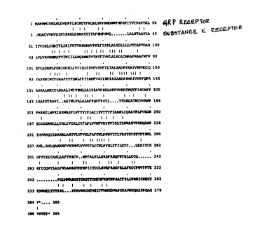
Figure 11 is a comparison of the amino acid sequen-
ces of the GRP receptor and the Substance K receptor.
Figure 12 shows the Northern hybridization analysis
of mRNA from human fetal lung cells (HFL).
Figure 13 shows the GRP ligand-dependent induction
of chloride current in a Xenopus oocyte expressing an ln
vitro transcript from the GRP receptor cDNA clone.
This invention provides the amino acid sequence and
DNA sequence of the gastrin releasing peptide (GRP)
receptor which were obtained after the GRP receptor was
purified and the amino acid sequence of tryptic fragments
of the GRP receptor was determined.
Partial amino acid sequences obtained from the
purified GRP receptor were used to deduce DNA probes
which were then used to isolate the GRP receptor cDNA
form of the gene. Some of the standard methods are
described, or referenced in T. Maniatis et al., Molecular
Cloninq. A Laboratory Manual (Cold Spring Harbor
Laboratory, Cold Spring Harbor, NY), or F.M. Ausubel et
al., Biolooy (Greene Publishing Associates, Brooklyn,
NY), all of which are incorporated herein by reference.
The procedure is broadly set forth below.
A cDNA library, constructed in lambda gtlO bac
teriophage, was prepared from RNA isolated from Swiss 3T3
cells. Several modifications and unique techniques had to
be utilized to overcome problems associated with
2~
W ~ /0~7 PCT/US90/06125
isolating a cDNA clone when probing the library with
oligonucleotides. In particular, it was necessary to
enrich the library for cDNA species encoding the GRP
receptor due to the under representation of such species
in unenriched cDNA libraries. Oligonucleotide probes were
designed having a nucleotide sequence based upon the most
likely codon usage. The cDNA library was plated out,
allowing the lambda phage containing cDNA inserts to lyse
their E.coli hosts and form plaques, each containing in-
dividual CDNA inserts. The plaques were screened for GRPreceptor DNA sequences with labeled oligonucleotide
probes. GRP receptor cDNA species were isolated, but
these did not encode the complete GRP receptor.
Polymerase chain reaction technology was used to
isolate additional cDNA species encoding portions of the
GRP receptor, and its 5' and 3' flanking regions. Gene
specific primer directed cDNA cloning was then used to
obtain a single cDNA clone encoding the entire GRP
receptor translation product. The actual cloning
techniques utilized herein are set forth in d~tail in
Examples 12 and 13.
once the cDNA for the GRP receptor was isolated from
mouse, it was sequenced. The nucleotide sequence revealed
the amino acid sequence of the primary translation
product of the GRP receptor, i.e., the amino acid
sequence before any post-translational modification.
The complete amino acid sequence is shown in Figure
8. As used herein, the term "GRP receptor" shall be
defined as a protein ox peptide having the amino acid
sequence shown in Figure 8 or a fragment thereof. The
term "GRP receptor" shall also be used herein to include
the GRP receptors of species other than mouse, for
example humans and other mammals.
..~ . .
: ..
W091/~7 PCT~US90/06125
2 ~ 6~7 6 6 8
This invention also encompasses proteins or peptides
having substantial homology with the amino acid sequence
in Figure 8, excluding any protein or peptide which
exhibits substantially the same or lesser homology, than
does the Substance K receptor. This is illustrated in
Figure ll.
Homology is determined by optimizing residue matches
by introducing gaps as required. This changes when
considering conservative substitutions as matches. This
definition is intended to also include natural allelic
variations in the GRP receptor sequence. Typical
homologous proteins or peptides will have from 25-100%
homology (if gaps can be introduced) to 50-100% homology
(if conservative substitutions are included~ with the
amino acid sequence of Figure 8. Some homologous pro-
teins or peptides, such as &RP receptor subtypes, will -
exhibit some biological activity in common with the GRP
receptor of Figure 8. As used herein, the term
"biological activity" is defined as including without
limitation, gastrin releasing peptide binding,
cross-reactivity with anti-GRP receptor antibodies raised
against the GRP receptor from natural sources, and -
coupling to guanyl nucleotide regulatory proteins.
This invention contemplates use of the isolated DNA
which encodes the GRP receptor or any fragment thereof,
to encode a biologically active GRP receptor polypeptide.
In addition, this invention covers isolated DNA which
encodes a biologically active protein having GRP receptor
activity and which is capable of hybridizing with the DNA
shown in Fi~ure 8. Said biologically active protein can
be the GRP receptor itself and have the amino acid
sequence shown in Figure 8. Further this invention covers
3S the use of isolated DNA which encodes proteins which are
homologous to the GRP receptor and which was isolated
using GRP receptor cDNA as a probe. The isolated DNA can
,
.
,
-:
2~ '7~
W~9~ 7 PCT/US90/06125
.~ .. - ~ . .. . .. .
~ -.-.......................................... ~ .. ..
9 ' ;.
have the respective regulatory sequences in the 5~ and 3'
flanks.
It is expected that the DNA which codes for the gas-
s trin releasing peptide receptor will be particularly
useful to identify genes, mRNA and cDNA species which
code for related or homologous receptors, along with
those which code for GRP receptor sub-types and for the
GRP receptor in tissue of different species. There is at
least one GRP receptor sub-type described with a
different selectivity towards bombesin-like peptides.
There are likely others. Various GRP receptor sub-
types are expected to be highly homologous. However, even
receptor proteins that have a more distant relationship
to the GRP receptor and no longer bind gastrin releasing
peptide, can readily be isolated using the GRP receptor
sequence if they are sufficiently homologous.
This invention further covers recombinant DNA molec-
ulec having a DNA sequence identical to the isolated DNA
set forth herein.
The isolated GRP receptor DNA can be readily modif-
ied by nucleotide substitutions, nucleotide deletions,
nucleo-tide insertions and inversions of nucleotide
stretches. These modifications result in novel DNA
sequences which encode the GRP receptor, its derivatives
or proteins naving GRP receptor activity. These modified
sequences can be used to produce mutant GRP receptor or
to enhance the expression of GRP receptor species. Such
mutant GRP receptor derivatives include the predetermined
or site-sp~cific mutations of the GRP receptor or its
fragments. Mutant GRP receptor is defined herein as being
a polypeptide otherwise falling within the homology
definition of the GRP receptor as set forth above, but
having an amino acid sequence which differs from that of
the GRP receptor as found in nature, whether by way of
''
W09l/0~7 PCT/US90/06125
Z~77~ o ~'
deletion, substitution or insertion. In particular, site
specific mutant GRP receptor is defined as having greater
than 50~ homology with the GRP receptor of Figure 8 and
as having biological activity in common with the receptor
S of Figure 8.
Although mutation sites are predetermined, that is
not a requirement. For example, in order to optimize the
performance of mutants at a given residue position,
random mutagenesis can be conducted at the target codon
and the expressed GRP receptor mutants can then be
screened for the desired activity. Methods for making
substitution mutations at predetermined sites in DNA
having a known sequence, are well known in the art such
as M13 primer mutagenesis.
GRP receptor mutagenesis can be conducted by making
amino acid insertions or deletions. Substitutions,
deletions, insertions or any subcombination may be
combined to arrive at a final construct. Insertions
include amino or carboxyl
terminal fusions. The mutations in the DNA must not place
coding sequences out of reading frames and preferably
will not create complementary regions that could
hybridize to produce secondary mRNA structure such as
loops or hairpins.
DNA which encodes the GRP receptor or fragments
thereof, can be obtained by chsmical synthesis, screening
cDNA libraries, or by screening genomic libraries
prepared from a wide variety of cell lines or tissue
samples.
This DNA ~an be expressed in a wide variety of host
cells for the synthesis of the full-length receptor or
fragments of the receptor which can in turn, for example,
be used to generate polyclonal or monoclonal antibodies;
-
: . .
WO~1/0~7 ~7';~ PCT/US90/06125
for binding studies; for construction and expression ofmodified receptor molecules; and for structure/function
studies. The GRP receptor or its fragments can be
expressed in host cells that are transformed or
transfected with appropriate expression vectors. These
molecules can be substantially free of protein or
cellular contaminants, other than those derived from the
recombinant host, and therefore axe particularly useful
in pharmaceutical compositions when combined with a
pharmaceutically acceptable carrier and/or diluent. The
receptor or portions thereof, may be expressed as fusions
with other proteins.
Expression vectors are self-replicating DNA or RNA
constructs containing the GRP receptor gene or its
fragments usually operably linXed to suitable genetic
control elements that are recognized in a suitable host
cell. These control elements are capable of affecting
expression within a suitable host. The specific type of
control elements necessary to effect expression will
depend upon the eventual host cell used. Generally,
genetic control elements can be a procaryotic promoter
system or a eucaryotic promoter expression control
system, and include a transcriptional promoter, an
optional operator to control the onset of transcription,
transcription enhancers to elevate the level of mRNA
expression, a sequence that encodes a suitable ribosome
binding site, and sequences that terminate transcription
and translation. Expression vectors also usually contain
an origin of replication that allows the vector to
replicate independently of the host cell.
The vectors of this invention contain DNA which en-
codes the G~P receptor or a fragment thereof encoding A
biologically active GRP receptor polypeptide. The DNA can
be under the control of a~viral promoter and can encode a
selection marker. This invention further contemplates use
' - ' ' ~ ~
W091/0~7 PCT/US90/06125
z~ 77~i6 12
of such expression vectors which are capable of
expressing eucaryotic cDNA coding for the GRP receptor in
a procaryotic or eucar-yotic host, where the vector is
compatible with the host and where the eucaryotic cDNA
coding for the GRP receptor is inserted into the vector
such that growth of the host containing the ve~tor
expresses the cDNA in question. Expression vectors need
not always replicate in their host cells to be useful,
however. Usually, expression vectors are designed for
stable replication in their host cells or for
amplification to greatly increase the total number of
copies of the desirable gene per cell. However, it is not
always necessary to require that an expression vector
replicate in a host cell. However, it is possible to
effect transient expression of the GRP receptor or its
fragments in various hosts using vectors that do not
contain a replication origin that is recognized by the
host cell. It is also possible to use vectors that cause
integration of GRP receptor or its fragments into the
host DNA by recombination.
Vectors comprise plasmids, viruses, bacteriophage,
integratable DNA fragments, and other vehicles which
enable the integration of DNA fragments into the genome
of the host. Expression vectors are specialized vectors
which contain genetic control elements that effect
expression of operably linked genes. Plasmids are the
most commonly used form of vector but all other forms of
vectors which serve an equiv- alent function and which
are, or become, known in the art are suitable for use
herein.
Transformed cells are cells, preferably mammalian,
that have been transformed or transfected with GRP
receptor vectors constructed using recombinant DNA
tec~niques. Transformed host cells usually express the
GRP receptor or its fragments but for purposes of
: - ~'
'':, .:, -.
:
WO9l/0~7 Z ~ S7 7 ~ ~ PCT/U~90/06125
13
cloning, amplifying, and manipulating its DNA, do not
need to express the GRP receptor. This invention further
contemplates culturing transformed cells in a nutrient
medium, thus permitting the GRP receptor to accumulate in
the culture. The GRP receptor can be recovered, first
from the culture and then from the culture medium.
For purposes of this invention, DNA sequences are
operably linked when they are functionally related to
each other. For example, DNA for a presequence or
secretory leader is operably linked to a polypeptide if
it is expressed as a preprotein or participates in
location of the polypeptide to the cell membrane or in
secretion of the polypeptide. A pro-
moter is operably linked to a coding sequence if itcontrols the transcription of the polypeptide; a ribosome
blnding site is operably linked to a coding sequence if
it is positioned to permit translation. Usually, operably -
linked means contiguous and in reading frame, however,
certain genetic elements such as repressor genes are not
contiguously linked but still bind to operator sequences
that in turn control expression.
Suitable host cells include procaryotes, lower
eucaryotes and yeasts or higher eucaryotes. Procaryotes
include both gram negative and gram positive organisms,
for exa~ple E. coli and B. subtilis. Lower eucaryotes
include yeasts, for example S. cerevisiae and Pichia, and
species such as Dictyostelium. Higher eucaryotes include
established tissue culture cell lines from animal cells,
both of non- mammalian origin such as insect cells and of
mammalian origin such as human, primates, and roden~s.
Procaryotic host-vector systems include a wide
variety of vectors for many different species. As used
herein, E. coli and its vectors will be used generically
to include aquivalent vectors for other procaryotes. A
-, :
WO91~06~7 - PCT/US90/06125
14 ~
z ~ ~q 7~;~epresentative vector for amplifying DNA is pBR322 or any
of its derivatives. vectors that can be used to express
the GRP receptor or its fragments include but are not
limited to such vectors as those containing the lac
promoter (pUC-series); trp promoter (pBR322-trp); Ipp
promoter (the pIN-series); lambda-pP or pR promoters
(pOT~); or hybrid promoters such as ptac (pDR540). See
~rosius et al., "Expression Vectors Employing Lambda-,
trp-, lac-, and Ipp-derived Promoters", in Vectors: A
Survey of Molecular Cloning Vectors and Their Uses, (eds.
Raymond L. Rodriguez and David T. Denhardt),
Buttersworth, Boston, 1988, Chapter 10, pp. 205-236.
Lower eucaryotes such as yeasts and Dictyostelium -
may be transformed with G~P receptor containing vectors.
For purposes of this invention, the most common lower
eucaryotic host is the baker's yeast, Saccharomyces
cerevisiae and it will be used to generically represent
lower eucaryotes although a number of other strains and
species are also available. Yeast vectors consist of a
replicatiQn origin (unless of the integrating type), a
selection gene, a promoter, DNA encoding the GRP receptor
or it fragments, sequences for translation termination,
polyadenylation and transcription termination. Suitable
expression vectors for yeast include such constitutive
promoters as 3-phosphoglycerate kinase and various other
glycolytic enzyme gene promoters or such inducible
promoters as the alcohol dehydrogenase 2 promoter or
metallothionine promoter. Suitable vectors include
derivatives of the following types: self-replicating low
copy number (such as the YRp-series), self-replicating
high copy n~mber (such as the YEp-series); integrating
types (such as the YIp-series), or mini-chromosomes (such
as the YCp-series).
Higher eucaryotic tissue culture cells are the pre-
ferred host cells for expression of the functionally
~. .
',
, . . .
WO 91/06647 ~?~i7'7~j5 PCr/US90/0612
active GRP receptor protein. In principle, any higher
eucaryotic tissue culture cell line is workable, whether
from an invertebrate or vertebrate source. ~owever,
mammalian cells are preferred. Transformation or
transfection and propagation of such cells has ~ecome a
routine procedure. Examples of useful cell lines include
HeLa cells, Chinese hamster ovary (CHO) cell lines, baby
rat kidney (BRK) cell lines, insect cell lines and monkey
(COS) cell lines. Expression vectors for such cell lines
usually include an ori~in of replication, a promoter, a
ribosome binding site, RNA splice sites (if genomic DNA
is used), a polyadenylation site, and a transcription
termination site. These vectors also usually contain a
selection gene or amplification gene. Suitable
expression vectors may be plasmids, YiruSes or
retroviruses carrying promoters derived from such sources
as from adenovirus, SV40, parvoviruses, vaccinia virus,
or cytomegalovirus. Representative examples of suitable
expression vectors include pCDNA1; pCD (Okayama et al.,
Mol.Cell Biol. 5:1136-1142, 1985); pMClneo PolyA (Thomas
et al, Cell 51:503-512, 1987); and a baculovirus vector
such as pAC 373 or pAC 610.
The GRP receptor can be solubilized from membranes
in an zctive form, and purified without loss of activity
by the methods outlined below. The source of GRP receptor
can be a eucaryotic or procaryotic host expressing
recombinan~ GRP receptor, such as is described above. The
source can also be a cell line such as mouse Swiss 3T3
fibroblasts, but other mammalian cell lines are also
contemplated by this invention, with the preferred cell
line being human.
The active GRP receptor was solubilized from
membranes containing the GRP receptor using a stabilizing
agent and a detergent. The stabilizing agent is
preferably a soluble cholesteryl ester. Particularly good
,
.
WO9l/0~7 ~ PCT/US90/06125
2~6'~7~, 16
results have been obtained using cholesteryl
hemisuccinate (CHS). The detergent can be non-ionic,
zwitter-ionic or the like. Particularly good results have
been obtained using the zwitter-ionic detergent
S 3-t(3-cholamidopropyl)dimethyla~monio3-1- propane
sulfonate (CHAPS).
Cellular membranes containing the GRP receptor are
prepared by lysis of a cultured GRP receptor containing
cell line such as Swiss 3T3 fibroblasts, followed by
centrifugation. The resulting pellets are washed by
resuspension and centrifuged again.
once the membranes are obtained from a suitable cell
line as described above and in Example 1, the final
concentration of protein is adjusted. A suitable final
protein concentration is about 15 mg/ml.
The membranes are then salt washed prior to solubil-
ization of the G~P receptor. The membranes are washedtwice with buffer and NaCl, then washed with a
solubilization buffer and finally suspended in the
solubilization buffer at an adjusted protein
concentration. A suitable buffer composition for the
first two washings comprises a medium such as 50
mM4-(2-hydroxyethyl)- l-piperazine ethane sulfonic acid
(HEPES), pH 7.5, a chelator such as 2 mM ethylenediamine-
tetraacetic acid (EDTA), and protease inhibitors. A
suitable NaCl concentration is 1.0 M. The solubilization
buffer, both for the washing and suspension, can be
typically comprised of 50 mM HEPES, pH 7.5, 2 mM EDTA,
another ch~lator such as lmM
[ethylenebis-(oxyethylenenitrilo)]tetraacetic acid
(EGTA), 100 mM NaCl and protease inhibitors. The protein
concentration is ad~usted to about 7 mg/ml, for example.
This salt washing step provides a 2 fold purification.
Similar results can be achieved by washing the membranes
W~9~ 7 ~ '77l3~; PCT/US9OtO6125
17 ~ ~
with 2 M urea, high pH buffers (pH 10) or chaotropic
salts such as KI. This procedure also increases the
stability of the GRP receptor in the extract. Other
constituents of the buffers may include, for example,
sucrose, and suitable protease inhibitors include,
without limitation, aprotinin, leupeptin, pepstatin,
bacitrin and phenylmethylsulfonyl fluoride (PMSF).
A mixture of detergent (CHAPS) and soluble
cholesteryl ester stabilizing agent (CHS) is then slowly
added to the membrane suspension to give a set final
detergent concentration. The weight ratio of detergent to
soluble cholesteryl ester can be within the range of
about 200:1 to 5:2, preferably about 10:1. Alternatively,
the detergent can be added to the membrane suspension,
followed by the addition of the soluble cholesteryl
ester. In that instance, initialiy there will be 100
detergent and the soluble cholesteryl ester is added
until the weight ratio of detergent to ester is within
the range of about 200:1 to 5:2, preferably about 10:1.
For solubilization of the GRP receptor, the concentration
of detergent should be 0.4 to 3.0% (w/v), and is
optimally set at about 0.75% (w/v) for a membrane
concentration (prior to the membrane washing steps) of
around 15 mg/ml. Similarly, the concentration of soluble
cholesteryl ester is within the range of about 0.0015% to
1.2% (w/v). Likewise, for a membrane concentration of
around 15 mg/ml, the concentration of soluble cholesteryl
ester is preferably about 0.075% (w/v).
The extract is then incubated at a temperature
within the range of about O to 37C, typically room temp-
erature such as 21C, and then cooled to O to 21C,
typically 4C, and the insoluble material centrifuged at
high speeds, preferably about 100,000 times gravity, in a
standard centrifuge for a suitable period of time
depending upon the volume involved to obtain an extract
W09l/0~7 PCT/US90/0612~
~67~ 18
containing the solubilized receptor (i.e., soluble
extract).
At the high detergent concentration (0.4 to 3.0%),
the receptor is not active. However, upon dilution with a
buffer solution, the receptor is reactivated. The
presence of the soluble cholesteryl ester, which acts as
a stabilizing agent, is necessary for the receptor to be
reactivated at the low detergent concentration. For
assays using the active solubilized GRP receptor, to
exhibit binding activity the final concentration of
detergent in the suspension should be diluted to within
the range of about 0.025 to 0.2% (w/v). The weight ratio
of detergent to soluble cholesteryl ester is still main~
tained within the range of about 200:1 to 5:2, preferably
about 10:1. Therefore, a suitable range for the soluble
cholesteryl ester is about 0.00012~% to 0.08% (w/v). The
preferable assay concentrations are 0.075% (w/v)
detergent and about 0.0075% (w/v) soluble cholesteryl
ester.
The solubilized receptor in its active form is then
purified and freed of contaminating proteins.
Purification of th~ GRP receptor involves a multistep
procedure which includes the following steps, which
follow the solubilization procedure set forth above.
(1) Polyethylene glycol precipitation. The GRP
receptor is precipitated from the soluble extract by
addition of polyethylene glycol (PEG). Addition of PEG is
preferably done to obtain a final concentration of 20%
(w/v). The precipitate is then collected by
centrifugation and resuspended in a buffer solution. The
buffer solution can typically be comprised of 25 mM
~5 HEPES, pH 7.5, 25 mM Tris/Cl, 2 mM EDTA, 0.075% (w/v)
detergent, 0.0075% (w/v) soluble cholesteryl ester, and
protease inhibitors. The final volume of the suspension
wo gl/o~ 77~ PCT/VS90/~61~5
is preferably 25~ that of the original soluble extract~
Proteins remaining insoluble in the suspension are
removed by centrifugation. This step provides a 2 fold
purification, and enhances the stability of the receptor.
(2) Wheat germ agglutinin chromatography. The sol-
uble extract is applied to a wheat germ agglutinin
affinity column equilibrated with a buffer solution
typically comprised of 50 mM HEPES, pH 7.5, 2 mM EDTA,
0.25% (w/v) detergent, 0.025% (w/v~ cholesteryl ester and
protease inhibitors. The column is eluted with column
buffer solution and 5 mM N-N'-N"triacetyl-chitotriose.
Fractions containing the GRP receptor are then identified
by 12sI-GRP binding assays. This step provides a 5 fold
purification by removing proteins that do not contain
carbohydrate. To obtain a good yield, it is necessary to
elute the column with chitotriose or chitobiose. The
yield may also be enhanced by maintaining the detergent
concentration above about 0.2% detergent and 0.02%
soluble cholesteryl ester.
(3) GRP-affinity chromatography. The wheat germ
agglutinin column eluate is further fractionated on a GRP
affinity column. In the preferred embodiment, the column
contains a beaded matrix with the peptide human
[Nlel4,27]GRP13-27 (the C-terminal portion of GRP)
coupled to it at 2 mg peptide/ml packed gel. Th~ column
is equilibrated with a solution typically comprised of 25
mM Tris, 25 mM HEPES, pH 7.5, 2 mM EDTA, 0.075% (w/v)
CHAPS, 0.0075% (w/v) CHS and protease inhibitors. The
concentration of detergent in the wheat germ agglutinin
column eluate is preferably adjusted to 0.075% (w/v) by
dilution with a solution typically comprised of 25 mM
HEPES, 25 mM Tris, pH 7.5, 2 mM EDTA and protease
inhibitors. After application of the sample and
extensive washing of the column, bound protein is eluted
with a salt at a concentration above 0.2 M. Particularly
.. . .
, ,: , ' ,
~ '
: . . , ~ , , ' ' '
', , ' ' ' ' ' :
,
WO91/0~7 ` `` PCT/US90/06125
~,
77~6 20
suitable is 0.5 M NaCl. Fractions containing the GRP
receptor are then identified by 125I-GRP binding assays.
The GRP peptide used ([Nlel4,27~GRP13-27) is an analog
made by Triton Biosciences Inc. (Alameda, CA) which is
resistant to oxidation. Other GRP peptides and matrixes
that will also work include, without limitation, GRP1-27,
GRP14-27 and ~Lys3]Bombesin. However the yield and
elution conditions may be altered. Elution of the bound
protein with salt is important because receptor binding
activity is preserved and a good yield is achieved. The
concentration of detergent in the sample loaded onto the
column is critical for optimal results. The suitable
range of detergent is about 0.025% to 0.2% (w/v). The
ratio of detergent to stabilizing agent is also the same,
being 200:1 to 5:2, preferably 10:1.
(4~ Second affinity column. Fractions containing the
GRP receptor eluted from the affinity column are desalted
and the sample is applied to a second GRP affinity
column, and eluted as described in step (3). Fractions
containing the receptor are then identified by binding
assays. Use of two consecutive affinity columns in this
step is required to give a high degree of purity.
(5) Gel filtration. This is an optional step that
yields a marginally purer product. The gel filtration
step is also useful to remove protease inhibitors, salt
and residual detergent from the receptor.
In general, the solubilized, unpurified and solubil-
ized, purified GRP receptor of this invention binds
gastrin releasing peptide with an affinity of at least
XD=10 nM. The GRP receptor from a mouse Swiss 3T3
fibroblast cell line, according to this invention was
found to have the following characteristics:
.. . .
'
~r~ '-7~:i6
WO~ K~47 PCT/US90/06125
21 `
runs as a broad barld on SDS-PAGE with an
apparent molecular weight of about 70 to 100
kilodaltons; binds selectively with
polypeptides of the ~ombesin type;
has a KD value of about 10-100 pM; is free of
coupled G proteins; contains N-linked
carbohydrates; when deglycosylated, has an
apparent ~olecular weight of 36+5 kilodaltons
on SDS-PAGE; and has a partial amino acid
sequence near the N-terminus of:
-Leu-Asn-Leu-Asp-Val-Asp-Pro-Phe-Leu-Ser-
Now that the sequence is known, the GRP receptor or
any fragments thereof can be prepared by conventional
processes for synthesizing peptides. These include
processes such as are described in John M. Stewart and
Janis D. Young, Solid Phase Peptide Synthesis (Pierce
Chemical Co., RocXford, IL 1984), M. Bodanszky and
A.Bodanszky, The Practice of Peptide Synthesis
(Springer-Verlag, New York, 1984) and M. Bodanszky, The
Principles of Peptide Synthesis (Springer-Verlag, New
York, 1984), all of which are incorporated herein by ref-
erence. For example, an azide process, an acid chloride
process, an acid anhydride process, a mixed anhydride
process, an active ester process (for example,
p-nitrophenyl ester, N~ hydroxysuccinimide ester, or
cyanomethyl ester), a carbodiimidazole process, an
oxidative-reductive process, or a DCC/additive process
can be used. Solid phase and solution phase syntheses are `
both applicable to the foregoing processes.
The GRP receptor is suitably prepared in accordance ~ ~`
with the above processes as typically employed in peptide
synthesis, generally either by a so-called stepwise
process which co~prises condensing an amino acid to the
terminal amino acid, one ~y one in sequence, or by
coupling peptide fragments to the terminal amino acid.
.. .... . .
'~ , . .
wOgl/0~7 PCT/US90/06125
2 ~ 7~ 22
Amino groups that are not being used in the coupling
reaction must be protected to prevent coupling at an
incorrect location.
If a solid phase synthesis is adopted, the
C-terminal amino acid is bound to an insoluble carrier or
support through its carboxyl group. The insoluble
carrier is not particularly limited as long as it has a
binding capability to a reactive carboxyl group.
Examples of such insoluble carriers include halomethyl
resins, such as chloromethyl resin or bromomethyl resin,
hydroxymethyl resins, phenol resins,
tert-alkyloxycarbonyl- hydrazidated resins, and the like.
;
An amino group-protected amino acid is bound in
seguence through condensation of its activated carboxyl
group and the reactiveamino group of the previously
formed peptide or chain, to synthesize the peptide step
by step. After synthesizing the complete sequence, the
peptide is split off from the insoluble carrier to
produce the peptide. This solid-phase approach is
generally described by Merrifield et al. in J. Am. Chem.
Soc. 85:2149-2156 (1963), which is incorporated herein by
reference.
The prepared receptor and fragments thereof can be
isolated and purified from the reaction mixture by means
of peptide separation, for example, by extraction,
precipitation, electrophoresis and various forms of
chromatography, and the like. The receptor of this
invention can be obtained in varying degrees of purity
depending upon its desired use. Purification can be
accomplished by use of the protein purification
techniques disclosed herein or by the use of the
~5 antibodies herein described in immunoabsorbant affinity
chromatography. This immunoabsorbant affinity
chromatography is carried out by first linking the
. .. , , :
:' :' ; ' '
wo 91/06647 2~ 7~ cr/us~o/n6l2s
. . : , , ~.,
23
antibodies to a solid support and then contacting the
linked antibodies with solubilized lysates of small cell
lung cancer cells, lysates of other cells expressing the
GRP receptor, or lysates or supernatants of cells
producing the GRP receptor as a result of DNA techniques
described below.
Derivatives of the GRP receptor included herein
include amino acid sequence mutants, glycosylation
variants and covalent or aggregative conjugates with
other chemical moieties. Covalent derivatives can be
prepared by linkage of functionalities to groups whirh
are found in the GRP receptor amino acid side chains or
at the N- or C-termini, by means which are well known in
the axt. These derivatives can include, without
limitation, aliphatic esters or amides of the carboxyl
terminus or residues containing carboxyl side chains,
O-acyl derivatives of hydroxyl group-containing residues,
and N-acyl derivatives of the amino terminal amino acid
or aminogroup containing residues, for example, lysine or
arginine. Acyl groups are selected from the group of
alkyl-moieties including C3 to C18 normal alkyl, thereby
forming alkanoyl aroyl spec~es.
A major group of derivatives are covalent con~ugates
of the GRP receptor or fragments thereof with other
proteins of polypeptides. These derivatives can be
syn~hesized in recombinant culture such as N- or
C-terminal fusions or by the use of agents known in the
art for their usefulness in crosslinking proteins through
reactive side groups. Preferred GRP derivatization sites
with cross-linking agents are at free amino groups,
carbohydrate moieties and cysteine residues.
This invention also contemplates the use of deriv-
atives of the GRP receptor other than variations in amino
acid sequence or glycosylation. Such derivatives are
...... . ... .
. . ~ .
~ : . ,
.
: .':
W091/06~7~ 67 7 ~ ~ PCT/US9OtO6125
24
characterized by covalent or aggregative association with
chemical moieties. The derivatives generally fall into
three classes: (l) salts, ~2) side chain and terminal
residue covalent modifications, and (3)adsorption
complexes, for example with cell membranes. Such covalent
or aggregative derivatives are useful as immunoqens, as
reagents in immunoassays or in purification methods such
as for affinity purification of gastrin releasing peptide
or other binding ligands. For example, the GRP receptor
can be immobilized by covalent bonding to a solid support
such as cyanogen bromide-activated Sepharose, by methods
which are well known in the art, or adsorbed onto
polyolefin surfaces, with or without glutaraldehyde
cross-linking, for use in the assay or purification of
anti-G~P receptor antibodies or gastrin releasing
peptide. The GRP receptor can also be labeled with a
detectable group, for example radioiodinated by the
chloramine T procedure, covalently bound to rare earth
chelates or conjugated to another fluorescent moiety for
use in diagnostic assays.
The solubilized GRP receptor of this invention can
be used as an immunogen for the production of antisera or
antibodies specific for the receptor or any fragments
thereof. The purified receptor can be used to screen
monoclonal antibodies prepared by immunization with
various forms of impure preparations containing the 5RP
receptor. The purified receptor can also be used as a
reagent to detect any antibodies generated in response to
the presence of elevated levels of gastrin releasing -
peptide receptor or cell fragments containing the GRP
receptor. A~ditionally, GRP receptor fragments may also
serve as immunogens to produce the antibodies of the
present invention. For example, this invention
contemplates antibodies having binding affinity to or
being raised against the amino acid sequence shown in
Figure 8, or any fragment thereof. In particular, this
'' .
.
WO91/0~7 ~Q677~,~ PCT/US90/0612~ i
invention contemplates antihodies having binding affinity
to or being raised against specific fragments which are
predicted to lie outside of the lipid bilayer. These
fragments include the following ten amino acid sequence
(residues 9-18, inclusive) near the N-terminus:
9 18
-Leu-Asn-Leu-Asp-Val-Asp-Pro-Phe-Leu-Ser-
In addition, as noted above, this invention covers
fragments of the GRP receptor which are predicted to
reside on the extracellular side of the membrane:
residues 1-39, inclusive; residues 98-115, inclusive;
residues 176-209, inclusive; and residues 288-300,
inclusive; and to the following portions of the receptor
which are predicted to reside on the intracellular side ;
of the membrane: residues 64-77, inclusive; residues
138-157, inclusive; residues 236-266, inclusive; and
residues 330-385, inclusive.
Antibodies can be raised to the GRP receptor, and
fragments thereof, both in its naturally occurring form
and in its recombinant form. Additionally, antibodies can
be raised to both the GRP receptor in its active form and
in its inactive form, the difference being that
antibodies to the active receptor are more likely to
recognize epitopes which are only present in the active
receptor.
Antibodies against predetermined fragments of the
GRP receptor can be raised by immunization of animals
with conjugates of the fragments with immunogenic
proteins. ~onoclonal antibodies are prepared from cells
secreting the desired antibody. These antibodies can be
screened for binding to normal or defective GRP
. .
. : .
: :.
. .
WO91/0~7 PCT/US90/06125 l~
~ 75~ 26
receptors, or screened for agonistic or antagonistic GRP
receptor activity.
The antibodies of this invention can have signifi-
cant therapeutic value. They can be potent antagoniststhat bind to the GRP receptor and inhibit ligand binding
to the receptor or inhibit the ability of gastrin
releasing peptide to elicit a biological response. They
also can be useful as non-neutralizing antibodies a~d can
be coupled to toxins or radionuclides so that when the
antibody binds to the receptor, the cell itself is
killed. Further, these antibodies can be conjugated to
drugs or other therapeutic agents, either directly or
indirectly by means of a linker.
The antibodies of this invention can also be useful
in diagnostics. As capture or non-neutralizing
antibodies, they can bind to the GRP receptor without
inhibiting ligand binding. As neutralizing antibodies,
they can be useful in competitive binding assays.
Receptor fragments may be joined to other materials,
particularly polypeptides, as fused or covalently joined
pol~pepti~es to be used as immunogens. The GRP receptor
and its fragments may be fused or covalently linked to a
variety of i~munogens, such as keyhole limpet hemocyanin,
bovine serum albumin, tetanus toxoid, etc. See for
example, Microbiology, Roeber Medical Division (Harper
and Row, 1969), Landsteiner, Specificity of Seroloqical
Reactions (Dover Publications, New York, 1962) and
Williams et al., Methods in Immunoloqy and
Immunochemistry, Vol. 1 (Academic Press, New York, 1967),
all of which are incorporated herein by reference, fo~
descriptions of methods of preparing polyclonal antisera.
A typical method involves hyperimmunization of an animal
with an antigen. The blood of the animal is then
- .
. ~ . .
-' ' , ~.
77~
W ~ 1/0~7 PCT/U590/Q6125
27
collected shortly after the repeated immunizations and
the gamma globulin is isolated.
In some instances, it is desirable to prepare mono-
clonal antibodies from various mammalian hosts, such as
mice, rodents, primates, humans, etc. Description of
techniques for preparing such monoclonal antibodies may
be found in, Stites et al., editors, Basic and Clinical
I~munolooY, (Lange Medical Publications, Lvs Altos, CA,
Fourth edition) and references cited therein, and in
particular in Kohler and Milstein in Nature 256: 495-497
(1975), which discusses one method of generating
monoclonal antibodies. Summarized briefly, this method
invol~-s injecting an animal with an immunogen. The
animal is then sacrificed and cells taken from its
spleen, which are then fused with myeloma cells. The
result is a hybrid cell or "hybridoma" that is capable of
reproducing in vitro. The population of hybridomas is
then screened to isolate individual clones, each of which
secrete a single antibody species to the immunogen. In
this manner, the individual antibody species obtained are
the products of single B cells from the immune animal
generated in response to a specific site recognized on
the immunogenic substance.
The antibodies of this invention can also be used
for affinity chromatography in isolating the receptor.
Columns can be prepared where the antibodies are linked
to a solid support, e.g., particles, such as agarose,
Sephadex, or the like, where a cell lysate may be passed
through the column, the column washed, followed by
increasing concentrations of a mild denaturant, whereby
the purif ied receptor protein will be released.
Both the naturally occurring and the recombinant
form of the GRP receptor of this invention are
particularly useful in kits and assay methods which are
~' " " ' ' ' ' ` ' .
WO91/0~7 PCT/US90/06125
28
Z C ~c~pable of screening compounds for binding activity to
the GRP receptor. Several methods of automating assays
have been developed in recent years so as to permit
screening of tens of thousands of compounds per year. The
development of suitable assays can be greatly facilitated
by the availability of large amounts of purified, soluble
receptor in an active state such as is attainable by the
process of this invention.
A kit for determining the binding affinity of a test
compound to the gastrin releasing peptide receptor would
typically comprise a test compound; a labeled compound,
for example a ligand or antibody having known binding
affinity for the gastrin releasing peptide receptor; a
source of gastrin releasing peptide receptor (naturally
occurring or recombinant); and a means for separating
bound from free labeled compound, such as a solid phase
for immobilizing the gastrin releasing peptide receptor.
Once compounds are screened, those having suitable
binding affinity to the GRP receptor can be evaluated in
suitable biological assays, as are well known in the art,
to determine whether they act as agonists or antagonists. -
This invention is particularly useful for screening
compounds by using the recombinant GRP receptor in any of
a variety of drug screening techniques. The advantages of
using the recom~inant GRP receptor in screening for GRP
receptor reactive drugs include: (a) improved renewable
source of the receptor from a specific source; (b)
potentially greater number of receptors per cell giving
higher signal to noise ratio in assays; and (c) receptor
subtype spacificity (theoretically giving greater
biological and disease specificity).
one method of drug screening utilizes eucaryotic or
procaryotic host cells which are stably transformed with
; ., . ., , - ;:
W~91/~7 ~ 75~ PCT/US90/06125
j
` 29 ~
recombinant DNA molecules expressing the GRP receptor.
Such cells, either in ~iable or fixed form, can be used
for standard receptor/ligand binding assays. Competitive
assays are particularly useful, where the cells (source
of G~P receptor) are contacted and incubated with a
labeled ligand having known binding affinity to the GRP
ptor such as 125I-GRP, and a test compound whose
binding affinity to the GRP receptor is being measured.
The bound ligand and free ligand are then separated to
assess the deqree of ligand binding. The amount of test
compound bound is inversely proportional to the amount of
labeled ligand binding measured. Any one of numerous
techniques can be used to separate bound from free ligand
to assess the degree of ligand binding. This separation
step could typically involve a procedure such as adhesion
to filters followed by washing, adhesion to plastic
followed by washing, or centrifugation of the cell
membranes. Viable cells could also be used to screen for
the effects of drugs on GRP receptor mediated functions,
for example, second messenger levels (Ca), proliferation,
etc.
Another method utilizes membranes from transformed
eucaryotic or procaryotic host cells as the source of the
GRP receptor. These cells are stably transformed with
DNA vectors directing the expression of the GRP receptor.
Essentially, the membranes would be prepared from the
cells and used in any receptor/ ligand binding assay such
as the competitive assay set forth above.
Still another approach is to use solubilized, un-
purified or solubilized, purified receptors from
transformed eucaryotic or procaryotic host cells. This
allows for a real "molecular" binding assay with the
advantages of increased specificity, the ability to
automate, and high drug test throughput.
:' . : ; .
- :
.
WO91/0~7 ` PCT/US90/06125
2~77~
Another technique for drug screening involves an
approach which provides high throughput screening for
compounds having suitable binding affinity to the gastrin
releasing peptide receptor and is described in detail in
Geysen, European Patent Application 84/03564, published
on September 13, 1984. First, large numbers of different
small peptide test compounds are synthesized on a solid
substrate such as plastic pins or some other surface.
Then all the pins are reacted with solubilized,
unpurified or solubilized, purified GRP receptor and
washed. The next step involves detecting bound GRP
receptor. -~
Purified GRP receptor can be coated directly onto
plates for use in the aforementioned drug screening tech-
niques. However, non- neutralizing antibodies to the GRP
receptor can be used as capture antibodies to immobilize
the GRP receptor on the solid phase.
This invention also contemplates the use of compet-
itive drug screening assays where neutralizing antibodies
to the receptor or receptor fragments compete with a test
compound for binding to the receptor. In this manner,
the antibodies can be used to detect the presence of any
polypeptide which shares one or more binding sites of the
GRP receptor and can also be used to occupy binding sites
on the receptor that might otherwise be occupied by
gastrin releasing peptide.
Additionally, neutralizing antibodies against the
receptor and soluble fragments of the receptor which
contain the high affinity ligand binding site, can be
used to inhibit gastrin releasing peptide receptor
function in cancerous tissues.
This invention also contemplates use of the GRP
receptor, fragments thereof, peptides, and their fusion
- . ~ . ......... , : .
- ~ .
.
::~
W091/0~7 ~7'7 ~ PCT/US90/0612S
.~''~ , .
t` ~ 31
products in a variety of diagnostic kits and methods for
detecting the presence of the gastrin releasing peptide
receptor.
A kit for determining the concentration of gastrin
releasing peptide receptor in a sample would typically
comprise a labeled compound (ligand or antibody) having
known binding affinity for the gastrin releasing peptide
receptor, a source of gastrin releasing peptide receptor
(naturally occurring or recombinant) and a means for
separating the bound from free labeled compound, for
example a solid phase for i~mobilizing the gastrin
releasing peptide receptor.
A method for determining the concentration of
gastrin releasing peptide receptor in a sample would
typically comprise the steps of:(1) preparing membranes
from a sample comprised of a GRP receptor source; (2)
washing the me~branes and suspending them in a buffer;
(3) solubilizing the GRP receptor by incubating the
membranes in a culture medium to which a detergent and a
soluble cholesteryl ester has been added; (4) adjusting
the detergent concentration of the solubilized receptor;
(5) contacting and incubating said dilution with
radiolabeled GRP to form GRP:GRP receptor complexes; (6)
recovering the complexes such as by filtration through
polyethyleneimine treated filters; and (7) measuring the
radioactivity of the recovered complexes.
Antibodies specific for the receptor or receptor
fragments are useful in diagnostics to detect the
presence of elevated levels of the receptor and/or its
fragments. Such diagnostic assays can employ lysates,
fixed cells, immunofluorescence, and further can involve
the detection of antigens related to the GRP receptor in
serum, or the like. Diagnostic assays may be homogeneous
(without a separation step between free reagent and
,,.~ , . ~
,. , ~ . ~
WO91/~7 PCT/US90/061~5
Z~i77~ 32
receptor-ligand complex) or heterogeneous (with a
separation step). Various commercial assays exist, such
as radioimmunoassay (RIA~, enzyme-linked immunosorbent
assay (ELISA), enzyme immunoassay (EIA),
S enzyme-multiplied immunoassay technique (EMIT),
substratelabeled fluorescent immunoassay (SLFIA) and the
like. For example, unlabeled antibodies can be employed
by using a second antibody which is labeled and which
recognizes the antibody to the GRP receptor or to a
particular fragment thereof. These assays have also been
extensively discussed in the literature.
Frequently, the reagents for diagnostic assays are
supplied in kits, so as to optimize the sensitivity of
the assay. For the subject invention, depending upon the
nature of the assay, the protocol, and the label, either
labeled or unlabeled antibody, or labeled receptor is
provided, usually in conjunction with other additives,
such as buffers, stabilizers, materials necessary for
signal production such as substrates for enzymes, and the
like. Desirably, the reagents are provided as a dry
powder, where the reagents may be reconstituted in an
aqueous medium having appropriate concentrations for
performing the assay.
Any of the aforementioned constituents of the drug
screening and the diagnostic assays may be used without
modification or may be modified in a variety of ways. For
example, by labeling such as covalently or non-covalently
joining a moiety which directly or indirectly provides a
detectable signal. In any of these assays, the ligand,
test compound, GRP receptor, or antibodies thereto can be
labeled either directly or indirectly. Possibilities for
direct labeling include label groups which include:
radiolabels such as 125I, enzymes (U.S.Pat.No. 3,645,090)
such as peroxidase and alkaline phosphatase, and
fluorescent labels (U.S.Pat.No. 3,940,475) capable of
. . . .
'., -
:. -':
Wo~9~ 7 '~?~7~,~ PCT/US90/06125
33
monitoring the change in fluorescence intensity,
wavelength shift, or fluorescence polarization.
Possibilities for indirect labeling include biotinylation
of one constituent followed by binding to avidin coupled
to one of the above label groups.
There are also numerous methods of separating the
bound from the free ligand, or alternatively the bound
from the free test compound. The receptor can be
immobilized on various matrixes followed by washing.
Suitable matrixes include plastic such as an ELISA plate,
filters, and beads. Methods of immobilizing the receptor
to a matrix include direct adhesion to plastic, use of a
capture antibody, chemical coupling, and biotin-avidin.
The last step in this approach involves the precipitation
of receptor/ligand complex by any of several methods
including those utilizing an organic solvent such as
polyethyiene glycol or a salt such as ammonium sulfate.
Other suitable separation techniques include, without
limitation, the fluorescein antibody magnetizable
particle method described in S.J. Rattle et al.,
Clin.Chem. 30(9): 1457-1461 (1984) and the double
antibody magnetic particle separation as described in
U.S~Pat.No. 4,659,678.
The methods for linking protein receptors or their
fragments to the various labels have been extensively
reported in the literature and do not require detailed
discussion here. Many of the techniques involve the use
of activated carboxyl qroups either through the use of
carbodiimide or active esters to form peptide bonds, the
forma~ion ~f thioethers by reaction of a mercapto group
with an activated haloqen such as chloroacetyl, or an
activated olefin such as maleimide, for linkage, or the
like.
.
: ' ~
WO91/0~7 PCT/US90/06125
z~677~i~
Another diagnostic aspect of t~is invention involves
use of oligonucleotide and polynucleotide sequences taken
from the GRP receptor sequence which can be used as
probes for detecting levels of the gastrin releasing
peptide receptor in patients suspected of having cancer.
The preparation of both RNA and DNA nucleotide sequences,
the labeling of the sequences and the preferred size of
the sequences has received ample description and
discussion in the literature. Normally an oligonucleotid~
probe should have at least about 14 nucleotides, usually
at least about 18 nucleotides, and the polynucleotide
probes may be up to several kilobases. Various labels may
be employed, most commonly radionuclides, particularly
32P. However, other techniques may also be employed,
such as using biotin modified nucleotides for
introduction into a polynucleotide. The biotin then
serves as the site for binding to avidin or antibodies,
which may be labeled with a wide variety of labels, such
as radionuclides, fluorescers, enzymes, or the like.
Alternatively, antibodies may be employed which can
recognize specific duplexes, including DNA duplexes, RNA
duplexes, DNA-RNA hybrid duplexes, or DNA-protein
duplexes. The antibodies in turn may be labeled and the
assay carried out where the duplex is bound to a surface,
~5 so that upon the formation of duplex on the surface, the
presence of antibody bound to the duplex can be detected.
The use of probes to the novel anti-sense RNA may be
carried out in any conventional techniques such as
nu~leic acid hybridization, plus and minus screening,
recombinational probing, hybrid released translation
(HRT) and hybrid arrested translation (HART). This also
includes amplification techniques such as polymerase
chain reaction (PCR).
This invention has significant therapeutic value. It
is expected that the GRP receptor (naturally occurring or
recombinant), fragments thereof and antibodies thereto,
. .
. . - . -
:
2~ 7~
WO~9~ 7 PCT/US90/06125
along with compounds identified as having binding
affinity to the GRP receptor, will be useful in the
treatment of cancerous tissues such as prostatic and
pancreatic tumors, and in the treatment of small cell
lung cancer. Additionally, it is further believed that
this invention will have therapeutic value in any disease
or disorder associated with abnormal expression or
abnormal triggering of the GRP receptor. For example, it
is believed that the GRP receptor plays a role in
lo neurologic function, and can affect gastrointestinal,
pulmonary, and brain tissue.
Recombinant GRP receptor or GRP receptor antibodies
can be purified and then combined for therapeutic use
with conventional pharmaceutically acceptable carriers or
diluents, alone with physiologically innocuous
stabilizers and excipients. These combinations can then
be sterile filtered and placed into dosage forms as by
lyophilization in dosage vials or storage in stabilized
aqueous preparations. This invention also contemplates
use of antibodies which are not complement binding.
Drug screening using the GRP receptor or fragments
thereof can be done to identify compounds having binding
affinity to the GRP receptor. Subsequent biological
assays can then be utilized to determine if the compound
has intrinsic stimulating activity and is therefore a
blocker or antagonist in that it blocks the activity of
gastr$n releasing peptide. Likewise, a compound having
intrinsic stimulating activity can activate the receptor
and is thus an agonist in that it simulates the activity
of gastrin releasing peptide. This invention further
contemplates the therapeutic use of antibodies to the GRP
receptor as antagonists.
The GRP receptor (recombinant), fragments thereof,
and antibodies to the receptor or its fragments,
WO91/0~7 PCT/US90/06125
7 7 ~ ~ 36
antagonists, and agonists, may be administered directly
to the host to be treated or depending on the size of the
compounds, it may be desirable to conjugate them to
carrier proteins such as ovalbumin or serum albumin prior
to their administration. Therapeutic formulations may be
administered in any conventional dosage formulation.
While it is possible for the active ingredient to be
administered alone, it is preferable to present it as a
pharmaceutical formulation. Formulations comprise at
lo least one active ingredient as defined above together
with one or more acceptable carriers thereof. Each
carrier must be both pharmaceutically and physiologically
acceptable in the sense of being compatible with the
other ingredients and not injurious to the patient.
Formulations include those suitable for oral, rectal,
nasal, or parenteral (including subcutaneous,
intramuscular, intravenous and intradermal) administra-
tion. The formulations may conveniently be presented in
unit dosage form and may be prepared by any methods well
known in the art of pharmacy. The therapy of this
invention may be combined with or used in association
with other chemotherapeutic or chemopreventive agents.
The broad scope of this invention is best understood
with reference to the following examples, which are not
intended to limit the inventions in any manner.
EXA~PLE 1
Preparation of Mouse 3T3 Fibroblast Membranes
Mouse Swiss 3T3 fibroblasts were grown to confluence
in Dublecco's modified Eagles medium supplemented with
10% (vollvol) fetal calf serum in T-850 roller bottles
(lots of 100) at 37C in a 10% CO2/90% air environment.
Upon harvest, the medium was poured off and each bottle
was rinsed twice with 50 ~l calcium/magnesium free
phosphate buffered saline (PBS-CMF). Cells were incubated
. .. ~ - :- . .
: . . -
' '
WO~l/o~7 2 g ~ PCT/US90/06125
:'. 't'l 37 '` ; ?
with 25-30 ml 0.04% (wt/vol) EDTA in PBS-CMF (warmed to
37C) for 15 minutes at room temperature. The cells were
then removed with firm knocks and pipetted into conical
250 ml centrifuge tubes on ice. Cells from six roller
bottles were combined into each centrifuge tube. Roller
bottles were rinsed a final time with 25 ml PBSCMF. Cells
were pelleted at 1800 rpm for 10 minutes at 4C in a
Sorvall RC-3B centrifuge. Each pellet was resuspended in
50 ml fresh PBS-CMF at 4C. Cells from 2-3 centrifuge
tubes were combined, pelleted and washed with an
additional 120 ml PBSCMF. The final cell pellets were
resuspended in 200 ml lysis buffer (50 mM HEPES, pH 7.5,
2 mM MgC12, 1 mM EGTA, 50 ~glml leupeptin, 2.5 ~g/ml
pepstatin, 10 ~g/ml aprotinin and 0.5 mM
phenylmethylsulfonyl fluoride (PMSF)). Cells were lysed
by N2 cavitation. Briefly, 100 ml of the cell suspension
was placed in ice in a sealed stainless steel container
which was pressurized to 900 psi of N2. The suspension
was slowly released from the chamber through a small
orifice into a collection tube, causing rapid
decompression and lysis of the cells. Cell lysis appeared
complete by microscopic visualization. Membranes were
pelleted at 39,000 x g for 30 minutes at iC, resuspended
in lysis buffer and pelleted again. The pellet was
suspended at a concentration of 15 mg membrane protein/ml
in a storage buffer (50 mM HEPES, pH 7.5, 1 mM EGT~, 0.25
M ~ucrose, 50 ~g/ml leupeptin, 2.5 ~g/ml pepstatin, 10
~g/ml aprotinin and 0.5 mM PMSF). Membranes were
aliquoted in volumes of 1 and 5 ml, flash-frozen in
liquid N2, and stored at -80C.
EXAMPLE 2
Comparison of Deteraents for
So~ ilization of the GRP Receptor
Several detergents employed for receptor extraction
in other systems were tested to measure their ability to
solubilize GRP receptor from Swiss 3T3 fibroblast
WO 91tO~7 ~ " ' ! PCT/US9~/06125
2~;77~ 38 ~
membranes. Digitonin, Triton X-100, CHAPS, and CHAPS with
CHS were all used to extract membranes at a detergent
concentration of 0.50% and all were effective in
solubilizing receptor that had been radio-labeled by
cross-linking to l25I-GRP. The binding o~ 125I-GRP (O.02
nM), measured as counts/minute (CPM) bound, was assayed
in the presence of the detergent (0.1%) used in the
extraction and several concentrations of the unlabeled
14-27 C- terminal amino acids of GRP (GRP14-27), as is
shown in Figure 1. Only extraction with C~APS plus CHS
yielded detectable binding activity. Since all detergents
were effective in solubilizing the GRP receptor, the
failure to observe binding activity in extracts prepared
with digitonin, Triton X-100 and CHAPS, was a result of
receptor inactivation during the solubilization process.
It was noted however, that partial reactivation of the
receptor extracted with CHAPS (without CHS) could be
achieved by subsequent addition of CHS. This established
that CHS acts as a stabilizer in promoting the active GRP
receptor.
Comparison of Deter~ent Concentration for
Solubilization of the GRP Receptor
Swiss 3T3 fibroblast membranes, prepared as in
Exa~ple 1, were incubated with various concentrations of
the detergent C9APS. After separation of insoluble
material by centrifugation, soluble GRP binding activity
was measured in the supernatant. When 0.75% (w/v) CHAPS
was used to solubilize the GRP receptor, maximal receptor
binding was observed, as is shown in Figure 2. However,
to obtain maximal solubilization of protein, CHAPS
concentration of 1.0% (w/v) or greater was required. The
GRP receptor binding declined steadily at higher
detergent concentrations. In order to observe specific
GRP binding to receptors solubilized by CHAPS, it was
necessary to include the stabilizing agent CHS~ The ratio
:
, . . : ; : .
:
. .
WO~ 7 2~,77~ PCT/US90/06125
. .
39
of CHAPS:CHS was maintained at 10:1 under both extraction
and assay conditions.
Comparison of Stabilizinq Aaent Concentration
for Solubilization of the GRP ReceptQr
Swiss 3T3 fibroblast membranes, prepared as in
Example 1, were solubilized with 0.75% (w/v) CHAPS in the
presence of various amounts of cholesteryl hemisuccinate
(CHS). After the removal of insolu~le material by
centrifugation, soluble GRP receptor binding activity was
measured in the supernatant at a 0.075% (w/v) CHAPS
concentration and a CHS conc~ntr~tion 10 fold less than
that used in the solubilization step. As shown in Figure
lS 3, the optimal ratio of CHAPS to CHS was about 10:1.
ComDarison of Deteraent Concentration for
Bindina Activity of the Solubilized GRP Receptor
The dependency of binding activity on the concen~
tration of detergent was studied. As is shown in Figure
4, GRP binding to the receptor has a narrow optimum
between 0.075% and 0.1% CHAPS, and specific binding falls
dramatically at CHAPS concentrations greater than 0.4%.
Detergent levels above a concentration of 0.4% also cause
a large increase in the nonspecific background in the
assay which is possibly due to the formation of detergent
aggregates. While the GRP receptor is maximally extracted
from membranes with detergent levels that are highly
inhibitory (0.75~), the inactivation of receptor
molecules by CHAPS appeared to be reversible. Complete
binding activity of the receptor incubated in 0.75% CH~PS
and 0.15% CHS could be recovered upon reducing the con-
centration of detergent by dialysis.
O~timum ~H ~or GRP Bindinq
l25I-GXP binding was determined in 500 ml of 20 mM
MES, 20 mM CHES, 20 mM HEPES, 2 mM EDTA, 10 mg/ml BSA, 30
.. . . .
:- . .-
.. ..
: . .... :-, ~
. .-, . . - , :
.:. -:, ,~: ,
..
W091/0~7 , ~ PCl/US90/06125 ~
~7~ 40 *;
~gtml bacitracin, 0.02 nM l25I-GRP and 5 ~g CHAPS
extracted membrane protein at several pH values, ranging
from pH 5-10. After incubation at 15C for 30 minutes,
samples were cooled on ice. This was followed by the
addition of 5.0 mL of 50 mM HEPES, pH 7.5, to normalize
the pH before the separation of bound and free ligand.
Receptor binding was found to be optimal at a pH of 7.5.
However, the receptor was able to tolerate incubation at
a pH of 10 for at least 24 hours at 4C without loss of
activity. In contrast, incubation of the receptor with a
pH 5 buffer at 4C caused a rapid loss of binding
activity.
EXAMPLE 3
Solubilization of the GRP Receptor for Assays
Swiss 3T3 fibroblast membranes, prepared in Example
1, were suspended at 15 mg protein/ml in 50 mM HEPES, pH
7.5, 1.0 mM EGTA, 100 mM NaCl, 0.25 M sucrose, 50 ~g/ml
leupeptin, 5 ~g/ml pepstatin, 10 ~g/ml aprotinin, 30
~g/ml bacitracin, and 0.5 mM phenylmethylsulfonyl
fluoride. A mixture Of 3-E (3cholamidopropyl)
dimethylammonio]-1- propane sulfonate (CHAPS) and
cholesteryl hemisuccinate (CHS) in a ratio of 10:1 was
added slowly to yield a final concentration of 0.75%
CHAPS. The extract was incubated at 21C for 30 minutes,
cooled to 4C and the insoluble material was removed by
centrifugation at 100,000 x gravity for 60 minutes. The
clear supernatant was frozen in liquid N2 and stored at
-80C without loss of activity.
EXAMPLE 4
Ligand Bindinq Assays
Specific 125I-GRP (3-(125Iodotyrosyll5~ gastrin
releasing peptide, 1900-2000 Ci/mmol) binding to intact
or detergent solubilized membranes (20-50 ~g, prepared as
.
. - , ::~ . . :-
:. . . : : -
WO~ 7 z~ 7~,~ PCT/US90/06125
41
in Example 3) was assayed in 50 mM HEPES, pH 7.5, 2 mM
EDTA, lo mg/ml bovine serum albumin (BSA), 30 ~g/ml
bacitracin, and 0.02 nM l25I-GRP. For assays of detergent
solubilized membrane extracts, the final CHAPS detergent
concentration was adjusted to between 0.050~ and 0.20%.
The concentration of CHS was maintained at 1/5 to l/lo
the concentration of CHAPS. Samples were also prepared
omitting the BSA. After incubation at 15C for 30
minutes, samples were cooled to 0C. Bound ligand
(l~sI-GRP:GRP receptor complex) was recovered by rapid
filtration through polyethyleneimine treated Whatman GF/B
filters, followed by four washes with 4 mls of ice cold
Tris buffer (50 mM Tris/Cl, pH 7.5). The filters were
counted in an Isodata 500 gamma counter. Nonspecific
backgrounds were determined by inclusion of 100 nM
unlabeled GRP in the assay to compete for specific
binding sites and typically represented 1.5-2% of the
specific radioactivity bound. The nonspecific binding
could be attributed to a small degree of binding of the
125I-GRP to the filters. It was found that binding
activity of the solubilized receptor is highly dependent
on the concentration of the detergent. As shown in Figure
4, GRP binding to the receptor has a narrow optimum
between 0.075~ CHAPS/0.015% CHS and 0.10~ CHAPS/0.02%
CHS, and specific binding falls dramatically at CHAPS/CHS
concentrations greater than 0.4%/0.08%. Detergent levels
above about 0.4% CXAPS with 0.08% CHS present also cause
a large increase in the n-- 3pecific background possibly
due to the formation of detergent aggregates. Since the
receptor is maximally extracted from membranes with
detergent levels that are highly inhibitory (O.75%
CHAPS~, inactivation of the receptor by CHAPS appeared to
be reversible. Indeed, complete binding activity of
receptor incubated in 0.75~ CHAPS plus 0.15~ CHS could be
recovered upon reducing the concentration of detergent by
dialysis.
' ~, '
. '
W091/0~7 PCT/US90/06125
7 ~ 42
EX~E~ 5
ReceDtor Kinetics
Assays were performed for various times of incu-
bation and BSA (lO mg/ml) was either included in the
assay or omitted. l25I-GRP binding to the soluble receptor
at 15C leveled off by 20 minutes and remained constant
for up to 2 hours. omission of the B5A that had been
added to prevent proteolysis of the ligand had no
significant effect on the binding kinetics.
EXAMPLE 6
G Protein Com~lex
The GRP receptor in Swiss 3T3 fibroblast membranes
was found to be G protein coupled. Therefore, the effect
of guanylnucleotides on 125I-GRP binding to soluble
receptors was studied. The final detergent concentration
was 0.075% CHAPS and 0.015% CHS was present. The G
protein coupling of the GRP receptor in intact Swiss 3T3
fibroblast membranes was inferred from the observation
that the ligand affinity of the receptor was reduced
about ten fold in the presence of the nucleotides GDP and
GTP and the non-hydrolyzable G~P analogue GMPPNP. In the
presence of M~+2, guanylnucleotides are presumed to induce
the dissociation of G proteins from the high affinity
ligand/ receptorJG protein ternary complex, resulting in
formation of the ligand/receptor complex that displays
lower affinity. The GRP receptor extracted from ~embranes
by CHAPS showed no change in their ligand binding
properties in the presence of Mg+2 and GTP or GMPPNP a~
levels that reduce GRP binding to membranes by about 80%.
The lack of an effect of GTP on GRP binding in the
presence of Mg~2 indicates that interaction of the
receptor with its G protein is not maintained in the de-
tergent extract. The control in the following table,
contains ~gCl2.
.
.
-
. .
w~/o~7 X~,'77~ PCT/US90/06125
43
Solubilized Membranes .
Counts/minute
Bound
Guanylnucleotide Measured as % of Total
5 Added
control 28
control + 10 ~M AMPPNP 27.8
control + 10 ~M GTP 27.5
control ~ 10 ~M GMPPNP 26.5
control + 10 ~M GMPPNP
+ 100 nM GRP1-27 2.0
Intact Membranes
Counts/minute
Bound
GuanylnucleotideMeasured as % of Total
Added
control 28.9
control + 5 ~M ATP 29. 7
control ~ 5 ~M AMPPNP33.4
control + 5 ~M GTP 10. 7
control ~ 5 ~M GMPPNP10. 5
control + 5 ~M GMPPNP
. + 100 nM GRP1-27 1.4
EXAMPLE 7
Scatchard Analysis of the Soluble GRP Receptors
Scatchard analysis of l25I-~RP binding to intact and
solubilized Swiss 3T3 membranes was done. One particular
experiment is discussed below, where the binding
parameters of the solubilized and the membrane bound form
of the receptor are determined under similar conditionsO
Assays were determined at 15C. For assays of solubilized
or intact membranes, ~he binding reactions were
terminated at 30 and 180 minutes, respectively. The
following are the binding parameters, where KD is the
dissociation constant and Bm is the maximum binding
capacity:
KD ( intact membranes) = 37 pM
KD (solubilized membranes) = 10 pM
B~ (intact membranes) = 0.79 pmoltmg
protein
B, ~solubilized membranes) = 1.0 pmol/mg
protein
,. , . .. . ~
,
:
W~91/0~7 , ~ PCT/US90/06125
44
Z ~ 67 ~c~chard analysis revealed the presence of a high
affinity binding site. Some non-linearity and scatter in
the data was observed at low values of bound/free ligand
where determination of precise binding data is most
difficult. The dissociation constant of the ligand
binding to the soluble receptors (lO pM) was less than
that exhibited by the receptors in intact membranes (37
pM) despite the lack of G protein coupling to the soluble
receptors that was observed. As noted above, such G
protein coupling boosts the affinity of the mem~rane
receptors by an order of magnitude. However, the assay
was performed under conditions that had been optimized
for GRP binding to the soluble receptor which may have
compensated for the affinity lost by G protein
interactions. In other experiments, the dissociation
constant of the solubilized receptor was calculated to
range from lO pM to 30 pM. The data demonstrated that the
functional conformation of the receptor binding site was
maintained in detergent solution.
The Scatchard data from this experiment also
indicated that there were 0.79 pmol receptors/mg protein
in crude Swiss 3T3 cell membranes and about 50% of the
receptor binding sites were solubilized by extracting the
membranes with detergent. Some of the factors that were
found to be necessary for the most efficient
solubilization of receptor activity were inclusion of ;~
NaCl (>lO0 mM), elimination of divalent cations and the
extraction of membranes at room temperature. Although
NaCl was necessary for the optimal solubilization of the
receptors, the salt inhibited GRP binding to both the
Swiss 3T3 fibroblast membranes and detergent solubilized
receptor (IC50= approx. 50 mM). However, the inhibition of
the receptors by NaCl at concentrations up to l.0 M was
found to be completely reversible.
. , . : , .
. .
., . -
;. : ~ : . ,
WO~1/0~7 2~77~ PCT/US90/06125
,, ,,~
EXAMPLE 8
Liqand Specificity of GRP Bindina Sites
in Soluble Membrane Extracts
The binding of ~25I-GRP to solubilized 3T3 membranes
was assayed in the presence of various unlabeled
competitor peptides. The C-terminal eight amino acids of
GRP (G~P20-27) were found to be essential for high
affinity binding to the GRP receptors in whole cells. The
complete GRP sequence (GRP127), the N-terminal portion of
GRP (GRP1-16), substance P, substance P antagonist,
physalemin (all of which were from Peninsula
Laboratories, Belmont CA), and the C- terminal portion of
GRP with norleucine substituted for methionine referred
to as [Nlel~'27]GRP13-27 ((i.e.Lys-Nle-Tyr-Pro-Arg-
Gly-Asn-His-Trp-Ala-Val-Gly-His-Leu-Nle-NH2), were tested
for their ability to compete for ~25I-GRP binding to
soluble 3T3 fibroblast membrane extracts. The
concentration of [Nlel~'27]GRP13-27 required to cause 50%
inhibition of 125I-GRP binding to the soluble receptor
(IC50= 0-3 nM) was slightly higher than that of GRP1-27
~IC50= 0.1 nM). In contrast, the N-terminal portion
(GRP1-16) was unable to compete with l25I-GRP for binding
to the soluble receptor. Additionally, substance P,
substance P antagonist and physalemin had no inhibitory
effect at the concentrations tested (up to 1000 nM).
These results parallel closely that which was found in
similar studies in whole cells and isolated membranes.
EXAMPLE 9
Cross-linkinq of l25I-GRP Receptors
The molec~lar weight of the GRP receptor in solu-
bilized Swiss 3T3 membranes was estimated by covalently
crosslinking it to bound 125I-GRP via the homobifunctional
crosslinking reagent bis(sulfosucoinimidyl) suberate
(BS3) and analyzing the affinity of labeled receptor by
SDS-PAGE (sodium dodecyl sulfate-polyacrylamide gel
: ' ' ,
WO91/~7 PCT/US90/06125
,. ~
2~'~77~;~ 46
electrophoresis). This cross-linker is specific for
primary amino groups. Soluble 3T3 fibroblast membrane
protein (40~g) was incubated for 30 minutes at 15C in a
final volume of 500 ml of 50 mM HEPES, 2 mM EDTA, 0.075%
CHAPS, 0.015% CHS, 30 ~g/ml bacitracin, and 0.2 nM
l25I-GRP. The binding reaction was cooled to 0C and BS3
was added to yield a final concentration of 3 mM. Cross-
linking was quenched by addition of 0.10 ml of Tris
buffer (1.0 M Tris/Cl, pH 7.5). After another 10 minute
incubation, 0.1 ml TCA (100%) was added and the solution
was further incubated at O~C for 30 minutes. Precipitated
material was collected by centrifugation, washed with ice
cold acetone, and heate~ at 95C for 3 minutes in
SDS-PAGE sample buffer. The samples were subjected to
SDS-PAGE on a 7.5% gel and the gel was fluorographed. A
detailed description of the SDS-PAGE technique is found
in Laemmli et al., Nature 227:6~0 (1970), which is
incorporated herein by reference. Figure 5 illustrates
the gel display.
Lane Com~osition
A no addition
B 0.1 nM unlabeled GRP
C O.5 nM unlabeled GRP
~5 D 1.0 nM unlabeled GRP
E 100 nM unlabeled GRP
A strongly labeled species migrated in a diffuse band
with an apparent Mr f about 75-100 kDa. Low levels of
unlabeled GRP inhibited the labeling of this species,
indicating that the labeling is highly specific. The
broadness of the labeled band is consistent with the fact
that the GRP receptor has been found to contain
carbohydrate. The labeled product is very similar to that
derived from whole cell or membrane crosslinking
experiment. N-Glycanase treatment of samples derived from
. . .
-- . , ~ , -. .
~ 1/0~7 z~77~ PCT/US90/06125
cross-linked whole cells indicated that the labeled
protein contained N-linked carbohydrates. The
deglycosylated protein displayed an apparent Mr of 38 kDa
on SDS-PAGE.
EXAMPLE 10
Purification of the GRP Rece~tor
Solubilization of the GRP Receptor
Swiss 3T3 fibroblast membranes (2-3 g of protein)
were prepared as described in Example 1 and suspended in
200 ml storage buffer (see Example 1). The membranes were
mixed with 50 ml of NaC1 (5.0 M), bringing the NaC1
concentration to about 1 M, pelleted by centrifugation at
40,000 x g for 30 minutes, and washed twice at 4C with
200 ml o~ high salt buffer (50 mM HEPES, pH 7.5, 2 mM
- EDTA, 1.0 M NaCl, 25 ~g/ml leupeptin, 10 ~g/ml aprotinin,
2.5 ~g/ml pepstatin and 0.5 mM PMSF). The~ membranes were
then washed with low salt buffer (50 mM HEPES, pH 7.5, 2
mM EDTA, 25 ~g/ml leupeptin, 10 ~g/ml aprotinin, 2.5
~g/~l pepstatin and o.5 mM PMSF) and resuspended in 200
ml 50 mM HEPES, pH 7.5, 2 mM EDTA, 1 mM EGTA, 100 mM
NaCl, 0.03 mg/ml bacitracin, 25 ~g/ml leupeptin, 10 ~g/ml
aprotinin, 2.5 ~g/ml pepstatin and 0.5 mM PMSF. A stock
solution containing a mixture of CH~PS and CHS was added
slowly to the membranes to give a final concentration of
0.75% CHAPS and 0.075% CHS. The mixture was incubated for
30 minutes at 21CC, cooled to 4C and centrifuged at
' ,.:
. . . .
WO91/0~7 , PCT/US~0/06125
z~ 77~ 48
lOO,Ooo x g for 60 minute~ at 4:C. The supernatant
contained the solubilized GRP receptor.
Pecipitation by Polyethvlene Glycol
To the solubilized extract (190 ml), 126 ml of ice
cold polyethylene glycol (PEG) 8,000 (50 w/v% in H20) was
added. After thorough mixing, the precipitate that formed
was collected by centrifugation at 100,000 x g for 10
minutes. The pellet was suspended in 25 mM HEPES, 25 mM
Tris, pH 7.5, 2 mM EDTA, 0.075% CHAPS, 0.0075% CHS, 5
~g/ml leupeptin and 10 ~g/ml bacitracin in a total volume
of 50 ml with the aid of a Potter-Elvehjem homogenizer.
The suspension, which contained some insoluble protein,
was centrifuged at 69,000 x g for 10 minutes, and the
pellet was discarded.
Wheat Germ Aqqlutinin ChromatogFaphy
Following precipitation by PEG, the GRP receptor was
further purified by lectin affinity chromatography. A
column (1.6 x 9 cm) containing wheat germ
agglutinin-agarose resin (3-5 mg lectin/mg of wet gel)
(E-Y Laboratories, San Mateo, CA) was equili~rated with
50 mM HEPES, pH 7.5, 2 mM EDTA, 0.25% CHAPS, 0.025~ CHS,
5 ~g leupeptin and 10 ~g bacitracin at 4C. The soluble
extract was diluted with one volume of column buffer, and
the final detergent concentration was adjusted to 0.25%
CHAPS and 0.025% CHS. The sample was applied to the
lectin column at a flow rate of 1.5 ml/min. The column
was then washed with about 10 column volumes of buffer,
and eluted with column buffer plus 5 mM N,N',N''-
triacetylchitotriose. Fractions containing the GRP
receptor binding activity were pooled and diluted with
2.3 volumes of 25 mM HEPES, 25 mM Tris, pH 7.5, 2.0 mM
EDTA, 5 ~g/ml leupeptin and 10 ~g/ml bacitracin.
"~, .. .. . . .
: . . ~ , : ,
, ' :
WO~I/0~7 PCr/US90/06125
2~67~
GRP Affinity Chromatoqraphy
Actigel superflow resin (10 ml)(Sterogene, San
Gabriel, CA) was washed with 5 volumes of 100 mM KP04, pH
7Ø The resin was incubated with 10 ml of 100 mM KP04,
100 mM NaCNBH3, pH 7.0 containing 2 mg/ml
[Nlel~'27]GRP13-27 for 2 hours with gentle agitation. The
resin was washed with 100 mM KP04, pH 7.0, followed by
alternating washes with 100 mM KAc, pH 4.0, 0.5 M NaCl
and 100 mM Tris pH 8.0, 0.5 M NaCl. A column of the
resin (1.6 x 5 cm) was equilibrated with 25 mM Tris, 25
mM HEPES, pH 7.5, 2.0 mM EDTA, 0.075% CHAPS, 0.0075% CHS,
5 ~g/ml leupeptin and 10 ~g/ml bacitracin at 4C. The
crude GRP receptor eluted from the lectin column was
loaded onto the GRP affinity column at 0.1 ml/min. The
column was then washed with about 20 volumes of the
equilibration buffer. The bound receptor was eluted from
the column with equilibration buffer plus 0.5 M NaCl at a
flow rate of 0.2 ml/min. Fractions containing the
receptor were identified by assays of 125I-GRP binding
activity and were pooled (10-13 ml). The elution pool was
concentrated to about 1 ml by ultrafiltration using a
Centriprep-10 device (Amicon, Danvers, MA). The sample
was then desalted by dilution of the sample with 15
volumes of affinity column equilibration buffer and
re-concentration of the sample to 1 ml. This desalting
step was repeated and the resulting 1 ml sample was
diluted to 5 ml with affinity column equilibration
buffer. PAGE analysis of the purified GRP receptor
revealed ~he presence of a significant level of
contamination.
This solution o~ semi-pure receptor was loaded onto
a second [Nlel4'27]GRP13-27-actigel superflow column (l.0
x 3 cm), prepared as described above, at 1.8 ml/h. The
column was washed with 20 column volumes of equilibration
buffer, and the bound receptor was eluted with
: -
-
WO91/~7 PCT/US90/06125
2~77~6 50
equilibration buffer plus 0.5 M NaCl at a flow rate of
0.1 ml/min. Fractions containing GRP receptor binding
activity were pooled and concentrated to 0.3 ml by
ultrafiltration.
Gel Filtration
The purified receptor was desalted by chromatography
on a Superose-6 HR 10/30 column (Pharmacia LKB,
Piscataway, NJ). The column was equilibrated with 20 mM
HEPES, pH 7.5, 2 mM EDTA, 0.075% CHAPS, 0.0075% CHS and
100 mM NaCl. The receptor was chromatographed at o.4
ml/min. The receptor was eluted from the column in about
2 ml.
Characterization of the Purified GRP Receptor
,
The overall yield of the pure GRP receptor from the
crude solubilized extract ranged from 10-20%, based on
recovery of high affinity 125I-GRP binding activity.
Scatchard analysis of binding data obtained with the
purified receptor indicated that its affinity for GRP (Kd
= 10-30 pM) was essentially the same as the receptor in
the crude detergent solubilized extract. The data show
that 30-50 pmoles of receptor sites are typically
obtained in the final purified fractions of the receptor,
as outlined in this example. This corresponds to about
1-2 ~g of receptor protein, taking into account that the
deglycosylated receptor exhibits an apparent molecular
weight of 36%5 kilodaltons on SDS PAGE gels.
A silver stained SDS-PAGE gel of the receptor pre-
paration showed a single intensely staining diffuse band
with an apparent molecular weight of 70-100 kD. The
receptor preparation was essentially free of
contaminants. Figure 6 illustrates the silver stained gel
display of the purified GRP receptor. The relative level
.
, . - ~ - . . :
.. ~ . . . . .
.. . . ~ . . . .
- . -
. . . . . -
. .~ : . . . -
~- ' . :'' - - ' ' :
.. . . .. ;...... -
7 ~
WO91~0~7 PCT/US90/0~125
51
of silver staining of the GRP receptor band was compared
with known amounts of protein to determine the
approximate amount of receptor protein loaded on the gel.
The rough value obtained was in the range of that
estimated to be present by Scatchard analysis of l25I-GRP
binding data, which confirmed that the intensely staining
band on the gel was the GRP receptor. Furthermore, the
apparent molecular weight of the purified GRP receptor
corresponded to that obtained with af f inity labeled
receptor. This was obtained by binding 125I-GRP to the
receptor in whole cells, intact membranes, or crude
soluble extracts, and cross-linking the receptor-ligand
complex with a homobifunctional cross-linking reagent.
The diffuse nature of the GRP receptor band on SDS
PAGE is characteristic of proteins containing
carbohydrate. A small portion of the purified receptor
was radiolabeled by iodination with l25I-NaI in the
presence of Iodogen (Pierce, Rockford, IL) to enhance the
detection of the receptor on gels. Treatment of the
radiolabeled receptor with N-glycanase resulted in loss
of the 70-100 kD band, and the generation of a new band
at about 36+5 kilodaltons, representing the degly-
cosylated receptor.
Determination of Partial Amino Acid
Sequence of the GRP Receptor
A partial sequence near the N-terminus of the
purified GRPreceptor was determined by sequential Edman
degradation. The sequence obtained for residues 8-17 was: `
-Leu-Asn-Leu-Asp-Val-Asp-Pro-Phe-Leu-Ser-
W091/~7 PCT/US90/06125
Z C'~7q ~ ~ 52
EXAMPLE 11
Try~sinization of the Purified GRP Receptor
and the Isolation of Tryptic ~raqments
Purified GRP receptor was prepared as described in
Example lO. After Superose-6 chromatography, 40
picomoles of receptor were obtained based on Scatchard
analysis of ~25I-GRP binding data. This corresponded to
about l.6 ~g of protein. The sample (3 ml) was
concentrated to about lOO ~l by ultrafiltration using a
Centricon-lO device (Amicon). The sample was then diluted
with 2 ml of H20, and concentrated to lOO ~l. Once
again, the sample was diluted with 2 ml H20, and concen-
-15 trated to lOO ~l, and was finally diluted with l ml of
H20, and concentrated to 138 ~l. To digest the receptor
with trypsin, O.l ~g of trypsin was added, and the sample
was incubated at 37. After 2 hours, an additional O.l ~g -
of trypsin was added, followed by another 0.2 ~g of
trypsin after 5 hours of incubation. After 22 hours at
37C, the sample was rapidly frozen in liquid N2 and
stored at -80C.
Trypsin digested GRP receptor was thawed to room
temperature and reduced with dithiothreitol (DTT) at a
final concentration of lO mM for 30 minutes at ~7C. The
entire DTT treated tryptic digest was then fractionated
by reverse phase high pressure liquid chromatography
(HPLC) using a 2.l mm X 3 cm C4 column (Brownlee,
Aquapore Butyl, 300 angstrom pore size), and a linear
gradient of 0.05% trifluoroacetic acid (TFA) in water
(solvent A) to 0.05% TFA in 100% acetonitrile (solvent B)
(Figure 7). The conditions for the HPLC gradient were 0%
solvent B to 100% solvent B in 60 minutes at a flow rate
of 0.2 milliliters per minute. Effluent fractions were
detected at 215 nm, collected at one minute intervals,
and stored at 4C.
.... .. . . .
: . -- . .: :. : : -, -, : -
' ~ . . : :'
WO9l/0~7 2 ~ 7~ 6 PCT/US90/06125
53
For peptide sequence analysis, consecutive fractions
were pooled and concentrated on a Speed Vac (Savant,
Farmingdale, NY) to a final volume of approximately 50
~1. The sample was loaded in entirety onto a glass fiber
filter which had been treated and precycled with Biobrene
(Applied Biosystems (ABI), Foster City, CA). Automated
amino acid sequence analysis was performed on an ABI
model 475A gas phase sequencer (Hewick et -~1.,
J.~iol.Chem. 256:7990-7997, 1981) equipped with an ABI
model 120A on-line detection HPLC system for
identification of phenylthiohydantoin (PTH-) amino acids.
Quantitation of PTH-amino acids was performed by an ABI
model 900 data system using 60 picomoles of a set of
known PTH-amino acid standards (ABI). In this manner, the
combined tryptic HPLC fractions 56 through 59 gave the
amino acid sequence MASFLVFYVIPLAII (designated T56/59);
the tryptic HPLC fraction 44 yielded the amino acid
sequence QLTSVGVSV (designated T44), and the tryptic HPLC
fraction 50 gave the amino acid sequence PNLFISXLALG
(designated T50), where X denotes a residue that could
not be identified.
NH2-terminal sequence analysis was performed on the
intact purified GRP receptor following washing of the
sample with H20 and concentration of the sample on a
Centricon 10 ultra~iltration device (Amicon, Danvers,
MA). The sample (95% or approximately 95 ~1 was loaded
onto a Biobrene (ABI) precycled glass filter and
NH2-terminal sequence analysis was performed through 30
cycles of automated Edman degradation on an ABI 475A gas
phase sequencer ~Hewick et al.). PTH- amino acid
identification and quantitation were performed using an
ABI 120A PTH-amino acid analyzer and ABI 900 data system.
Following two separate NH2-terminal sequence runs on two
puri~ied preparations of the GRP receptor, the following
consensus NH2-terminal amino acid sequence was obtained
for 17 residues, where X denotes a residue for which an
., . , . ~
WO9l/0~7 ~ - PCT/US90/06125
2~?577~6 54
accurate assignment of a specific amino acid was not
made:
1 5 10 15
A P N X X S X L N L D V D P F L S
EXAMPLE 12
Identification of cDNA Clone
Encodinq the Swiss 3T3 GRP Rece~tor
Preliminary studies established that a murine
embryonal fibroblast cell line (Balb 3T3) expressed a
repertoire of mRNAs very similar in abundance and
distribution to the GRP receptor- expressing Swiss 3T3
murine fibroblast cell line, but did not have any cell
surface GRP receptors detectable in standard binding ~`
assays (Kris et al., J.Biol.Chem. 262:11215-11220, 1987;
Zachary et al., Proc.Natl.Acad.Sci. USA 82:7616-7620,
1985). These observations suggested that the GRP receptor
mRNA would be one of a limited number of transcripts
present in Swiss 3T3, but absent from Balb 3T3 mR~A.
Polyadenylated mRNA was isolated from both Swiss 3T3 and
Balb 3T3 cell lines and was used to generate a Swiss 3T3
subtracted cDNA library enriched for cDNAs derived from
Swiss 3T3 mRNA but not represented in Balb 3T3 mRNA using
published methodology (Timlin et al., Nuc.Acids.Res.
18:1587-1593, 1990, incorporated herein by reference).
The cDNA inserts whose length exceeded 300 base pairs
were ligated into the lam~da gtlO bacteriophage cDNA
cloning vector and the library amplified using
established methods (Davis et al., Basic Methods in
Molecular Bio~ogy, Elsevier Science Publishing Company,
New York, 1986).
The library was screened with an oligonucleotide
probe whose sequence was based on the amino acid sequence
of an internal tryptic fragment (T 56/59) purified by
HPLC from a tryptic digest of the purified GRP receptor
protein. The amino acid sequence (MASFLVFYVIPLAII) of the
.: .,- . .
~' . ,"' ~ - : -
.
wo~l/o~7 2~'77~,~ PCT/US90/06125
;~
internal peptide was used to design a long non-degenerate
antisense oligonucleotide whose sequence was based on
optimal codon usage frequency as described in the
literature (Lathe, Mol.Biol. 183:1-12, 1985~, resulting
in a 44-base long probe referred to as I3: (5'ATGA-
TGGCCAGGGGGATCACATAGAAGACCAGGAAGGAGGCCAT 3'). The I3probe was labelled by phosphorylation of the 5' end using
gamma 32P- ATP and polynucleotide kinase employing
established techniques (Davis et al.). The labelled probe
lo was used to screen 100,000 member clones from the
subtracted library using hybridization and wash
conditions previously described (Wood, Chapter 48 in
Methods in Enzymology 152:443-447, 1987). Duplicate
screening identified five positive clones, which were
plaque purified. The EcoRI inserts from the five clones
were subcloned into the plasmid vector pGEM 4 (Promega),
and the nucleotide sequence of the hybridizing inserts
was determined using the Sequenase 2.0 double stranded
sequencing kit (US Biochemical). Two of the five clones
(T1 and T2) had an identical region of overlapping DNA
sequence which encoded the internal peptide used to
design the oligonucleotide probe. The fragment was
-emoved from the plasmid vector by EcoRI digestion and
purified by gel electrophoresis and electroelution as
described (Davis et al.). The purified insert fragments
were labelled by random primer extension using a
commercially available kit and the supplier's
recommendations (Bethesda Research Laboratories~ to
generate a probe to identify other overlapping cDNA
clones from the subtracted library in a second screening
of the 100,000 library me~bers. Nucleotide sequence
analysis of the nine additional clones identified
revealed a single long -n reading frame whose predicted
translation product inc~:led the internal tryptic
fragment amino acid sequence, which ended in a
termination codon within the composite sequence. The
amino terminal end of the open reading frame was not
: ~ .
WO91/06~7 ? PCT/US90/06125
~'~ 7 7 5 6
present in any of the clones isolated from the subtracted
library.
To obtain the 5' end of the cDNA and at the amino
terminal end of the open reading frame, an in vitro
polymerase chain reaction amplification (PCR) cDNA
cloning procedure (5' RACE) was performed essentially as
described in Frohman et al., Proc.Natl.Acad.Sci. USA
85:8998-9002, 1988) using two nested gene-specific
oligonucleotides (EXT 3: 5' GGGGAGCCAGCAGAAGGC 3'; EXT 2:
S' CCATGGAATGGATTTTA) derived from the known nucleotide
sequence of the cDNA clones previously analyzed. EXT 3
was used as a gene-specific primer for reverse
transcription of Swiss 3T3 mRNA, and EXT 4 was used as a
gene specific primer for Taq DNA polymerase catalyzed
PCR. Nineteen 5' RACE cDNAs were isolated and
characterized, and five of the clones that extended the
longest distance were sequenced as described previouslyO
Nucleotide sequence analysis revealed an extension of the
long open reading frame encoding the internal tryptic
peptide amino acid sequence, beginning with an initiator
methionine codon. The predicted amino acid sequence of
the open reading frame was compared with amino terminal
sequence derived from the purified G~P receptor (Example
ll). The experimentally determined amino acid sequence
did not contain the methionine at position l of the
deduced sequence, but corresponded well to residues 2-18.
Deduced amino acids 2-4 and 8-18 (Figure 8A) were
idantical. ~he amino acids that did not match (amino
acids 5-7, Figure 8A) were ambiguous in the original
amino acid sequence, probably because they are located at
an N-linked glycosylation site (Asn-Cys-Ser). In
addition, the amino acid sequence from internal tryptic
peptides T44 (QLTSVGVSV) and T50 (PNLFISXLALG), derived
from the purified 5wiss 3T3 GRP receptor (Example ll),
matched seg~ents within t~e long open reading frame of
the composite GRP receptor cDNA. ^
.
WO 91/06~7 ~ 77~ PCT/US90/06125
t ` r 5 7
Gene-specific primer-directed cDNA cloning was used
to obtain a single cDNA clone which encodes the entire
uninterrupted open reading frame. In this procedure, a
genespecific oligonucleotide (EXT 7: 5'
TACTTTGAGATACAATGG 3') complementary to an 18 nucleotide
segment of the 3' untranslated region of the GRP receptor
mRNA was used to prime the synthesis of first-strand cDNA
by MuLV reverse transcriptase. Double-stranded cDNA was
generated, and cloned into lambda gtlO using standard
methodology (Davis et al.). Five hundred thousand clones
were screened with a cDNA fragment probe derived from one
of the 5' RACE cDNA clones which extended into the 5'
untranslated region of the cDNA. Over twenty clones were
identified, and ten were plaque purified and subcloned
into plasmid vectors by standard methods (Davis et al.).
Nucleotide sequence analysis confirmed that the clones
contained the entire uninterrupted open reading frame of
the GRP receptor protein. The DNA sequence of the GRP
receptor and its deduced amino acid sequence is shown in
Figure 8.
Analysis of the nucleotide sequence of the open
reading frame revealed several interesting features of
2S the predicted protein. The predicted molecular weight of
the protein is about 43,100 daltons, in good agreement
with that r~orted for the N-glycanase reduced GRP
bi~ding pro~ein from Swiss 3T3 cells, described in
Example 10. Hydrophobicity analysis predicts the presence
of seven putative transmembrane domains (Figure 9),
consistent with earlier observations that the GRP
receptor is coupled to a guanine-nucleotide binding
protein (G-protein) (Fischer et al., J. Biol. Chem.
263:28082816, 1988). The superfamily of G-protein coupled
receptor genes typically share certain conserved residues
within or adjacent to the seven transmembrane domains
(Masu et al., Nature 329:836-838, 1987). These conserved
~ ,:
, .
. ~ . . .
W09l~7 ~ PCT~US90/06125
Z~'~i77~S 58 ~
amino acids are found in the predicted locations within
the open reading frame of the GRP receptor sequence
(Figure 8). Five potential sites for N-linked
glycosylation (Asn-X-Ser/Thr) are noted (Figure 8),
consistent with the observation that the GRP receptor is
heavily glycosylated, and that N-glycanase treatment of
the GRP receptor glycoprotein reduces the apparent
molecular weight of the protein in SDS polyacrylamide
gels from about 70-100 kilodaltons to about 38+5
kilodaltons (Example 10).
Northern blot analysis (Davis et al.) was undertaken
to identify the nature of the transcripts encoding the
Swiss 3T3 GRP receptor (Figure 10). One microgram of
polyadenylated mRNA derived from Swiss 3T3 and Balb 3T3
cells was purified and resolved by electrophoresis on a
formaldehyde-containing one percent agarose gel, which
was subsequently kransferred to a nitrocellulose filter.
The filter was hybridized with a 450base pair cDNA
fragment probe encoding the carboxy terminal
transmembrane domains 5, 6, and 7 as well as a portion of
the 3' untranslated sequences. The probe was labelled
with 32P to a specific activity 500 cpm/picogram using a
commercially available random primer extension kit
(Bethesda Research Laboratories). Two mRNAs specifically
hybridized to the probe, whose sizes were estimated to be
7.2 kb and 3.0 kb by comparison to mouse 28S (5.0 kb) and
18S ~2.0 kb) markers (Figure 10). As expected, the two
mRNA forms were only detected in mRNA from Swiss 3T3,
with no GRP receptor transcripts observed in mRNA from
Balb 3T3 cells.
. .
EXAMPLE 13
Human mRNA Species Homolo~ous to
Mouse GRP Receptor cDNA
Northern blot analysis was performed to determine
the degree of homology between the GRP receptor expressed
. . .
,' . ~
2!~;'7~5~
WO~1/0~7 PCT/US90/0612
59
in human fetal lung cells (Kris et al., J.Biol.Chem.
262:1121511220, 1987~ and the Swiss 3T3 cell receptor.
Polyadenylated mRNA was isol~ted from human fetal lung
cells, and subjected to Northern analysis as described in
Example 12, using the same 450-base pair cDNA fragment of
the Swiss 3T3 cell GRP receptor as a probe, except that
the stringency of the hybridization filter washing steps
was reduced. Two mRNA species of approximately 7.2 and
3 .0 kb were detected in the human cell line,
corresponding to those observed in mouse Swiss 3T3 cell
mRNA (Figure 12). Based on the conditions used for the
blot, the mRNA species identified were at least 80%
homologous to the Swiss 3T3 GRP receptor probe. The
results indicate that the mouse GRP receptor cDNA,
described in Example 12, can be used to readily isolate
cDNAs or genomic DNA fragments encoding the GRP receptor
in other mammalian species, including humans.
EXAMPLE 14
Expression of the Mouse GRP Receptor Derived from
the cDNA Clone in Xenopus Oocytes to Demonstrate
Receptor Function
A sense in vitro transcript was prepared from the
mouse GRP receptor cDNA protein coding region (Figure 8)
cloned in the transcription vector pGEM 4 (Promega) using
sp6 RNA polymerase and established methods (Davis et al.,
1986). The synthesized transcript (about 20 nanograms)
was injected into XenoDus oocytes. Sixteen hours later,
the oocytes were voltage clamped and bathed in a solution
containing 10_9 M GRP. As shown in Figure 13, a GRP ligand
dependent chloride current (magnitude of about 160
nanoamperes), was coincident with addition of the ligand.
These results demonstrate the expression of an in vitro
transcript dependent GRP receptor on the Xeno~us oocyte
cell surface, which is coupled thr-ough Gproteins to a Ca++
dependent chloride channel. The liqand dependent chloride
current was not observed in control oocytes injected with
': :
.
.
W091/0~7 PCT/US90/06125 ~
2~ 7~3~ 60
an antisense in vitro transcript demonstrating
specificity of the response.
, ' , ,