Note: Descriptions are shown in the official language in which they were submitted.
~ 2~788~
TITLE .
E~AC~CGROUND OF THE INVENTION
Medical/surgical procedures requiring the step of
tunneling subcutaneously between two locations for
administration of a fluid to a patient are well Xnown in the
art.
Illustrative of such procedures is the relatively recent
advancement in relieving intractable pain, e.g. in terminal
cancer patients, by long-term epidural catheterization. In such
procedures, an epidural catheter for administering a narcotic
such as morphine extends subcutaneously from the paravertebral
entry site where the catheter is introduced into the epidural
space to an exit site on the flank where it is attached to a
syringe or other source of narcotic to be introduced into the
epidural space for pain management.
The tunneling step may be performed from the exit site for
connecting the external portion of the catheter to the drug
source toward the paravertebral entry site of the epidural
catheter or, alternatively, it may be performed from the entry
site to the desired exit site.
In either case, the tunneling device should not only be of
sufficient length to provide a subcutaneous passageway between
the two points in a single pass, but it must also be malleable
sQ as to be capable to being shaped to conform to the shape of
the body between these two points before tunneling is
commenced.
To illustrate the state of the art pertaining to long-term
epidural catheters, mention may be made of the Du PenlTM)
Long-Term Epidural Catheter commercially available from Davol,
Inc., a subsidiary of C. ~. Bard, Inc.
The Du Pen cath~ter system consists of three component
parts: ll) an epidural catheter segment placed through a
needle into the epidural space; (2) an exteriorized line
equipped with an external luer connection and a subcutanPous
Dacron cuff; and 13) a small splice segment to join the two
catheter segments.
patO5129 2
2~67~2
.,
In view of the luer connection and cuff, it will be
readily understood that the tunneling step must be from the
exit site on the flank, e.g. from a suocostal location on the
mid-nipple line toward the paravertebral incision provided for
introduclng the epidural catheter segment.
To accomplish subcutaneous tunneling procedures of the
foregoing brief description, essentially two types of tunneling
devices have heretofore been employed: (1) a solid tunneler
of metal or plastic in which one end of the catheter to be
tunneled is slipped over the trailing end of the tunneler (the
end opposed from the leading end having the cutting tip) and
then dragged through the pa~sageway created by the tunneler; or
(2) a hollow tunneler open at the trailing end and having an
opening in the cutting tip of sufficient diameter to permit
passage of the catheter therethrough, in which case after the
tunnel is ma~e and with the tunneler still in place, the
catheter may then be threaded through the openins in the tip
and out the trailing end of the hollow tumbler.
While either type of these malleable tunnelers is quite
satisfactory most of the time, each does nevertheless possess
inherent properties which may adversely affect the tunneling
step.
Since the ~olid tunneler functions by dragging the
catheter behind it through the passageway created by tunneling,
it fQllows that the catheter is dragged through the debris of
host origin caused by the tunneler. This may, in turn, cause
certain problems re~u~ring the tunneling and, in some instanc~s
the insertion of the epidural catheter itself to be repeated.
First, kinkin~ o~ the catheter may be caused. Secondly, any
undue or sudden resistance in the advancement of the catheter
behind the tunneler may cause the catheter to slip off the
trai-ing end of the tunneler. Finally, if the epidural
catheter is the component to be tunneled (as will be the case
in the preferred long-term epidural catheter system
contemplated for use with the present invention and which will
be discussed in more detail hereinafter), any such resistance
may cause the distal end of the epidural catheter to become
dislodged from its position within the epidural space. Such
dislodgement may or may not require the catheter to be removed
patO5129 3
2~7~2
and re-introduced into the epidural space, depending upon the
extent of the dislodgement.
The second type of tunneling device heretofore used,
namely the hollow tunneler having an opening in the cutting
tip, does not suffer from the inherent dangers noted above.
However, it may instead cause different proolems.
Since the cutting tip at the leading end of the tunneler
has an opening permitting passage of the catheter therethrough,
there is a tendency for flesh, blood and/or other debris from
the tunneling to enter tha hollow tunneler through this opening
at the leading end. This in turn may at least partially clog
up thP passageway within the tunneler, notably at the leading
end, thereby impairing threading the catheter therethrough and
possibly causing kinking within the tunneler. Additionally,
some of this debris of host origin may enter the leading end of
the catheter, thus providing an environment for infection due
to bacterial contamination.
Stated simply, the task of the present invention is to
provide a subcutaneous tunneling device which obviates the
aforementioned inherent dangers when employing the tunnelers of
the prior art, thereby providing a consistently efficacious
device for accomplishing subcutaneous tunneling between two
sites for the pain management or other medical procedure
contemplated.
~RIEF DESCRIPTION OF ~E INVENTION
In accordance with the present invention, this task is
solved in an elegant manner by providing a malleable tunneler
consisting of a hollow shaft having a solid cutting tip
releasably secured to one end thereo~, e.g. by threading.
After tunneling, the tip is removed and the catheter then
passed through the hollow shaft.
patO5129 4
~78~2
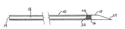
BRIEF DESCRIPTION OF THE DRAWINGS
Fig. 1 is a longitudinal sectional view illustrating a
pre~erred from of the hollow tunneler cf this invention.
Fig. 2 is a similar view of the shaft member of the device
of Fig. l;
Fig. 3 is an enlarged cross-sectional view taken along
lines 3-3 of Fig. 2; and
Fig. 4 is a longitudinal view of the solid cutting tip of
the device of Fig. 1.
DETAILED D~SC~IPTION OF T~E INVENTIO~
As was heretofore mentioned, the present invention is
directed to a subcutaneous tunneling device, which device finds
particular use in the preparation of long-term epidural
catheters which may be considered as having a distal internal
end inserted dorsally into the epidural space and an external
proximal end extending through the skin at a location on the
flank of the patient where it can be more conveniently atta~hed
to a syringe or other source of the liquid drug to be
administered epidurally.
The novel tunneling device of this invention will be
readily understood by reference to the accompanying drawing.
~ s shown herein, the tunnelin~ device of this inventlon
consists essentially of a hollow shaft 10 and a solid cutting
tip 12. Shaft 10 defines a chamber 18 extending between
opposed ends 14, 16 of the shaft~ In its preferred form, shaft
10 is tapped or contains internal threads 20 adjacent end 16.
Solid cutting tip 12 has external threads 22 at its base 12a to
mate with the internal threads 20 of hollow shaft 10 so that
the respective elements 10, 12 can be screwed together for
tunneling and thereafter readily separated by unscrewing. As
shown, tip 12 is preerably beveled or chamfered at its cutting
end 24.
While mating of the threads so the respective elements may
releasably engage one another is the preferred means, it will
be appreciated that the invention ~s not restricted thereto and
patO5129 5
2~7~2
"
other means for doing so will be readily suggested to those
skilled in the art. By way of illustration, they may
releasably engage one another for tunneling by a friction fit
wherein the base 12a of the cutting tip fits within end 16 of
ahaft 10.
As was alluded to previously, the tunneler must be
mallea~le. In this context, it may be made o~ a suitable
medical grade semi-rigid vinyl or other plastic material.
However, a metal such as stainless steel is preferred.
It will be appreciated that the dimensions of the tunnelex
may vary in accordance with the contemplated usage and are
accordingly not capable of pxecise ~uantification. In general,
the tunneler should of course be of sufficient length to
traverse the whole area to be tunneled in a single pass.
Likewise, the diameter of the tunneler must be sufficiently
large to present a passage for the catheter, cannula or other
tubular article to be tunneled subcutaneouslY-
~ y way of illustration, the preferred long-term epidural
catheter for relieving intractable pain contemplated to be
provided by the novel tunneling device of this invention is
that described and claimed in Applicant's concurrently filed
copending application, Serial No. (P.F. 1695).
As is disclosed therein, an epidural catheter of one-piece
construction is provided extending from the paravertebral entry
point subcutaneously to the exit site, in combination with a
protective enf~rcement sleeve adapted to be tunneled
suhcutaneously within the exit site for a short distance over
the proximal end portion of the catheter. The pro~imal end of
the sleeve is permanently attached to a catheter connector
having a luer fitting for securing the catheter in fluid
communication via the connector to the source of liquid drug to
be administered epidurally. After the catheter is tunneled
through the skln and out the exit site, the tunneler is
removed. Thereafter, the distal or free end of the sleeve is
then inserted over the free proximal end of the catheter and
then paxt way into the passage created by the tunneler. ~fter
securing the catheter to the catheter connector and closing
patO51~9 6
~0~8~2
"
both the entry and exit sites in the skin with sutures and
,terile dressings, the system is then ready for use.
With the novel long-term epidural catheter system
described and claimed in the aforementioned copending
application, the epidural catheter may for example be a 20
gauge (0.036 inch) and the protective sleeve may have an outer
diameter of on the order of 0.144 inch (11 French). A tunneler
in accordance with the present invention intended to be used
with a catheter system of these general dimensions may, for
purposes of further illustration, be on the order of 10-12
inches in length and possess an inner dlameter of at least 0.06
inch and an ou~er diameter of at least the same si~e as the
sleeve, i.e. 0.144 inch.
The following description is illustrative of the
preparation of a patient for intractable pain relief utilizing
the long-term epidural catheter of the aforementioned copending
application in combination with the tunneler of the present
inveDtion.
The epidural catheter is first threaded through a needle
into the epidural space in per se known manner. Wlth the
needle still in place to avoid inadvertent damage to the
catheter, a small incision is made with a scalpel extending
cranially and caudally approximately 0.5-1.0 cm. A11 tissue is
dissected away from the needle to allow the catheter to fall
freely into the incision as the tunneler is later advanced.
The epidural needle is then removed. If a wire stylet is used
for insertion of the catheter, it is also removed.
The malleable tunnele~ of the present invention is then
manually shaped to match the contour of the flank. The skin at
the paramedial incision is lifted and the shaped tunneler is
introduced subcutaneously and then guided laterally toward the
contemplated exlt site on the flank.
When the tlp of the tunneler has reached the desired exit
point laterally, the tunnelex is turned away from the patient,
thereby forcing the cutting tip up against the skin. A scalpel
is then used to cut down to expose the tip, after which the
tunneler is advanced through the thus provided exit site.
patO5129 7
2al678~2
Following advancement of the leading end of the tunneler
through the skin, the tunneler tip 12 is removed and the
catheter passed through the chamber or lumen 18 within hollow
shaft 10 and out through end 16. Shaft iO is then remaved
through the exit site.
Next, the protective sleeve (provided with a cuff of
Dacron or cther suitable material adjacent its proximal end
portion) is advanced over the free proximal end of the cathetex
extending above the skin and down to the exit site. The sXin
is then lifted with forceps and the sleeve advanced through the
exit site into the passage provided by the tunneler until the
cuff is approximately two inches beneath the skin.
The epidural catheter may then be trimmed to fit within
the adapter preattached to the sleeve and the adapter then
rnoved to the closed position to secure the catheter.
Both the paramedical incision and ventral exit sites are
then closed with suitable sutures and sterile dressings
applied. After attaching a removable morphine filter~injection
cap assembly, a saline solution may be injected to confirm the
catheter integrity.
The long-te~m epidural catheter is then ready to commence
introducing narcotic into the epidural space on an as-needed
dosage for pain management.
From the foregoing description, it will thus be seen that
the novel tunneling device oi this invention protects the
catheter from biological debris of host origin, kinking or
other damage during the tunneling step from the paravertebral
entry point to the exit site. Additionally, since it threads
easily through the tunneler, there is no resistance or pulling
which can cause the catheter to become dislodged from its
placemPnt within the epidural space, a problem noted previously
in discussing the prior art solid tunnelers. of course, it
can't possibly become separated from the tunneler beneath the
skin.
~ hile reference has been made throughout the description
to subcutaneous tunneling procedures for long-term epidural
catheterization, lt will be appreciated that the invention is
patO5129 8
J, 2~67~2
not limited thereto and is applicable to any tunneling
procedures where the cutting tip of the tunneler projects
externally following tunneling so as to be removable. It may,
for example, be used in tunneling procedures wherein a central
venous catheter (CVC) is tunneled from a suitable exlt sit,
e.g. at the midline, toward the venous insertion site, e.g. the
subc~arian entry point where the CVC is to be introduced into
the blood vessel.
Since certain changes may be made without departing from
the scope of the invention herein contemplated, it is to be
expressly understood that the foregoing description, including
the drawing, is by way of illustration and not by way of
limitation and the invention i5 limited only as indicated in
the appended claims.
patOS129 9