Note: Descriptions are shown in the official language in which they were submitted.
w ~ 62l~ rcT/~ o
?~ ~
~;L 3R;~ON~; ANAL'S ~ OF 3~NES
$hLs invenc~on relates to a ~e~hod and appara~us fcr the
non- nvasive eva'uatLon Oe d sease~i or f-acture~ ~one, and in
par-icular eor idenc .~lns ehe s~:uctural in~egrL~v of lo~
bcnes in-vivo
3~C~G~UN~ OF ~E ~NVE~TI~N
~ t present the cLinician decides whe~ the injured o_
diseased bcne can resume nor3al u3suppor-~ed u~ctic~ or e~e
~asis cf physical e~àmina~ion, rac ographs, the passa~e o,
t me anc satlent evaluatao~ of pai- as a -esult o. stress
placec or the ho~e
Vlbrational analysis o_ bonee has bee~ use~ ir the
expe-i~entaL environment for the examinatio~ of diseased and
._aceured bones a3d ?athological or~hopaedic disorders
however rhe pri~arv -easo~ for ~he lac~ of acce~ta~ce o- this
method of bone analvsis bv clinicizns has been the lack or
reliability of the -esults Most i~porta~tly the clinician
has not been able to -eliaDly assess from the results o~
tests uhen and i the bone unde: examination has regained
st-uctlral intes-ity
~ e lac~ of -eliabil tv ard _eproducibility of the ~rio-
v ~ra.~oral analvsis ~ethods resul_ ~rom a ru~be- OL ractors
ldentlied by the inveneo-s ~hes2 rac-ors incluce
i~suf'iciene -ange of exc tatior --ecuency, inappropriata
excita' on means, inconsis.ene detecelor anc meehodologv o-
~e sure~en~, a lac~ o- ap~recia~:or of ehe need ~o assess
~ora than ~ust one mocda of e~cit-~ on and the cnolce of
unn-cessa:ilv co~pllca~ed sei~fness c:ie~ria for c~i~ical
evaluatior~
Also of i~portance ls the me~hocd o suppor- provided to
.he ~one unde- esam:na_ or ard ~ e assessmen- o- he ef-ace,
of e~te-na' or i~ta- al '-~ation cevices wt'ch ~oe~
cone-i~u~ ~o the accu:ac~, _ep::c, ci~i!i.v ard p~aceica
clinical ~52 0 - p ' O- aralvsis me_-ods and means
'''' - ' ' ' ' ' ' `
.
~' .
~,. ~ ,
'
' ' ' ~ ` ~ ` ,' ~
W( ) ') ~ 62~ ~ PCr/ ,~ /1)050h
2~9~
a R ~ _ ^ S U Mt~iR '~ 0 ~ E I NV '` ~ N
~herefore thas lnven~Lon alms ~o overcome the
a~oremen~ioned probLems and ~rovide a method and apparatus
suiea~le ,or use bv cl~nLc an~ 2r:~a-ily fo_ e~e Ln-vivo
moni~o~ 3~ of bone f-ac~ure heal~ OUt which may aLso be
us~d for eh~ assessmen~ of othe~ pat~olo~icaL bon~ co~citions
such as ~u~ not confinedi to osteoporosis, prima~f ancL
secondarf tumour deposits, other bone lesions other bones and
orhe- boc,il~ beam Like structures such as teeth.
P-eferably the apra-atus comprises si3ple ele~ents that
a:e ~uic~ to apply and by adherence to the metho~i of the
invention, ~rov:des reLiabie, reproducible and accurate
-esul~s ~h~ch a-e consisten~ with empirlcaL cLinicaL
evaluatio~s o~ the st~ucturaL intec_ity o- the bo~y-eLement
unde- e~a~natio~ by a methad of compa-ison with a nor~21 or
contralate-aL body element
In its broadest for~ the i3vention is direc~ed to æ
method for applying in-vivo a non- rvas~ve vibrational 30tion
to a selected 'ractured or disease~ ele~ent o a body fo
determini~g the stif}ness state of the element, said metho~
comprislng the staps o~;
a) placing a vibration transduce- means in ~ r3
mechanical con~act with the selec~ed ele3ent at o- adjacent
ar~ enc region t~e_eof, for de~ec~iag ~ibratorr energy,
b) coc~act~ng a vi~r:torf deYice aqains~ the seleceed
eleme3t a. a firs. loca~ion re~ore _-om said erd -egion,
c) d:ivin :he said vibrator~ device ~o v~ra;e ov~- a
f:esuer.cv _anqe betweer. 20 ~o 2,000 Hert7 a~ a predete~~ine~
rate o~ ':e~uency cnarge,
d) us~'n$ a co~pu~e: device to s~ore a r irst mode of
vi~ratory rosponse ~-om the vibra~ion t-ansducer means,
e) contac~inq a vi~:3:0r~ dev~c_ agains~ ~~e seler.ed
elemen~ a. a second lcca~ on aLso ~emot~ _-om said er.d
rec~ion~
f) repea;lng s~eps c) and d) so as to s~ore a second
moda o~ vi_ra-orv r~s~ons~ f~om saic vibra~:on .-ans~uce-
means, anc
~; ' ' . ' .................... .
.
,'. . :. . ::
.~: ., . .~ -
W() ~1/1)621` PCr/,~ 91\~)051~h
g) comparLn~ ~ne ~ 'erence ~ec~een ~he peak ~:eauency
of eacn :esponse maae w.cn ~hat o. a cor-esponding reerence
mode rep~esentatLve o~ .he sticrness of a no~aL bocy ~le~ent
to ~rovide an indica~Lon o the s~a~e of the stifCness of the
elemen~.
The invention is also dl-ectad tO an apparatus for
appLyinc in-vivo a nan-l~vasive vi~rationaL motio~ tc~ a
selecte~ Çractu-e~i or diseased eLe~en~ o~ a hody~ Çor_
dete~inin~ the- s.iffness. state of the eLement~ sai~
apparatus co~prisins;
a vibrat~o~ transduce~ means which is applied; ta the
selectec, element at or adjacen~ an end re~ion the~eof,
a vi~rato~r device applied t~ ~he selectec'i eLement at æ
location remote ~ro~ saic end res_on,
sis~ L output means to drive tne vi~rator~r ~evice t~
vi~rate ove- a frequenc- r n~e Der~ee~ 20 to ~,000' Hie-t~ at
predere~ined rate of fre~uency change,
-eceiver means for receiving a signaL out~u~ fso~ the
said vi~ration transducer means representing a mode o~
vlbr~tion of the selected element,
storage means having a plurality of stora~i vibration
tsanscuce~ response modes o,' a nor~al hody ele~ent seored.
~herzi~,
compasison means to compase the ci'fe-ence je~ween t~e
pea~ ~:eaue~cy of each _esponse mode with tha~ of a
cor-esponcins refe-ence ~oae -epresentative o_ tae s~ 3ess `' '''
of a nor~aL body elemer.~ o provide an indica.ion of the
stata of the stiffness of the element.
BRTEF DESC~STION OF ~_ DR~WI~GS
In orde: that the invention may be clearl~ unde:stood
and readily carri-d in to e'fece, a prefe:red e~bodiment w~ll '
now ~e described bV wav of e~a~pLe onlv wi~h -e~a-_nce to rhe ''
accompanvina represen~ .ions, whe:ain:
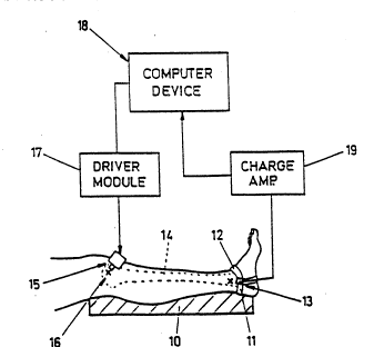
F'g. ' depices an a~30di~er.; of the appar ~us cf rhe
inveneLon in clinical use to ~easu:a the s.i ness
charac~e:istics o~ a tib:a in-v~o;
- .- ,
.
;.: . ,: , .. , . , - , . . . .
~;'.' ~` ' - ' '' , '-: ', ' , ' ' ' ' - ' '"' ' '' ": '
' ' ' ` ' ' ', :- :- . ' ' . .:
-: ' ' ' ' ' ' ' '~ "
W() 9~ 2~ 9~ PCl'/AU~ 05l~h
.- :g. ' depLc~s ~ cro~s seC~l~n ae ~he p oxLm~l en~
re~Lon o~ ~.e ~LbL~ ana LndLca~es .he pre~er3~1e Loca~Lon~
for appL ca~ion o a v ~racory de~ice;
F:g. ' depic~s a ~unc~-onal bLock diag~am Oe the
appa_~eu, o~ an emDodL~en~ of ~he :nvent on;
~:g. 4 depic_s an e~ample of ~he outpu~ of a cha~
recor~ina whlch dis~lavs .he resonance characteristiCs of. a
firs~ ~ode of resonance o~ a~ injured; and can~ralate_aL
nor~aL tibia;
Fiç. ~ depicts a~ exa~ple of the output of a cha~t
recordilg of a ~irst ~ode o~ resonance of. a bone dur nq the~ ~ -
union p_ocess;
~~5. o depacts an exa3ple of ;he OU.~UT; of. a cha-~
record~ns of a seco~d; ~ode!of resonance o~ a bone ~urinS .he
union process; - -
Fi5. ~ shows a typicaL graph~cal r~presenta~io~ o~ the
resonance characteristics of a healed tlhlaL fracrure and
t~at oS a nor~al cont-alateraL bone for the first mode o_
resonance; and
Flg. 8 depicts an e~odi~ent of the vibratio~ de~ice.
DET~I~c3 ~ESCaI~TION OF ~E I~VE~TTON
The appa:atus as show~ i3 Fig. 1 ca~ ~e appl~ed to a~y ~-;
access-ile iody ele~ent, for exa3~le teeth, sof ~ ~' ssue,
inge~ boQes and in pa-__cular _o- the ~urpose o- .his
desc: p~ on any long ~one and for ease oS desc_iptlon, the
~ethod a..d apparatus will De shown as applied to the t~ia
DUt is ecually applicable to othe_ aones such as e;~e fe~u-,
hume:~s, :3dius and ulna. '-
R-ga~dless o~ th- ~one to be exami~ed it Ls prefera~le
that i~ be supoortad so that the pa-e o~ the li~b u~der StUGy
has re~ucec conse:ai~ ~:o~ efects of the ~ocy~s lin~ages of
~hat lL~b .o ot~e- pa~t~ o t~e aocy anc places ehe
su-_o~nd ec ~uscle and ;issue ineo ~ rela~ed state.
In ~h~; e~bodi~en~ a cushicn a~ 10 ~n Fiç. I suppor-s
the u~pe~ la5 at ~he knee w~;h aoth hip anG knee sli~ht!y
le~ec thus ~e!axLn5 th_ ~uscles ad~acen~ ~he knee ~oint.
hher. _~.a `~-~2- lec, p2-~:cu!3~ e ;:~a, s suppo ~ec. bv
,., = ~.. ... .
''': ' '` :' ~' ' '' . ' ' ' '.~; '- '
Y .,
~*,-, . -
~:: . - . . :; .
~ . . .. .
. ~ . .
~,-, ' ' ' ''
~, ,
~ - .
,.,,: ~ ,
w(~ ~"~ ,` 2~ pCr/,~ ~'V~/l)OSl~h
~3e ~oc~ o~ ~ne cusn~s~ e hee' , '~kew:se supoor~ec.
alLow~ns e~e ooc ~ssel' so assume a ~eLa~ed pos~ure and
t~e~eb~ -eLa~Lnq muscle and tissue ad~acen~ the ankLe joint.
Suppar~ o~ ~he Lea in ~hls fashlon alLows ~he eL~ia to
S ap~:oach an unbound s~e. Ln ~;~is e~hodi~es.t 2 ~nit2r'~
sc~_'pe~:ed cushLo~ is used to provldc the reculred suptort
The foLL~wins steps comp~se a methc~ o measu~ement anc
analysLs which i, applicable to ei~her ror~aL or injure~
limbs. ~he~ comparisons are made the reference ~ata usad- i~
the ~ethod may co~sis~ o' data collectad '_om a~ measuremerts
made on a nor~aL cort-alater 1 lim~ Ot the same pariert or h)
data re?resentative o~ the ~ost Li~elv ecui~alen~ ele~ent
~hich ~a~ be compiled 'ro~ a number o~ othe- pat~en~s or
inrer-ed f~om ~he bone beins exa~i~ed.
Ste~ A comprises the place~ent o~ the vi~ratior;
transducer. General guldelines are appLicable '3 relatior; to-
t~is step wne-ein choice of location _equires tha. t~e-e
exist least possible depths o~ ski~ and suhcutaneous tissue
between the ~-ansducer and the bone under examination,
generally refer:ed to as t~e ~ost pro~inent subcutaneous ` ,'~
point o. the bone. This is preferably locatea a; or near the
end regions o, the bone and i3 the example o~ a fractured
bone at least at opposite sides of the '~ac.ure. In t~is '~
embodi~ent the ~edial malleolus Ot the dis~al regio~ Ot the ",
ti~i~ i 5 t~e mose ie,eal loc~tior. 'or t~e vibr~tion
t-anscucer.
FLs. 1 depict3 the vlbration ~-ansducer w'nich in ~is
embodimert is a 3rueL and ~jae: 4382 accele:ometer 11 navins
a compliar.: se:ap 12 loc~ted upon the ~ed~ alleolus 13 o~
~he cistaL end _e~Lon o~ the tLbia lt. I- Ls impor~an~ that
th- aceel~:o~ete: is palpablv ir. the ~ir~es. contac. with t~.e
~cno underlying t~e so~t tissue and the accelerometer
posi~ion is ~ainta:ned cons.~nt dur~n~ the e~amina~ion. ~he
str~p 1~ ~s~ ~e com_o_:ablv com?l~an_ to he shape 5~ the
patian~'s ankle bu~ mus. also re.aLn ;he accelerometer in it3
posi.son an~ o:Lenea~Lo~. T; ~ be a~pa~en~ .~a~ ;he
accel2-ome~2: has been locatec. suc~ as to ?:i~a:ilv de~act
vibrational energv ac;i~g i^. ;;e mecio-la~2r3i piar.e o- the '~ `
t:~a. , `,
., .
.. , ~ ~ .
`: '~ , . : '' ' '
- : . .~
' . ., . ' ` : , ` :~ ' , .
,: ' ` ':. ' '.
:'' . ' , :'
W~` 91/~)6'1' 2~~ PCr/~U9/)~00506
Ste~ 3 COmprLSes ~he placemenC o the vibraeLon d~vice
Ln accordance wieh ehe previously descrlbed guidelLnes co
detect a first mode of resonant vL~ratlon of ehe bone. The
vi~ratorv device 15 in t~is em~odi~ent is a ~rueL and Rjae -
v~ra~ion exciter ~odel 4310 and :s 4irst loca~ed on the
medial su~Cace of the medial t~bial plateau 16 of the
proxLmaL end reqion o the ti~ia 14. ~hLs ~i_st Locatio~ ic
also displa~ed; as ~0 in F;g. ~.
I~ this location the vibratory energy imparted i5 i~ a-
medio-latera~ plane of the tihia. Figure T depicts, an
em~odi~ent o_ the vihration excite- whlch has bee~ adapte~
for hand-held operatio~. _ncorjporated irto the vibratio~
exciter is a sp-ir,~ tensioned mechanical pre-loadi means whic~
ensures that each user applies a consistent pressure lying
~etween a predeter~i~e~ range o~ 1 to 20 NewtoQs.
Step C co~prises arivin~ the vibratory device. ~he
vibration exciter is drive~ bv a constant a~nlitude
sinusoidal wave ,or~ provided ~y a ~rive- ~odule ~T wQich is ` .' ~'-
cont:olle~ by a voitage signal supplie~ by a digitaL to
r~alogue output of a conpute~ device 18. ~he freouency
excursio~ of t~e sinusoidal wave form comyrises a linear (or
alternatively a lo~arith~$c) sweey from 20Hz to 2,000Hz ove
a pe:lod of ti~e between 2 to 30 seconds.
Step 3 comp_ises s-o:i~ the vi~ratorv response of the -i.',
.i~ia. Vi~ra~ory :esponse of the '~ia L- detec~ed by the ,--
acceleromete- 11 whic~ is connected to a cha-ge am~li ie:.
The charge a~plifie- a~pli_ies the elect-ical signal output
o: the acceleromete: a~c converts the signal into a ~aryi~g
analogu- voltage suita~le for connec lon to the analo~ue to
dlgltal input of a co~pu~e- dev~ce 18 which is then sto:ed
for recall or ~urtho- ?socessin~ as -ecuired.
~his signal may then be displayed as a fi:st vibratory
response on a plotter and or a visual display sc-een. In
this e~bodi~ent an ampli~ude versuS _re~uency plot oS the
~brato:v :esponse is p~ovided, an e~amp1e of which is shown ''
in Figs. 4-o. ~. is t~e ':e~uer.cv oS the peak o} the
response which provides ~ne mos. r-levar.t s iSCness
charac~e_is-~c o- the l:~b unc,e: exami~a~on and is generallv
r_r-2--ed t~ as modal -espons2 c-' ehe ~one ur.de: e~amLn_.lor
~_ .
~.: - :.
. . .
-.- - -.' :,. ~ , .
-
h-
wo9l/n621 ~'~3 ',~ PCI/ a l,"~)/005l~h
~e!i de~'~ned peak ~s an ~deal mOCdl respanse wh~le a
indLs~Lngulshahle peak Cre~uenc~f response LS a non-l~eal
modal ~espanse.
r~ s noe unusual for these steps to be repea~ed to
5 pro~ide the clinicLan the o~por~un ~ to varr the Location o
, the vi~ra~or~ device at points l~ca~ed ac~oss the re~ion i~ a
pla~e or~hosonal to the longitudinaL axis, o. hody eLement
un~e- esamira.ion. ~his step may provide~ se- es o~
displays whic~ exhihie in most instances a single peaked
-esponse and it is impo~tant that ~he clinicia~ ohtai~ the
s~oothes- and most clea~!y peakedi -esponse possi~Le
-epresenta~ive- o~ an ideal modaL _esponse ~-o~ t~e hone un~er
exami~ation.
A~ ancillary paramete~ is tha= o~ amplituce w~ic~ is
purely _elative to the amplitude o~ the d-ivi~c sisnaL
provideti to the vibratory device. This para~et ~ ca~ be
cont-olleL by the c~inicia~ to ensure that ehe peak is- of
s~ficient ~rominenca fo- assess~ert of its ~recuency value - '-
but need not be any greater. ~owever, it is importa~t to
have a similar amplitude response wnen comparisons o~ the
contralateral results are made sc as to simplify statiseicaL
analysis. `'
Step ~ then comprises the plzcemeQt of the vibratory
device to aetect a secona mode of -esonant vibrati-an. This
seconca=~ ~ode w~ll be senerated _s a esul t of a?plyin~ the
vibratorv energ~ on a di_2e~ent lccation o~' ~he ~:oximaL
region o_ the tibia. This seconc ~ode will be found ir. mos~
lons zr.d short beam like me~bers o_ the body, particularly
long ~on-s and is now understood ~o ~e a second ~lexural mode
of :esona~ce.
0~ particular impor~ to this inventior. is that ie is now
~ecogni--d that both modes o. resonance need to exhibit
nor~a' :esponses within acceptable variance be.2ore proper ,'
union or re~air of diseased bone -an be said to have
occ~--ed.
_n this embodimer.t ~he vibra~ory device is e~en loc ted
on a ~econd proximal end -eqion o' the ti'~La 1~ at the
anee-omedia' sur:ace of the media' ~ibia' ~la.ea~. This
sacor._ loc~.ion i~ ~lso c:s?~aye_ as 'I ~ c. ' ~he
- . : . ~ .
.
' - : . , ,
'~9~ ~
W(~ 91 /062J~ PCr/ A U9n/0050h
prevlous~-, desc_:bed s ~nals are p~o~tLded ~o ~:ive ~he
vLbra~ory devlce.
The vibratorv device pre~erabl~ applies its vibrationaL
ene:Sy ir. a plane whLch l~es at an acute ansLe to the mediaL
late_al olane.
The signal output os the accelerome~er is stored and
dis~laye~ as a secon~ moce o vL~ratory respo~e.
As prevlously desc=L~e~ it is 30t unusu~L 'o~ thie step.
to be :epeate~ unt~L a consistent sinsLe pea~ecL response is
obtained which will have a peak frequency hiqher than that
ob~aine~ -or the location chose~ 'o~ Step a.. D~e aLlowanc&
is made ove- the period of union oS a fractured. bone fo~ the
al.e-at~on o~ muscle tone and the like which is likely to
marsinally affect the amplitude response, but, in generaL not - ~-
a-ece the-peak freouency cetected.
The desc--bed methoa of modal response measurement is. ~ . `
also used to store the stiffness characteristics or a normal
bone. Il the siven example the co~t-alateraL ti~ia is used
although a modal response represen~at~e of a comparable ~ `~
tib$a may suffice.
r dee~ the contralateral tihia of a person may have a
slig~t ~a~iance '_om its pair even hefore it was fractured or
diseased so the compa_ison yet to ~e perfor~e~ be~ween the
resu~ts will account for this ex~ected ~ariance.
Ste~ G compa-es in the ~_s~ insta~ce of thLS e~bod'ment
the ?eak f_eauency o ~he measured -esults. Comparison o: ~ -
the moda! response may be achie~ed in a nu~oe: of ways. Mos.
con~enien~ and of simpler implementation is t~e method of
compa:~ng the pea~ freauency o each mode wi:h the
correspo~d'n~ cont:alater~l mode and if both compa-isons fal!
w'th;3 a p:~deter~in-d rang-, the stif-ness of the bones are
si~ilar and the clinician can be confident that the bones are
compa:a~lv stif~.
~!tz-nati~ely, a statistical analysis of the recorded
moda' :-sponses, may be con~ucted to produce a corralation
coe~-icient which if for each mode is within a predeter~inec.
range will likewise indicate to t~e clin~ cian tha~ the
sti~-ness of the bones are simila-.
. .. , . . -, - . -
- - - . -: . . - . - .
::, . : . - -
.
. .
'- - .-,
'
:'
2~
W 91/06~ P~/AU91)/00SOh
I_ LS i~partan~ that ~oe~ ~qdes of vibratory respcns~
are detected and their comparisons fall within the
predetermine~ range since one withou~ the other in~icates
~-om ~nysical e~a~Lnation that normal se~_fness has not yet
been ach~eved.
Step ~ is the first in a series of additLonaL steps, that
prcvide .urther assurance to the clinicia~ that the banes
unde- compar$son are comparahLy sti~. T~e placement o~ the
vibraeory device is relocated to detect a thi-~ mode~ o_
_esonant vi~ration. This third. 30d~ may na~ ~e found i~ alL
bones or indeed in every body ele~e~t however it is
recognised in ehe tihia as bein~ æ torsional ~ode of -'
resonance whick generalLy occurs at a higher freguency tha~ '-
the previousLy discussedi fLexuraL ~odes, of resonance.
The tihial exa~ole $n~$cates that the anterior surface~ ~
o the tibial tuberosity is the~ preles-e~ location 'or ''detecting the third mode of -esonance.
Steps 1, J an~ ~ are iden~ical to steps C, D a~d G as
previously discusse~.
Fig. 3 depicts an embodiment of a functional hloc~ ,'
diagram of an apparatus suitable for im~lementation of the ' '
iDvention. The circuits which co~prisa the function bloc~s ',
a:e of a uell kncwn nature and reguire no fulther
desc-iotion. _t wilL De a~arent t~at these ci-cuits ~ay be `'
~ariouslv configured, whesein, for exam~le the computer ',
device 2~ ~zy contai~ withi~ its housins on stancard,plu~ ,'
compatible carGs, the char~e am~ ier ~3 and/or the circ~it
moaules 23-2~, while the vibration exci~e- 29 and t~e ,~ ' '
accelero2e~e- 30 are simply plussed i~to extar~al connector ,",
sockots, the:eby providing a~ exceedingly si~ple ar_a~gement
for clinical us~
Fig. 4 depicts an example of the output o' a chart
:ecorce- showing a logar.thmic d~s~lay of frequencv along the
x-axis and r-lative a~l~tude alonc the y-axis. The trace
shown as ~Nor~al~ indica~es the o~pical C~equency to
a~pli~ude characteristics o~ the patients un-injured or
os~ens~bly normal bone, LndLca~:n~ '~y its s~ooth t-ansit'on
and c!ea- ~eak that the bone has a _~-s~ modal -esonance of
approx:~at21v '60 H-. T~e me~hcc, c_ ~e embodi~en~ shown has
~ - .
g: ~
z~
Jl/062J~ PCI /AU9~\/OOSo6
I~
exhLbl~ed an accuracy o~ :5 H- even when app~i~d by diffsrsnt
cLinicians
In co~arison t~e erace shown as "I~JURED~ typically
i~dicates the f-equencf .o amplitude characteristics o an
S acutel~ L~jured ~one, lndicat2d b~ its irrecula~ an~
imprecise Çre~uency Its fre~uency and amplitu~e however, is --
~ecognisahly lower and is deter~i~sd hy averasi3g th~
~se~ueucy o~ maxi~al amplitude- rsspanse over cocsecutive
m-asussments which i~ this case is approxi~ately 84 Hz
Nor~al cLi3ical tests confir~e~ that t~e injured bone ha~ no
evidence of signi~cant bony union
;~ has been 'ound that i~ seneral, acutely f ractused
bones exhibit a~ i--esular, Lo~ fre~uency ;eso3ance usuaLly ~ -~
beiow 100 Hz wheseas un-i~jure~ or united hone exhihits a -~
lS resonance bet~ee~ 200 Hz and 500 ~z and typically a~out 350 ~ ; -
~z for a nor3al adult male and 300 ~2 for normaL adult
;~emaLe I' has ~een fou~d that the shape of .he normal
response curve is typical for each i~di~idual and that the
fre~uency at pea~ amplitud- respon-~ (resonance) remains
consta~t ~or that indi~idual
Meas~red resonance characteristics of ~one are generally
unaf-ected ~Y the ~resence of surgical nails, screws, plates
a~d pi~s It is found that the resonahce of t~ese devices is
usuallv easily identi_iable as a ciscrete lessar amplitude
~ea~ tha~ that of the excitation -esponse of a ~one unde_
exami3at on a~d t~eir cont_ ~ution becomes less noticeable as
the bone union develops Thus it is found tha~ the
characterist~c resona~ce of unitqc ~one resem~les that of the
30r~1 con~-alateral li~ eve~ wit~ t~e internal or exter~a!
i;xation d-vices in place~
F'~ 5 hows a typical graphical represe~tation of the
firs: ~odal -esonance characterist cs of a f-actured ~one
The modal _esponse showi~q an i--ecula- t_ace IINJURED 1"
exhibits a low peaX ampli-ud- ~alu- and a lower ~requency of
:esonance of 84 H~ four weeks af.e: the ~n~urv The
inte-mediare stage of bone union :s depiceed as "~NJURE0 2~
eigh~ wee~s afrer injurV which has a more regula_ t_ace, an
inc-eased a~pl tude and a h_gher _esonan~ f_equencv of 148
i; ind~c-~es un:or lS prog-esslns anc .~e s~:;~;ness of
.
. -' ~ . .
.. . . .
.. . - - ~ . :
;- . ~ - - .. .
i - .
2'~3~
~4 ~1/062 1` PCr/AU90/0050h
L~
the bone unLon has L~proved althaugh has no~ yet roached that
expected o~ a con~:alateraL nor~taL limb. T~.e ~race denote~
~N0RM~L~ which has a regular shape exhi~itlng, the greatest
ampli~ude and a resonan~ f_eouency of ~5a H-
Fig. 6 shows a cor-espondinq second modaL resonance
characterLstic o~ the same fractured bone desc~ihe~ i~ Fi~. -
5. ~he peaX reso~ances o, each trace are cor~espondinsly
hi~her than those o Fi~. 5 and are characteristic o this
sscond 'lexuraL made o the bono.
L0 ~is. T shows a typicaL graphicaL represe~tatio~ o the ~:`
-esa~ance characte:ist~cs o a healed tibiaL .-actlre an~
-~hat o a normal cont_alateraL bone using the 'i-st ~odal
response as an exa~ple.
~he concordance o' t ese traces and their peak
f-ecuencies pro~idee COA~irmation to the cliaicia~ that ~one
union has occur~e~ and that it has su~icie~t sti2fness to -~
resuue nor~al activity.
It has bee~ found by usi~g the i~ve~tion that an average
variation of the resonance value ~eeween normal contralateral ~ -
li3bs i5 2 . 9~ with a ~axi3t~ acceptable ~ariation of 8.0~.
Althouçh abnormal variation between contralateral li~bs ~ ~
has noe bee~ ound in elderly persons, allowance for typical :
~or~aL resonance values needs to be made or the bones of
elde-ly pe:sons. It has ieen ou~a t~at the lowe: density o
bones caused by age ana osteoporos_s resul.~ in a lower
-esonan~ --equency than ~ould be expected bv li~D length and
circu~stance. ~owever thi 5 charac~eristic is consistent,
r-sul;ins in a rssponss which has a sha~ and well de_ined
p-a~, suc~ that co~pariston to frzc~ured or diseased bone is
Jtlll poJslbl- and th- ltethod and apparatus still applicable
to th-L: assess~e~t.
~ 2wise the resonance wilt shi't downwards by a
predictable a~ount where _he li~b has become shortened as a
rosult o~ the ~:acture an~ neali..g process.
Fig. 8 depict~ an em~odiment o_ the Vi~ra.iOA device,
which as previously desc_:~ed, comprises a voice coil and
~açnet vibra~ion exci~e: 1c. mO e~-ect ease o. handlinq and
a~p!ication to ~he ~a~ ene, the ~J:2r3t:0n device has ~een
,~_........ . ..
~; . , - .~ - ,,
, :. ., , . :
. ~ , . . . .
.
.; ~ ~. - . . - :
, .
-- ; . - ~
u 7l/0624~ PCT/AU90/OOSO~
1~ ,
modelled in~o the shape shown Ln plan view in 'ig 8, whlch
however is indicative of its substantially cylindricaL shapo
Wires supplyinq moeive energy enter the bady of the
device via stress relief member 40 and connect ~o the encased
exci~er 45 in the normal manne~ Shaft 41 ls the
reciproca~Ln~ memher o; the device an~ its hea~ 4~ is the ~ -
por~ion of the device which is applied to the bone o bady !. ' '
member unde; exa~ination A sprins 4~ is a_-ansed with its
lonsitudinaL axis coaxiaL with the sha4t 41 an~ provides a
predeter~ineti pre-loa~i force when the heai is app~ieti to the
body A pre-load force of between 1 to 20 Newtons '~as bee~
fou d adequate for enerqy t-ansference and also comfor;abLe
for patients
When ~pplyi~s the methoci o~ the invention it is
convenient for the cLinicia~ to initiate the step of a~ivinq
the vibratorv devic- when it has be~n fi 31y located O~L the
~ocy ~hus, switch 43 is located im easy reach of t~e ehu~ :
of ~le hand holding the vibratory ~e~ice Alter~atively, the
step of drivins th devic~ ~ay b~ automatically repeated by
th- conput-r d-~ice until a suitable trace of resona~t ~;
respons- is obtaine~ and it5 results stored for analysis
~is inventian therafore provides a reliable a~d
acc~rate clinical tool for non-invasive assassment of
disease~ or f_actusad bo~e in-vivo
,, . . ~ , . . - - , . , - .
r ~. . . . : . ,; , - :
.
~; '' . ' - . ~ ,
.. , ~ , ' .
~, , , ' , ' , ~. ' .
t