Note: Descriptions are shown in the official language in which they were submitted.
207~615
EndoProsthesis for cancer dama~ed hiP bones
The invention refers to an endoprosthesis for cancer d~maged
hip bones according to the preamble of claim 1.
A variety of endoprostheses is known to replace the damaged
joint ball of tha femur and/or the joint socket of the hip
bone when these parts do not anymore properly function. In
cancer damaged hip bones, a large portion of the bony
structure of the hip bone is attacted in many cases and is
thus incapable to support a prosthetic hip socket in the
usual manner. In such cases a unique bone substitute must be
formed including a recess in the distal portion to receive a
prosthetic hip socket. The individual forming of endo-
prostheses of this type is costly and takes too long a time.
It is known to first form a model of the bone portion to be
substituted using X rays, in particular computer tomography,
and then to form the endoprosthesis from the model.
The object of the present invention is to provide an
endoprosthesis for cancer damaged hip bones which can b~
used in a wide variety of individual shapes o~ the hip bone.
The object referred to is solved by an endoprosthesis
according to claim 1.
The present invention is based on the knowledge that it is
inevitable to have a photographic survey when cancer damaged
parts lie within the region of the natural hip socket or
close thereto. It is further important that the pros~hetic
recess for receiving the prosthetic hip socket finds
sufficient support and is safely secured to the hip bone.
: . . :,. . ; : . . ..
~, - :: : .- : - ,.
2~7~
.
The endoprosthesis according to the invention provides for a
unique structure, but allow~ an individual ad~ustment wikh
respect to the dimensions of the hip bone. According ~o ~e
invention, the endoprosthesis comp~i~e~ a distal and a
proximal part which can be rigidly secured ~ogether by
screws. The distal as well as the proximal part include
mounting extensions to be secured by screws to the hip bone
or, respectively, to a vertebra. Preferably, the proximal
part is secured to the fifth vertebra and to the satrum.
The two-part structure of the endoprosthesis according to
the invention facilitates making it in a die-casting
process. Furthermore, the surgical fixing of the prosthesis
parts i5 simplified due to the bipartite structure. For
example, after secting certainbone portions of the pelvis,
the proximal part is first fixed and then the distal part is
secured to the proximal part, whereupon the proximal part is
screwed to the vertebra.
For a safe connection between the distal and the proximal
part, the invention provides for an interlaced connection.
According to a further embodiment of the invention, both
interlaced portions include interengaging faces, such as a
nose, formed on the first part engaging a corresponding
recess of the second part. The noses prevènt a relative
rotation of the parts even then when a single screw only is
used for connecting both parts. Preferably, a pair of screws
is used and the heads thereof are preferably embedded.
According to a still further embodiment of the invention,
the distal part of the prosthesis is formed to comprise a
pair of supporting portions of which one engages a pubic
bone branch and the other an ischium branch, each supporting
portion including a securing bracket. The supporting
portions thus define bridge portions between the pubic bone
branch or, respectively/ ischium branch left and the portion
,, "~
: ~ . . , :. . .
207~
of the distal prosthesis part in which the recess is formed
to receive the prosthesis hip socket.
According to a still further embodiment r the proximal paxt
includes a pair o~ securing brackets of which one having a
BCS surface laterally engages the sacxum, and the oth0r
engages the vertebra in front leaving still a nerve passage.
Each bracket preferably has a number of screw bores for
receiving spongiosa screws to be secured to the associated
vertebra.
A preferred embodiment of the present invention is described
in more detail with reference to the drawings, which show:
Fig~ 1 a perspective view of an endoprosthesis according to
the invention;
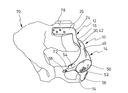
Fig. 2 a perspective view of a part of a hip bone from the
front including an endoprosthesis mounted thereon;
Fig. 3 a view of the hip bone from the rear and a
prosth~sis mounted;
Fig. 4 a left-hand view of a hip bone and a prosthesis
mounted.
The endoprosthesis 10 illustrated in Figs. 1 to 4 comprises
a proximal part 12 and a distal part 14. Both parts 12 and
14 are die-cast and are made of a body-compatible material.
The mold is made in a known manner using computer tomo
graphy, for example, to survey the bone region to be
substituted and making the mold according to the data
obtained this way.
Th~ distal part includes a plate-shaped portion merging via
a bent-off portion 18 in a flat mounting bracket 20
: ., .................................... ;
-
, .,
-
- 207~
including four bores 22 to receive bone screws not shown.
~he bracket e~ten~ion 20 extends under a small angle with
respect to the plane of the plate portion 16. The mounting
portion 20 extends beyond a ben~ portion 24 to a second
curved mounting bracket 26 including four bores 28 alike to
receive bone screws not shown. The plate portion 16 includes
an extension 30 in which two bores, 32 are provided. A
rounded nose 34 is provi,ded at the free end of the plate
portion 16. A recess 36 which is complementary with respect
to the nose 34 is formed at a shoulder of the extension 30.
The distal part 14 comprises a plate portion 40 including an
extension 42 including two bores 44. The extensîons 30, 42
are complemantarily shaped, and the nose 46 of the portion
40 engages the recess 36 when the extensions 30, 42 are
mounted in overlapping relationship. Screws 48 are used to
secure the plate portions 16, 40 together, wherein the screw
heads are received in the bores 44.
A recess 50 to receive a prosthetic hip socket made of a
suitable plastic material for example is provided in the
center region of the distal part 14. A first supporting
portion 52 and a second supporting portion 54 including a
lateral mounting bracket 56 and 58 each having a bore 60 to
receive a bone screw, are formed on the side of the recess
50 opposite the plate portion 40. The supporting portions
52, 54 are provided with obtuse ends and surfaces having a
microspherical structure for facilitating fixing to the
bone. A number of bores is provided. The supporting portion
54 includes a bore which end 64 opens into the recess 50.
The supporting portion 52 is provided with two bores as
shown in Fig. ~. The bores receive screws for mounting the
distal part to proper bone sections of the pelvis.
According to Fig. 2 a pelvis bone 70 is shown in a front
view; the pubic bone and the ischium are resected on the
207~
left~hand side. Thus a pubic bone branch 72 and an ischium
branch 74 are left. The surgical cut is selected sueh that
the obtuse end of the supporting portions 5Z, 54 ob~usely
contacts the branches 72, 74. The mounting takes place
through the mounting brackets 58 and 56. Furthermore, the
supporting portions are secured by screws which are inserted
through the bores opening into the recess 50.
In performing the surgical operation, the proximal part 14
is first mounted in the manner described and then the part
14 is screwed to the distal part 12 by using the screws 48.
The mounting bracket 20 thus laterally engages the corre-
lated vertebra (5th lumbral vertebra~ which has to be shaped
correspon dingly. The mounting bracket is then secured to
the vertebra by means of screws. The other mounting bracket
26 engages the front side of the vertebra and is mounted
alike by means of screws, as particularly shown in Figs 2
and 4. The arched portion 24 leaves space for a nerve
channel 76.
'
. . : : ~ ~
:,
