Note: Descriptions are shown in the official language in which they were submitted.
WO 91/09958 PCT/US90/07607
24'~1~1
METHOD OF DELIVERING MOLECULES
INTO EUKARYOTIC CELLS
Description
Background of the Invention
05 In eukaryotic cells, the nuclear envelope
isolates the central genetic processes of DNA
replication and RNA synthesis (i.e., transcription).
The cell nucleus contains nucleic acids (i.e., DNA
and RNA) and a variety of proteins. Some proteins
.LO are structural proteins that bind to and organize
the DNA (e.g., hist:ones). Other proteins are
regulating proteins. that bind to DNA and thereby
positively or negatively regulate transcription
(e. g., activator and repressor proteins). Still
7_5 other proteins are enzymes that carry out DNA
replication and RNA synthesis (e.g., DNA and RNA
polymerase).
At the present time, there are several
techniques available for introducing molecules, such
0 as proteins and oligonucleotides, into cells. Such
techniques are of interest, for example, in genetic
engineering and as .a possible means of introducing
into cells drugs or other substances potentially of
value diagnostically, therapeutically or
25 prophylactically.
Presently-available genetic engineering
transfer techniques include membrane fusion (e. g.,
fusion of cells with other cells or with liposomes),
incubation of cells with a calcium phosphate
WO 91 /09958 PCT/US90/07607
~~'~1~~ :~
-2-
precipitate of DNA fragment, DEAE-dextran-mediated
transfection, electroporation, direct intracellular
micro-injection of DNA fragments (e. g., via glass
capillaries), and infection of cells with modified
05 vectors (e. g., viral vectors).
All of these techniques can only be used in
vitro to incorporate genetic material into cells in
culture. In addition, the techniques are unreliable
and nonspecific. Not all cells are altered and many
cells do not survive the harsher treatments.
Finally, these techniques are only used to transfer
nucleic acids (e. g., DNA or RNA) into cells.
At the present time, there is no simple
technique for delivering molecules of any type
(e. g., proteins or peptides, nucleic acids) directly
into the nucleus of cells in vitro or in vivo. It
would be very useful if it were possible to deliver
such molecules reliably and specifically into the
cell nucleus.
Summary of the Invention
The present invention pertains to the use of
HIV Tat protein (Tat protein) to deliver a molecule
of interest into eukaryotic cells, particularly into
the cell nucleus, in vitro or in vivo. It further
pertains to conjugates, which include a molecule of
interest and HIV Tat protein, which are useful in
the method of the present invention. The method of
the present invention is based on the unexpected
finding that when Tat protein is present extra-
cellularly, it is readily taken up by cells and
WO 91/09958 PCT/US90/07607
~~ 1J1
-3-
specifically introduced into the cell nucleus, as
evidenced by the fart that cells treated with Tat
exhibit high levels of transactivation.
In the method of the present invention, a
molecule of interest. and Tat protein are brought
into contact with cells into which the molecule of
interest is to be introduced, under conditions .
appropriate for its entry into cells. As a result,
Tat protein and the molecule of interest enter into
10' cells, in which they pass specifically into the
nucleus.
In one embodiment of the present method, a
molecule of interest-Tat protein conjugate, which
includes a molecule of interest (i.e., a molecule to
15 be introduced into cells) and Tat protein is brought
into contact with cells into which the molecule of
interest is to be introduced, under conditions
appropriate for its entry into cells. As a result,
the conjugate enters into cells, in which it passes
20 specifically into the nucleus.
In a further embodiment of the present method,
the molecule to be delivered into cells is a
protein, a peptide or an oligonucleotide. The
present method is particularly useful for delivery
25 of proteins or peptides, such as regulatory factors,
enzymes, antibodies, drugs or toxins, as well as DNA
or RNA, into the cell nucleus.
A stabilizing agent, which serves to increase
Tat stability and uptake, can be brought into
30 contact with cells, in conjunction with the molecule
of interest and Tat ;protein. For example, metal
CA 02071214 2003-11-12
Z
-4 -
ions which bind to Tat protein and increase its
stability and uptake, can be used for this purpose.
In a further embodiment, a lysosomotrophic
agent is provided extracellularly in conjunction
05 with Tat protein and a molecule of interest, in
order to enhance uptake by cells. The lysosomotro-
phic agent can be used alone or in conjunction with
a stabilizer. For example, lysosomotrophic agents
such as chloroquine, monensin, amantadine and
10 methylamine which have been shown to increase uptake
of Tat in some cells by a few hundred fold, can be
used for this purpose.
In another embodiment, a basic peptide, such as
Tat 38-58 or protamine, is provided extracellularly
15 with Tat and a molecule of interest to enhance
uptake of Tat. Such basic peptides can also be used
alone, in combination or with stabilizing agents or
lysomotrophic agents.
Through use of the present method, it is
20 possible to introduce into cells and, particularly
into the cell nucleus, a drug or other substance
which is of diagnostic, therapeutic or prophylactic
value.
In a further embodiment, there is provided an
25 isolated and purified molecule of interest-tat protein
conjugate comprising a non-tat molecule of interest
covalently linked to an HIV tat protein or peptide
having cellular uptake activity, excluding a conjugate
consisting of amino acids 5-86 of tat linked to any
fusion protein consisting of an E.coli trp leader (L)
sequence fused to an E.coli trp E sequence.
CA 02071214 2003-11-12
- 4a -
In a further embodiment, there is provided a
molecule of interest-tat protein conjugate comprising a
non-tat molecule of interest covalently linked to an
HIV tat protein or peptide having cellular uptake
OS activity, excluding the conjugates consisting of amino
acids 1-58 of tat linked to a peptide having the amino
acid sequence LWLTKEPTA; amino acids 1-56 of tat linked
to the carboxy terminal 104 amino acids of an HIV
art/rev protein; and amino acids 5-86 of tat linked to
any portion of an E.coli trp LE protein.
Brief Description of the Drawings
Figure 1 is the amino acid sequence of the
HIV-1 Tat protein.
Figure 2 fs a thin layer chromatogram (TLC)
showing chloramphenicol acetyl transferase (CAT)
activity, a measure of Tat uptake, resulting from
WO 91 /09958 PCT/US90/07607
~~'~~21~
-5-
incubating HL3T1 cells with Tat for 24 hours at the
indicated concentrations.
Figure 3 is a TLC showing CAT activity
resulting from incubating HL3T1 cells with 5 ~g of
OS Tat and a variety of lysosomotrophic agents.
Figure 4 is a graph showing the cellular uptake
and nuclear localization of 1251-labeled Tat.
Figure 5 is a gel of nuclear fractions from
HL3T1 cells treated with Tat protein in the absence
(-) or presence (+) of chloroquine.
Figure 6 is a series of graphic representations
showing chloroquine stimulation of Tat transactiva-
tion.
Figure 6A is a graph showing the effect of
chloroquine concentration on uptake and transactiva-
tion by 2 ~g Tat added to the medium.
Figure 6B is a graph showing the time course of
uptake and transactivation by 2 ~g of Tat and 100 ~cM
chloroquine.
Figure 6C is a graph showing the effect on
uptake and transactivation by several concentrations
of Tat with 100 ~M chloroquine.
Figure 7 is a TLC showing CAT activity from H9
lymphocytes, U937 promonocytes and HeLa cells
treated with Tat protein in the absence or presence
of chloroquine.
Figure 8 is a TLC showing CAT activity from
HL3T1 cells and illustrating the extent of
activation of the CAT reporter gene following
various time periods of exposure to 1 ~g Tat + 100
~M chloroquine.
WO 91/09958 PCT/US90/07607
-6-
Figure 9 is a schematic representation of the
murine sarcoma virus (MSV) retroviral vector used to
establish the H938 reporter cell line from H9 cells.
The transcription start sites from the SV40
05 promoter, the HIV and MSV LTRs are indicated by
arrows, and the location and size of the fragments
protected in the RNase analysis are indicated by
bars.
Figure 10 illustrates the results of an RNase
protection experiment in H938 cells, using an a-32P
UTP-labeled HIV-1 LTR probe corresponding to a 200
by fragment from -120 to +80 of the viral LTR,
prepared by in vitro transcription.
Figure 11 is a graph illustrating the
enhancement of transactivation in H938 cells upon
addition of increasing amounts of the Tat 38-58
peptide with 1 ~g of Tat as assayed by CAT activity
after a 24 hour incubation with peptide and Tat.
Figure 12 is a graph illustrating
transactivation of a tPA reporter gene under the
control of the HIV-1 LTR in HeLa.318 cells upon
addition of exongenous Tat or a TatE2C fusion
protein.
Detailed Description of the Invention
The present invention is based on the unexpec-
ted finding that Tat protein from immunodeficiency
virus (e.g., HIV-1, HIV-2, SIV) is readily taken up
into cells and subsequently into the cell nucleus.
Tat is a potent viral transactivator and is
essential for viral replication. In light of the
WO 91/09958 PCT/US90/07607
_7_
fact that proteins and peptides are typically poorly
taken up (Sternson, L.A., Ann. N.Y. Acad. Sci.
57:19-21 (1987)), th.e finding that Tat is readily
taken up into cells is surprising.
p, As a result of this finding, it is now possible
to use Tat protein to deliver molecules (e. g.,
proteins, peptides, nucleic acids) into cells and,
specifically, into the cell nucleus. The present
invention is a method of delivering a molecule of
lp interest into cells and, particularly, of targeting
a molecule to the cell nucleus, as well as a
conjugate useful in the method. Any molecule can be
delivered into cells, especially into the cell
nucleus, using the method of the subject invention.
l~~ In one embodiment of the method of the present
invention, a molecule of interest-Tat protein
conjugate, which includes a molecule of interest
(i.e., a molecule to be introduced into cells and
delivered to the nucleus) attached to HIV Tat
2p protein, is brought into contact with cells into
which introduction of the molecule of interest is
desired. In another embodiment, a molecule of
interest and HIV Tat protein are contacted with
cells into which the molecule of interest is to be
25 introduced. The method can be used to deliver a
molecule of interest either in vitro or in vivo.
For example, delivery can be carried out in vitro by
adding a molecule of interest-Tat conjugate to
cultured cells, by producing cells that synthesize
3p Tat or Tat conjugate or by combining a sample (e. g.,
blood, bone marrow) obtained from an individual with
WO 91/09958 PCT/US90/07607
_8-
the conjugate, under appropriate conditions.
Delivery can be carried out in vivo by administering
the molecule of interest and Tat protein to an
individual in whom it is to be used for diagnostic,
05 preventative or therapeutic purposes.
The following is a description of: the HIV Tat
protein; uptake of Tat into the cell nucleus; the
molecule of interest-Tat protein conjugate; and the
method by which the conjugate is used to deliver a
selected substance into cells.
HIV Tat protein
Figure 1 shows the amino acid sequence of the
Tat protein. The full-length Tat protein is 86
amino acids long and is encoded by two exons; the
N-terminal 72 residues are encoded by the first exon
and the C-terminal 14 residues are encoded by the
second exon. Tat protein contains a highly basic
region (with 2 lysines and 6 arginines in 9
residues) and a cysteine-rich region (with 7
cysteines in 16 residues).
The basic region (i.e., amino acids 49-57) is
thought to be important for nuclear localization.
Ruben, S. et al., J. Virol. 63:1-8 (1989); Hauber,
J. et al., J. Virol. 63:1181-1187 (1989). The
cysteine-rich region mediates the formation of
metal-linked dimers in vitro (Frankel, A.D. et al.,
Science 240:70-73 (1988)); (Frankel, A.D. et al.,
Proc. Natl. Acad. Sci, USA, 85:6297-6300 (1988));
and is essential for its activity as a
transactivator (Garcia, J.A. et al., EMBO J. 7:3143
WO 91/09958 PCT/US90/07607
1~l C.. '~
-9-
(1988); Sadaie, M.R. et al., J. Virol. 63:1 (1989)).
Like other regulatory proteins, the N-terminal
region may be involved in protection against
intracellular proteases (Bachmair, A. and A.
0'' Varshavsky, Cell, 56:1019-1032 (1989)).
It will be appreciated that the entire 86 amino
acids which make up the Tat protein may not be
required for the uptake activity of Tat. For
example, a protein fragment or a peptide which has
fewer than the 86 amino acids, but which exhibits
uptake into cells and uptake into the cell nucleus,
can be used (a functionally effective fragment or
portion of Tat). As is shown in the Examples below,
Tat protein containing residues 1-72 is sufficient
for uptake activity and Tat residues 1-67 are shown
to mediate the entry of a heterologous protein into
cells. In addition, a synthetic peptide containing
Tat residues 1-58 has now been shown to have uptake
activity. A Tat peptide comprising the region that
mediates entry and uptake into cells can be further
defined using known techniques. (see, e.g.,
Frankel, A.D., et al., Proc. Natl. Acad. Sci, USA,
86: 7397-7401 (1989)).
The Tat peptide can be a single (i.e., continu-
ous) amino acid sequence present in Tat protein or
it can be two or more. amino acid sequences which are
present in Tat protein, but in the naturally-occur-
ring protein are separated by other amino acid
sequences. As used herein, Tat protein includes a
naturally-occurring amino acid sequence which is the
same as that of naturally-occurring Tat protein, its
WO 91/09958 PCT/US90/07607
2~'~~2~~
-i0-
functional equivalent or functionally equivalent
fragments thereof (peptides). Such functional
equivalents or functionally equivalent fragments
possess uptake activity into the cell and into the
05 cell nucleus that is substantially similar to that
of naturally-occurring Tat protein. Tat protein can
be obtained from naturally-occurring sources or can
be produced using genetic engineering techniques or
chemical synthesis.
The amino acid sequence of naturally-occurring
HIV Tat protein can be modified, by addition,
deletion and/or substitution of at least one amino
acid present in the naturally-occurring Tat protein,
to produce modified Tat protein (also referred to
herein as Tat protein). Modified Tat protein or Tat
peptide analogs with increased stability can thus be
produced using known techniques. Therefore, Tat
proteins or peptides may have amino acid sequences
which are substantially similar, although not
identical, to that of naturally-occurring Tat
protein or portions thereof. In addition,
cholesterol or other lipid derivatives can be added
to Tat protein to produce a modified Tat having
increased membrane solubility.
Variants of Tat protein can be designed to
modulate the intracellular location of Tat and the
molecule of interest following uptake into the cell
or when expressed in the cell. When added
exogenously, such variants are designed such that
the ability of Tat to enter cells is retained (i.e.,
the uptake of the variant Tat protein or peptide
WO 91/09958 PCT/US90/07607
-11-
into the cell is substantially similar to that of
naturally-occurring HIV Tat). For example,
alteration of the basic region thought to be
important for nuclear localization (see e.g., Dang,
OS C.V. and Lee, W.M.F., J. Biol. Chem. 264:
18019-18023 (1989); Fiauber, J. et al., J.Virol. 63:
1181-1187 (1989); Ruben, S.A. et al., J. Virol. 63:
1-8 (1989)) can result in a cytoplasmic location or
partially cytoplasmic location of Tat, and
therefore, of the molecule of interest.
Alternatively, a sequence for binding a cytoplasmic
component can be introduced into Tat in order to
retain Tat and the molecule of interest in the
cytoplasm or to confer regulation upon nuclear
uptake of Tat and the' molecule of interest.
Demonstration of U take of Tat into the Cell Nucleus
In an attempt to develop a convenient assay for
the Tat protein, various ways of introducing Tat
protein into cells containing a reporter gene (i.e.,
a gene that can be transcribed to express an
assayable protein, e.g., the chloroamphenicol
acetyl-transferase or CAT gene) were tried. Scrape
loading, as described by McNeil and co-workers, was
shown to be a convenient method which gave quanti-
tative and reproducible transactivation of the HIV-1
promoter (McNeil, P.L. et al., J. Cell Biol.,
98:1556-1564 (1984)). It i.s believed that scrape-
loading transiently damages the cell membrane and
allows molecules present in the culture medium to
equilibrate with the cytoplasm. An unexpected
WO 91/09958 PCT/US90/07607
-12-
result was seen when Tat was simply added to the
culture medium of HL3T1 cells (HeLa cells containing
the integrated LTR-CAT plasmid): Expression of CAT
from the integrated HIV-1 promoter increased and was
05 proportional to the Tat concentration, indicating
that Tat was taken up and transactivated the HIV-1
promoter. This result was surprising because
proteins and peptides are generally believed to be
poorly taken up by cells. Sternson, L.A., Ann. N.Y.
Acad. Sci., 5719-21 (1987).
To measure cellular uptake directly, HL3T1
cells were treated with 1251-labeled Tat in the
presence or absence of chloroquine, and the amount
of radioactive Tat present in various cellular
fractions was determined. This work is described in
greater detail in Example III.
Figure 2 shows results of assessment of the
expression of CAT by HL3T1 cells incubated with Tat
protein for 24 hours, at the indicated concentra-
tions. Expression of CAT from the integrated HIV-1
promoter increased and was proportional to the Tat
concentration. CAT activity did not increase
further after 24 hours. Additional small increases
in activity (2- to 3-fold) were observed upon
addition of 10 mM zinc or 1 mM cadmium, suggesting
that metals might stabilize Tat either during uptake
or once inside the cell.
To explore the uptake process further, various
lysosomotrophic agents were added to the culture
medium. Lysosomotrophic agents are thought to
WO 91/09958 PCT/US90/07607
-13-
inhibit receptor-mediated endocytosis. Mellman, I.
et al., Ann. Rev. Biochem., 55:663-700 (1986).
Figure 3 shows the effect that a variety of
lysosomotrophic agents have on uptake and subsequent
OS transactivation by Tat placed in tissue culture
medium. HL3T1 cells were incubated with 5 ~g of Tat
(100 nM) and each agent for 24 hours, the medium was
replaced, and CAT activity was determined after 60
hours. Activity frown untreated cells, cells
incubated with Tat alone and cells incubated with
ch.~oroquine alone are also shown. The level of
uptake and subsequent transactivation in HL3T1 cells
by 5 ~g of Tat with chloroquine present was about
7000-fold compared with untreated cells, whereas
chloroquine.gave little increase in promoter
activity in the absence of Tat. Monensin,
amantadine and methy:Lamine also significantly
increased transactivation, whereas ammonium chloride
only slightly increased activity. No lysosomo-
trophic agent tested significantly activated the
promoter in the absence of Tat. The parameters of
chloroquine-stimulated Tat activity are explained in
more detail in Example IV.
Figure 4 shows that within 6 hours after
treating HL3T1 cells with Tat and chloroquine, a
significant amount of radioactive Tat (about 3% of
the total) had been taken up by the cells. Most of
this Tat (<80%) was localized in the nuclear frac-
tion. Trypsin-sensitive counts, representing Tat
bound to the cell surface, remained relatively
CA 02071214 2003-11-12
-14-
constant and by 12 hours were less than 20% of the
counts found in the nucleus.
Figure 5 shows nuclear extracts run on an SDS
gel. A radioactive band comigrating with intact Tat
05 is readily apparent. HL3T1 cells treated with Tat
but without chloroquine showed similar kinetics of
uptake and nuclear localization when assayed by
counting the cellular fractions but only degraded
Tat was seen on the gel.
10 The ability of Tat to directly enter lympho-
cytes or monocytes was also assessed; Tat readily
entered both types of cells, as demonstrated by the
high levels of transactivation in cells treated with
Tat, alone or with chloroquine. H9 lymphocytes and
15 U937 promonocytes (106 cells) containing an inte-
grated HIV-1 LTR-CAT plasmid (H938 and U38 cells,
respectively (Felber, B.K. and G.N. Pavlakis,
Science, 239:184 (1988)) were incubated in RPMI 1640
medium containing 10% fetal bovine serum (1 ml in 25
20 mm wells) at 37°C (no tat), treated with 5 ~cg of Tat
protein (tat) or treated with 5 ~g of Tat and 100 um
chloroquine (tat + CQ). Cells were harvested 24
hours after Tat treatment and assayed for CAT
activity (Gorman, C.M. et al., Mol. Cell Biol.,
25 2:1044 (1982). HeLa cells (106 cells) containing an
integrated HIV-1 LTR-CAT plasmid (HL3T1) (Felber,
B.R. and G.N. Pavlakis, Science, 239:184 (1988)),
were incubated in Dulbecco~s modified Eagle's medium
(DMEM) with 10% fetal bovine serum (1 ml in 25 mm
30 wells) and similarly treated with Tat protein, or
with Tat and chloroquine, and assayed for CAT
WO 91/09958 PCT/US90/07607
-15-
activity. Unacetylated (cm) and acetylated (ac)
forms of 14C chloramphenicol were separated by thin
layer chromatography.
Figure 7 shows the results of this assessment
05 of Tat entry into lymphocytes and monocytes. High
levels of transactiv~ation were seen in all three
cell lines. In the HeLa cells, the addition of
chloroquine resulted in a significant stimulation of
Tat activity. However, in contrast to the case with
HeLa cells, chloroquine had little effect on Tat
entry into lymphocytes or monocytes. The
chloroquine-independent entry into lymphocytes and
monocytes may suggest a different mechanism of
uptake.
The time course of binding was determined in
HeLa cells containing an integrated HIV-1 LTR-CAT
plasmid (HL3T1 cells) (Felber, B.K. and G.N.
Pavlakis, Science, 239:184 (1988)). Cells (2 X 10⁶)
were grown to confluence in 12 well tissue culture
plates (l2mm well diameter), and washed with
phosphate buffered saline (PBS). Cells were
incubated in fresh DMEM with 1 ~cg Tat (1-72) and 100
~M chloroquine at 37 oC for different lengths of
time. Following two washes with PBS to remove Tat,
fresh medium was added and transactivation was
measured 24 hours after Tat addition. CAT activity
was used as a measure of transactivation.
The results of this analysis are shown in
Figure 8. The basal level of expression from the
HIV-1 LTR in the absence of Tat is shown in the "no
Tat" lane. Maximal levels of transactivation were
WO 91/09958 PCT/US90/07607
-16-
observed after a five minute exposure to Tat. Thus,
binding is rapid, and a brief exposure can result in
uptake by cells, as assayed by transactivation.
The time required to observe a response to
05 exogenous Tat was determined in H938 cells. H938
cells were derived from the H9 lymphoid cell line by
infection with a murine sarcoma virus (MSV)
retroviral vector. (Felber, B.K. and G.N. Pavlakis,
Science, 239:184 (1988)). The integrated MSV vector
contains the CAT gene under the control of the HIV-1
LTR, and the neo gene under the control of an SV40
promoter (Figure 9). H938 cells were maintained in
RPMI 1640 medium supplemented with 10% fetal bovine
serum, penicillin (250 U/ml), and streptomycin (250
~g/ml). The cells were treated with 10 ~g/ml of Tat
protein (amino acids 1-72) in the presence of 100
~cg/ml protamine, and RNA was prepared and analyzed
by RNase protection. An a-32P UTP-labeled HIV-1 LTR
probe corresponding to a 20o by fragment from -120
to +80 of the viral LTR was prepared by in vitro
transcription. These procedures are further
described in Example V.
The results of the RNase protection assay are
shown in Figure 10. Two major fragments were
protected. The 80 nucleotide fragment is derived
from transcripts expressed from the HIV LTR and the
200 nucleotide fragment is derived from transcripts
expressed from either the upstream MSV or SV40
promoters. Transcription from the HIV LTR increased
after 15 minutes of exposure to Tat, and reached a
maximum by 2-6 hours. In contrast, transcription
WO 91/09958 PCT/US90/07607
-17_
from the upstream MSV and SV40 promoters was not
increased by Tat addition, indicating that
exogenously added Tat retains specificity for the
HIV promoter. When Tat pratein is added exogenously
05 to cells, there is a significant increase in
transcription in 15 minutes, indicating that Tat can
enter cells, become localized to the nucleus, bind
to its target site TAR specifically, and promote
transcription within 15 minutes. (The short
transcripts, which may be degradation products from
incompletely elongated RNAs, were not affected by
Tat.)
Several peptide fragments of Tat were tested
for their ability to compete for Tat binding and
uptake in HL3T1 and H938 cells. In these
experiments, 0.5 X 10⁶ H938 cells were pelleted and
resuspended in 0.5 ml. fresh RPMI 1640 medium. Cells
were incubated at 37 oC with 1 ~g Tat (1-72) and
increasing concentrations of peptide. Extracts were
prepared after 24 hours and assayed for CAT
activity. Surprisingly, Tat 38-58, which contains
the basic region of Tat, actually enhanced the
effect of exogenous Tat and increased
transactivation in a concentration dependent manner.
Figure 11 shows the results of this experiment in
the H938 cell line. The data was quantitated by
cutting the spots from the TLC plates and counting
the associated radioactivity in a scintillation
counter.
Protamine (protamine sulfate, Sigma), another
basic peptide, was also observed to enhance
WO 91 /09958 PCT/US90/07607
i~ '~..i ~ i C_ ~i
-18-
transactivation by extracellular Tat when present at
a concentration of 100 ~g/ml. However, a smaller
Tat peptide, containing only the basic region from
47-58, had no effect on transactivation under the
conditions used. A mixture of two peptides, Tat
05 38-47 and Tat 48-58, the products of chymotryptic
digestion of Tat 38-58, also had no effect on
transactivation under these conditions. No
enhancement of activity by protamine was seen when
HL3T1 cells were scrape-loaded with Tat, suggesting
that protamine directly affects the uptake process.
Other cell lines were also tested for Tat
uptake and transactivation activity. Jurkat T cells
showed significant transactivation when Tat was
added to the medium and showed further
transactivation in the presence of chloroquine. A
Vero line (VNHIV-CAT; Mosca, J.D. et al., Nature,
325:67-70 (1987)) also showed significant
transactivation upon incubation of cells with Tat
and chloroquine; no activity was seen with Tat
alone. However, since the basal expression of CAT
was low in this cell line, a several fold increase
in CAT activity would still have been undetectable.
To directly follow the entry of Tat into live
cells, Tat was labelled with rhodamine (TRITC-Tat)
and its movement was followed by fluorescence
microscopy. Punctate staining was observed on the
surface of HL3T1 and H938 cells immediately after
incubation with TRITC-Tat, similar to that seen in
receptor-mediated endocytosis. After one hour,
clear nuclear staining was observed in HL3T1 cells.
WO 91 /09958 PCT/US90/07607
-19-
Punctate cytoplasmic staining was also observed,
suggesting that Tat may be localized within
endosomes. Incubation at low temperature, which
blocks endocytosis, also blocked entry of
05 rhodamine-labeled Tat. After six hours, most of the
Tat was in the nucleus of HL3T1 cells, but was
excluded from the nucleoli. Remarkably, every cell
in the culture was labeled with TRITC-Tat,
indicating that the uptake of exogenous Tat is
efficient. (Cellular localization was also examined
in H938 cells, however, since the nucleus
constitutes most of the lymphocytic cell, it was
difficult to distinguish nuclear from non-nuclear
compartments.) When tested for transactivation,
TRITC-Tat was found t:o have the same specific
activity as unmodified Tat.
Tat-mediated uptake of a heterologous rotein
A preliminary assessment of the ability of Tat
to mediate the uptake of a molecule of interest was
carried out. Additional details of this analysis
are provided in Example VII. The E2 open reading
frame of the bovine papillomavirus-1 (BPV-1; Chen,
E.Y. et al., Nature 299: 529-534 (1982)) encodes
both positive and negative acting transcriptional
regulators (regulatory factors; Sousa, R. et al.,
Biochim. Biophys. Acta 1032: 19-37 (1990); Lambert,
P.F et al., J. Virol. 63 7 3151-3154 (1989);
Lambert, P.F. et al., Cell 50: 69-78 (1987)). A
fusion gene was constructed in which the HIV-1 tat
gene was linked to the carboxy-terminal region of
WO 91/09958 PCT/US90/07607
-20-
the E2 open reading frame. The construct which
encodes the fusion protein, pFTE103 (constructed by
Dr. J. Barsoum, Biogen, Inc.), was designed to
express a protein comprising amino acids 1 through
05 67 of Tat at the amino terminus, followed by the
C-terminal 105 amino acids of E2 (residues 306
through 410 of BPV-1 E2), Which contain the DNA
binding domain of the E2 open reading frame (EP
0,302,758, Androphy et al., (Feb. 6, 1989); Giri, I.
and Yaniv, M., EMBO J., 7 9 2823-2829 (1988);
McBride, A.A. et al., EMBO J. 7 2 533-539 (1988);
Androphy, E.J. et al., Nature 325: 70-73 (1987)).
pFTE103 was introduced into E. coli and the TatE2C
fusion protein was expressed using the T7 RNA
polymerase expression system as described by Studier
et al. (Studier et al., Methods in Enzymology
185:60-89 (1990)). The purified TatE2C fusion
protein migrated with an apparent molecular weight
of 20,000 to 21,000 daltons on protein gels. Uptake
of the TatE2C fusion protein was tested following
introduction into the culture medium of animal
cells.
Uptake of the Tat portion of the fusion protein
(molecule of interest-Tat protein conjugate) was
assayed by measuring transactivation of a
Tat-responsive reporter construct integrated into
HeLa cells (HeLa.318 cells). The Tat-responsive
reporter construct (pXB318) present in HeLa.318
cells contains the human tissue plasminogen
activator (tPA) cDNA reporter gene from pTPAll4
(Fisher et al. J. Biol. Chem. 260:11223-11230
WO 91/09958 PCT/US90/07607
-21-
(1985)) under the control of the HIV-1 long terminal
repeat (LTR) from pU3R-III (Sodroski et al. Science
227:171-173 (1985)). Tat protein (amino acids 1-72)
or the TatE2C fusion protein were added to the
05 culture medium of Heha.318 cells in 24 well plates
at concentrations ranging from 2.5 nM to 250 nM, in
the presence of 100 ~M chloroquine, essentially as
described (Frankel, A..D. and Pabo, C.O., Cell
55:1189-1193 (1988)). The culture medium was
harvested 24 hours later and assayed for tPA
activity by the method of Granelli-Piperno and Reich
(J. Exp. Med. 148:223-234 (1978)). Cell numbers
were determined and tPA secretion was expressed as
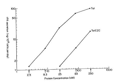
ng/106 cells per day. Figure 12 shows the results
obtained from a tPA assay of HeLa.318 media 24 hours
after the addition of Tat or TatE2C protein to
culture medium. In the absence of Tat or the
TatE2C protein, tPA activity was undetectable (less
than 0.1 ng/106 cells per day). However, addition of
either Tat or TatE2C protein led to an increase in
tPA production (Figure 12). Thus, it appears that
Tat (residues 1-67) can retain the ability to enter
cells when linked to a heterologous protein.
Although transactivation upon addition of the
TatE2C protein was somewhat less efficient than that
observed upon addition of Tat, the TatE2C fusion
protein was also less active than Tat in
transactivation assays when the proteins were
produced intracellularly after transfection of the
genes into HeLa.318 cells. Thus, it is not clear
whether the apparent reduction in activity is
CA 02071214 2003-11-12
-22-
attributable to reduced uptake or reduced activity
of the fusion protein produced by E. coli and added
exogenously. It is possible that some Tat activity
may be lost during the denaturatfon and refolding of
05 the TatE2C fusion protein during purification.
Uptake of the E2 portion of the conjugate was
determined by indirect immunofluorescence using
rabbit polyclonal serum raised against E2-C85 (the
C-terminal 85 amino acids of the E2 protein produced
10 in E. coli). For indirect immunofluorescence, mouse
TM
3T3 cells were seeded into LAB-TEK four chamber
tissue culture chamber/slides. The next day, TatE2C
fusion protein was added at 250 nM to the culture
medium, in the presence of 100 ~M chloroquine. Six
15 hours later, immunofluorescence was performed as
described in Example VII.
While only very faint background fluorescence
was seen when E2.C85 protein was added to cells (at
the same concentration and in the presence of 100 ~M
20 chloroquine), addition of the TatE2C fusion protein
led to very intense fluorescence in all cells
observed. These cells displayed fluorescence on the
plasma membrane, in the cytosol and in nuclei. The
staining was present in bright patches rather than
25 evenly dispersed throughout the cells. The amount
of E2 fluorescence obtained following addition of
TatE2C protein to culture medium was far greater
than the immunofluorescence observed when a TatE2C
gene was expressed in these same cells. These data
30 indicate that the Tat protein is capable of
efficiently carrying a heterologous protein present
WO 91/09958 PCT/US90/07607
-23-
as part of a molecule of interest-Tat conjugate into
cells.
The molecule of interest-Tat protein conjugate
A molecule of interest, which will generally be
05 a protein ar peptide, a nucleotide sequence, or
other chemical which has diagnostic, prophylactic or
therapeutic application (referred to herein as a
drug) is combined, as described below, with HIV Tat
protein to produce a molecule of interest-Tat
protein conjugate the resulting conjugate is brought
into contact with the extracellular surface of
cells.
In one embodiment of the present invention, the
molecule of 'interest is a protein, such as an
enzyme, antibody, toxin, or regulatory factor (e. g.,
transcription factor) whose delivery into cells, and
particularly into the cell nucleus is desired. For
example, some viral oncogenes inappropriately turn
on expression of cellular genes by binding to their
promoters. By providing a competing binding protein
in the cell nucleus, viral oncogene-activity can be
inhibited.
In a further embodiment, the molecule of
interest is a nucleotide sequence to be used as a
diagnostic tool (or p:robe), or as a therapeutic
agent, such as an oligonucleotide sequence which is
complementary to a target cellular gene or gene
region and capable of inhibiting activity of the
cellular gene or gene region by hybridizing with it.
In yet another embodiment, the molecule of interest
WO 91/09958 PCT/US90/07607
-24-
is a drug, such as a peptide analog or small
molecule enzyme inhibitor, whose introduction
specifically and reliably into the cell nucleus is
desired.
05 The molecule of interest can be obtained or
produced using known techniques, such as chemical
synthesis, genetic engineering methods and isolation
from sources in which it occurs naturally. The
molecule of interest can be combined with or
attached to the Tat protein to form the molecule of
interest-Tat protein conjugate which is a subject of
the present invention.
The attachment of the molecule of interest to
Tat to produce a molecule of interest-Tat protein
conjugate may be effected by any means which
produces a link between the two constituents which
is sufficiently stable to withstand the conditions
used and which does not alter the function of either
constituent. Preferably, the link between them is
covalent. For example, recombinant techniques can
be used to covalently attach Tat protein to
molecules, such as by joining the gene coding for
the molecule of interest with the gene coding for
Tat and introducing the resulting gene construct
into a cell capable of expressing the conjugate.
Alternatively, the two separate nucleotide sequences
can be expressed in a cell or can be synthesized
chemically and subsequently joined, using known
techniques. Alternatively, the protein of
interest-Tat molecule can be synthesized chemically
as a single amino acid sequence (i.e., one in which
WO 91/09958 PCT/US90/07607
-25-
both constituents are present) and, thus, joining is
not needed.
Coupling of the two constituents can be
accomplished via a coupling or conjugating agent.
05 There are several intermolecular cross-linking
reagents which can be utilized (see, for example,
Means, G.E. and Feeney, R.E., Chemical Modification
of Proteins, Holden-Day, 1974, pp. 39-43). Among
these reagents are, for example, J-succinimidyl
3-(2-pyridyldithio) propionate (SPDP) or N,
N'-(1,3-phenylene) bismaleimide (both of which are
highly specific for sulhydryl groups and form
irreversible linkages); N, N'-ethylene-bis-(iodo-
acetamide) or other such reagent having 6 to 11
carbon methylene bridges (which relatively specific
for sulfhydryl groups); and 1,5-difluoro-2,4-dini-
trobenzene (which forms irreversible linkages with
amino and tyrosine groups). Other cross-linking
reagents useful for this purpose include: p,p'-di-
fluoro-m, m'-dinitrod:iphenylsulfone (which forms
irreversible cross-linkages with amino and phenolic
groups); dimethyl adipimidate (which is specific fcr
amino groups); phenol-1,4-disulfonylchloride (which
reacts principally with amino groups); hexamethyl-
enediisocyanate or di:isothiocyanate, or azophenyl-
p-diisocyanate (which reacts principally with amino
groups); glutaraldehyde (which reacts with several
different side chains) and disdiazobenzidine (which
reacts primarily with tyrosine and histidine).
WO 91/09958 PCT/US90/07607
-26-
Delivery of a molecule of interest using the present
method
The present method can be used to deliver a
molecule of interest into cells, particularly into
05 the cell nucleus, in vitro or in vivo. In in vitro
applications in which the molecule is to be
delivered into cells in culture, the molecule of
interest in combination with Tat protein or the
molecule of interest-Tat protein conjugate is simply
added to the culture medium. This is useful, for
example, as a means of delivering into the nucleus
substances whose effect on cell function is to be
assessed. For example, the activity of purified
transcription factors can be measured, or the in
vitro assay can be used to provide an important test
of a molecule's activity, prior to its use in in
vivo treatment.
Alternatively, the molecule of interest in
combination with Tat protein or the molecule of
interest-Tat protein conjugate can be used for
prophylactic or therapeutic purposes (for the
treatment, prophylaxis or diagnosis of a disease or
condition). For example, a selected molecule of
interest in combination with Tat protein or the
molecule of interest-Tat protein conjugate can be
combined with a sample obtained from an individual
(e.g., blood, bone marrow) in order to introduce the
molecule of interest into cells present in the
sample and, after treatment in this manner, the
.sample returned to the individual. A series of
treatments carried out in this manner can be used to
WO 91/09958 PCT/US90/07607
-27-
prevent or inhibit the effects of an infectious
agent. For example, blood can be removed from an
individual infected with HIV or other viruses, or
from an individual with a genetic defect. The blood
05 can then be combined with a molecule of interest in
combination with Tat protein or a molecule of
interest-Tat protein conjugate in which the molecule
of interest is a drug capable of inactivating the
virus or an oligonucleotide sequence capable of
hybridizing to a selected virus sequence and
inactivating it or a protein that supplements a
missing or defective protein, under conditions
appropriate for entry in cells of the conjugate and
maintenance of the sample in such a condition that
it can be returned to the individual. After
treatment, the blood is returned to the individual.
Alternatively, t:he molecule of interest in
combination with Tat protein or a molecule of
interest-Tat protein conjugate can be delivered in
vivo. For example, cells that synthesize Tat or Tat
conjugate can be produced and implanted into an
individual so that Tat or Tat conjugate is
constantly present. In another embodiment, the
conjugate can be used much like a conventional
therapeutic agent and can be a component of a
pharmaceutical composition which includes other
components useful, far example, for delivery,
stability or activity of the conjugate. In this
embodiment, a selected molecule of interest in
combination with Tat protein or a molecule of
interest-Tat protein conjugate, such as a selected
CA 02071214 2003-11-12
-28-
oligonucleotide sequence-Tat protein conjugate, can
be administered in sufficient quantity to result in
entry into cells, particularly cell nuclei, and
inhibition (reduction or elimination) of the
causative agent (e.g., virus or bacterium) or
OS provision of a missing or defective protein.
Administration of a selected molecule of interest in
combination with Tat protein or of a molecule of
interest-Tat protein conjugate may be by a variety
of routes. For example, administration may be by
injection, infusion or other parenteral routes
(e. g., subcutaneous, intravenous, intramuscular,
intrasternal and intracranial injection or infusion
techniques). Similarly, administration may be
topical (administered topically), that is, applied
locally to a particular part of the body (e. g.,
skin, lower intestinal tract, vaginally, rectally)
where appropriate. For example, in the case of a
papillomavirus infection, topical administration
would be an appropriate mode of administration.
A selected molecule of interest in combination
with Tat.protein or a molecule of interest-Tat
protein conjugate can also be used in making a
vaccine. For example, the molecule of interest can
be an antigen from the bacteria or virus or other
infectious agent that the vaccine is to immunize
against (e. g., pg120 of HIV). Providing the antigen
into the cell cytoplasm allows the cell to process
the molecule and express it on the cell surface.
Expression of the antigen on the cell surface will
WO 91/09958 PCT/US90/07607
Q
-29-
raise a killer T-lymphocyte response, thereby
inducing immunity.
For example, for in vivo applications, a
selected conjugate can be formulated in appropriate
05 compositions which include pharmacologically
appropriate carriers, adjuvants and vehicles. In
general, these carriers include aqueous or
alcoholic/aqueous solutions, emulsions or
suspensions, including saline and buffered media.
Parenteral vehicles can include sodium chloride
solution, Ringer's dextrose, dextrose and sodium
chloride, lactated Ringer's or fixed oils. In
addition, intravenous vehicles can include fluid and
nutrient replenishers, and electrolyte replenishers,
such as those based on Ringer's dextrose.
Preservatives and other additives can also be
present, such as, for example, antimicrobials,
antioxidants, chelating agents, and inert gases.
See, generally, Remington's Pharmaceutical Sciences,
16th Ed., Mack, ed. 1980. The amount of conjugate
administered will vary and will depend on such
factors as the condition or disease in question, the
mode of administration, and the individual's size.
Examples
The subject invention will now be illustrated
by the following examples, which are not to be seen
as limiting in any way.
WO 91/09958 PCT/US90/07607
-30-
Example I Bacterial Expression and Purification of
Tat
Two plasmids were constructed to produce the
Tat protein in E. coli; one expresses amino acids
05 1-g6 (the entire coding sequence) and the other
expresses the first coding exon of Tat (residues
1-72). It is known that the second exon is not
required for activity (Cullen, B.R., Cell, 46:
973-982 (1986); Muesing, M.A., et al., Cell, 48:
691-701 (1987); Sodroski, J., et al., Science, 229:
74-77 1985); Frankel, A.D., et al., Proc. Natl.
Acad. Sci., USA, 86:7397-7401 (1989)). Synthetic
tat genes were constructed and ligated into the NdeI
site of pET-3a, a plasmid that uses a strong
bacteriophage T7 promoter to express cloned genes
(Studier, F.W. and B.M. Moffat, J. Mol. Biol., 189:
113-130 (1986); Rosenberg, A.H., et al., Gene, 56:
125-135 (1987)). The resulting plasmids, ptat72 and
ptat86, express Tat (residues 1-72 or 1-86,
respectively) as 1%-5% of total E. coli protein.
Both proteins gave similar results in all
experiments. BL21(DE3) cells were used for
expression and these cells also contained a plasmid
expressing the T7 lysozyme gene to inhibit any T7
RNA polymerase expressed prior to induction (F. W.
Studier, personal communication). Tat was induced
with isopropyl ~-D-thiogalactopyranoside (IPTG)
(Studier, F.W. and B.M. Moffat, J. Mol. Biol., 189:
113-130 (1986)) and purified essentially as
described (Frankel, A.D., et al., Science, 240:
70-73 (1988)) except that Tat was extracted from the
CA 02071214 2003-11-12
-31-
polyethyleneimine pellet with 10~ ammonium sulfate
instead of 700 mM RCl, and the S-Sepharose
chromotography was eliminated.
Example II ~nthetic Tat Peptides
OS Syntheses were performed using Fmoc chemistry
TM
on a Milligen/Biosearch model 9600 peptide
synthesizer with a peptide amide linker-norleucine-
4-methylbenzhydrylamine (PAL-Nle-MBHA) polystyrene
resin (Milligen/Biosearch; 0.5 g). The benzotria-
10 zolyloxytris(dimethylamino)phosphonium hexafluoro-
phosphate/1-hydroxybenzotriazole (BOP/HOBt) coupling
method (Hudson, D., J. Org. Chem., 53: 617-624
(1988)) was used with coupling times of 1-4 hours
and with double coupling of His-33. Protecting
15 groups were t-butyl ester (for Glu and Asp),
2,2,5,7,8-pentamethylchroman-6-sulfonyl (Arg),
t-butyloxycarbonyl (Lys), trityl (His and Cys),
t-butyl (Ser, Thr, and Tyr), and trimethoxybenzyl
(Asn and Gln). All peptides were synthesized as
20 their C-terminal amides. After synthesis was
completed, protecting groups were removed and the
peptide chains were cleaved from the resin with tri-
f luoroacetic acid/ethaneidithiol/thioanisole/anisole
(90:3:5:2, vol/vol). The mixture was filtered and
25 the products were obtained by addition of cold
anhydrous diethyl ether to the filtrate. The
precipitate was collected by filtration, thoroughly
washed with ether and dried.
Peptides were treated with 0.5 M dithiothreitol
30 at 37°C for 30 minutes to ensure complete reduction
WO 91/09958 PC1'1US90/07607
-32-
of the cysteines and were purified on a C4 HPLC
column (vydac) using an acetonitrile gradient in
0.1% trifluoroacetic acid. Amino acid composition
was determined by hydrolysis in 6 M HC1 containing
05 0.5% phenol at 100°C and analysis on a LKB Alpha
Plus analyzer. Peptide purity (>90%) was determined
by HPLC using an acetonitrile gradient of <0.5% per
minute.
Example III Uptake of 125-I_Labeled Tat
Tat (residues 1-72) was labeled with 1251 by
treating 500 ~g of protein with 0.5 mCi 1251 and
IODO-BEADS (Pierce) in 0.1 M Tris-HCI (pH 7.5) at
room temperature for 5 minutes. The sample was
dialyzed to remove unreacted 1251. The specific
activity was approximately 106 cpm/~g protein.
HL3T1 cells (2 x 10⁶ cells per dish) were treated
with 5 ~g radioactive Tat in the presence or absence
of 100 uM chloroquine. Medium was removed at
various times, cells were washed with PBS and EDTA,
and cells were trypsinized for 10 minutes.
Pancreatic trypsin inhibitor was added (5 ~g/ml),
cells were chilled to 4°C, centrifuged at 100Xg, and
the supernatant was saved. The cell pellet was
washed twice with serum-free DMEM, once with PBS and
nuclei were isolated by lysis in 0.5% NP-40 as
described (Ausubel, F.M. et al., Current Protocols
in Molecular Biology (New York: John Wiley and Sons,
1980 ). 1251 was counted using an LKB gamma
counter.
WO 91 /09958 PCT/US90/07607
-33-
Example IV Chloroquine Stimulated Tat Uptake
The parameters of chloroquine-stimulated Tat
activity were studied in more detail. Figure 6A
shows that the concentration dependence of
05 chloroquine is a rather sharp dose response with
maximum transactivation observed at 100 ~M
chloroquine. This concentration is typically used
to raise vacuolar pH (Mellman, I. et al., Annu. Rev.
Biochem. 55:663-700 (1986)).
The time course ~of Tat transactivation in the
presence of chloroquine showed a plateau after 24
hours (Figure 6B), and transactivation in the
presence of chloroquine increased with increasing
Tat concentration (Figure 6C). Transactivation was
detectable with Tat concentrations as low as 1 nM.
Controls were dome to determine whether
transactivation was dependent on an intact TAR site,
to determine whether ~a heterologous promoter could
be stimulated by Tat, and to determine whether any
of the effects seen with chloroquine occurred when
Tat was produced intr~acellularly. After transient
transfection of HeLa cells with an HIV-LTR plasmid
(p-167/+80; Rosen, C.A. et al., Cell 41:813-823
(1985)), high levels of transactivation were seen
when Tat was introduced by cotransfection with a Tat
expression plasmid (pSV2tat72), by scrape-loading
purified Tat, or by treatment with Tat and
chloroquine. However, expression from the HIV-LTR
containing a mutant TAR site (p-167/+21; Rosen, C.A.
et al., Cell 41:813-823 (1985)) or from the SV40
early promoter (pSV2-CAT; Gorman, C.M. et al., Mol.
CA 02071214 2003-11-12
-34-
Cell. Biol. 2:1044-1051 (1982)) was not stimulated
when Tat was introduced by these methods. Thus,
introducing Tat by scrape-loading or by uptake with
chloroquine appears to transactivate the HIV-LTR by
OS the same mechanism that occurs when Tat is produced
intracellularly. Chloroquine had no effect when Tat
was produced intracellularly; chloroquine treatment
of HL3T1 cells transiently transfected with
pSV2tat?2 showed no additional transactivation.
Example V RNA Isolation and AnalYSis
For the RNase protection experiment, total RNA
was isolated by the hot acidic phenol method (Queen,
C. and D. Baltimore, Cell 33:?41-?48 (1983)).
HIV-1-specific probes for all hybridizations were
prepared by in vitro transcription (with c-32F UTP)
of an EcoRV-linearized plasmid containing the EcoRV
(-120) to HindIII (+80) fragment of the viral LTR
(cloned into the plasmid sp?3; Promega). RNA probes
TM
were purified on Sephadex G-50 spin columns
(Boehringer-Mannheim).
_RNase protection experiments were perforaed as
described (Ausubel, F.M. et al., Current Protocols
in Molecular Biology (New York: John wiley and Sons,
198?)). Twenty ~g of cellular RNA were hybridized
overnight with 5 X 10⁵ cpm of the RNA probe at 38 oC
in 40 ~1 of 80~ formamide, 40 mM PIPES (pH 6.?), 200
mM NaCl, 1 mM EDTA. Single-stranded RNA was
digested with RNase A (10 pg/ml) and RNase T1 (45
U/ml) (Boehringer-Mannheim) in 400 ~l of 10 mM
Tris-HC1 (pH ?.5), 300 mM NaCl, 5 mM EDTA for 1 hour
CA 02071214 2003-11-12
-35-
at room temperature. Protected fragments were
analyzed by electrophoresis on 6t polyacrylamide-7M
urea sequencing gels. Protected RNAs were
visualized by autoradiography with intensifying
OS screens and were quantitated using a Betascope 603
(Betagen).
Example VI Localization of Tat by Fluorescence
Purified Tat protein was labeled at lysine
residues with tetramethyl rhodamine isothiocyanate
(TRITC) by incubating 200 ~g of Tat (amino acids
1-72) in 0.1 M Na2C03 pH9.0 with 5 ~g of TRITC
dissolved in 5 pl dimethylsulfoxide (DMSO), for 8
hours at 4 °C. Unreacted TRITC was quenched with 50
mM NH4C1. The pH was lowered to 7.0 With HCl and
15 rhodamine-labeled Tat was purified from free TRITC
by dialysis against 50 mM Tris, pH 7, 1 mM DTT.
HL3T1 cells were grown on glass coverslips and
incubated for various lengths of time with
rhodamine-conjugated Tat (TRITC-Tat) in DMEM. H938
cells in suspension were incubated with rhodamine-
conjugated Tat in RPMI. Cells were washed three
times with phosphate buffered saline and viewed live
TM
on a Zeiss Axiophot fluorescence microscope.
Example VI Uptake of TatE2C Fusion Protein
25 Cell lines. The mouse embryo fibroblast cell line
Balb/c 3T3 (clone A31; Aaronson and Todaro, J. Cell
Physiol. 72:141-148 (1968)) was obtained from the
American Type Culture Collection. HeLa cells were
WO 91/09958 PCT/US90/07607
~~?~.2~4
-36-
obtained from Dr. Alan Frankel (Whitehead Institute,
MIT). Both cell lines were propagated in Dulbecco~s
minimal essential medium (GIBCO) supplemented with
10% donor calf serum (Hazelton) and 4 mM glutamine
05 (Whittaker). Cells were grown in a 5.5% C02
incubator at 37°C. Passaging of cells was performed
by washing with phosphate-buffered saline and
treating with trypsin (both GIBCO) to remove cells
from plates followed by addition of culture medium
and dilution of cells into plates containing fresh
culture medium.
The HeLa cell line containing a Tat responsive
reporter construct (HeLa.318) was generated by the
introduction and stable selection of plasmid pXB318
(described below) by electroporation as described by
Chu et al. (Chu et al., Nucleic Acids Res.
15:1311-1326 (1987)). pXB318 DNA was electroporated
together with the selectable marker pSV2-neo
(Southern, E.M. and Berg, P. J. Mol. Appl. Genet.
1:327-341 (1982)). Stable transfectants were
selected in the presence of 6418 (Southern, E.M. and
Berg, P. J. Mol. Appl. Genet. 1:327-341 (1982)), and
the presence of pXB318 DNA was confirmed by Southern
blot hybridization analysis (Southern, E.M., J. Mol.
Biol. 98:503-517 (1975)).
Vector Constructions. All molecular cloning
reactions were carried out by methods described by
Maniatis et al. (Maniatis, T., Fritsch, E.F., and
Sambrook, J., Molecular Cloning: A Laboratory
Manual (Cold Spring Harbor Laboratory, NY (1982)),
WO 91/09958 PCT/US90/07607
-37-
using enzymes obtained from New England Biolabs
(Beverly, MA).
The TatE2C fusion protein (protein TatE2C), in
which HIV Tat was fused to the carboxy terminal
05 portion of BPV-1 E2, was expressed from the
bacterial expression plasmid pFTE103. This plasmid
was derived from ptat72 (see Example I) by insertion
of a StyI-SpeI fragment which was isolated from
vector pC0-E2 (Hawley-Nelson et al., EMBO J.
7:525-531 (1988)) and which encodes the C-terminal
portion of the E2 protein. Four synthetic
deoxyoligonucleotides were used in the construction
described below in detail.
The plasmid ptat72 was cleaved with the
restriction endonucleases NdeI and BamHI releasing
the Tat encoding portion of the vector. The 4603
base pair (bp) vector fragment was purified by
agarose gel electrophoresis, and a 169 base pair
(bp) NdeI-AatII fragment of the Tat encoding
fragment was isolated. The 3' portion of the E2C
coding sequence was isolated as a 375 by StyI-SpeI
fragment from pC0-E2 (Hawley-Nelson et al., EMBO J.
7:525-531 (1988); obtained from Dr. Elliot Androphy,
Tufts University/New England Medical Center
Hospitals). The E2C fragment was connected to the
Tat fragment and to the expression vector by use of
two pairs of complementary deoxyoligonucleotides
(synthesized according to standard procedures using
an Applied Biosystems 380A DNA Synthesizer). One
complementary pair of oligonucleotides was designed
WO 91/09958 PCT/US90/07607
-38-
to join the AatII overhang of the Tat fragment to
the StyI overhang of the E2C fragment:
oligo 374-3
5' CGTCCGCCGCAGGGATCCCAGACCCACCAGGTTCCGGTTACTCTGC 3'
05 3' TGCAGCAGGCGGCGTCCCTAGGGTCTGGGTGGTCCAAGGCCAATGAGACGGTTC 5'
oligo 374-4
A second pair of complementary oligonucleotides was
designed to link the SpeI overhang of the E2C
fragment to the BamHI overhang of the 4603 by vector
backbone isolated from ptat72:
oligo 374-5
5' CTAGTGGCTCGAGATTCCG 3'
3' ACCGAGCTCTAAGGCCTAG 5'
oligo 374-6
The Tat fragment, the E2C fragment and the two pairs
of oligos were inserted into the 4603 ptat72 vector
backbone to create pFTE103. The resulting fusion
gene is designed to express a protein comprising
amino acids 1 through 67 of Tat at the amino
terminus followed by the C-terminal 105 amino acids
of E2 (residues 306 through 410 of BPVO1 E2).
The Tat responsive reporter construct pXB318
was constructed in three steps. The starting
plasmid was pBG312 (Gate et al., Cell 45:685-698
(1986)). Two oligodeoxynucleotides were
synthesized, which when annealed have an
AatII-compatible overhang at the 5' end and an
WO 91/09958 PCT/US90/07607
-39-
EcoRI-compatible overhang at the 3' end, and form a
polylinker with internal XhoI, HindIII and BamHI
restriction sites:
5' CTCGAGAAGCTTGA.CGGATCCG 3~
05 3' TGCAGAGCTCTTCGAACTGCCTAGGCTTAA 5'
pBG312 was cleaved with AatII and EcoRI to remove
the promoter, and the above polylinker was inserted
into the vector to farm the promoterless vector
pXB100. The HIV-1 long terminal repeat (LTR) from
pU3R-III (Sodroski et al. Science 227:171-173
(1985)) was excised as a XhoI-HindIII fragment and
was inserted into XhoI and HindIII sites of the
polylinker of pXB100 to create pXB301. The human
tissue plasminogen activator (tPA) cDNA reporter was
excised as a BamHI fragment from pTPA114 (Fisher et
al. J. Biol. Chem. 260:11223-11230 (1985)) and
inserted into the BamHI site of pXB301 to create
pXB318.
Expression and purification of TatE2C. The TatE2C
fusion protein was expressed in E. coli using the
vector pFTE103 and the T7 RNA polymerase expression
system precisely as described by Studier et al.
(Studier et al., Methods in Enzymology 185:60-89
(1990)).
Virtually all of the TatE2C protein was found
in the insoluble fracaion. The following
purification was performed:
WO 91/09958 PCT/US90/07607
-40-
1. E. coli were pelleted, resuspended in ten
packed cell volumes of 25 mM Tris-HC1 pH 7.5, 1 mM
EDTA, 10 mM DTT, and 1 mM PMSF and lysed with two
passages through a French press.
05 2. The membrane fraction was pelleted by
centrifugation at 10,000 rpm for 30 minutes.
3. This membrane fraction was resuspended in 6 M
urea.
4. Solid guanidine-HC1 was added to a final
concentration of 6 M and DTT was added to a final
concentration of 10 mM.
5. After 30 minutes at 37°C, the solution was
clarified by centrifugation at 10,000 rpm for 30
minutes.
6. The sample was loaded onto an A.5 agarose gel
filtration column in 6 M guanidine-HC1, 50 mM sodium
phosphate pH 5.4, and 10 mM DTT.
7. TatE2C-containing fractions were loaded onto a
C18 reverse phase HPLC column and eluted with a
gradient of 0-75% acetonitrile in 0.1%
trifluoroacetic acid.
TatE2C protein appeared in a single peak. On
protein gels, the TatE2C fusion protein migrated
with an apparent molecular weight of 20,000 to
21,000 daltons.
Assay of TatE2C uptake by Tat activity. Uptake was
detected either as Tat activity (activation of a
Tat-dependent reporter in HeLa.318) or by indirect
immunofluorescence using anti-E2 antibodies.
CA 02071214 2003-11-12
-41-
Tat activity was determined by adding Tat
protein (amino acids 1-72) or TatE2C fusion protein
at 2.5-250 nM along with chloroquine at 0.1 mM to
the culture medium of HeLa.318 cells in 24 well
05 plates essentially by the method of Frankel and Pabo
(Frankel, A.D. and Pabo, C.O., Cell 55:1189-1193
(1988)). The Culture medium was harvested 24 hours
later and assayed for tPA activity by the method of
Granelli-Piperno and Reich (J. Exp. Med. 148:223-234
(1978)). Cell numbers were determined and tPA
secretion was expressed as ng/106 cells per day.
tPA secretion was undetectable in the absence of
added Tat or TatE2C protein (less than 0.1 ng/106
cells per day). _
Assay of TatE2C uptake by E2-specific immunofluor-
escence. For indirect immunofluorescence, mouse 3T3
cells were seeded into LAB-TEK four chamber tissue
culture chamber/slides. On the next day, TatE2C
protein and chloroquine were added to the culture
medium to final concentrations of 250 nM and 0.1 mM,
respectively. Six hours later, immunofluorescence
was performed as follows:
1. Medium was removed and wells were washed twice
with phosphate-buffered saline (PHS).
2. Cells were fixed by treatment with 3.5%
formaldehyde for 10 minutes at room temperature.
3. Cells were permeabilized in 0.2% Triton
X-100/2% bovine serum albumin (BSA) in PBS with 1 mM
CA 02071214 2003-11-12
-42-
MgCl2/0.1 mM CaCl2 (PBS+) for 5 minutes at room
temperature.
4. Cells were blocked by treatment with whole goat
serum (Cappel (5506-1380) at a 1:30 dilution in
p5 pBS+/2% BSA for one hour at 4~C.
5. The primary antibody was an affinity purified
rabbit polyclonal which had been raised by injection
of purified protein E2.C85 (in this case the carboxy
terminal 85 amino acids expressed in bacteria using
10 the T? polymerise expression system) into a rabbit,
followed by purification by passage of the bleed
over an E2 affinity column. This antibody was added
to the wells at a 1:100 dilution in PBS+/2% BSA for
one hour at 4°C.
15 6. The secondary antibody was a rhodamine
conjugated goat anti-rabbit IgG (Cappel (2212-0081).
This antibody was added at a 1:100 dilution in
PBS+/0.2% BSA for 30 minutes at 4°C.
7. Wells were washed three times with P8S+/0.2%
TM
20 Tween-20/2% BSA.
8. Slides were mounted in 50% glycerol in PBS and
viewed with a fluorescent microscope with a
rhodamine filter.
As a control, purified E2C protein (the carboxy
25 terminal 85 amino acids which were found to be
recognized by the polyclonal antibody preparation)
was added to wells in the same manner as the TatE2C
fusion protein.