Note: Descriptions are shown in the official language in which they were submitted.
~ITLE: MICROBORE Ch~ l~K WITH SIDE PORT( S)
BAC~G~OUND OF ~HE ~NVFNIION 2 a 713 b 3
~ atheters with side ports are per se well known in the
~rt. For instance, epidural catheters currently commercially
available from Kendall Healthcare Company, a division of The
Kendall company, assignee of the present invention, include
side holes or ports in order to provide ~etter drug dispersion
than that obtained from a single opening at the distal end of
the catheter lumen. Such side ports are produced by mechanical
methods, e.g. skiving with a sharp instrument or drilling with
a small bit.
While entirely satisfactory for epidural catheters (or
larger ones), the known mechanical procedures are not
applicable for so-called microbore or small bore catheters,
i.e. catheters no greater than 24 gauge (outer diameter = 0.022
in). For example, the preferred microbore catheters
contemplated by the present invention hre almost two times
smaller than the tyFical 20 gauge epidural catheter:
EPIDURAL MICRO~ORE SPINAL
(20 gauge) (28 gauge)
OD 0.036 in OD 0.014 in
ID 0.020 in ID 0.007 in
OD = outer diameter
ID = inner diameter
By way of further explanation, the epidural catheter's
side port diameter is typically on the order of 0.01~ inch or
on the order of fifty-five percent (55%) of the catheter's ID.
As is recognized in the art, the tendency for a patient to
have rather severe headaches following spinal anesthesia is
markedly reduced if a smaller needle is used. This in turn
means that in conventional systems where the catheter is
introduced through the needle, a smaller bore catheter is also
required.
2071 353
Whlle for thls reason mlcrobore catheters are
greatly preferred, lf also deslred that the mlcrobore splnal
catheter, llke the larger epldural catheter have one or more
slde ports along wlth lts dlstal end port to lnsure proper
drug dlsperslon wlthln the subarachnold space. In the llght
of the state of the art at the tlme the present lnventlon was
made, these two characterlstlcs appeared to be mutually
excluslve so that one must elther elect to have a mlcrobore
catheter wlthout slde ports or to have a larger bore catheter
wlth slde ports.
Stated slmply, the task of the present lnventlon was
to devlse a way to have the deslred slde port(s) ln a
mlcrobore catheter.
SUMMARY OF THE INVENTION
The present lnventlon provldes a mlcrobore catheter
for lntroduclng a drug lnto the subarachnold space of a
patient characterlzed ln that the catheter has at least one
slde port at lts dlstal end, each sald slde port belng
substantlally clrcular and belng characterlzed as belng
substantlally free of any burrlng or craterlng.
The lnventlon also provldes a catheter for splnal
anesthesla, the catheter havlng a dlstal and a proxlmal end,
an outer dlameter no greater than about 0.22 lnch and a slngle
lumen extendlng from the proxlmal end for llquld communlcatlon
wlth a source of llquld anesthesla and a dlstal end adapted to
be lnserted wlthln the subarachnold space of a patlent, the
catheter further havlng at least one slde port near lts dlstal
end and an openlng at lts dlstal tlp, the slde port(s) and the
opening at the dlstal tlp communlcatlng wlth the lumen for
-- 2
64407-133
C
207 1 353
dellverlng anesthesla lnto the subarachnold space of the
patlent, each slde port belng substantially clrcular and belng
substantlally free of any burrlng or craterlng.
From another aspect the lnventlon provldes the
method of maklng a mlcrobore splnal catheter comprlslng: (1)
formlng a catheter comprislng an elongated flexlble wall
materlal havlng opposed dlstal and proxlmal ends and havlng a
single lumen extendlng between the ends and termlnatlng wlth
an openlng at lts dlstal end, the catheter havlng an outer
dlameter no greater than about 0.022 inch; and (2) thereafter
formlng at the dlstal end proxlmal to the dlstal openlng at
least one slde port characterlzed as belng spherlcal and
substantlally free of any burrlng or craterlng by contactlng
the catheter wall materlal wlth a laser beam selected wlth
respect to the catheter wall materlal so that: (a) the
radlatlon emltted by the laser ls ln a wavelength such that
llttle or no radlatlon ls transmltted through the target
catheter wall materlal; and (b) the radlatlon does not emlt
enough heat to cause the target catheter wall materlal to melt
rather than belng vaporlzed.
The inventlon wlll further be descrlbed, by way of
example only, wlth reference to a preferred embodlment thereof
as lllustrated in the accompanylng drawlngs whereln:
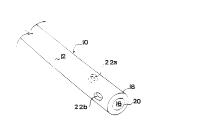
FIG. 1 ls a fragmented perspectlve vlew, greatly
enlarged of the dlstal portlon of a novel catheter of thls
lnventlon havlng two slde ports; and
FIG. 2 ls a longltudlnal sectlonal vlew of the novel
mlcrobore catheter as shown in FIG. 1.
As heretofore mentloned, the present lnventlon ls
- 2a -
64407-133
C
~07 1 3~3
dlrected to mlcrobore catheters havlng partlcular utllity ln
admlnlsterlng splnal anesthesla whereln the catheter has at
least one slde port or opening ln additlon to the opening at
the distal end of the catheter. Catheters of thls descrlptlon
provlde lmproved drug dlsperslon over that which may be
obtalned wlth catheters havlng a slngle opening at the
termlnal end of the lumen ln fluid communicatlon wlth the drug
source.
- 2b -
64407-133
~J
CA 020713~3 1998-0~-04
A preferred form of a catheter of this general
description is shown in the illustrative drawings~
As shown therein, catheter 10 (greatly enlarged for
purposes of illustration) has inner and outer walls 14 and 12,
respectively, defining a lumen 16 extending from its proximal
or trailing end ~not shown), where it can be placed in fluid
communication with a source of liquid drug, to its distal or
leading end 18 terminating in opening 20 for drug administration,
e. g. within the subarachnoid space. In accordance with this
invention, catheter 10 is shown to have two additional openings
22a and 22b on opposed sides near the distal end 18.
In order to maximize drug distribution while not
adversely decreasing tensile strength, ports 22a and b are not
opposite one another but instead are offset longitudinally with
port 22a proximal to port 22b. Since for administering spinal
anesthesia the catheter is generally inserted no more than 2 cm
within the subarachnoid space, it will be appreciated that port
22a should be no further than about 1.5 cm from the distal end
18, and preferably on the order of about 1.0 cm. Port 22b may
be on the order of 0.5 cm from distal end 18.
Preferably, the size of ports 22a and 22b should be
on the order of a 1:1 ratio with the catheter wall thickness.
For instance, with a 28 gauge catheter as mentioned above, the
wall thickness is the difference between the OD and ID of about
0.0007 in. divided by two or 0.0035 in. Accordingly, for a 28
gauge catheter the hole size should preferably be on the order
of 0.0035 in + 0.001 inch in tolerance.
While the ports are shown to be generally spherical,
the shape is not critical and other shape holes which can be
provided to the microbore catheter are equally applicable.
In the preferred form shown in the drawing, two side
ports are provided. However, it is contemplated that 1-6 side
ports of varying sizes may be utilized in the practice of this
invention.
64407-133
~ s heretofore mentioned, side ports in the sma~l cat~
which this invention is directed cannot be provided by
~echanic~l means such as skiving which are employed to make
noles in larger cannulas, e.g. epidural catheters.
Accordingly, an essential part of the present invention is to
provide a way to make side ports of the foregoing general
description in a microbore catheter. While the initia~ concept
of the present invention was to utilize a laser to provide the
side ports, initial efforts using a laser beam were
unsuccessful.
Apart from the size restrictions, there were additional
requirements that the holes provided should extend through only
one wall of the catheter and sho~lld not result in cratering or
~urring which could be detrimental to the contemplated usage.
If cratering is external, it will increase the OD and may
prevent the catheter from passing through the needle; while
internal cratering can reduce the flow rate. Although burring
is of lesser concern than cratering, it is still to be avoided
because the burrs or small pieces of the catheter tubing can
come off wit~in the patient, e.g. in the dura mater during
insertion or wi~hdrawal and/or in the subarachnoid space.
In the initial efforts to create the side ports 22a, 22b
with a laser beam, a carbon dioxide laser was selected. The
carbon dioxide laser employed emits radiation in the infrared
(IR) range at around 1000nm and creates holes or voids by
vaporizing a material on absorption of the radiation. It
possesses a high energy output and is typically used in the
industrial setting.
While the holes in the catheter produced by the carbon
~ioxide layer were of proper size as anticipated, quite
unexpectedly it was found that cratering and burring effects
occurred surrounding the hole, rendering the catheter so
produced impractical for sale for the contemplated primary use
for spina~ anesthesia.
Accordingly, another solution was required.
It was found that the pro~lem was not with ~asers per se
but with the particular laser selected. It was believed that
CA 020713~3 1998-0~-04
the burring was caused because the particular catheter material
employed, nylon, did not absorb substantially all of the laser
radiation so that sufficient radiation from the carbon dioxide
beam was transmitted to cause the burring rather than being
absorbed by the material. The cratering was determined to have
been caused by the amount of heat emitted by the laser beam,
which heat was absorbed by the catheter material.
It was accordingly theorized that a laser beam could
indeed be utilized to obtain the desired side ports not
previously obtainable in catheters this small in OD by the
known mechanical means if the proper laser could be selected.
The selected laser beam should be characterized as being one:
(1) whose emitted radiation is in a wavelength such that little
or no radiation is transmitted through the target material so
as to cause objectionable burring; and (2) does not emit enough
heat to cause cratering by heat emitted by the laser being
absorbed by the catheter materials, causing it to melt rather
than being vaporized.
In the foregoing discussion it will be appreciated
that one cannot state unequivocally that no radiation is
transmitted, nor can one say that no heat from the laser is
absorbed by the catheter material. What one can say is that
if any radiation is transmitted or heat absorbed it is
insignificant as not adversely affecting the desired physical
characteristics, e. g. size, shape, etc. of the hole so
produced. In this sense, the selected laser can be character-
ized as being innocuous in that it does not cause any physical
properties to the area surrounding the port which would be
regarded by an anesthesiologist or other person skilled in the
art as deleterious for use in administering spinal anesthesia.
With this theoretical consideration in mind, it was
concluded that if Applicants were correct, what was in fact
needed, at least for the nylon catheter employed, was a laser
beam of a shorter wavelength and specifically one whose wave-
length was sufficiently small that no detrimental heat is
emitted.
64407-133
CA 020713~3 1998-0~-04
Excimer lasers meet that description. Excimer lasers
emit in the ultraviolet (W) range and are sometimes referred
to as "cold lasers" because they do not create thermal warming,
as other lasers do, that can cause severe damage to surrounding
material. For this reason they have previously found use in
medical procedures such as laser angioplasty where plaque
causing blockage of an artery is vaporized with the laser with-
out harm to the fragile artery tissue. Since harmful heating
of the area peripheral to the area to be vaporized would be
obviated, it was then postulated that the Excimer laser would
solve the task to which this application is directed.
It is known that Excimer laser energy exhibits a
wavelength in the UV ranging from 40-400 nanometers (nm).
Excimer laser energy can be provided, for example, by a laser
medium such as argon-chloride having a wavelength of 193 nm;
krypton-chloride (222 nm); krypton-fluoride (240 nm) or xenon-
chloride (308 nm). The output energy from this type of laser
appears inshort bursts or pulses that can last for 10-85 nano-
seconds and have a high peak energy. Although the destruction
mechanism involving this form of energy may not be completely
understood, it has been reported in the literature that one
pulse of the Excimer laser produces an incision which destroys
the target area without accompanying thermal injury to the
surrounding area. This result is thought to be due to either
or both of the following phenomena: (1) the delivery of the
short duration, high energy pulses may vaporize the material
so rapidly that heat transfer to the non-irradiated adjacent
tissue is minimal; and/or (2) W photons absorbed in the
organic material might disrupt molecular bonds to remove target
material photochemically rather than by thermal mechanisms.
Having theorized that an Excimer laser could solve
the task of providing the desired side ports, the initial
effort was made with a 308 nm Excimer. The hole was produced
in about 75 seconds. Holes provided by this Excimer were
characterized as being generally cone-shaped with yellow edges.
--6--
64407-133
CA 020713~3 1998-0~-04
While satisfactory from the performance standpoint, they were
concluded to be unacceptable from the marketing standpoint.
Since it was thought that this might be obviated by
increasing absorption of the laser beam still further and,
moreover, that production time should be decreased, it was next
decided to employ a 193 nm Excimer. The desired generally
circular holes (no discoloration, burring or cratering) was
produced in only three seconds.
While the foregoing test results were performed with
a nylon catheter it is believed that the 193 nm Excimer may be
equally applicable for providing the side ports in other
catheter materials as well. In any case, in the light of the
foregoing discussion the selection of the appropriate laser
for the particular catheter material employed will involve
routine experimentation readily within the expected knowledge
of the skilled worker.
While in the foregoing discussion reference has been
made to so-called "open tip" catheters having an opening at
the tip or terminal end of the lumen along with one or more
side ports, the invention also contemplates so-called "bullet"
or "closed tip" catheters in which the only openings are the
side ports.
From the foregoing discussion it will be seen that
the present invention makes it possible to obtain better
distribution of drug in the subarachnoid space, e. g. providing
a wider spread of drug over the lumbosacral curve.
Since various changes may be made without departing
from the scope of the invention herein contemplated, it is to
be understood that the foregoing description and accompanying
drawing are to be taken as being illustrative and not in a
limiting sense.
64407-133
