Note: Descriptions are shown in the official language in which they were submitted.
SURGICAL ToRoTDAL SNARE
The present invention relates to a surgical instrument
for seating an intraocular lens in a posterior ch~mber of an
eye in place of a cataracted natural lens removed during a
cataract operation. In particular, the present invention
relates to a surgical snare for seating an intraocular lens in
a posterior capsule that remains after extracapsular removal of
a cataxacted natural lens.
1.
1 0
A surgical snare for cutting a cataracted lens of a
: human eye is disclosed in my U.S. Patent 4,538,611. The
surgical snare cutter disclosed therein comprises a loop which
is adapted to snare tha natural lens of an eye, crosswise Of
:15 ~uch lens, i.e., along a diameter thereof, and to cut the ~ame
upon movement of the loop member, which is displaceable within
the bore of the shank of the instrument, to its retracted
condition.
A suryical instrument for seating an intraocular lens
in a posterior chamber of an eye is disclosed in my U.S. Patent
4,530/117. ~enerally, an intraocular lens used to replace a
cataracted natural lens includes a lens and a pair of opposed
position fixation members called haptic~ for retaining the
intraocular lens in the po~terior chamber. UOS. Patent
?7~,7/s~,
4,530,117 disclosed an instrument for æeating an intraocular
lens of the type described, in the posterior chambex. The
instrument has a longitudinal shank portion and a hooked end
portion. The hooked end portion has a tip portion adapted to
S ~ngage the upper haptic of the lens for displacing the upper
haptic toward the lower haptic during the insertlon of the lens
through the pupil of the eye. After the lower haptic of the
lens is seated in the lower portion of the posterior capsule,
the instrument is operated to move the upper haptic through the
pupil into the posterior capsule and let the upper haptic
expand to its undeformed condition in the posterior capsule.
While the above-described hooked instrument sub tantially
sLmplified the lens seating procsdure, it, neverthele~s,
continued to be a difficult procedure to properly ~eat ~oth
haptics in the capsular bag.
It is an object of the invention to provide a new and
improved surgical instrument that avoids at least some of the
disadvantages o~ the prior art instruments.
According to a first aspect of the present invention,
there is provided a surgical instrument for seating an
intraocular lens in the posterior capsule of an eye and
comprising:
a longitudinally extending hollow shank ha~ing a
body and a tip portion;
flexible band means forming a loop extending
forwardly of said tip portion $or e~bracing the intraocular
lens,
said shank tip portion having a generally S-shape
to allow said tip portion to curl around the iris of the eye to
position said loop with the intraocular lens held therein
inside the posterior capsule in a plane ~enerally parallel to
the posterior wall of the capsul~; and
support means displaceable within said hollow
shank and connected to said band means;
said displaceable support means having in aid
hollow shank a forward position in which said loop is larger
than the lens to be held therein, an intermediate position in
dt~'s~.
2a
which said loop ~nugly retains the lens, and a rearward
position in which said band means releases the lens to enable
withdrawal of ~aid band mean from the eye without disturbing
the seating of the lens in the posterior capsule of the eye.
According to another aspect of the present invention~
there is provided a surgical instrument for seating an
intraocular lens in the posterior capsule of an eye, said
instrument comprising:
a longitudinally extending hollow shank having a
tip portion;
~ lexible band means forming a loop extending
forwardly of said tip portion for embracing the intraocular
lens;
said tip portion haviny a generally S-~hape to
allow said tip portion to curl around the iris of the eye to
position said loop with the intraocular lens held therein
inside the posterior capsule in a plane generally parallel to
the posterior wall of the capsule; and
support means connected to ~aid hand means and
displaceable in said hollow sh~nk relative thereto to provide
for release of the lens by said loop to enable withdrawal of
said band means from the eye without disturbing the seating of
the intraocular lens in the posterior capsule o the eye.
According to another aspect of the present invention,
there is provided a method of inserting an intraocular lens
into the posterior capsule of an eye with a surgical instrument
for holding th~ intraocular lens and having a longitudinally
extending hollow sha~k having a gen~rally S-shaped tip portion,
and band mean attached to ~aid shank and forming a loop
extending forwardly of 6aid tip portion for embracing the
intraocular lens, said method comprising the steps of:
inserting the ~urgical instrument with the
intraocular lens held in said loop through an incision made in
the cornea of the eye and positioning said loop with the
intraocular lens held therein inside the posterior capsule in a
plane generally parallel to the posterior wall of th~ capsule,
2b
the S-shape of the tip portion allowing the shank to clear the
iris of the eye when said loop is positioned in the plane
generally parallel to the posterior wall of the capsule;
breaking the loop to release the len~ and enable
withdrawal of the band means from the eye without di~turbing
the seating of the lens in the posterior capsule of the eye;
and
thereafter, withdrawing the surgical instrument
from the eye.
Preferably, the present invention provides a new and
improved surgical instrument that can both hold the lens and
seat it in the posterior capsule of an eye.
Preferably, the present invention also provides a new
and improved surgical instrument that is easily disengaged from
the lens once the lens is seated in the posterior capsule.
These and other advantages of the invention that will
become apparent hereafter, are achie~ed by providing a surgical
snare for holding the intraocular lens during insertion into
the eye and for seating it in the posterior capsule. The snare
according to the invention comprises a hollow longitudinal
shank with a tip portion from which a toroidal band loop
extends outwardly. The band loop has a ~ize adapted to accom-
modate the lens and the deformed opposed hapticR of the lPns.
In one embodiment, one end of the band loop is fixedly secured
to the shank, preferably at the tip portion thereof. ~he other
end of the band loop extends into the tip portion mouth and is
free to be pulled further into the mouth in order to close,
iOe., reduce the size of, the loop for peripherally embracing
an intraocular lens therein. The length of the band is so
selected that it will, in the contracted condition of the loop,
fit snugly about a 6 mm diameter lens. The other end ~xtending
into the top porti~ , in this ~mbodiment, secured to a
member movable within the ~hank bore between forward and
retracted positions. The band is preferably a thin, very
flexible plastic and, in accordance with an aspect of my
invention, m~y have a weakened portion at one point in its
periphery. The movable member has, in the shank, a forward
position in which the loop is of a si~e facilitating loosely
placing an intraocular lens therein, an intermediate position
in which the loop snugly fits around the periphery of the lens,
and a rearward position. With the movable member in the
intermediate position, the tip of the instrument, with the
loop-held lens in front, is inserted into the eye. The tip is
generally S-shaped so that the shank will clear the iris while
the loop-held lens is positioned parallel to and adjacent the
~0 posterior wall of the posterior capsule. When the lens is
seated in the posterior capsule, and the snare has to be
removed, the movable member is retracted toward its rearward
position in the shank body, tearing the weakened portion of the
band to release it from around the lens, and the band is
removed when the instrument is withdrawnO The tearing is
accomplished against the slight opposing pressure of the lens
positioned in the loop.
In another embodiment, the band has a hole at its end
secured to the movable member through which a metal or nylon
wire extends, the wire being connected to a front end of the
movable member within the shank bore. Upon movement of the
member to its rearward position, the wire cuts the band,
releasing it from its conn~ction with the movable member. In
both embodiments, the S-shape of the tip of the instrument is
proportioned to allow the tip to accommodate the iris of the
eye by extending around the iris.
Providing a surgical snare with a breakable lens
holding loop according to the invention substantially facili-
tates seating of an intraocular lens in the posterior chamber
of an eye and withdrawal of the snare from the eye without
disturbing the seating of the lens.
The present invention, both as to its construction ~nd
as to its mode of operation together with additional object~
and advantages thereofl will be be6t understood fro~ the
following detailed description of the preferred embodLments
when read with reference to the accompanying drawings.
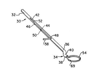
Figure 1 is a perspective viPw of a surgical snare
according to the invention;
Figure 2 is a partial longitudinal cross-sectional view
of a surgical snare according to a first embodiment of the
invention;
Figure 3 is a partial longitudinal cross-sectional view
of a surgical snare according to a second embodiment of the
invention;
Figure 4 is an enlarged fragmented sectional view of a
human eye with the surgical snare according to the invention
shown holding an intraocular lens just prior to withdrawing of
the band from zround the lens;
Figure 5 is an enlarged fragmented sectional view of a
2~ human eye with a surgical ~nare accordi~g to the invention in
its partially withdrawn position and with the intraocular lens
partially sea~edt
Figure 6 is an enlarged transverse sectional view of
the toroidal band loop with the lens held therein.
In the drawings, Figures 4 and 5 show a fragmented
sectional view of a human eye 10 with some portions omitted for
the sake of clarity. The eye 10 has a cornea 12 in which a
surgeon makes an incision opening 14 for removing the catar-
acted natural lens and inserting an intraocular lens. The eye
10 has anterior and posterior chambers 16 and 18 defined by the
position of an iris 20. ThP iris defines a central opening or
pupil 22. After extracapsulaxy removal of the cataracted
natural lens, the posterior capsule 24 and peripheral portion
of the anterior capsule remain intact. The capsule 24 is
normally connected to the ciliary body 26 of the eye by zonulas
28 at the opposed e~ds of the _apsule 24.
One em~odiment of the surgical snare 30, in ac~ordance
with my invention, for eating an intraocular lens in the
posterior capsule 24 is shown in detail in Figures l and 2 and,
as shown, comprises a longitudinal hollow shank 32 having a
preferably substantially S-shaped tip portion 34. The tip
portion 34 consists of two arms 36 and 38 connected by a
transition portion 40 for accommodating the iris. As showr. in
Fig. 1, the hank has a first longitudinal slot 42, a second
transverse slot 44, and a third transverse slot 46. The axial
planes of slots 44 and 46 intersect at a~ angle of 90at th~
axial plane of the slot 42. The slots 42,44 have respective
front surfaces 48 and 50 which define, respectively, forward
and intermediate positionC of a movable support member 52 that
provides for securinq a band loop 54 in the mouth of the tip
portion 34. The member 52 has an axially extending part or
plunger 56 and a projection 58 extending transverse to the axis
of the plunger 56. In the forward position of the movable
member 52 with the projection 58 against ~urface 48, the 6ize
of the loop is such that an intraocular lens with haptics can
be easily positioned th~rein. In the intermediate positio~ of
the movable member 52 with projection 58 against ~urface 50,
the size of the band loop 54 is reduced so that it snugly fits
around the periphery of the lens.
The plunger 56 extends into a bore 60 of the shank 32
and is displaceable herein. The projection 58 is displaceable
in the slot 42 and projects through the transver~e slots 44 or
46 for engagement by a surgeon. The plunger 56 of the movable
member 52 may represent a friction plunger that presses a free
end 62 of the band loop 54 radially against the interior wall
of the tip portion 34. The other end 64 of the band loop 54 is
generally fixedly secured to the interior wall of the sh~nk 32.
In another embodiment ~hown in Fig. 3 ? the free end
52 of the band loop 54 has a hole 66, while the other end 64,
not seen in F.ig. 3, ,~ again fixedly attached to the interior
wall of the shank 3~. The plunger 56 in this embodLment is
much shorter and has a wire 68 connected at its one end to the
front face of the plunger 56. The other end of the wire
-engages the hole 66 at the end of the band loop 54.
The band, in the embodiment shown in Fig 1, has a
weakened portion, such as a smaller cross-section part 69.
Alternatively, the weakened portion may be provided by the hole
66.
The band forming th~ loop 54 i5 preferably O.5 mm
wide and is preformed into a shape that curYes with a radius of
about .25 mm transversely to its longitudinal direction ~orming
thus essentially a toroidal loop at the tip of the instrument.
The cross-section o~ the loop 54 which is substantially
C-shaped is shown in Fig. 6. The band loop 54 holds a lens 70
with opposed deformed haptics 72 and 74. The band is made of
polyethylene, nylon, mylar or similar thin flexible material
compatible with the interior of the human eye.
In operation, when the member 52 is in its forward
position with the projection 58 abutting the front face 48 of
the slot 42, the lens is placed into the oversize loop 54~
Thereafter, the surgeon retracts the member 52 to its interme-
diate position, in which the projection 58 abuts the front face
50 of the æecond slot 44, to contract the band loop so that it
snugly holds the lens, and pivots the projection 58 by 90 into
the slot 44. Then, the tip portion 34 with the contracted band
loop 54 with the lens therein is inserted through the incision
14 in the eye cornea. Further insertion of the 5-shaped tip
portion of the instrument through the incision allows the
surgeon to position the loop inside the posterior capsule in a
generally flat position with respect to the posterior wall of
the capsule. This flat positioning is possible as a result of
the S-shape of the tip portion 34. Thus, the offset of the
axis of the mouth of the tip portion with respect to the axis
of the handle of the instrument, an offset of approximately 1-
1/2 mm, compensates for the spacing along the optical axis
between the iris and the interior of the posterior capsule.
Once the loop-encircled lens i5 in this flat position within
the capsule, the surgeon, breaks the band forming the loop at
its weakened portionn This permits the surgeon to withdraw the
tip portion of the instrument, to~ether with the now released
band, from the eye. As the band is removed from around the
lens, the hapties are able to expand into proper seating
position within the capsule. The release of the band is
effected by movement of the projection 58 rearward in the slot
42 until the projection 58 is received in the slot 46.
The instrument, according to the invention, can be
made relatively cheaply and can, therefore, be disposable.
Alternatively, the instrument may be in a more permanent form
with a disposable, i.e., replaceable, band.
While the particular embodiments have been shown and
described, various modification thereof will be apparent to
those skilled in the art. Thus, one end of the band loop may
be fixedly secured to the movable member and the other end of
the band loop may be releasably attached to the interior wall
of the shank. Therefore, it is not intended that the invention
be limited to the disclosed embodiments or to the details
thereof, and that departure may be made therefrom within th~
spirit and scope of the invention as defined in the appended
claims.
