Note: Descriptions are shown in the official language in which they were submitted.
W O 91/08711 2 ~ 3 PCT/US90/07406
~ethod and Apparatus for Re-Profiling the Cornea
-
BAC~CGROUND OF THE INVENTION
This invention relates to a method and apparatus ~or
adjusting the shape of components of thle eye and more
particularly to making fixed changes in thP corneal curvature.
Deviations from the normal shape of the corneal surface
produce errors of refraction in the visual process. The eye
in a state of rest, without accommodation, focuses the image
of distant objects exactly on the retina. Such an eye enjoys
distinct vision for distant objects without effort. Any
variation from this standard constitutes ametropia, a
condition in which the eye at rest is unable to focus the
image of a distant object on the retina. Hyperopia is an
error of refraction in which, with the eye at rest, parallel
rays from distant objects are brought to focus behind the
retina. Divergent rays from near objects are focused still
further back. In one aspect of hyperopia, the corneal surface
is flattened which decreases the angle of refraction of rays
as they pass through the refractive surfaces of the cornea,
causing a co~vergence or focus of the rays at a point behind
the retina. The retina is comprised partially of nerve fibers
which are an expansion o~ the optic nerve. Wa~es of ligh~
falling on the retina are converted into nerve impulses and
carried by the optic nerve to the brain to produce the
.~ .
.. . , : : :
: ~: , . .
W O 91/08711 ~r ~ )3 P ~ /~'S90/07406
sensation of light. To focus parallel rays on the ~et'na, the
hyperopic eye must either accommodate, i.e., inc~ease the
convexity of its lens, or a convex lens of sufficient strength
to focus rays on the retina must be placed before the eye.
Myopia is that refractive condition in which, with
accommodation completely relaxed, parallel rays are brought
to focus in front o~ the retina. One condition which commonly
causes myopia is when the corneal curvature is steepened, thus
the refraction of rays is greater as they pass through the
refractive surfaces of the cornea, and the over refracted rays
converge or focus in front of the retina in the vitreous of
the eye. When the rays reach the retina ~hey become
divergent, ~orming a circle of diffusion and consequently a
blurred image. A concave lens is used to correct the focus
o~ the eye for myopia.
The normal treatment of these classic forms o~ refractive
error of the eye is with the use of eyeglasses or contact
lenses, both of which have well-known disadvantagesi to the
user. Recent research has been directed to operative
techniques to change the refractive condition o~ the eye.
Such techniques are generally referred to as "keratorefractive
techniques". Two such techniques are more particularly called
keratophakia and keratomileusis. Keralomileusis involves the
regrinding of a corneal lamella into a meniscus or hyperopic
~ ,
,
.: .
. .
~ J~
WO91/08711 PCT/US~/07406
lens to correct myopia or hyperopia. A corneal optical lathe
has been especially developed for this procedure and is also
used in the keratophakia proc~dure, when a homograft ground
into a convex lens is placed interlamellarly to correct
aphakic hypermetropia. The homograft ~issue (corneal lamella)
is frozen with carbon dioxide. The homograft is cut as a
contact lens would be, i.e., to the optical power required to
effect the desired optical correction of the cornea. In
keratomileusis, the anterior corneal lamella is shaped by the
lathe and in keratophobia, it is the corneal stroma of a donor
eye that is shaped by the lathe. These techniques have a
broad application in the correction of high hyperopic and
myopic errors. These procedures require radial cutting of the :
cornea about the periphery of the graft which weakens the
cornea so that pressure from fluids below the incisions pushes
up under the cuts and flattens the curvature of the cornea.
This flattening of the cornea results in refractive errors to
the eye not compensated ~or by the graft. Suturing in these
operations also causes radi~l asymmetry of the cornea which
consequently promotes astigmatic error in this regard.
Sutures also cause scarring of the corneal tlssue, which scar
tissue loses its transparency. Surgical correction of
astiymatism is accomplishe~ by asymme~rically altering the
corneal curvatureS. The effect of a peripheral distorting
~ ' .
. . .
WO91/08711 ~ 3 PCT/US90/07406
force may be easily visualized ~y lmagining an inflated
balloon with a spherical surface being compressed between the
palms of the hands. Because the volume of air in the balloon
is constant, the surface area remains constant. The
previously spherical anterior sur~ace is distorted
meridianally as a result of compressing the diameter between
the hands so that the curvature changes without changing the
circumference of the surfacP. The meridian passing over the
balloon between the extended fingers steepens, while the
uncompressed meridian at right angles thereto flattens as its
diameter lengthens in proportion to the shortening of the
compressed diameter. This demonstrates the effect ~hat may
result from slight variations in the symmetrical patterns or
intentional asymmetrical patterns attempted to be accomplished
during surgical procedures and attendance suturing. It is
thus seen that present procedures in keratore~ractive
techniques are best limited to situations where other more
standard'corrective practices are found in effective. It is
readily seen that the limiting factors in such surgical
techniques is the gross complexity involved not anly with
multiple incisions in oorneal tissue for affecting the
procedures but also co~plex suturing patterns, resulting in
gross restructuring of the eye. The eye is thus faced with
a difficult job of adjusting ~o this trauma.
'.'., ' .
WO91/08711 2~7 ~ ~, PCT/US90/074~6
Over the past few years developments have been made in
the use of lasers as a means to reshape the cornea in an
attempt to get rid of refractive errors. In these processes,
pulsed lasers remove tissue ~rom the cornea, the most common
type being an Exemer laser. The fundamental effect of such
a laser on tissue is a photochemical one, the breaking of
molecular bonds with so much energy that the tissue fragments
fly fro the sur~ace at supersonic speeds, leaving behind a
discreet space. The process has b~en designated as ablative
photodecomposition or photoablation.
The use of Exemer lasers require delivery of the beam to
the eye in a controlled manner requiring that the homogenous
beam be appropriately managed and focused because the optical
elements must withstand the high energy photons, and because
the beam must be shaped to a non-uniform configuration to
create the new non-uniform optical surface of the cornea.
Such delivery system contains multiple components including
lenses to expand or focus the beam, mirrors to direct the
beam, modulators to homogeni~e the beam, masks to shape the
beam, and detectors to measure the intensity and configuration
of the beam. Current models range from a simple collection
of lenses and masks to complex robots with components that
control not only the laser parameters but also the optical and
mechanical components. Because the process is dealing with
'
: :
- .
: - :
,
,
W~91/08711 ~ PCT/US90/07406
6 :
submicron (less than .00001 of a meter) accuracy, grea.
demands are placed upon such systems for stability, even
thought he interaction of the laser and t:issue lasts only
microseconds.
Using the system requires exquisite technical and
biological control to modulate corneal wound healing.
,
,
'
':
:
' ''.. '."':'
~' '' ;`' '
.' i ' .
: . ' .~ '~' ''.
~. ...
WO91/08711 ~ ~q ~ PCT/US90/07406
SUMMARY OF THE INVENTIOM
It is therefore an object of the present invention tc
provide a new and improved keratorefractive technique
involving method and apparatus for changing the shape of the
optical zone of the cornea to correct refractive errors of
hyperopia ~far-sightedness), myopia (near-sightedness), an~
astigmatism, whereby a minimum disturbance is imposed on the
eye system and the simplicity of the technique virtually
eliminates the chance of error or further complicationc
resulting from gross disturbances of the eye system.
With this and other objects in view, the present
invention contemplates a method and apparatus for sculpting
or scarifying the cornea for the purposes of correcting
refractive error.
Another object of the invention is to provide mechanical
apparatus capable of easily being used by a surgeon for
sculpting or scarifying the cornea in order to correct for
hyperopia, myopia, and astigmatism which includes means to
provide consistency in depth and con~iguration of the surface.
Specifically, the method ~bjects of this invention
involve the surgical reprofiling of the corneal portion o~ the
eye to change the corneal radius and thus correct refractive
errors. The steps include creating a placido ring keratograp~
o~ a simulated cornea having the correct refractive qualities.
~ ~ .
' ' ~:'.. '' ,'
:; ':, .
~ :
WO91/08711 2~ 3r3 ~ P~T/US90/07~06
Next, an actual keratograph of said cornea is created. The
two kerotographs are compared to determine the amount OL
refractive error, i.e., whether lt would be hyperopia, myopia,
or astigmatism. A profiling tool is constructed that includes
a plurality of incising blades of shape sufficient to sculpt
the cornea and thus change its corneal radius to that o:E the
simulated cornea. The profiling tool is then positioned
within a holding sleeve that is contiguously positioned upon
said eye such that the incising blades will contact the
cornea. The profiling tool is then rotated or cscillated
until the corneal radius has been corrected to that of the
simulated cornea. The profiling tool includes means for
making precise axial depth changes as needed during the
operational procedures.
The apparatus used to achieve the objects of this
invention specifically includes a circular positioning ring
having a resilient vacuum rin~ means on its bottom side for
temporary attachment.to the sclexa portion of an eye which
surrounds the cornea that is to be reprofiled A plurality
of positioning pins exist on the top side o the positioning
ring and A vacuum means is provided ~or communication with the
vacuum ring. A cylindrical holding sleeve includes means at
the bottom of the holding sleeve to interconnect with the pins
: of the circular positioning ring.... Fine Screw threads of a
' ': ';
W091/08~11 2~7 1,?~`; ' PCT/US90/07406
given pitch, preferably about 40 threads per inch, are forme~
on the exterior portion of the holding sleeve. Threadably
conne~cted thereto is a guide sleeve having screw threads of
the same pitch which are formed interiorly thereof for
rotatable attachment with the holding sleeve. A profiling
tool is adapted to be rotatably and axially received wit~i~
the positioning ring, the holding sleeve, and the yuide
sleeve. A collar means existing on the profiling tool allows
it to be rotatably supported upon the guide sleeve. A
plurality of scarifying blades at the bottom of the profili~g
tool are designed to be of a shape sufficient to sculpt or
form the desired corrective curvature in the corneal porticn.
Another object of the invention is to provide a means to
incise, sculpt, and scarify the outer anterior surface of a
cornea to raprofile same to correct for refractive error, and
to do so with a minimum or no in~lammation and with regrowth
of the epithelium layer o~ the cornea in a minimum amount cf
time.
Another object is to achieve a reprofiled cornea, as set
forth in the previous object, that will permit regrowth of
the epithelium layer ~rom unshaped areas of the cornea,
witbout returning to the original curvature.
-
., , , : : . . .:, , . ,, :: : , : : .
WO91/08711 ~ ;J PCT/US90/07406
- ,
BRIEF DESCRIPTION OF THE DRAWINGS
Figure 1 is a schematic illustration of a horizontal
section of the eye.
Figure 2 is a schematic illustration of a hyperopic eye
showing adjustment of the cornea to shorten the radius of
cur~ature.
Figure 3 is a schematic illustration of a myopic eye
system showing adjustment of the cornea to increase its radius
and thus flatten the corneal slope.
Figure 4 is a detailed schematic illustration of a
horizontal section of the frontal portion of an eye showing
the various layers of the cornea.
Figure 5 is an exploded view showing the basic components
of the apparatus of this invention.
Figure 6 is a bottom end elevational view of the
profiling tool taken along the line 6-6 of Figure 5.
Figure 7 is a top elevational view of the positioning -
ring of the invention.
Figure 8 is a partial sectional view of an alternate
profiling tool.
Figure 9 is a side elevational view of an alternate
scarifying tool.
Figure lO is a front sectional view taken along the line
1~-10 of Figure ~. ;
:
" .,;
:
'
WO91/08711 f~ PCT/US90/07~6
2~ls3~J.~.3
Figure 11 is an assembly view of the apparatus of the
invention with an electrical indicating means.
Figure 12 is a partial sectional vie~ of an alternate
embodiment.
Figure 13 is an end elevationalal view taken along line
13-13 o~ Figure 12.
Figure 14 is an enlarged partial sectional view of the
positioning ring on an eye.
~' '.
WO91/08711 ~ 7 ~ PCT/US90/07406
DETAILED DESCRIPTION OF THE PREFERRED EMBODIMENT
Before explaining the present invention in detail, i. is
to be understood that the invention is not limited in its
application to the details of the construction and arrangement
of parts illustrated in the accompanying drawings. The
invention is capable of other embodiments and of being
practiced or carried out in a variety of waysO It is to be
understood that the phraseology and terminology employed
herein is for the purpose of description and not of
limitation.
Referring first to Figure 1 of the drawings, a horizontal
section of the eye shows the globe of the eye resembling a
sphere with an anterior bulged spherical portion 12
representing the cornea. Thus the eye is actually comprised
of two somewhat modified spheres placed one in front of the
other. The anterior of these two segments is the smaller more
curved cornea.
The globe of the eye consists of three concentric
coverings enclosing the various transparent media through
which the light must pass before reaching the sensitive
retina. The outermost covering is a fibrous protective
portion, the posterior five-sixths of which is white and
opaque and called the sclera 13, and sometimes referred to as
the white of the eye where visible to the front. The anterior
WO91/08711 ,~ 7 _~ k ~ PCT/US90/07406
one-sixth of this outer layer is the transparent cornea 12.
A middle covering is mainly vascular and nu~ritive in
function and is comprised of the choroid 14, ciliary body 15
and iris 17. The choroid generally functions to maintain the
retina. The ciliary muscle is involved in suspending the lens
and accommodation of the lens. The iris is the most anterior
portion of the middle covering of the eye and is arran~ed in
a frontal plane. It is a thin circular disc corresponding to
the diaphragm of a camera, and is perforated near its center
by a circular aperture called the pupil l9. The size of the
pupil varies to regulate the amount of light which reaches the
retina. It contracts also to accommodation, which serves to
sharpen the focus by diminishing spherical aberration. The
iris divides the space betwe~en the cornea 12 and the lens 21
into an anterior chamber 22 and posterior chamber 23. The
innermost portion of covering is the retina 18, consisting of
nerve elements which form the true receptive portion for
visual impressions.
The retina is a part of the brain arising as an outgrowth
from the ~ore-brain, with the optic nerve 24 serving as a
fibre tract connecting the retina part of the brain with the
fore-brain. A layer of rods and cones, lying just beneath a
pigmented epithelium on the anterior wall of the retina, serve
as visual cells or photoreceptors which transform physical
:
'' ~
WO91/08711 PCT/US90/07406
14
energy (light) into nerve impulses.
The vitreous 26 is a transparent gelatinous mass ~hich
fills the posterior four-fifths of the globe. At its sides
it supports the ciliary body 16 and th~ retina 18. A frontal
saucer~shaped depression houses the lens 21.
The lens 2l of the eye is a transparent bi-convex body
of crystalline appearance placed bet~een the iris 17 and
vitreous 26. Its axial diameter varies markedly with
accommodation. A ciliary zonule 27, consisting of transparent
fibers passing between the ciliary body 16 and lens 21 serves
to hold the lens in position and enable the ciliary muscle to ,
act on ito . .
Referring again to the cornea 12, this outer~ost fibrous
transparent coating resembles a watch glass. Its curvature
5 i5 somewhat greater than the rest of the globe and is ideally
spherical in nature. However, often it is more curved in one
meridian than another giving rise to astigmatism. A central
third of the cornea is called the optical 20ne with a slight
flattening taking place outwardly thereof as the cornea
thickens towards it periphery. Most of the re raction of the
eye takes place on the surface of the cornea.
Referring next to Figure 2 of the drawings, the globe of
an eye is shown having a cornea 12 with a normal curvature
represented by the solid line 39. If parallel rays of light
''
" ~ : : .
WOg~ 71~ PCT/US90/07406
41 pass through the corneal surface 39 of Figure 2, they are
refracted by the corneal surfaces to converge eventually near
the retina 18 of the eye. The diagram of Figure 2 discounts,
for the purposes of this discussion, the refractive effect of
the lens or other portions of the eye. The eye depicted in
Figure 2 is hyperopic and thus the rays of light 41 are
refracted to converge at point ~2 behind the retina. If a
peripheral band of pressure is applied inwardly at the chord
43 of the cornea, the walls of the cornea are caused to
steepen. This is because the volume of fluids within the
anterior chamber 22 remains constant, thus the anterior
portion of the cornea, including the optical zone ~inner third
o~ the cornea) steepens in slope to form a curvature (shown
in exaggeration) following the dotted line 44. The rays of
light 41 are then refracted from the steeper surface 44 at a
greater angle to direct the refracted rays into focus at
shorter distance, such as directly on the retina 18.
Figure 3 shows a similar eye system to that of Figure 2
except that the so-called normal corneal curvature of Figure
3 causes the light rays 41 to refract into focus at a point
46 in the ~itreous which is short of the retinal surface 18.
This is typical of a myopic eye. If chord 43 of the cornea
is expanded uniformly outw~rdly as shown by the arrows, the
walls of the cornea are flattened. Light rays 41 refracted
: '
. ,, , ,.,:
W091/08711 PCT/~iS90/07406
,~J ~ 6
by the now flattened corneal surface will be refracted at a
smaller angle and thus converge at a more clistant point such
as directly on the retina 18.
Referring now to Figure 4, a more detailed drawing of the
S anterior portion of the globe shows the var.ious layers of the
cornea comprising an epithelium 31. Epithelial cells on the
surface thereo~ function to maintain transparency of the
cornea. These epithelial cells are rich in glycogen, enzymes
and acetylcholine and their activity regulates the corneal
corpuscles and controls the transport of water and
electrolytes through the lamellae of the stroma 32 of the
cornea.
An anterior limiting lamina 33, referred to as Bowman's
membrane, is positioned between the epithelium 31 and the
subs~antia propria or stroma 32 of the cornea. The stroma is
comprised of lamella having bands of fibrils parallel to each
other and crossing the whole of the cornea. While most of the
fibrous bands are parallel to the surface, some are oblique,
especially anteriorly. The fibrous bands within alternate
lamella are at a near right angle to bands in the adjacent
lamella. A posterior limiting lamina 34 is referred to as
Descemet's membrane. It is a strong membrane sharply defined
from the stroma and resistant to pathological processes of the
coFnea- ' ~,, . '
~ ~.
.
. . . . . ... . ..
. . .. , . , . .. . . :
, : . . , ., . ~ .: . :
. .: -. . . - .
WO91/08711 ~ PCT/~S90/07406
The endothelium 36 is the most posterior laye- of the
cornea and consists of a single layer of cells. The lim~us
37 is the transition zone between the conjunctiva 38 and
sclera 13 on the one hand and the cornea 12 on the other.
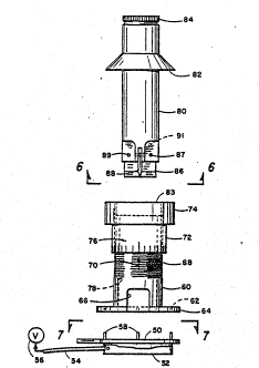
Referring now to Figure 5 wherein thle assembly of the
basic parts of the apparatus are shown in an exploded view.
These parts comprises a cylindrical positionlng ring 50 having
a resilient vacuum ring 52 extending from the botto~ side of
the positioning ring for contact with the eye of the patient
I0 being treated. A vacuum hose 54 provides communication from
the inside of the resilient ring 52 and a vacuum pump source
means 56 as a means to retain the assembled parts upon the eye
for surgical procedures herein described and to remove
scarified portion of the cornea. A plurality of positioning
pins 58 are pro~ided on the top side of the positioning ring
to receive the cylindrical holding sleeve 60, the pins being
adapted to be re~eived through openings 62 in the flange
portion 64. A visual inspection opening 66 is provided for
use by the surgeon. The exterior of the cylindrical holding
sleeve 60 includes a plurality of screw threads 68 along its
length, the threads being a very fine pitch thread, e.g., of
a pitch equal to 40 threads per inch. ~n indicia or marker
70 is provided in the body of the cylindrical holding sleeve
so as to provide a visual measuring point for the surgeon
. ;~ ;:
.:
:: :
~ ~ : '" ~., '
WO91/08711 PCr/US90/07406
,~i3
18
: .
relative to the rotatable position of a micrometer-like guide
s~eeve 72 which includes interior threads to match threads 6~
of the cylindrical holding sleeve. The gui~e sleeve includes
an outer knob portion 74 and indicia generally designated by
the numeral 76, e.g. millimeter or micrometer like markings
on the lower portion of the guide sleeve. 'rhe interior 78 of
the cylindrical holding sleeve is adapted to rotatably receive
a profiling tool 80. The profiling tool includes a collar 82
whioh is adapted to rest upon the top surface 83 of the guide
sleeve 72 for movement upwardly or downwaxdly therewith. The
top end of the profiling tool can include a knurled portion
84 for rotation and/or oscillation by the surgeon. At the
bottom of the profiling tool are a plurality of scarifying
surgically sharp Xnife-edge blades 86 and 88 which are
retained within the body of the profiling to~l 80 by pins 87,
89 and 91. The blades 86 and 88 are retained transverse to
the longitudinal axis of the profiling tool 80. The blades
86 and 88 as used in the invention are of surgical steel.
The profiling tool 80 of Figure 5 is adapted to provide
a scarifying or sculpting operation upon the cornea over the
top center thereof for myopia refractive error, i.e., near-
sightedness, which will effectively lengthen the corneal
radius of curvature as shown in Figure 3.
To correct for hyperopia (far-sightedness), the profiling
"
WO91/087]] ~ ~7 ~ PCT/~90/07406
tool as shown in Figure 8 is utilized, the tool having a shank
90 of similar design to tool 80 shown in Figure 5, except that
the bottom end of the tool includes a plurality of surgical
steel knife-edge blades 92, 94 and 96 which are positioned
transverse to the axis of the tool at an angle of
approximately 30 with respect to the horizontal axis (or 60
to the vertical axis). The blades are adapted to contact the
outer anterior portion of the cornea in order to shorten the
effective radius thereof, that is, the blades will be adapted
to contact and scarify the corneal area A as shown in Figure
2 whereas the profiling tool 80 of Figure 5 will be adapted
to sculpt or scarify the area B of Figure 3. ; -
.
`,~'. .,
'',
WO9l/0871] PCT/US90/07406
r3 2 0
OPERATION
The operation of the apparatus and ~ethods of surgery are
accomplished by first taking optical measurements of the eye
as to what shape the cornea should have in order for that eye
to operate in an optically correct manner~ i.e., correct
refractive errors. Typically, a kerotograph photographic
image using a placido-ring target is used. The photograph is
of reflected light from the placido rings upon a standard
spherical surface of the same size as the cornea in question,
creating an image in the same manner as a topographic contour
map. Subsequently, the topographic survey of the eye to be
corrected is made for comparison purposes and to provide the
surgeon with the necessary dioptic information for correcting
the refractive errors. once this occurs, then the operation
will proceed by placing the positioning ring 50 over the eye
as shown in Figure 14. The size of this ring may vary for
di~ferent operations but is preferably of size wherein the
resilient vacuum ring 52 will rest upon the sclera portion of
thP eye concentric about the cornea. Once the circular
positioning ring 50 is in place, the cylindrical holding
sleeve 60 is then positioned thereupon by the engage~ent of
openings 62 with positioning pins 58. The profiling tool 80
is then inserted within the cylindrical holding sleeve 60 to
a posltion where the bottom of the knife-edge blades 86 and
:
, .. ., ~ , . . ~ . ,
W O 91/08711 PCT/~lS90/07406
88 will lnitlally contact the cornea. ~y rGtating the guide
sleeves 72 in incremental amounts as dictated by the caliper
or measuring scales 70 and 76, the surgeon can continue ~o
increase the depth of the sculpting operation. The scarifying
or sculpting of the cornea occurs by hand rotation or
reciprocation of the profiling tool 80 although other
mechanical or motor operated means are within the scope of
this invention.
In myopic conditions, the profiling tool 80 of Figure ~
is utiliæed. During the operation, the knife-edge blades
press upon the corneal surface which becomes depressecl and
thus gives a larger contact surface with the blades resulting
in a larger diameter of sculptured surface. The scari~ying !i
or sculpting action is accentuated in proportion to the
pr~ssure between the cornea and the blade. The resulting i-
effect is a langthening of the refractive radius in that
portion of the cornea under the blade. When the tool is
removed, the cornea returns to its normal contour except that
the radius over the top center thereof is now longer than it
was initially. As a result, refractive light through the
cornea now focuses upon the retina. The scari~ying action
occurs by the surgeon in incremental movement by rotating or
reciprocating the guide sleeve 72 relative to cylindrical
~holding sleeve 60 utilizing the incremental ~easuring indicia
: `'. ':" .
': ' '.
''' '' ""
: .,, ,' '
WO91/08711 2 ~ PCT/US90/07406
76 relative to a pointer or other indicia 70. Typically, the
guide sleeve is graduated into 25 or 50 micrometer divisions
to provide one hundredth millimeter adjustments ~or each
marked division of rotation. Through use, the surgeon begins
to decide the amount of downward movement: needed ~o achieve
the required changes in the cornea by the rotation and/or
oscillation of the knives. The rotation for a period of a few
seconds will result in removal of small amounts of corneal
material from the cornea. The tool can be removed and/or
kerotographic photographs taken to determine if the
refractive error has been corrected. Since the apparatus and
the surgical methods deal with vexy small increments of
movement in the corneal reprofiling process, it is essential
that the first contact setting be precise and accurate. Many
times this can be done by visual means by the surgeon and in
other instances electrical detecti~g means can be provided
between the cornea and the tool blade to provide an exact
setting of the tool which permits repeatable amounts of
corneal removal.
The profiling.tool of Figures 9 and lO represent a
modified form comprising a body 90 with an indented handle 92
and a knurled fingar knob 94. In this embodiment an internal
sleeve clamp is comprised of scissor elements 96 and 98 which
are pivoted at pir 10D. The outer ends of the elements are
' , ' " .
WO91/08711 ~ ~ 7 "~ ~ ~3 PCT/~'S90/0~406
grooved at 99 and l0l to provide a gripping ac~ion against the
internal diameter of the tool guide or holding sleeve 60.
spring 102 normally biases the blade handles 104 and lO~
outward. Pinching the handles 104 and l0l5 inward retracts
respective elements 96 and 98 so as to be able to be inserte~
into the cylindrical holding sleeve 60~ Release of th~
handles causes the elements 96 and 98 to frictionally en~ge
with the internal periphery of sleeve 60. ~!-
Figure ll provides an electrical indication means for the
surgeon to determine the initial contact of the tool blades.
A first contact electrode ll0 is removably connected with ~he
conductive tool 90. A second electrode is grounded to the
patient at 112. The leads are connected to a low voltage
power source 114 including an indicator lamp 116. Once the
blade touches the cornea, the light will go on which provides
the initial contact polnt from which downward movement
measurements begin. Typically a predetermined amount o~
corneaI material is set into the tool by rotating the guide
tool 72 downwardly. The rotation or oscillation of ~he
profiling tool 80 then begins to change the contour of the
cornea. Measurements are then taken to determine if more
corneal material removal is necessary. If so, a new depth is
set, and the process is repeated. The profiling tool i~
designed to be removed and replaced without cha~ging the depth ; '
'
: . .
~'091/08711 PCT/~S90/~740~
2 ~ ~ L ~ t
~4
setting of the sleeve 72. Typically the amoun~ cf de~th
removal is about two thousandths of an inch (.002"). Many
times it is necesary to operate in several cycles with
measurements being taken after each cycle. A nomo~ram useà
with a computer generatDd set of curves of the cornea before
and after each contouring procedure permits the surgeon tO
constantly monitor the amount of removal of the epithelium
layer and/or in some cases portions of Bowman's layer. It has
been found that the epithelium will return over the surface
of the contoured portion in a period of 2~ to ~8 hours.
owever, there will be~ no xegrowth of the Bowman's laywer which
will cause the changed radius to remain. The epithelium will
return and regrow to its same thickness and clarity but with
a changed radius.
':
'
' ~:
' .
'
,
WO91/08711 25 ~7-g~ PCT/~IS9o/b7406 : -
TEST EXAMPLES
A series of tests have been made upon rabbit eyes and
have resulted in uniform repeatable changes in corneal shape.
In these experiments the apparatus of Figure 5 and the
profiling tool of Figure 8 were utilized. The rabbits were
anesthetized and the pro~edure aibove desc~ibed was performed
to the cornea of the animals. The following chart describes
the results of the corneal changes in terms of the amount or
pre and post corneal curvature change relative to the depth
of the cutter setting:
RADIUS MM
CORNEA NO. PRE-CURVE POST-CURVE; BLADE-DEPTH
l 7.05 7.70 .002"
2 7.40 7.90 .002"
3 8.00 8.70 .002"
4 8.00 8.60 .00l"
7.22 7.70 .00l"
~ 7.l0 7.60 .00l"
Figures 12 and 13 represent a modified form of profiling
tool blade design, shown here for correcting hyperopia, but
the same concept is applicable to myopia correcting tools.
The body 120 includes a plura1ity of radially intersecting
'; '~
,.
~ ' , ` . '
:. :
WO91/08711 ~ ~ 7 ~ PCT/US90/07406
26
blades 122. The extreme tip 124 of the sharpened end of each
blade is bent at an angle prererably of 120. The bend of the
edge of the blades are in alternate directions as shown by the
arrows in Figure 13.
Figure 14 is an enlarged view of the positioning ring 52
as positioned on an eye forming a small vacuum pocket for
maintaining the ring on the eye during surgery.
. ' . "