Note: Descriptions are shown in the official language in which they were submitted.
W~9~03967 PCT/US91/06914
~7 3~
Description
A METHOD OF MEASURING BLOOD PRESSURE WITH A
PHOTOPLETHYSMOGRAP~
Technlcal Field
This invention relates generally to blood
pressure measurements. More particularly, it relates to a
method of non-i~vasively determining blood pressure using
a photoplethysmograph.
Back~round of the Invention
Arterial blood pressure measurements provide
valuable information about a patient's condition. The
heart's cyclical action produces a blood pressure maximum
at systole, called systolic pressure, and a minimum
pressure at diastole, called diastolic pressure. While
the systolic and diastolic pressures are themselves
important in gauging the patient's condition, other useful
parameters are the mean (average) blood pressure during a
heart cycle, and the pulse pressure, which is the
arithmetic difference between the systolic and diastolic
pressures.
The importance of arterial blood pressure has
spurred the development of numerous methods of determining
it. The most widely used method is probably the familiar
blood pressure cuff, which consists of an expandable ring
(l) inflated to stop arterial blood flow and (2) then
gradually contracted. Using a stethoscope, medical
personnel listen to the artery to determine at what
pressure blood flow begins, establishing the systolic
pressure, and at what pressure flow is unrestricted,
establishing the diastolic pressure. More advanced blood
pressure monitoring systems plot the arterial blood
pressure through a complete heart cycle. Typically, these
systems use catheters having piezoelectric pressure
transducers that produce output signals dependent upon the
S-18STITUl E SHEET
-~ .
.
... .. . . .
.. . . . . . . .
'' , ' ~ :.
, . ~, ,, ~ . , .
W092/03~67 ~ 9 PCT/US91iO6914
~ ~Q~ ~ 2
instantaneous blood pressure. The output signals are
monitored and used to determine the arterial blood
pressures over a complete heart cycle. These systems are
advantageous in that the blood pressure is continuously
measured and displayed.
While prior art methods are useful, they have
disadvantages. Cuff-type systems require restricting
arterial blood flow and are not suitable for continuous
use. The piezoelectric-type systems generally require
undesirable invasive techniques, costly disposable
materials, and time and skill to set-up. However, during
certain critical periods, such as surgery, continuous
arterial blood pressure monitoring is highly desirable.
Therefore, it would be beneficial to have a method of
continuously and non-invasively measuring a patient's
blood pressure.
Photoplethysmographs are well-known instruments
which use light for determining and registering variations
in a patlent's blood volume. They can instantaneously
track arterial blood volume changes during the cardiac
cycle. Since photoplethysmographs operate non-invasively,
much work has gone into using them to determine blood
pressure. In 1983, inventor Warner was issued U.S. Patent
No. 4,418,700 on a method of determining circulatory
parameters, wherein signals from a photoplethysmograph
were used to determine arterial blood pressure.
Significant problems were found when
investigating the Warner method. Therefore, it is clear
that the need for a practical method of continuously and
non-invasively monitoring arterial blood pressure has
remained.
Summarv of the Invention
It is an object of this invention to provide an
improved method for continuously and non-invasively
measuring arterial blood pressure.
,
SUBSTITUTE SHEET
; ' ' , " ,~,
W092/0396~ PCT/US91/06914
~ 3 2~73~
It is another object of the present invention to
provide an improved method and system for non-invasively
determining arterial systolic and diastolic blood
pressures with a photoplethysmograph.
These and other objects, which will become
apparent as the invention is more fully described below,
are obtained b~ providing a method and apparatus for
determi~ing arterial blood pressures using a
photoplethysmograph. The inventive method comprises the
steps of calibrating the photoplethysmograph output with
a patient's arterial blood pressure to determine an
arterial constant k in the formula,
~ ~inf(l~KeXp(-kp)~
where ~ is the arterial blood volume, ~inf is a conversion
lS constant corresponding to arterial blood volume at
infinite pressure, K and k are arterial constants for the
patient, and P is the instantaneous arterial blood
pressure; gathering data from the photoplethysmograph
output during a measurement period; and computing the
arterial systolic and diastolic pressures at the
measurement period using the evaluated arterial constant k
and the data gathered during the measurement period.
Brief Description of the Drawinqs
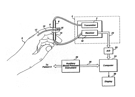
Figure l is a partial cutaway view, partial
application depiction, and partial block diagram
illustrating a preferred method in operation.
Figure 2 is a sketch of the output waveform from
a photoplethysmograph receiver over two cardiac cycles.
Figure 3 is a block diagram illustrating the
basic procedural steps of the preferred method of
Figure l.
Figure 4 is a flow diagram of the pref erred
procedure for calibrating the photoplethysmograph output
to a patient according to the inventive method.
SUBSTITUTE SHEE~ -
.. . . . . : . .,:. .
, :
,
.: .
,, , ~ . . , .,, , .. ,. .. . ~ . ..
W~92/03967 PCT/US9~/OS914
~ 4 ~
Figure 5 is a flow diagram of thP output
monitoring and data acquisition steps according of the
inventive method.
Figure 6 is a flow diagram outlining the
preferred procedural steps for arterial blood pressure
determination according to the inventive method.
Figure 7 is a flow diagram of an alternative
procedure for calibrating the photoplethysmograph output
to a patient according to the inventive method.
Figure 8 is a flow diagram outlining alternative -~
procedural steps for arterlal blood pressure determination
according to the inventive method.
Detailed Descri~tion of the Invention -~
A preferred embodiment of the present invention,
shown in Figure 1, uses a transmitter 2 portion of a
photoplethysmograph 4 to cause monochromatic light 6,
preferably in the red and IR ranges, to be emitted from a
photodiode light source 8. The emitted monochromatic
light 6 travels through a patient 9, along a light path
which includes blood 10 in an artery 12, to a photodiode
light detector 14. While artery 12 has been described,
and is shown in Figure 1, as a single artery, in all
practical cases the light path actually passes through
many arteries. These arteries can be lumped together and
treated as if only one artery 12 existed. Therefore, for
simplicity, the remainder of this application will only
discuss one artery 12, but it is to be understood that it
represents the composite effects of many individual
arteries. The light path is also through background
tissue 16. The transmitter 2 controls the amount of
monochromatic liyht 6 emitted by varying the amount of
current through the light source 8. In the preferred
embodiment, the transmitter 2 regulates the monochromatic
light 6 at a fixed level.
As the monochromatic light 6 travels along its
light path it is partially absorbed by the background
SUBSTITUTE SHEET
, . . . ~
, . .
. .
W092/03967 PCT/US91/06914
~ 5 207~ 9
tissue 16 and the blood 10. A portion of the
monochromatic light 6 is not absorbed and impinges on the
light detector 14, creating electrical signals which are
applied to a receiver 18 of the photoplethysmograph 4.
The magnitudes of these electrical signals depend upon the
amount of monochromatic light emitted by the light source
8, the path lengths through the background tissue 16 and
the blood 10, the amount of light absorbed per unit length
by the blood lo and tissue 16, the conversion efficiency
of the light detector 14, and various lumped losses such
as poor focusing of the monochromatic light 6.
Since the artery 12 i5 pliant, as blood pressure
increase so does the volume of blood 10 within the artery
12. As the heart beats, its cyclical action causes the
arterial blood pressure to change. This causes the
electrical signals to change since the path length through
the blood 10 changes, causing the amount of monochromatic
light 6 absorbed by the blood 10 to change. Therefore,
the electrical signals from the light detector 14 applied
to the receiver 18 is a function of the arterial blood
pressure.
The receiver 18 amplifies the electrical signals
to a usable level and applies them as analog signals, via
a receiver line 22, to an analog-to-digital converter A/D
23. The A/D 23 converts the outputs of the receiver 18 to
time sampled digital signals which are applied to the
computer 24 via a computer bus 25.
The signals on the receiver line 22 can be
represented by the photoplethysmograph output waveform 26,
shown in Figure 2 for two cardiac cycles. The horizontal
axis designates time and, in the present apparatus, the
vertical axis designates volts, but current levels would
also be suitable. Times tO and tl, denoting the beginning
of each cardiac cycle, are clearly marked. The waveform
26 can be described mathematically as a function of time,
with the description being f(t). The voltage waveform is
inverted from the common pressure waveform becaus~ the
SUBSTITUTE SHEET
r ' ' . ... ~
~,
W092/03967 ~ PCT/~S91/0~914
voltage corresponds to transmitted light. The highest
voltage obtained over a cardiac cycle, Vd, coincides with
the diastolic pressure and the lowest voltage, Vs
coincides with the systolic pressure. Between Vs and V
is a mean pressure voltage Vm, which corresponds to the
mean, or average, arterial pressure over a full cardiac
cycle. The duration of the cardiac cycle, td is the time
between reoccurrences of the diastolic or systolic
voltages. The area between the waveform function f(t) and
the diastolic voltage line, shown in crosshatch in
Figure 2, is called the IIARCo 1l The particular values for
Vs~ Vm, Vd, as well as the waveform function f(t) and the
area A~C, change with different patients,
photoplethysmographs, sensor locations, and
photoplethysmograph settings. However, these parameters
are functions of the arterial blood pressure.
In a preferred method of the present invention,
three major steps are used to determine arterial blood
pressure, shown in Figure 3. The first, shown in
block 310, is the calibration of the photoplethysmograph
output to the patient. Referring now to Figure l, the
calibration is accomplished by matching the
photoplethysmograph output on the computer bus 25 at the
time of calibration with the systolic, Psl and diastolic
Pd, blood pressures from the auxiliary blood pressure
instrument 20. In the preferred embodiment, these blood
pressure measurements are entered via a keyboard to the
computer 24. However, preferably this information would
be entered directly via an instrument bus 28. The
photoplethysmograph output is compared with the systolic
and diastolic pressures, Ps and Pd, from the auxiliary
blood pressure instrument 20 and several constants are
determined, as is subsequently discussed.
~s is shown in Figure 3, block 320, the next
step is the measurement of the phokoplethysmograph outputs
during a measurement period to determine various
information. This information includes the systolic,
SUBS~ITUTE SHEET
:, .. . . - ~ . . . ..
" , . . . ~ I ' . ' ! . , ~
''' ' ',, ' ' . ,, "' ' .' ' ': ~
" ' , '" ' " ' " ; '' ' ,
' :'
, ~ ~ . ,
.
. ' ' ' ' ' ' .
W092/03967 PCT/U591/06914
~ 7
mean, and diastolic photoplethysmograph voltages Vs~ Vm
and Vd, respectively, the cardiac duration td, and the
ARC. The final steps, shown in Figure 3, blocks 330 and
340, are the calculations of the systolic and diastolic
blood pressures, Ps and Pd, respectively, using the
determined photoplethysmograph information and the
constants determined in blocks 320 and 310. After the
systolic and diastolic blood pressures are determined, the
information is output to medical personnel on a display
30. If more measurements are desired, decision block 350
causes blocks 320, 330, and 340 to be repeated. However,
only one calibration phase 310 is required. These major
steps are expanded upon below.
Derivation of the Mathematical Model
The principle of the inventive method is derived
from the Beer-Lambert law of analytical chemistry. The
Beer-Lambert law gives the rela-tionship between the
absorption of monochromatic light by a concentration of a
material in a solution as a function of the path length
through the solution. Mathematically, the Beer-Lambert
law is expressed as:
I = IOexp cex
where I is the intensity of transmitted light, Io is the
intensity of incident light, c is the concentration of
material, e is the extinction coefficient of monochromatic
light at a wavelength ~, and x is the light path length
through the medium.
The present invention analogizes blood lO and
tissue 16 density to concentration, modifies the Beer-
Lambert law so that the light intensity terms are given in
terms of receiver 18 output voltages, and breaks the light
SUBSTITUTE SHEET
" ' , ' ' ., ~ . ' ~ ' '
. .
., , ~ ;, .,: ,............. . .
W092t03967 PCT/~S91/06914
~ ~3~ 8 ~
path into individual lengths containing the background
tissues 16 and the arterial blood 10. Therefore, the
modified version of the Beer-Lambert law is:
V = ZI exp(~Ctetxt - c e x )
where the t refers to the background tissues 16, a refers
to the blood 10 in the artery 12, V is an equivalent
transmission voltage corresponding to the transmitted
light, and Z is a constant relating light intensity to the
receiver 18 output voltage.
This can be simplified to:
V AOexp~ Ctetxt)exp(-cae x )
where Ao = ZIo-
This version has separable components,
AOexp( Ctetxt) which relates to the conversion constant and
the background tissues 16, and exp( Caeaxa)~ which relates
to the arterial blood 10. For simplicity, the first
component can be given as VO = AOexp( Ctetxt)~ the
background transmission voltage. Therefore, the equivalent
transmission voltage can be calculated as:
V = VOeXp( a a a) -~
It is convenient to express the above formula in
terms of arterial blood volume rather than light path
length. Therefore, letting ~ be the arterial blood
volume, and substituting for the light path xa
V = VOexp (-b~
where b is equal to caea(4/~L)~, and L is the light path
width through the artery 12. Taking the natural logarithm
results in:
lnV = -b~ + lnVO
;'''' ''," '
- SUE~STITUTE SHEET
: ., . . , . . .... ,: ~ .' . , .
,. , : ' "'.
~ .' ' , " ',' , ' ' ' . . '
,, ., , ':
'~ '' ,' '~ ' ' ' , ' ' .
W092/039~7 PCT/US91/06914
9 2 a7 ~ O l9
This version becomes more useful after
incorporation of the arterial volume-pressure
relationship~
~ = ~inf(l~KeXp(-kP))
where ~ is still the arterial blood volume, ~inf is a
conversion constant corresponding to the blood volume at
infinite blood pressurP, and K and k are constants for the
artery 12, and P is the instantaneous arterial blood
pressure. This arterial volume-pressure relationship is a
good approximation at the pressures of interestO
Substituting this formula for ~ in the logarithmic
version:
lnV = -b(~inf)~ Kexp(-kP))~ + lnVO
This can be expanded using a Taylor series.
Expanding and eliminating higher terms results in:
lnV = f + (n)exp(-kP)
where f is equal to lnVO - b(~inf)~, and n is equal to
(Kb(~inf)~)/2. This can be converted to:
V = (u)exp((n)exp(-kP))
where u is equal to exp(f~. In terms of systolic,
diastolic, and mean pressures:
Vs = (u)exp((n)exp(-kPs)) for systolic Pressure
~ = (u)exp((n)exp(-kPd)) for diastolic Pressure
V~ = (u)exp((n)exp(-kPm)) for mean Pressure ~ ~
Vinf = U ~ ''
V0 = (u)exp(n)
Vo/Vinf = exp(n)
SUBSTITUTE SHEET
W092/039~7 PCT/US9~/06914
Where Vinf is the equivalent receiver voltage at infinite
pressure and V0 is the equivalent receiver voltage at zero
pressure.
Establishing various ratios:
Vd/Vs = exp((n)(exp(-kpd)-exp(-kps)))
Vd/Vm = eXP((n)(eXp(-kPd)-exp(-kpm)))
ln(Vd/Vs) = ~n)(exp(-kPd)-exp(-kPs))
ln(Vd/Vm) = (n)(exp(-kPd)-exp(-kPm))
and
ln(V0/Vlnf) = n
leads to useful ratios: ~-
ln(Vd/Vm) exp(-kPd)~eXP(~kpm)
=
ln(Vd/Vs) exp(-kPd)-eXp(-kps)
1-exp(-kPp/3)
1-exp(-kPp)
and:
ln(Vd/Vs)
= exp(-kPd)-eXp(-kps)
ln(Vo/Vinf)
= [exp(-kPd)](l-exp(-kPp)
where Pp is termed "pulse pressure" and is equal to
Ps - Pd~
. -
Details of the Preferred Method
The previous ~ection derived various .
relationships useful in the preferred method as outlined .
in Figure 3. The step of calibrating the
photoplethysmograph outputs to the patient 9, shown in
Figure 3, block 310 is shown in expanded detail in Figure ~:
4. The first two steps, shown in block 410 and block 420
SUBSTITUTE SHEET ~-
~,~
,., . : , ,,. ,, . . ,,, .,
.,, .. , , .. :, . . ..
' ' ' '' ' .
, .' , . . .
,., , ~,, . ' ,
W092/03967 PCT/US91/0691~
l:L 2~73~
are the determination and entering of the systolic and
diastolic blood pressures, Ps and Pd, respectively, at
calibration into the computer 24. As previously indicated
and as shown in Figure 1, these blood pressures are
determined by an auxiliary blood pressure instrument 20,
preferably an accurate blood pressure cuff having direct
inputs to the computer 24 via the instrument bus 28.
The next two steps, shown in blocks 430 and 440
of Figure 4 are the determination of the
photoplethysmograph voltages, Vs and Vd, from the
receiver 18 output at the calibration systolic and
diastolic blood pressures, respectively. These
photoplethysmograph voltages are readily determined since
they are the minimum and maximum output signals,
respectively, from the A/D converter 23. Next, as shown
in block ~50, the duration of the cardiac cycle, td is
determined from the output of the A/D converter 23. This
is also readily accomplished by using a counter to
determine the time between the diastolic voltages, times
to and t1 of Figure 2.
To determine various patient arterial constants,
the preferred method requires that the area between the
diastolic voltage Vd and waveform function f(t), or ARC,
be determined. This step is shown in block 460 and is
preferably accomplished by determining the integral of the
photoplethysmograph voltages over the cardiac cycle
using: tl
ARC - (Vd)(tl ~ to) f(t)dt
tO
where ARC is the area bet1 ~een the waveform f(t) and the
diastolic voltage line Vd, time to is the time at the
start of a cardiac cycle, t1 is the time at the start of
the next cardiac cycle and (tl - to) is the cardiac cycle
duration td. The calculation of ARC is easily performed
using a digital computer since the output of the A/D
converter 23 is a series of digital representations of the
SUBSTITUTE SHEE~T
W092/03967 PCT/US91/06914
~,Q~ 3~-9 12
photoplethysmograph signals over time. Using the Simpson
approximation to determine the integral is particularly
expedient because the digital magnitudes can be multiplied
by the sampling time between readings, then summed, to
arrive at ARC. While ARC is preferablv determined using
integral equations, other methods of determining it are
also acceptable.
Next, as shown in block 470, the
photoplethysmograph voltage, Vm corresponding to the mean
pressure is determined from the formula
Vm = Vd - (ARC/td) i
where all terms are as previously given.
With Vm known, the next steps, shown in block
480 and 490, are to determine the patient's arterial
constant k, solved numerically, and the ratio Vo/Vinf
solved using either algebraic or numeric methods:
ln(Vd/Vm) exp(-kPd)~eXP(~kpm)
ln(Vd/Vs) exp(-kPd)-exp(-kps)
and
ln(Vd/Vs)
= exp(-kPd)-eXP(~kPs)
ln(Vo/Vinf)
With the above patient arterial constant k and
Vo~Vinf in memory, the patient's arterial blood pressures
can be determined only from the photoplethysmograph
output. This requires that various information be
determined during a measurement period, as shown in block
320 of Figure 3 and with expanded detail in Figure 5.
35 Referring to Figure 5, when arterial blood pressures are ~-
to be determined, the computer 24 monitors the
photoplethysmograph outputs to determine, at the time of
measurement, the systolic voltage Vs~ the diastolic
voltage Vd, the duration of the cardiac cycle td and the
SUBSTITUTE SHEET
-. . ,. , . . , .~ ............. :,. : '
, - . . . . . . .
.,, ~ . .
W092/03967 PCT/US~1/06914
`13 ~ .
2~730~L~
ARC, as shown in blocks 510, 520, 530 and 540, of Figure 5
respectively. With the information Vd, td and ARC
determined, the computer 24 th~n determines, as shown in
block 550, the equivalent photoplethysmograph voltage Vm
usi.ng the formula:
Vm = Vd - (ARC/td)
With the arterial constant k and the ratio
Vo/Vinf determined according to th~ flow chart of Figure
4, and the photoplethysmograph information determined
according to the flow chart of Figure 5, the computer 24
determines the patient's systolic and diastolic blood
pressures as shown in the flow chart of Figure 6, which is
a more detailed description of blocks 330 and 340 of
Figure 3. The most efficient method of determining
systolic and diastolic blood pressures appears to be, as
shown in block 610, to first calculate the pulse pressure
Pp, using numerical methods, from the formula:
ln(Vd/Vm) 1 - exp(--kPp/3)
ln(Vd/vs) 1 - exp(-kPp)
Next, the diastolic blood pressure Pd is determined, as
shown in block 620, using the formula
ln(Vd/vs)
= [exp(-kPd) ] [l-eXp(-kPp) ]
30ln (vo/vinf )
The determination of the systolic blood pressure Ps~ is
then readily accomplished, as shown in block 630, using
the equatiOn Ps = Pd + Pp-
While the above is the preferred method of calculatingarterial systolic and diastolic blood pressures from the
photoplethysmograph outputs, other schemes are possible.
The systolic and diastolic blood pressures are
then available for output to medical personnel as shown in
SUBSTITUTE SHEET
:, - : . '
- , ;
.
~, .. .
' ' ' ,.' , '. `' '
, ' . .
W~92/03967 PCT/VS91/~6914
~ 3~; 14 ~
block 640, in a variety of way such as by digital or
analog readouts, chart recorders, voice synthesis, or as
in the present embodiment on a display monitor 30. If
another sPt of measurements is desired then decision block
650 causes the flow shown in Figures 5 and 6 to be
repeated.
The preferred embodiment described above is
useful, can be readily implemented on a digital computer,
and provides accurate and rapid measurements of arterial
blood pressures non-invasively and in a manner suitable
for continuous measurements. However, in some patients
and under some conditions, the preferred method leads to
inaccuracies because of time variations in Vinf, the
equivalent receiver voltage at infinite pressure. Vinf,
in the preferred method was part of the ratio Vo/V
determined during calibration and presumed constant. The
preferred embodiment can be modified to compensate for
changes in Vinf but at the expense of additional
computation difficulty and time.
The alternative embodiment follows the same
three major steps as shown in Figure 3 for the preferred
embodiment. However, the calibration procedure of Figure
4 is modified to that shown in Figure 7. These
calibration procedures, shown in Figure 7 blocks 710
through 780, are identical until Vinf is determined in
block 790. It can be shown that Vinf is determinable by
the following formula~
Vinf = exp{[ln(Vs) - (lnVd)exp(-kPp)]/[l-exp(-kpp)]}
With Vinf thus determined in block 790, V0, the
equivalent receiver voltage at zero pressure, is
determined, as shown in block 799, from the formula:
ln(vd/vs)
- - = [exp(-kPd)][l-exp(-kPp)]
ln(Vo/Vinf)
SUBSrlTUTE SHEET -
: : , . .
.
.. ..
.
,: '
W~92/03967 PCT/US91/~691~
~ 15 2~7~0i ~
After the photoplethysmograph output is
calibrated according to the alternative embodiment, as
shown in Figure 7, the patient constants k and V0 are
known.
According to the alternative embodiment, the
data gathering steps depicted in Figure 5 remain the same.
However, during blood pressure determination, the flow
diagram of Figure 6 is modified to the procedural steps
shown in Figure 8. Referring now to Figure 8, after
determination of the pulse pressure Pp in block 810, in
the same manner as it was determined in block 610, the
Vin~ at the time of measurement is determined, as shown in
block 820, from equation:
Vinf = exp{ln(Vs) - [exp(-kPp)]lnvd]/~l-exp(-kpp)]}
where Vs and Vd are also the values at the time of
measurement.
This new Vinf is then used in the equation of
block 830, along with the previously stored value of V0,
to determine the diastolic pressure Pd. This alternative
embodiment reduces the effects of changes in Vinf. The
calculation of the systolic pressure Ps~ shown in Block
840, and the output of the systolic and diastolic
pressures, Pd and Psl respectively, as shown in block 850
are performed in the same manner as they were in blocks
630 and 640, respectively, of E'igure 6. Likewise, the
decision block 860 operates in the same manner as the
decision block 650 in Figure 6.
The apparatus for practicing the present
invention uses a modified pulse oximeter-type
photoplethysmograph 4 having numerous user controls, such
as receiver 18 gain and light source 8 current settings.
It outputs an analog voltage representation of the
photodiode output to an analog-to-digital converter A/D 23
which digitizes the receiver 18 output and applies it to
an IBM-AT type personal computer 24 under the control of
- SUBSTITUTE SHEET
: : . .. .
'~' .
: ,
W092/03967 ~ PCT/US91/06914
~ ~`3 16
software stored in a hard-disk drive. The display 30
output is on a computer monitor. The required auxiliary
blood pressure instrument 20 readings are input by
keyboard when directed by software programmed prompts. In
future applications, the separate photoplethysmograph 4,
A/D converter 23, and computer 24 will probably be replace
by similar structures within a single chassis and
calibration data will be automatically inputted by an
automatic blood pressure cuff.
From the foregoing, it will be appreciated that
the invention, as described herein for purposes of
illustration, provides an advancement in non-invasive
blood pressure instruments. Although alternative
embodiments have been described herein, various
modifications may be made without departing from the
spirit and scope of the present invention. Accordingly,
the scope of the invention extends to the broad general
meaning of the appended claims.
;....
SUBSTITUTE SHEE~
.
.. .. .. . .
. .- . . . ` :
. ,
, :, . :' , : . ` . ` ,