Note: Descriptions are shown in the official language in which they were submitted.
91/1~704 ~ 0 7 ~ PcT/us9o/06913
Method for inspecting density of carbon fibers in a substrate by
infrared imaging
BACKGROUND OF THE INVENTION
-Fuel cells electrodes and other articles, which are
basically matrices of carbon fiber and a binder such as
phenolic resin, for a variety of reasons, desirably are of
uniform density. In the particular case of fuel cell
electrodes, if the fabrication process results in regions of
the substrate being too high in density, it is a problem. More
particularly, at the completion of the processing, when the
electrode substrate is placed in service, overly dense regions
may restrict the diffusion of oxygen or hydrogen gas. Thus,
the performance of the fuel cell may be impaired.
In the case of aircraft structural part substrates,
such as airframe parts, uniformity of density is desirable
because it translates into uniformity of strength per unit
measure of material. r
Currently, there is no easily applied method to
nondestructively test resin bonded carbon fiber substrates such
as fuel cell electrodes except to visually inspect them. Upon
examination, small areas can be tested. However, the present
invention solves a particular problem in infrared imaging by
creating a steady state temperature gradient around areas of
high density. Other methods of heating produce temperature
gradients in a transient state making it very difficult to
capture and record an infrared image for analysis. More
generally, the present invention improves the diagnostic value
of infrared imaging of carbon composites over that of eddy
current testing and ultrasonic testing.
~............................................... , . ~ .
2074~31 1
-2- 6289B-1429
SUMMARY OF THE INVENTION
According to one aspect, the invention provides a method
of nondestructively identifying regions of high density in a
substrate of a bonded matrix of carbon fibers comprising the
procedural combination of steps of:
connecting conductor terminals at opposite extremities of an
area of a substrate of a bonded matrix of carbon fibers to be
imaged;
connecting the conductor terminals to an electrical power
supply to apply a current through the terminals and across the
area of the substrate, thereby to heat and to create contrasting
regions of temperature gradients which clearly distinguish regions
of high and low density;
identifying and recording the regions of high and low density
by photographing or scanning the area with infrared ray emission
imaging equipment.
According to another aspect, the invention provides a
method of nondestructively identifying regions of irregular
structure in a substrate of a bonded matrix of carbon fibers by
29 performing the steps of resistively heating the substrate and
taking an infrared image of the heated substrate.
The importance of the novel combination of procedural
steps is the method of heating. It takes advantage of the fact
that carbon is conductive yet resistive enough for I2R heating
which solves the problem of the part, if otherwise heated,
becoming uniformly heated over the entire area. Visual
inspections previously required primarily a subjective decision
whether to accept or reject a carbon composite part. The need for
2074~1 I
-2a- 62898-1429
a non-destructive test method to quickly and quantitatively
identify regions of high density or each of homogeneity has led to
applying thermal imaging in the past. However, the results have
been unsuccessful. The reason for this seems to be because some
form of contact or convection heating has been relied upon to heat
the electrodes for subsequent infrared (IR) imaging. The poor
results occur because the fuel cell electrode being tested, for
example, reaches thermal equilibrium (i.e., constant temperature
throughout). There is but a short window of time to capture an
image that shows temperature gradients formed due to regions of
high and low density. Moreover, this time window may vary for
different areas of the same part.
In contrast, the instant invention, because of the
resistive heating, reverses the problem of reaching steady state
thermal equilibrium. By electrically heating a fuel cell
electrode, for example, the reqions of high and low density
correspond to regions of high and low conductivity. Similarly, if
regions of non-homogeneity, voids, pitting or cracks are present
in a substrate, they will show as regions of steady state thermal
gradients. As current is passed through the electrode, current
density increases in regions of high density and results in
greater resistive heating. With this method, temperature
gradients exist in steady state instead of in the transient
response. The infrared images in this situation clearly
distinguish regions of high and low density. When photographed or
scanned with infrared ray emission imaging equipment attached to a
20748 1 ~
-2b- 62898-1429
video recording machine, pictures or tapes showing contrasting
areas of temperature gradients which
.
~'0 91/11704 ~ i PCT/~S90/06913
clearly distinguish regions of high and low density are
provided.
BRIEF DESCRIPTION OF THE DRAWINGS
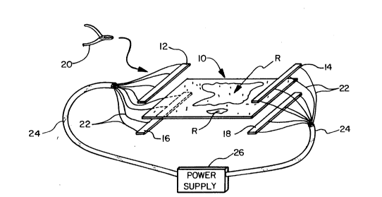
Fig. 1 is a schematic exploded view of a fuel cell
electrode substrate of phenolic resin bonded carbon fibers
showing the mode of application of alternating current from one
substrate area extremity to the other in performing the method
of nondestructively identifying regions of high density in the
substrate area and recording them by photographing or scanning
the area with infrared ray emission imaging equipment.
DETAILED DESCRIPTION OF THE INYENTION
The method of nondestructively identifying high
density reglons "R" of a fuel cell electrode 10 made of
phenolic resin bonded carbon fibers includes a procedural
combination of steps of no critical sequence except that the
photographing or scanning with imaging equipment follows a step
of resistance heating to create contrasting areas of
temperature gradients.
Typically, the method is performed by connecting
J' copper or aluminum terminals or bars 12, 14, 16 and 18 at
opposite extremities of and on opposite sides of the electrode
10. The bars 12 and 16 on opposite sides from each other on
one end and the bars 14 and 18 on opposite sides from each
other on the opposite end. The bars 12 and 16 are held at
`~ their end of the electrode substrate area by means of a
plurality of clamps 20. In similar manner, the bars 14 and 18
are held at the opposite end of the electrode substrate area 10
to be imaged.
The bars 12, 14, 16 and 18 have attached thereto,
wires 22 which are part of conductor cable 24 such that a
substantially even current density is distributed within the
bars 12, 14, 16 and 18. The cables 24 are suitably connected
to a power supply 26.
By means of this arrangement, a current is applied
through terminals and.across the area of substrate 10, thereby
W 0 91/11704 2~7 ~7 ~ PCT/US90/064~.-
to resistively heat and to create contrasting high density
regions "R" of temperature gradients which clearly distinguish
from the regions of the rest of the area of electrode 10 which
is, relative to regions "R", of low density.
The electrodes 10 in their as-fabricated state
typically can be heated by applying approximately 230 volts of
alternating current (AC) across the substrate area. Once the
electrodes are carbonized, the conductivity becomes very high.
The electrodes 10 can still be tested using this method,
however a power supply 26 capable of high current (120 amps or
3 amps per inch) is needed.
The electrodes 10, after electrical heating, as
described, is photographed by a camera using infrared ray
emission sensitive film or is scanned. The scanning can be
performed with a Model 525 hand held or tripod mounted
electronic imaging apparatus obtained from "INFRAMETRICS" of
Billerica, Massachusetts 01862, which for certain high
resolution shots can be supplemented with a 0.8 micron infrared
filter from the same source. The Model 525 electronics pack
attaches to a commercial VCR video cassette recorder to create
magnetic recording tapes of the images produced from the scan.
The electrode 10 may be heated at any stage in the
processing so that it can be thermally imaged to identify
regions of high density.