Note: Descriptions are shown in the official language in which they were submitted.
Dkt. No. 2~.1690/JCG
BIOMEDICAL MAGNETISM IMAGING APPARATUS AND METHOD
1 ~A~P~ O~
2 1. FiQl~ o~ th~ I~o~tior
3 The present invention r~lates to an imaging apparatu3 and
4 method for displaying magnified images o~ sequentially e~timated
areas of current sources for ~3timating thQ position of an active
6 ar~a wi~hin a living body by u~in~ a biom~dical ~agnetism
7 measuring apparatus, including a highly sen~itive magnetic ~ield
8 s~nsor, for sxampl~, a SQUID (Superconducting QUantum
9 Interferenc~ Device) ~or ~ea~uring a ~agnetic fiQld gsnerat~d
from a living body.
11 2. Do~c~iption of t~ R~l~t~ Ar~ ,
12 With recant dvanc~s in superconductivQ d~vic2 technology,
13 highly sensitive biomadical magneti~ m~a~uring ~pparatus
14 utilizing a SQUID havs r~cently been ~mployed in ~edical
diagnostic appar~tu~. Such apparatu~, which ar~ ~190 referred to
16 as bio~n~to~et~rs, SQUID co~puted tomography (SQUID ~T),
17 magn~tic our~ i~aging (~SI~ t bio~agnetic i~aging ~BHI),
18 magn~to~ncerh~logra~ ~E~), and ~agn~toc~rdiogra~3 (~CG) operate
19 as follow~. Electric sourcQ~ within a living body si~ultaneou31y
generate a low 19v~1 ~agn~tic field. Th~re~ore, per~or~lng an
21 inv~rqa e timation (or inv~rse proble~) of th2 active area within
22 a living body by mQa~uring th~ distri~u~ion o~ thi~ magnetic
,r l ~ o ~ r ~
1 field i~ eXpected to be u~eful for diagno~ of diseased regions
2 within a living body. The terms in~erse estimation and inverse
3 problem refer to an algorithm in which a m~asured magnetic field
4 is employe~ for estimating the location and/or distribution of
tha electric sources within the body. Tha position o~ a current
6 source which act~ as a magnetic fi~ld generating source i~
7 estimated to analyze heart dis~ase and brain function disease
8 from tha ~easured ma~netic field. For thi~ purpose, a current
9 dipole i3 used a~ a model of a biolo~ic~l ~ourcQ and i~ e~timated
within ~ ho~ogeneou~ condu~tor using a computation model. A
11 current dipole i~ a short s~gment of current which i8 used to
12 illustrate a tran~i~nt curr~nt flow in a s~all ar~a. Th~ inverse
13 proble~ approach i~ Pmployed for determining the current
14 distribution for which the computed ~agnetic ~i~ld beco~e~ equal
to the measured magnetic ~ield. In accordanc~ with the inverse
16 problem approach, an algorith~ i~ u~d to move the e~timated
17 value of th~ current dipol~ to thQ po~ition for which the
18 corresponding comput~d ~agnetic fiQld approximate tha maasured
19 magnetic field. The algorith~ which is employed i~ based on th~
l~ast square ~rror solution (sea Equation (5) b810w~ .
21 ThQ abov~-describ~d bio~agnetic ~easuring appar~tu~ should
22 not ba con~u~d with ~agnetic r~sonan~ imaging (~RI) which
23 detect~ only th~ con~igur~tion o~ a struc~ur~. Instuad, thQ
24 subject inYention i~ dirQcted to d~t2r~ining the functional ~tate
2S of an organ by cl~tecting current path~ in th~ body an~
26 particularly th~ brain (~o-called neuromagnetis~) and the heart
27 (so-called cardiomagn~ti~). To indicat~ th~ m~gnitude o~ the
-- 2 --
25307-296
1 magnetic fields created by such current flow in a living body,
2 the neuro~agn~tic field is approximat~ly 10 14 tesla, while the
3 cardiomagnetiG fiald is 10 12 tesla. The magnatic ~ield is
4 measured in order to determine the~ current amplitude and position
of the equivalent current dipole.
6 Biomagnetometers are current:Ly available in the art. For
7 example, Biotechnology Incorporatlad (BTi) produces a
8 neuromagnetometer utilizing a SQUID. Other biom~gnetometers are
9 manufactured by Sie~ens and CT~ of Canada.
In prior art biomedical m~ynetism
11 measuring apparatu~, a homoganaous semi-infinita conductor model
12 of tha tor-cs, for exa~ple, i9 u~ed for ~ ti~ation o~ thR current
13 source in the heart. Alt~rnatively, a ho~ogQneou~ or
14 multilayer conc~ntric conductor sphere can be u~ed a~ a mo~el for
the head. Tho paramaters of the current dipoles for the heart
16 model ars then e~ti~ated and the magnetic field ~c based on
17 the e estimated dipols~ i~ calculated. Th~ ~agnetic fi~ld
18 of the heart in tha living body i~ mea~ured and the measured
19 data B~ i~ input to thQ computer. A current dipole which
minimi7e~ an objective function equal to thQ ~quared difference
21 between th~ m~a~urad ~a~netic field and th~ comput~d magnetic
2~ field, ha~ ~o~n consid~rad a~ ~he esti~ated position oP th~
23 current ~ourc~ (also re~errQd to above a8 ~h~ inv~r~ problem or
24 inverse e~timation). Th~rQ~oro, th~ ~ea~ured value B~ and the
computed valu~ 3c are co~pared and i~ th4 s~uar~d error is
26 ~ini ized th~n the di3tribution of the current dipoles is
25307 296~ , L '
1 displayed. I~ the 2rror i5 large then the param~ters
2 of the test dipole3 are modifisd and the magnetic Eield Bc
3 i~ recalculated based on the newly set dipole~.
4 The above-described approach for determining the amplitudes
and positions of current dipoles i.s used in a numb~r of different
6 fields including the above-described biomedical magnetism
7 apparatus and in other field~ where the determination of current
8 dipoles i9 desirable. However, c6!rtain problems are inherent in
9 this approach. Local minima in th~ squar~d error between the
measured and computed ~agnetic ~ield~ can produce incorrect
11 solutions to the dipole parameters. To ov~rco~ thi~ proble~, a
12 long time has been r~quired to obtain a~urat~ current sourCQ
13 localization becau~e the co~putation doe~ not converge within a
14 finite amount og time to th~ corr~ct ~olution (i~o I the global
minimum of the objectivQ ~unction in tha inverse problem).
16 A~ explained ab~v~, t~Q l~a3t 3quara error ~ethod for
17 solving 3uch a non-linear ~yst~ r~quire~ repeated computation.
18 As a method of avoiding such rep~ated co~putation, it has been
19 suggested to fix the po~ition of current dipole~ on grid points,
thereby making ths proble~ linsar. Such ~eth~ds are de~cribed
21 in, for exa~ple, J~ a ~,'An E~wfio~ of M~x~ for N.~ ic Imag~
22 R~ , E~~ ~ ' onBi ' ~ F~ ~, Vo~ 34, No. 9, 5~ b
23 /~7,pp. ~13~ ~ ~,'Lin~ F ~9 App~ fo t~ R- , of o 3-D
24 Vcaor Currcn~ D~ ', AppUot Op~icJ, Vo~ 29, ~lo. S (1990), pp. 6~8-667; and Sann~,
~Basicl~ 'crl a~l EL~ fc Conc~p~ of she Bi ~ ~:r Invcrsc Pro~ , 1989,
26 Phys. Mcd. ~io. 32, pp. 11-22 .
2530~ 6 ,~
1 A ~ethod for obtaining a lea~t square
solution by relating the measured magnetic field to the intensity
3 of the current dipol~ by using si~ultaneou~ linear equations can
4 be then for~ulated. To express the meas-~red magnetic field
obtained by picXup coils and the a:mplitude of the current dipole
6 on aach grid point witn ~imultaneous linear eguations, the
7 distribution of the current dipoles i~ d@fined on a set o~ fixed
8 grid point~. The a~plitude~ in three ~irection o~ n current
9 dipoles are defined a~ x~ q1y~ ql3) ~-- (q~ qn2); th~
positions of the corre~ponding grid points are d~fined a3 (xl',
11 Yl ~ (Xn~ ~ Ynl ~ znl ); ~he amplitude~ in three dir~ction~
12 of the magnetic field measured by m pickup coils at m points are
13 defined as (b1~, bly, b~2) ... (b~, b~, b~); th~ positions of tha
14 corresponding pickup coil~ ar~ dQfined a~ (x1, y1, z1) ... (x~, y~,
15 z~), the current dipole vector Q = {ql, q2~ ~.. qn}T and the
16 measured magnetic field vactor B~ b2, ... b~)T. Based on
17 the Biot-Savart law, ths simultaneous linear equation B - AQ can
l~ be solved, where th~ ~atrix of coefficientc A i~ given by:
19
all al2 ~ ~ aln
2 l a21 a22 ~ ~ a2n
2~ A ~ . . . . . (1)
23
2 4 a~l a~
1 Moreover, the elementS of the equation B~ - AQ can b~
2 expres~ed a~ follow~:
3 bl a~1 al2 . . aln ql
4 b2 a21 a22 ~ a2n q2
. = . . . . . . (2)
7 b~ a~1 a~ . . a~ qn
8 Here, from the ~iot Savart law, element~ o~ each coe~ficient
9 matrix can bs obtainsd ~ro~ the following expr~s~iQn:
~0
a~J = 4 ~r {~Xj-X3 ) + (yi-yjl) ~ (Z~_Zj~)Z}5/C
3 (3)
14 0 Z~~Zj~ ~(Y~~YJ~)
1S x --(Z,--ZJ ~ ) o X,~-XJ
16 y~-yj -~X~-Xjl~ 0
17 The eguation B~ ~ AQ i8 ~ linear eguation which is
18 determined by th~ curr~nt dipole po3ition~ and pickup coil
19 po ition~. Th~re~or~, the current dipol~ values Q can be
obt~ine~ by ~olving this equation wh~n m, the number of
21 meaSUre~QntM, agual~ n, th~ number o~ unknown~ th~
22 coefficient matrix A i~ a non-singular matrix, t~e inver3~ matrix
' :.
'
-
25307-296
~ ~ 7 ~
l A1 exist.q and a current dipole di~tribution Q can be solved
2 directly from
3 Q = AlB~ (4)
4 However, if the coe~ficient matrix A is singular or if
n > m, the inver~e matrix cannot bQ obtainad and a unique
6 solution doe~ not exist. However, in thi~ case, the product ATA
7 o~ the coefficient matrix A and tran~posed ~atrix AT beco~es a
8 square matrix and ATA can be inverte~ when the column vectors of
9 A are independent. In this ca~e, tha lea~t squares solution,
denoted Qc~ which rin1~; ze~
11
12 i~l (b~ - bc,i (~) ) (5)
14 which i~ th~ squared su~ of the difference o~ measured values B~
and co~puted value~ Bc ~ AQC~ giv~n by tha nor~al Pquation
16
17 Qc ' (~TA)1ATB~ (6)
18 can bQ obtAine~ a~ de cribad by ~n~8, ~Li~ Ig~bra ~ ~# ~t.'i~ s~,
19 l~gO, Ncw ~, AG~DEAIIC ~;, IN::.
Under tha lea~t square error method
21 of Equation (5), th~ parameter ~ d~fin~ t~e te~t dipole
~ ,
25307-296
h~ 3
position x ', y ' and z ' and dipol~ strength qx, qy and qS .
2 Equation (6) refers to the formulation which is one case o~ the
3 least square error solution using singular value decomposition.
4 Thus, the terms inver~e estimation and inverse problem used above
5 ara based on the least ~quare solution o~ Equation (5).
6 Moreover, when the matrix A is singular, where the column
7 vectors of matrix A are not indepe!ndent ( i . 8., rank (A) < n), the
8 inverse matrix o~ ATA does not ~xi~t and th~refore a unique
9 ~olution cannot be obtained. In thiR case~ singular value
10 deco~position can be utilized.
11 In accordanc:e with ~ingular value decompo~ition, a de~ired
12 (m x n) matrix A can b~ decomposed to
13 A ~ U~VT ( 7 )
14 with the m x m orthogonal ~natrix U, m x n diagonal matrix A and
15 (n x n) orthogonal matrix V. ~ diasonal matrix where th~
16 el~ment~ or singular value~ 1, 2 m) are the ~quare
17 roots of th~ eigenvalue~ o~ A~T and ATA which are arranged on the
18 diagonal in de~csn~ing ord~r and U and V ars eigenvectors o~ AAT
19 and ATA, re~pectively, as de~cribed in Fon~he ~ al, 'Com~urcr Mah~ds foT
2 0 M~r ~ Ncw Je~s~y, (1978~,
21
f ,' ~ s-
1 I n this case, the least~quare minimu~nor~ solution Q~ of
2 ~quation t4) above can be obtained from the following equation
3 for the generalized inverse matrix
4 Q - V~ UTB~ - A~3~ (8)
Here, A~ is a diagonal ~atrix who~e element i5
7 when A~ is not equal to 2ero, ~j=O when ~=0. A~ is a p~eudo
8 inverse ~atrix, where th~ inverse matrix A~ extended to an
9 arbitrary (~ x n) matrix from an (n x n) square matrix.
A generalized inverse matrix ~ethod utilizinq normal
11 equations and a method utilizing ~ingular value deco~position ar~
12 effectiva ~or obtaining the den~ity di~tribution of m~ny current
13 dipole~ becaus~ a ~ulti-dipola ~odel i~ assumed. In thi~ case,
14 once the inverse ~atrix i~ obtain~d by u~ing the normal ~quations
or singular valuQ ~ gition, tha curr~n~ ~ourc~ density
16 ~istribution Q can b~ obtainq~ ~imply by ~ultiplying tha measur d
17 values ~ a~thQr by the co~f~lcient A~ a~ in Eguation (8~ or by
18 (ATA~ 1 AT a~ in Equ~tion (6~. Th~ di tribution of current
19 dipoles ean then b~ obtain~d with high~r 3peed than with the
i~erative ~eth~d ~or solving ~h2 nonlinear least squar~ ~y te~ in
21 which ~h~ position ig esti~ated by moving currsnt dipol~.
_ g _
,
; :
"'~
:
1 In the technique for expreSSing the relationship between
2 magnetic field intensity and current dipole locations with
3 simultaneou~ linear equatlons and obtaining a generalized inverse
4 matrix from tha normal equations, a method for dividing the
higher current dipole intensity area based on the initial
6 estimated value is known, and is described in O~u~ ~ ~.,'C~e~
7 Densiry Imaging as a Mcthod of ~lsualizJng l~cu~onal Aai~ty of Ih~ Brain', So~i~ry for
8 l~woscicnc~ AbsJraas, 505~.16:1241 fl9~0) .
g However, in this ~ethod, curren~
dipole resolution can be improved while leavinq grid poin~
11 ~xisting in the peripheral area, but sinc~ the n~mb~r o~ grid
12 point~ of current dipolQ location3 increa3ea through division
13 into subsQction~, the influence of ~he li~ited numb~r of pickup
14 coils limit~ ths re~olution. In addition, Okad~'s ~ethod as~ume~
a current ~ipole plane (source plane) which i parallel to the
16 measurement plane and doe~ not describs a ~lexibl~ grid point
17 distribution method and a display ~thod therefor.
18 As described above, th~ numb~r of ~en~or~ m mu3t b~ equal to
19 or larger than the nu~bQr of curr@nt dipole~ n in order ~o
accurately obtain th~ inten~ity di~ribu~ion o~ current dipole
21 from th~ st square ~olution o~ such 8imultaneou8 linear
22 ~qua~ion8. Ther~for~, a larg~ num~er o~ pickup coils and SQUID
23 magn2to~t~rs ~or mQa~uring ~agnetic field int~n~ity ars rsquired
24 ~or estimating the ps~ition Or current dipole~ with an accuracy
of several millim~ter~ required for ~Qdical dia~nosi~. It has
-- 10 --
2530~
also been impossible to realize a real-time apparatus for display-
ing movement of dipole sources.
SUMMARY OF THE lNv~l~ION
It is an object of the present invention to provide a
biomedical magnetism imaging method and apparatus for displaying
a desirably magnified image of the distribution of current sources
having high resolution by using a comparatively smaller number of
pickup coils than is needed for an equivalent resolution over the
entire region of interest.
It is a further object of the present invention to
provide a biomedical magnetism imaging apparatus which obtains the
distribution of current sources by solving the least square
solution of simultaneous linear equations using ~he normal
equations and singular value decomposition as the methods of
estimating current sources.
It is a still further object of the present invention
to provide a method and apparatus for displaying medical imaging
data including MRI data and biomagnetic imaging data (BMI or MSI
data).
Therefore, in accordance with an aspect of the present
invention, there is provided a method of imaging current sources
within a subject, comprising the steps of: (a) defining a grid
having a first region and having multiple gxid points; (b~
measuring a magnetic field generated by current sources located
within a portion of the subject corresponding to the defined grid;
(c) determining a distribution of current sources on the defined
grid based on the measured magnetic field; and (d) modifying the
defined grid to have a second region which is smaller than the
2 5 3 ~S,7
first region to improve the resolution oE the current sources
distributed on the grid.
According to another aspect, the present invention
provides a method of displaying medical imaging data, comprising
the steps of: (a) providing biomedical magnetism image data;
(b) providing medical image data other than biomedical magnetism
image data; (c) selecting contour image data from the medical
image data; and (d) generating a display by superimposing the
biomedical magnetism image on the selected contour image.
According to a further aspect of the invention, there
is provided a biomedical magnetism imaging apparatus for imaging
current sources within a subject, comprising: a sensing unit for
sensing a magnetic field generated by current sources located in
the subject and for providing sensing signals corresponding to
the sensed magnetic field; means for defining a grid having a
first region and having multiple grid points, for processing the
sensing signals to determine a distribution of current sources on
the defined grid, for modifying the defined grid to have a second
region which is sma~ler than the first region, and for determining
the distribution of the current sources on the modified grid; and
display means for displaying the distribution of current sources
on the defined grid and the distribution of current sources on
the modified grid.
The method and apparatus of the present invention
provide a number of advantages over the prior art. In particular,
currently available biomagnetic imaging apparatus are only
- lla.:--
' ' ~ :
- : ' ~ i.'- ' . :
2530?-296
capable of displaying a single dipole. While multiple dipoles
have been discussed, it has been thought that multiple dipole
display arrangements would require many pickup coils in order to
- llb -
,: ,
~' ~
~ ~ ,
~ 3~3~ 3
1 achieve acceptable re~olution. The present invention overcomes
2 these deficiencie~ by providing a biomagnetic imaging method and
3 apparatus which is capable of displaying a desira~ly magnified
4 image of multiple dipoles, while using a relatively small number
of picXup coils.
6 These together with other o~jects and advantages will become
7 apparent from the following descr:iption of the pre~erred
8 embodiments in conjunction with tlle acco~panying drawings,
9 wherein like reference numerals refer to like p~rts throughout.
~ 8C~IP~I0~ 0~ R~
11 FIGS. l(a), l(b) and ltC~ are diagrams for explaining the
12 principle of th~ presQnt inventisn in which a current dipol~ grid
13 for a particular current source di3tribu~ion is ~ucces~ively
14 reducPd in size;
FIG. 2 is a schsm~tic diagram of an embodi~ent of the
16 present inv~ntion;
17 FIG. 3 is a block diagra~ showing t~ d~tails of th~ signal
18 processor 8, tha control unit 9 and thQ di play 10 of FIG. 2;
19 FIG. 4 is a flowchart for describing thR operation of th~
circuitry o~ FIG. 2;
2~ FIGS. 5(a~, 5(b) and 5(c) are diagra~ o~ exa~ple~ for
2~ reconstructing a two-di~n~ional dipol~ grid in accordance with
23 the present inv~ntion;
25307-296
FIGS. 6(a), 6(b) and 6(c) are graphical displays for
illustrating the results of the reconstruction of FIGS. 5(a),
5(b) and 5(c), respectively;
FIGS. 7(a) and 7(b), on the fourth sheet of drawings,
are diagrams illustrating examples of reconstruction in accordance
with the present invention, wherein one of the current dipoles is
located between grid points;
FIGS. 8(a), 8(b) and 8(c) are graphical displays for
illustrating the results of reconstruction in accordance with
FIGS. 7(a) and 7(b);
FIGS. 9(a) and 9(b) are diagrams for illustrating the
use of a three-dimensional grid in accordance with the present
invention;
FIGS. lO(a), lO(b) a'nd lO(c) are diagrams for illustrat-
ing the use of a polar coordinate g-id, a brain-shaped grid and
a heart-shaped grid in accordance with the present invention;
FIG. ll is a diagram for illustrating a display device
having two display screens in accordance with the present
invention;
FIG. 12 is a diagram illustrating a display device
having four separate display areas with different levels of
resolution on a single screen;
FIG. 13 is a diagram for illustrating a display having
multiple display screens for displaying different planes of a
subject to be imaged and having the capability of selecting the
depth of the plane being displayed;
FIG. 14 is a diagram for illustrating a display having
a primary screen and having a secondary screen for selectively
- 13 --
: . ' .
displaying a plurality of portions of the primar~ screen at
different times, in order to simulate movement of the current
dipoles within the subject;
FIG. 15 is a block diagram for illustrating a system
for superimposing an MRI image and a biomagnetic or MSI image
on a single CRT in accordance with the present invention;
FIG. 16 is a flowchart for describing the operation
of the circuit of FIG. 15 in accordance with the present
invention;
FIG. 17 is a block diagram of an alternative system
for superimposing an MSI image on an MRI image which is a hardware
version corresponding to the circuit of FIG. 15; and
FIG. 18 is a flowchart for describing the operation of
a prior art biomagnetic display device.
DESCRIPTION OF THE PREFERRED EMBODIMENTS
Referring to FIG. 18, in prior art biomedical magnetism
measuring apparatus, a homogeneous semi-infinite conductor model
of the torso, for example, is used for estimation of the current
source in the heart (Sl). Alternatively, a homogeneous or multi-
layer concentric conductor sphere can be used as a model for thehead. The parameters of the current dipoles for the heart model
are then estimated (S2~ and the magnetic field Bc based on these
estimated dipoles is calculated (S3). The magnetic field of the
heart in the living body is measured (S4) and the measured data
B is input to the computer (S5). A current dipole which
minimizes an objective function equal to the squared difference
between the measured magnetic field and the computed magnetic
field, has been considered as the estimated position of the
- 14 -
~,
25307-296~ J~
current source (also referred to above as the inverse problem or
inverse estimation). Therefore, the measured value sm and the
computed value B are compared (S6) and if the squared error is
~;n;r; zed (S7) then the distribution of the current dipoles is
displayed (S8). If the error is large (S7) then the parameters
of the test dipoles are modified (S9) and the magnetic field sc
is recalculated based on the newly set dipoles (S3).
In the present invention, an estimated current dipole
grid 1 is first roughly or coarsely set to enclose the entire
region in which it is probable that one or more current dipoles
2 exist, as shown in FIG. l(a). A generalized pseudo inverse
matrix A+ is obtained by the normal equations or singular value
decomposition of Equations (6) and (8)l and reconstruction of
current dipole Qc is carried out by multiplying such matrix with
the measured magnetic field Bm for the initial estimation of the
distribution of current sources. Next, as shown in FIG. l(b) the
estimated
- 14a -
' ' . : ~ ' ' : .
:' ' ': .,' : , .'
- ~
:
1 current dipole ~rid 1 i~ reduced to produce a reduc~d e~timated
2 current dipol~ grid 1' which includes the active region o~
3 reconstructed current sources or ~ipoles 2 for the purpose of
4 improved current dipole position e'stimation. And, as illustrated
in FIG. l(c~, the reduced estimated current dipole grid 1' is
6 further reduced to producs a twice reduced estimated current
7 dipole grid 1' which includQs the ,activ~ region o~ r~constructed
8 current sources. This further improve~ ths currant dipole
9 position esti~ation accuracy.
A~ explain~d above, inver~e estimation with higher
11 resolution can be realized with a small~r nu~b~r o~ pickup coils
12 by gradually reducing the estimated current ~ipole grid 1 to
13 always include the activ~ area of the current source, whil~ th~
14 number of point3 of potential current dipols~ (grid point~) is
kept constant. That i~, th~ number o~ grid points per unit area
16 is increa~ed, thereby i~proving re301ution. Th~ dipole grid can
17 be reduced as m~ny time~ aR nece~ry to achi~vQ a de~ired
18 resolution.
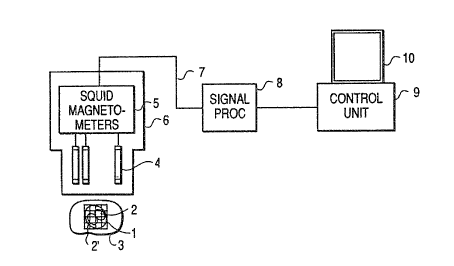
19 FIG. 2 is a ~che~atic diagra~ of a pr~ferred e~bodi~ent of
the present invention, illustrating z bio~dical magnetism
21 measuring apparatus ~or ~ti~ating the po~ition of current
22 source~ or dipoles 2 ~ithin an area o~ electrical activity 2'
23 based on th~ mea~urad ~agnet$c ~ield of the heart or oth~r
24 current producing 30urce. In accordanc~ with ~ha invention, a
magnetic field g~nerated by c~rrent ~ourc~ in a living b~dy 3 is
26 sensed ~y pickup coils 4 which generat~ ~agn~ic signal~ which
~J ~
1 are th~n converted to proportional electrical ~ignals or sensing
2 signals 7 by SQUID magnetometers 5. The pickup coils 4 and SQUID
3 magnetometer3 5 together form a sen~ing unit 6. The proportional
4 electrical signal 7 output by the SQUID magnetometers 5 are
processed by a signal processor 8 which processes th~
6 proportional electrical signals 7 by performing the inverse
7 estimation and produce~ display data as an output. A control
8 unit 9 r2ceives the di~play data and provide3 an output signal
9 which is used to drive a di~play 10.
A~ illustrated by the block dia~ra~ of FI~. 3, th~
11 proportional el~ctrical signals 7 fro~ the SQUID ~agnetometers 5
12 are multiplexsd on a tim~ ~haring b~si~ by a ~ultiplexer 11.
13 Each signal 7 is then convert~d to a digital ~ignal by an
14 analog/digital converter 12, an~ th~ valu2 of each channel i~
then applisd to a buffar 13. ThQ value~ for the coefficients of
16 the matrix A (Equation (1)) whiçh ar~ d~te~ ine~ by the shape and
17 spacing of th~ pickup coil~ 4 and the initial grid point
18 arrange~ent, ars previou31y ~torQd in a ~emory 13', and
19 generaliz~d invers~ matrix A~ i3 co~put~d by th~ nor~al equations
or singular value d~co~po~ition of Equations (6) and (8) in an
21 arithmatic unit 14 b~se~ on ~uch stored valu~, with the results
22 being stor~d in th~ ~s~ory 13'. In th~ preferr~d ~ ho~i~ent, the
23 arithmatic unit 14 i~ a T~8 320-s~ri~ digital aign~l processor.
24 One software package which can b~ us~d ~or singular value
decomposition is thQ so-called EISPA~ so~twar~ distribut0d by
- 16 -
25307-296
~,i33~3~
IUa~onal En~rgy So.~warc Cen-~r of Argonn~, Illinois and IMSr, of ~ou5ton, Te~u which
2 performs Equation~ (~) and (8). The general use of normal
3 equations and sin~ular value decomposition is described in Golub,
4 Golu~ e~ al., 'Singular Valu~ Dc .,~ it;-;. and l,cas~ Squa~ Jions~ Nu~ . Ma~h, 14, pp~.
4~3~20 (1970).
6 The arithmetic unit 14 conv~rts the estimated current dipole
7 distribution to image data based on th~ value~ obtained by
8 inverse estimation ~rom the magnetic measurement3, and these data
9 are then stored in a graphics buffer 15 in the control unit 9. A
C~T-controller 16 then control3 operation o~ the CRT 10 to
11 display the image data. The ov~rall operation o~ the arithmetic
12 unit 14, graphic~ buffer 15 and CRT controll~r 16 i~ controlled
13 by a master CPU 18. Any standard graphic~ ~ackage can be
14 employed as the control unit 9 in combination with a compatible
CRT 10. Th~ CRT 10 in addition to displaying ths position of tha
16 dipoles i also capa~le of displaying the amplitude or strength
17 of the dipoles by showing v~riation~ in ~ize, color, hu~,
18 brightnes~ or thres-~i ?~ional illustration.
19 FIG. 4 i~ a flowchart for describing thQ operation of th~
circuit of FIG. 3, including the in~erse estimation algorith~. A
21 grid width and an ar~a ~or estimation o~ currant dipoles are
22 initiali2ed to cover th~ region of interest (S20). The
23 coe~ficiGn~ ma~ix A i~ co~pu~ed ~a~ed on ~h~ position~ o~ th~
24 pickup coil~ 4 and dipole grid point ~S21). Then, th~ magnetic
field i3 measurQd (S22a) and a ganaralized inverse matrix A~ is
- 17 -
5,~. ~ r,~
1 obtained from the normal equations or sinqular value
2 decomposition of Equation (6) and (8) (S22b). An initial
3 estimated distribution Q0 is obtainPd by multiplyinq magnetic
4 field distribu~ion B~ measured in step S22a by the inverse matrix
A~ ~S23). If the grid interval or spacing doe~ not reach the
6 desired resolution (S24), then th~ width and area o~ tha grid are
7 changed (i.e., r~duced) b~sed on the initial estimated value Q0
8 (S25). Step~ S21-S25 are rep2atecl until the theoretically
9 limited resolution or desired accuracy i~ obtained. ThQ
di~tribution Q0 o~ th~ current dipoles finally obtained i~
11 displayed ~n th~ display unit 10 (S263 to show tha po ition and
12 strength o~ multiple dipole~.
13 FIGS. 5(a), 5(b), 5(c), 6(a), 6(b3 and 6tC) illu~trate the
14 op~ration of a praferred ~hodi~ent of the pre~ent invention
where th~ esti~ated current dipole grid 1 is set in tha form of a
16 plan~, and current sourc~ normal to thQ plane (y-component) are
1~ ~stimatad by 3inqu1ar value decomposition. In the cas~ where the
18 s~nsor~ or pickup coil. 4 ar~ arrang~d lin¢arly and measure the
19 normal (z-component) of th~ magn4tic field for position
estimation for two or ~or~ curr2nt dipol~ 19 on ths grid 1 as
21 illu~tr~t~d $n FIG. 5(a), a recon~truction i~ag~ 20 o~ a paix of
22 unresolvQd ~ dipoleR 19 can ba ob~ainsd a~ shown in FIG.
23 6(a) by executing curr~nt source po~ition e~ti~ation with a rough
24 or coarsQ setting width for th~ area wher~ current dipole ~ay
exist. FIG. 6~a) i5 a graphical display for better visualizing
26 the position and amplitud~ o~ ~h~ curren~ dipolas 19.
- 18 -
~ ~ r~ r
1 A reconstruction image 20' of two separated current dipole~
2 19 as shown in FIG. 6(b) can be obtained by e~timating, as shown
3 in FIG. 5 (b), the current dipole position with a grid 1', having
4 a half-grid width for the m~asured data . Moreover, as shown in
FIG. 5(c), a reconstruction image 20~ having twice the resolution
6 can be obtainad, as shown in FIG. 6(c), by reducing the width of
7 the current dipole estimation grid to half that shown in FIG.
3 5(b~.
9 In FIGS. 5(a)-5(c), the x axi~ in th~ ~igure indicates the
horizontal direction, while the z axis indicate~ the vertical
11 direction and th~ y axis indi~ates the dop~h direction into he
12 paper. Moreover, the grid 5~tting interval i3 reduced in th~
13 sequence of 10 ~ (FIG. 5(a)), 5 mm tFI~, 5(b~), 2.5 ~ (FI~.
14 5(c)) and po~ition~ of tha current dipole~ 19 are ~t to ~0.005,
0.0, 0.01) and (-0.005, o.no, o.ol) con~idering the center
16 position o~ ~h~ s~nsvrs 4 a3 tha origin. Th~ sen~ors 4 ar~
17 arranged in such a mann~r that 128 3en30rs are arranged at equal
18 intervals within a total width of 80 ~m. Morsover, the number of
19 current dipol2 grid point3 is ~ixed to 64 points (8 x 8) although
~h~ numb~r can ~ varied in accordanca with the ~ircumstanceR of
21 a particular u~.
2i In FIGS. 5(a), 5(b) and 5(c), tha current dipol~3 19 exist
23 on grid point~ of th~ ~ti~ated grid 1. ~owevar, i~ a current
24 dipole 19 doQs not sxist on a grid point a~ ~hown in FIG~ 7(a~,
and if reconstruction is carrisd out u~ing all ~ingular value~
'' ' :;; . ~ ~ '
:
,: :
1 A = (~ ,) (10)
2 up to ~he rank r of th~ coefficient matrix A, the influenc~ of
3 the current dipol~ 19 not existing on the grid becomes large as
4 shown by the graphical display of the reconstruction in FIGo
8(a)0 This reconstruction contains gross artifa~ts because the
6 model is not obeyed. Therefore, reconstruction is pPrformed by
7 accumulating the laxger singular valu~ sequ~ntially from 1 to K
8 in accordance with the following relation
Q ~ v ~-1 T
11 i=l
12 where v; and u; are th~ column~ of U and V, r~pactively, in
13 Equation (8) and B~ i~ a mea~ured valu~. A~ shown in FIG. 8(b),
14 a reconstruction image 120' which is in~erior in resolution but
1~ more robust with respect to off-grid point 30urc~4 c~n b~
16 obtain2d by employing a Rmallsr number of the singular values.
17 The number of singular values used should b~ suf~icient to
18 display a w211 dsfined peak in the recon~tr~otion. Moreover, by
19 reducing th~ grid arQa a~ shown in FIG~ 7(b), a racon truction
imaga 120~ having a fin~r resolution grid a~ shown in FI~. 8(c)
21 can be obtainad by reducing th~ int~rval of estima~ion ~rid 1 in
22 the direction of th~ obs2rved distribution of th~ current dipole
23 known fro~ FIG. S(b). Accordingly, (1) in th~ initial estimation
24 where the coars~ grid 1 (FI~. 7(a)) i~ ~ployed, a ~11 number
~~J ~ ilJ~
1 of singular values is used for reconstruction to avoid artifacts
2 caused ~y off-grid dipoles. The probability that a current
3 dipole exists on a grid point i5 low for coarse grids. The
4 estimation i~ carried out with coarse resolution under th~
condition than an element existing~ outside the grid make~ a small
6 contribution to the measured magn~!tic field and the approximate
7 current dipole position is estima~:ed by the location of th~
8 maximum peaX in the reconstruction. Then, (2) e~timation with
9 higher accuracy can b~ r2alized on a smallsr grid 1', whilQ the
resolution of the estimat~d current dipols i9 i~proved by
11 executing th~ reconstruction over a smaller ~ized region. The
12 accumulated number o~ ~ingular values ~uitable for a given grid
13 siz~ can bs deter~ined by executing the recon~tructio~ while th~
14 number o~ ~ingular value~ i being incre~9ed, until a wQll-
d2fined peak i9 observed in th~ recon~truction. Thi~ peaX will
16 exist in the vicinity of thQ current activity and indicates the
17 location about w~ich thQ n~xt ~tage of ~agnification will occur.
18 As th~ nu~bsr o~ magnific~tion ~tep~ increase~, the artifact~ in
19 the reconstruc ion ar~ due les~ to mod~l errors (of~-grid
sources) than to errors in the measur~d ~agnetic field. At these
21 latter staga~ of ma~ni~ication, the number of qingular values i5
2i limitQd by th~ ratio of ~ignal power to m~asurement error pow~r.
23 Thi~ ratio da~ine~ a thre~hold l~vel. Singular valu2~ ~low the
24 threshold lev~l canno~ b~ u3ed in th~ r~con~truc~ion. Th~
magnification procedur~ ter~inatQs whe~ no ~urther impLuvl-~nt in
2~ localization or r~olution oc~r3~ which can b~ iden~i~ied by
- 21 -
1 noting the number of ~ingular value~ that: arQ lo~t in the
2 magnification step. For exampls, in the planar grid case, if a
3 magnification ~actor of 2 per linear dim~nsion cause~ a reduction
4 of four in the number o~ singular valuei above th~ threshold
level, no improvement in the magnified reconstruction will be
6 realized. If multiplQ sources exi~t, ~h~ magnification procedure
7 tarminate~ with the minimum grid araa that includ~s all the
8 multiple sources.
~ In FIG. 1, the estimated grid of current dipoles is set on a
plane. How~ver, tha resolution can be improv~d by reducing the
11 estimat~d grid of current dipole~ in thQ direction of th~ activ~
12 ar~a 2' of a current source, whil~ the position i8 estimated for
13 a thr~e-~i ?~aional distribution by for~ing a thr~e-dimen~ional
14 cubic shap~ of th~ estimated grid 100 of current dipole~ as shown
in FIGS. 9(a~ and 9(b~.
16 In addition, in the case o~ setting the estimation points
17 for the current source, it i~ not neca~sary to include grid
18 points where current source~ cannot exi~t. In~tead, the grid may
19 b~ set in accordance with the shape of th~ ~stimat~d area. For
instance, in th~ ca~ o~ a circular material, thQ ~stimation
21 point~ can ~ s~t on a polar coordinat~ grid 101 as shown in FIG.
22 lO(a~. In t~Q ca3~ o~ a brain, inform~tion ~or~ u~fu- ~or
23 diagno~i~ oan ~Q obtain~d ~y dst~rmining ~ grid 102 having grid
24 point.~ in accordan~a with th~ shap~ of ~h~ p~riph~ry of the brain
as shown in FIG. ~0(~. In t~ ca~ o~ the heart, a solution
26 exhibiting a ~all~r error can be obtained by settiny a grid 103
- 22 -
.
~ tJ ~
1 having grid points so as to avoid the reg:ion of the heart whPre a
2 conductive ~y~te~ doe3 not Pxi~t, such as an atrium or ventricle,
3 as shown in FIG. lO(c). Thus, as used in this application, the
4 term grid refer~ to any defined set of grid points in a region
(two-dimen~ional or three-dimen~ional). In ord~r to implem~nt
6 the specifically shaped grids such a~ grids 102 and 103, magnetic
7 resonance imaging (MRI~, dia~nostic ultrasound imaging (US~ or X-
8 ray computad -tomography (X-RAY CT~ can bs used to determine the
9 specific grid point~ which ara most suitable for achiaving high
accuracy. Further, depending on th~ grid arrange~ent sel~cted,
11 it can be desirable to position the sensor~ 4 in a non-linear
1~ arrange~nt such a~ an arc or circla.
13 There ara a nu~ber o~ way~ for di~play~ng the result~ o~
14 biomagnetic imaging for di~gnostic purpose~ in accordance with
the present inv~ntion. For example, FI&. 11 illustrates that th~
16 display 10 for~ed by a di~play devica 21 having fir~t and second
17 display ~creens 22 and 23 for displaying a non-magnified i~age
18 and a magnifie~ image of the current dipoles obtained by the
19 inVersQ estimation. Bas~d on the invQrs~ e3timation, de~ired
area ~4 on scrQen 22 can bs 3~1~cted ~or display in a magni~ied
21 state on $crsen ~3. Th~ de~ired area 24 can b~ ~lectad ~y
2~ de~ign~ting ~orner point~ 25 and 26 on ~cr~n 23 ~y using an
23 operator input d~vic~ .uch a~ a k~ybo~rd or a mou3~. Ths ~cr~ens
24 22 and 23 can be u~ed for diagno~tic purpo~e~ to view ~he
po~ition and strength o~ th~ curr~nt dipole~. Th~ ~mple dl~play
26 o~ FIG. 11 is that o~ a portion of a heart and illustrat~
ti ~ ~ 3 ~ ~3 iJ
1 currant dipole~ lg in the c~nter wall of the heart. Based on
2 this display, it is possible for a physician to determin~ whether
3 el~ctrical activity in the heart i~ normal. For example, if the
4 resolution of the select~d area 24 of screen 22 is increased, a
physician might be able to better se~ a dipole 19' loca~ed along
6 the edge of the center wall of the heart, which may indicate a
7 malfunction in th~ heart.
8 In an alternata display mode in accordance with the present
9 invention, a singl~ display 122 i3 divided or ~plit into a
plurality of di~play ar~a~ as shown in FIG. 12. Th~ divided
11 area~ are designated sequ~ntially a~ 122a, 122b, 122c and 122d
12 and are capable o~ realizing ~ucces~ively magnificd di~play3.
13 Moreover, if a current dipole l9a exi~tR outside the selected
14 area as indicated in display area 122c, a curr~nt dipole l9b
within the fra~e can b~ e~ti~ated under th~ condition that the
16 currant dipole l9a existing out~ide th~ cted area proYide~ a
17 smaller influen~-e. In particular, th~ magnetic fi~ld B~t,
18 terminated by a current dipol~ in thQ region out~ide the frame
19 can be computQd u~ing thQ Biot-Savart law a~ preprocessing for
the next inver~a e~timatio~. Then invers~ esti~ation i executed
21 using th~ ~ollowing equation for subtracting th~ out~ide magnetic
22 field fro~ th~ ~aa~ured magnetic ~ield ~:
23 ~n 3 ~ B~t (12)
24 and Bin i~ then u~ed in thQ inverse e~timation.
- 24 -
:~ ' '
~ J~ a3 ~
1 Moreover, in the case of e~t.imating a three-dimenslonal
current dipola di~tribution~ planc~r sections which are mutually
3 vertical with re~pect to each oth~r can be displayed on adjacent
4 screens 29 as shown in FIG. 13. ]?urther, a desired cross-section
S within the three-dimensional di~tribution can b~ cons~ructed by
6 providing an indicator bar 31 whioh can ba changed in height
7 within a rectangle 30 in accordance with th~ depth position o~
8 the salected plane for display at a d~9ired three-dimensional
9 ssction. Further, estimation suitable for diagnosis can be
r2alized by designating a desired area u3ing ~rame 24 (de~crib~d
11 abovQ with respect to FIG. 12) within the scre~ns 29 and
12 recon3truction will be executed to produce ~agni~ied di~play~ on
13 lower screen~ 29'.
14 If it is de~ired to display dif~arent ~agni~ied rasults over
ti~e for diffPrant selectad areas 24, th~n th~ CRT 10 can be
1~ divided into scrQen~ 32 and 33, wh~r~in screen 32 displays a
17 tomographic i~ga provided by X-ray, ultrasound or an MRI i~age
18 of a sub3ect and di~f~rent framas 24a, 24b, 24c and 24d are
19 identified for ~agnifiQd viewing to ~how th~ location of the
reconstruction grid on th~ ~RI or tom~graphic imag~. No dipole
21 are display~d on scr~n 32. In~tead, dipol~ di~play are~s 33a-
22 33d corr~pond to ~ra~ 24a-24d, r~sp~ctivaly and ar~ displayed
23 at a firqt ti~ tl. Then, at a dQ~ignat~d tim~ ~:2 (B.g., 10
24 second~ after tl), a new ~t o~ diRpl~y areas 33a' 33d' is
generated, followed by an additional set o~ di play~ at a time t~
26 in display area~ 33a'-33sl'. This typ~ of display can ba u~ed to
-- 25 --
r'J ~
l better detect change~ in the strength or po~ition o~ curren~
2 sources or dipoleg over time~
3 In accordance with another aspect of the present invention,
images produced by ~agn~tic resonance imaging (MRI) and
biomagnetic imaging (BMI or MSI) can be superimposed a~ A
6 diagnostic tool. In tni~ aspect, image data showing major
7 contours or features of an MRI image are selectPd and bio~agnetic
8 imaging data generated in the manner de cribed above, i~ employed
9 to gsnerate an image which iY superimposed on the selected
portion on the MRI imaga.
11 FIG. 15 i3 a block diagram o~ an smbodim~nt of an imaga
12 superposition system which can be u3ad to sup~rimpose MSI and ~RI
13 i~age3. Referring to FI&. 15, an input device 150 3uch as a
14 mouse iR used to select a portion of an image which is to be
displayed at a m~gnified level (i.e., similar to ar~a 24 in FIG.
16 11). A hard di3~ 152 store3 data input via the input device. An
17 MRI and MSI dat~ reader 154 is used to read graphic data fro~ an
18 MRI, a~ w~ll a3 MSI data which i~ output, ~or exa~ple, fro~ th~
19 signal processor 8 o~ FIG. 2. Th~ data rQader 154 may be part of
a local area n~twork or ~ay b~ another kind o~ data input device,
21 such a~ ~ tapa r~ad~r. A c~ntral proc~s~ing uni~ 15~ controls
22 the op~ration~ o~ th~ i~aga superposition ~y~te~ of FIGo 15~ A
23 memory 158 ~tor~ Gy5a~ for imple~enting thQ imag~
24 superpo~ition operation. A graphic ~amGry 160 store~ th~ ~RI
data and th~ MSI dat~. A C~T controller 162 r~cQive~ ths graphic
26 data which has be~n proce~ad by tha CPU 156 and provides data
- 26 -
f~ 3 ~3
1 for a superimposed display on a CRT 164. Thu~, by employing the
2 system o~ FIG. 15, overlapping displays of an MRI i~age and a
3 current source image produce by biomagnatic imaging can be shown
4 on a ingle display. Further, portions of the display can be
selected for higher resolution in the manner de~cribed above with
6 respect to FIGS. 11-14. In tha preferred embodimen~, the system
7 of FIG. 15 i3 a computer-based system capa~le of operating a
8 multiple window syste~ software package such a~ the X WINDOW
9 software produced by MIT.
FIG. lS is a flowchart illustrating the operation o~ ths
11 system of FIG. 15. R~erring to FIG. 16, MRI image data i8 first
12 r2ad (S30) and contour extraction i~ carriad out to select a
13 contour ~rom th~ MRI i~age based on a conc~ntration threshold
14 valu~ (S32). Thi~ allow~ only major contours or feature~ o~ the
MRI image to be ~elected ~or display. Next, imaga interpolation
16 is perfor~d to fill in os interpolate betwQQn the extracted data
17 point~ depending on th~ selected ar~a for~ed ba~ed on such d~ta
18 (S33). This imag~ interpolation i~ ~imilar to that used in
19 ultrasonic diagnsstic~ to fill in data for selacted area~. Next,
the magnetic field o~ th~ ~ubject i~ me~urad ~S34) and
21 reconstruction is perfo~d to producs i~q~ data for displaying
22 the po~ition~ of current sourc~s by inversa e-~timation b~ed on
23 the measur~d ~agn~tic field data ~ (S353. MSI image data i~
24 generated to illu~trate intensity ba~ed on brightnes~ or hue
25 (S36). The MSI imag6~ data obtain~d in S3S and the MRI image data
26 obtained in S33 ara combined (S37) and ar~ displayed on the
-- 27 --
~o~J~ 'g~ '3
1 display 164 (S38). Thus, an overlapping image combining ~n M~I
2 image and an e3timated current dipole or MSI image can be
3 displayed concurrently. Further, if hi~her resolu~ion i~
4 desir~d, t~e area of the grid can b~ changed tS39) and th~
inverse matrix A recalculated (S40j 50 a~ to produce a new
6 display of higher resolution. It should b~ notad that when the
7 grid area is changed, it is required to again interpolat~ the
8 image based on the 8xtracted data for the MRI i~age (S33).
9 FIG. 17 i~ an altarn~tiv~ h~rdware em~odiment for achieving
the processing illustrated in FIG. 16. Re~erring to FIG. 17, a
11 magnetic field mea~uring circuit Z34 provid~s mea~red data which
12 i~ conv~rted to a current dipol~ den~ity distribution by an
13 invers~ eS~timation circuit or recon~tructing circuit 235. An
14 image data generating circuit~ 236 conv2rt~ the curr~nt dipole
den~ity di~tribution ts a contra~t imag~ which i~ provided to
16 imag~ com~ining circuits 251 and 253. ~eanwhile, th~ coordinate~
17 tX1~ yl), (x2, y2) of a magnifiad area or framQ (~imilar to frame
18 24) are obtainsd from count~rs 239 and 240 which are 3ynchronized
19 with the po~ition o~ a cur30r on th~ i~ag~ controlled by an inpu~
devic~ 238, such a3 a mOU~Q. Th~ output~ of the coun~er~ 239 and
21 240 ar~ proYided to an inver3~ matrix arith~etic circuit 237
22 which con~rol~ ~h~ recon~ructing circuit 235, a~d to a
23 magni~ication co~fficien~ arithmatic circuit 241 which provide~ a
24 magnification co~fficient Na~ to a ~ultiplier 243. An MRI image
reader 245 provide~ MRI image data to ~ contour extracting
26 circuit 246 whi.ch extract~ ~elected MRI image d~ta and provides
2~ -
, J ~
1 the selected MRI imag~ data to an image data generator 247.
2 counter 242 is used to generate an address to be used for image
3 interpolation and to addr~ss an image me~ory 248. In addition,
4 counter 242 provides an output to control the output o~ image
data generator 247. The counter 242 also provide~ an output to a
6 multiplier 243 which multiplies th~ magnification coe~icient "a"
7 by the value of counter 242 to provide an offset value to an
8 adder 244. Th2 output o~ the adder 244 i~ u~ed to addres~ th~
9 image m~mory 250 to identify the addre~ o~ the magnified image
in the image memory 250. The addre~sed image data of image
11 m2mory 248 ia s~nt to ~n imdge m~mory 250 via an interpolating
12 circuit 249 in which a vacant imag~ data ar~a ganerated by
13 magnification i~ filled in with int~rpolation data generatQd by
14 the interpolation circuit 249. Th~ ~xtraction o~ contour data by
contour extracting circuit 246 befor~ the ~RI image data enter~
16 tAe ima~e memory 248 i~ perform~d in ord~r to clarify th~
17 difference of image~ batween re3pectiv~ ~ia~ue~ and image~
18 d~pending on th~ co~bination o~ contrast change~ in the current
19 dipole. A ~agnified i~ag2 ~tored in thz i~age ~e~ory 250 and
non-magnifiQd i~aga data fro~ im~q~ ~e~ory 248 are sent to the
21 image co~b~ning circuit~ 25~ and 253 in ord~r to generata image
22 data which indicate~ the distri~ution of thQ ~RI image at the
23 time of ~agnification. Thi~ image data i~ co~bined with the
24 image data provided by th~ i~age d~t~ g~nerator 236 in the image
co~bining circuits 251 and 253 and the combined images are stored
26 in tha image or graphic me~o~ies 2S2 and 254 and ar~
25307-296
1 simultaneouqly displayed on the screen of a display 255. Thus,
2 the current dipole can be estimated while the de~ired area is
3 magnified.
4 To illustrate the operation of the subjQct invention,
simulations were conducted u~ing an 8 x 8 grid (128 unknowns)
6 situated perpendicular to a square planar array of 256 sensors
7 measuring the Z-component of the magnetic field. Simulations
8 were performed by u ing eith r a ~ingla randomly-plaoed dipole,
9 to test for location accuracy, or a pair o~ dipoles havin~ random
location, spacing and orientatisn to investigate the resolution
11 capabilities of the procedure. Magnetic measure~ents were
12 computed by uqing th~ Biot-Savart Law and a rando~ numb~r was
13 add~d to the ~ea3uraments to simul~ts th~ effect3 of a give~
14 signal-to-nois~ ratio. In cases whar~ a r~gion of activity wa~
observed within tha reconstruction r~gion, the 64 element qrid
16 area wa~ r~duced to 25S, that is, a 50~ reduction in each
17 dim2n~ion and ~hifted ~o that the -~i of th~ obs~rved
18 recon~truction fell near the center of tha reduced grid area.
19 The reconstruction wa~ then repeated u~ing thQ naw grid points~
The procedure ter~inated when no further improvsment in
21 resolution was ~chievedO The ~ingle dipole re ult~ using both
2~ simulat~d and r~al d~ta wera co~parad ~ith th~ dipol~
23 localization proc~dure u~ing ths ~rquardt algorith~ describ~d by
24 Rdda~u a al., En5 3 a~ Mchod~ , Jo~ Wiky ~ So~, ~cw
2 5 Yo* (1983)
-- 30 --
1 In the ~n ~h~ dipole experiments, a singlQ dipole 2 cm long,
2 was constructed and driv~n with a peak cu:rrent of 50 mA, to
3 produce Q - 103 A-m. The ~ingle dipole was placed at variou~
4 locations in a ~aline solution within a cubic container. The
magnetic field flux normal to the surface of the container was
6 measured with a one centimeter di,~meter coil having 100 turn~,
7 which was translated to simulate ;~ 15 x 15 ~en~or array ~225
8 sensors). The coil was attached 'tG a preamplifier fe~ding a
9 Biomation 8100 A/D convertQr. Di~ferent noise l~vel~ in thQ
measurement3 were obtained by averaging th~ appropriat~ num~er of
ll signals over time. These mea ur~ment~ were appli~d to the same
12 progra~ u~e~ in the si~ulation~. The sen~itivitie~ of thi~
13 approach ar~ describcd in terms of tha signal-to-noise ratio in
14 the measursm~nt~ and the depth of ths dipole(s) balow the sensor
plane. The ~ingl~ dipol~ results using the 6yRte~ of the present
16 invention wer~ comparable to thos~ obtained u~ing the Marquardt
17 algorithm. ~s th~ depth of th~ dipol~ incr~a~ed or the SNR
18 decr2asad, the loc~lization accuracy dacreased.
19 As explain~d pr~viou~ly9 according to thQ pres~nt invention,
th~ po~ition of current ~ource~ can b~ ~ti~a~ed with high
21 accuracy u~ng a s~ r nu~er of pickup coils fro~ the magnetic
2i fi~ld g~nQratad in a living body. Mor~ov~r, th~ ~zoo~ ~achnique
23 of th~ pr~ent invention allow~ a de~ir3d araa to ba zoom~d in
24 on, and ~agn$fied on th~ di~play ~cr~n. Th~r~fore, the present
inv~ntion i~ usuful for e~timating ths po~ition~ o~ ragion3
- 31 -
1 diseased by brain malfunction~ myocardial infarction or irregular
2 pulse and provides a significant improvement in the fi~ld o~
3 biomedical magneti3~ measuring apparatus. Further, the methsd
4 and apparatus of thQ present invention have applicability to any
field in which it is desired to generate an image of current
6 sources within a particular living body, subject or material.
7 ~ The foregoing de~cribe~ th2 preferred r hodl ents of the
8 pressnt invention and is con~idered illustrative of the
9 principle3 of the present invention. Further, since numerous
modification~ and changes will readily occur to tho~e skilled in
11 the art, it i3 not desired to li~it the invention to the exact
12 con~truction and applications ~hown and describ~d, and
13 accordingly, all ~uitable modification~ and equivalentc ~ay be
l~ rasort~d to, falling within th~ scop~ o~ thQ inv~ntion in the
appended clai~ and their equivalents.
- 32 -