Note: Descriptions are shown in the official language in which they were submitted.
' 20777~9
REAGENT COMPOSITIONS AND THEIR USE IN THE IDENTIFICATION AND
CHARACTERIZATION OF RETICULOCYTES IN WHOLE BLOOD
BACKGROUND OF THE INv~:NLION
1. Field of the Invention
The present invention relates to reagent compositions and
their use in identifying and characterizing cells in samples of
whole blood, and more particularly to reagent compositions and
their use in (i) identifying reticulocytes; and tii) simultane-
ously measuring the volume, hemoglobin concentration and
hemoglobin content of large numbers of individual reticulocytes
and erythrocytes, in a whole blood sample by light scatter and
flourescence flow cytometry techniques.
2. DescriPtion of the Prior Art
In all the higher animals, blood consists of an aqueous
fluid part (the plasma) in-which are suspended corpuscles of
various kinds: the red blood cells (erythrocytes), the white
blood cells (leukocytes) and the blood platelets. Plasma has a
composition comprising roughly 90% water, 9% protein, 0.9%
salts and traces of other materials such as sugar, urea, uric
acid and the like.
MS-1716
2077789
The cells or corpuscles of the peripheral blood (i.e. the
blood outside the bone marrow) are divided into two main groups:
erythrocytes, whose primary object is to transport osygen and
leukocytes, whose primary functions relate to the immune system
and the destruction of materials foreign to the body. In addi-
tion to these two main groups, the blood also contains the so-
called blood platelets which are important in hemostasis.
The final stages of erythrocyte maturation occur after their
release from the bone marrow while these cells are circulating
in the peripheral blood. These young red cells, or ~reticulo-
cytes~, have lost their nucleus, and thus, their ability to
divide or to synthesize ribonucleic acid (RNA). Although these
functions have ceased, reticulocytes are still metabolically
active and are capable of synthesizing protein, taking up iron
for the synthesis of heme, and carrying out the necessary
metabolic reactions required to maintain an energy-rich state.
These cells are usually most easily distinguished from mature
erythrocytes by esposing them to solutions of cationic dyes
which react with the anionic RNA in the reticulocytes and
precipitate into a fine or coarse stained ~reticulum~ within
the reticulocytes, which gives the reticulocytes their name.
Although reticulocytes normally comprise about 0.5 to 2
percent of the total red blood cell population, this percentage
can change dramatically under abnormal conditions. For esample,
MS-1716
- 2077789
reticulocyte counts have been used for many years as a diag-
nostic aid in studying blood dyscrasias, as an inde~ of red
blood cell regeneration following hemorrhage, as well as for
monitoring early toxicity in chemotherapy of certain malignant
diseases.
Nucleic acids (RNA and DNA) are polyanions which can be
stained with practically any cationic dye. The RNA in reticulo-
cytes can be stained with only a few cationic dyes [including
Brilliant Cresyl Blue (BCG), New Methylene Blue (NMB), Auramine
O (AuO), Acridine Orange (AO), Thiazole Orange (TO) and Pyronine
Y (PY)]. Among these dyes, only a sub-set can be made to pene-
trate the cells (and therefore stain) rapidly. The sub-set
includes NMB and AO. The rate of, and degree of staining of
reticulocytes depends upon the extracellular concentration of
the dye, the rate of penetration of the dye through the reticu-
locyte membrane, and the strength of the specific binding
constant between the cationic dye and the reticulocyte RNA.
The latter two properties are different, and not easily
predictable, for each dye, so that trial and error are
necessary to discover useful reticulocyte stains. Not all
cationic substances are capable of penetrating intact red cells
(and reticulocyte) membranes, and the nature of the anions
which necessarily accompany the cations, can effect whether or
not the cationic substance penetrates rapidly, slowly or not at
all. Hydrophobic molecules generally penetrate red cell
MS-1716
20777~9
membranes faster than hydrophilic molecules, and small molecules
generally penetrate membranes faster than large molecules. Only
a sub-set of salts or buffers mised with those cationic dyes
which can stain reticulocytes permit rapid staining; that is the
~riqht~ dye with the ~wrong~ buffer can take ~forever~ to stain
reticulocytes. Again, trial and error are necessary to discover
useful formulations of reticulocyte staining mistures. Thus,
despite various ~rules~ which can be used as guides, it is not
yet possible to predict, a priori, whether, and under which
conditions any particular cationic dye may rapidly penetrate
and stain reticulocytes.
The fundamental concept of flow cytometry is essentially
the passing of cells, one at a time, through a specific sensing
region. Typically, by means of hydrodynamic focusing, single
cells are passed through the sensing zone, which consists of a
focused light source and a detection system for the measurement
of scattered, absorbed or fluorescent light. The effect a
particle has on the light it intercepts can be detected in a
number of ways. In general, the particle has a refractive
inde~ which is different than that of the medium in which it is
suspended. It will therefore scatter light with which it is
illuminated through a range of angles, and with varying
intensities, that depend upon that refractive indes difference,
the particle's size, its shape and any internal variations in
MS-1716
2~777~9
refractive indes and structure as well as upon the wavelength
of the illuminating light. (For homogeneous spheres, Mie
Scattering Theory provides a complete description of the
distribution and intensities of scattered light.) A particle
may also absorb some of the incident light. In the latter
case, a portion of the absorbed light may be reemitted as
fluorescence, typically at a longer wavelength than the
wavelength of the absorbed light.
These and other effects can be measured with light
detectors arranged to measure different angular intervals of
scattered light, of unscattered light and of fluorescent light.
When particles are as small as cells, typically less than
15 micrometers in diameter, the numbers of photons in the
illuminating beam affected by their passage at high speed
(typically hundreds to thousands of widely-spaced cells per
second), and especially compared to the number of photons per
second falling on the illuminated part of the suspension
stream, tand compared to the background illumination of an
absorption detector (and even a fluorescence detector)] can be
very small. Therefore, the limits of sensitivity of detection
of small particular differences between particles depends
critically on the photon flus (which depends at least on the
intrinsic ~brightness~ of the light source) and how large the
MS-1716
~ 2 0 7 7 7 ~ ~
perturbations of the ~hoton flu~ are that are produced by other
small and large differences between particles.
The main sources of interfering noise in absorption, scatter
and fluorescence flow cytometry signals can be quite different
for each kind of signal. To a first order appro~imation, the
magnitudes of fluorescence signals from stained or unstained
cells are almost uninfluenced by shape or orientation of the
cells from which the signals arise, whereas scatter and absorp-
tion signals are ~ery strongly influenced by shape and orienta-
tion. As an estreme e~ample, the nati~e biconca~e shape of
human erythrocytes has a profound effect on the absorption and
scatter signals they generate; effects larger than the small
absorption signals of typical classically stained reticulocytes.
This is the main reason why, prior to the present co-pending
invention described in Canadian application No. 2,077,788
entitled ~Reagent Compositions and Their Use in the
Identification and Characterization of Reticulocytes in Whole
Blood~ filed concurrently herewith and assigned to the assignees
of ~he present in~ention, absorption flow cytometry methods
ha~e not been useful for reticulocyte counting or generally for
the measurement of low concentrations of a~sorbing molecules in
cells. On the other hand, weakly fluorescence materials in
cells or (for e~ample, unbound fluorescent dyes) in their
MS-1il6
, ),
~ 207778 ~
surrounding medium has virtually no effect on absorption or
scatter signals.
But in fluorescence flow cytometry, when the light source
is sufficiently intense, and stray light and system fluorescence
have been minimized, the limit of sensitivity for the discrimi-
nation of selectively stained cells from unstained cells is set
by the relative magnitude of the apparent ~background
fluorescence~ from the components within the unstained cell
and/or the level of fluorescence of the medium in the sample
stream surrounding the stained and unstained cells. With
reticulocyte stains, there is always some equilibrium concentra-
tion of dye in solution in the sample stream surrounding the
cells. For a given level of binding to reticulum RNA, the ratio
of e~ternal dye to reticulum-bound dye decreases as the specific
binding constant of the dye to RNA increases. Therefore, the
higher the binding constant, the lower the necessary e~ternal
dye concentration, the lower the ~background~ signal and the
higher the signal to noise. Also, if the fluorescence effi-
ciency of the unbound dye is lower than the fluorescence effi-
ciency of the reticulum-bound dye, improved signal to noise is
achieved. In fact, the methods which utilize AuO, TO and
Thioflavine*T, depend upon differences in bound and unbound
fluorescence efficiency. The preferred methods described
hereinafter, however, depend mainly on high specific binding
*Trade-mark
MS-1716
D
20~77~
constants for the high signal to noise achieved, and in one
case, the lower non-specific fluorescence of unstained cells at
long (red) wavelengths.
Several semi-automated methods are available which can be
used for counting the percentage of reticulocytes in an anti-
coagulated sample of whole blood. In each of the esisting
methods, a diluent containing an organic cationic dye, such as
AO, AuO or TO, is used to stain the RNA within the reticulo-
cytes. The dye penetrates the cell membrane and binds to the
RNA and usually precipitates a ~reticulum~ within each
reticulocyte. The amount of the signal from stained RNA is
roughly proportional to the RNA content. After proper staining,
a fluorescence flow cytometer, eguipped with the proper
escitation light source (typically an argon ion laser emitting
at 488 nm), and emission detection system, can be used to
determine the percentage of reticulocytes in the effluent.
Illustrative methods for differentiating reticulocytes in
whole blood samples using fluorescent dyes and flow cytometric
methods are disclosed in the patent literature.
For e~ample, U.S. Patent No. 3,684,377 to Adams and
Kamentsky discloses a dye composition for differential blood
analysis including an aqueous solution of AO, and having a pH
MS-1716
~778g
factor and osmolality within normal physiological ranges for
human blood. The dye composition can be used for counting
reticulocytes by measuring the presence or absence of a
fluorescence signal with an erythrocyte scatter siqnal.
U.S. Patent No. 3,883,247 to Adams discloses a similar
method to that of Adams and Kamentsky using a dye composition
including AO having a concentration of between 10 6 and
10-5 grams per ml.
U.S. Patent No. 4,336,029 to Natale discloses a reagent
composition comprising an aqueous solution of the dye AO,
citrate ion and paraformaldehyde at a pH of about 7.4 and an
isotonic osmolality. The concentrations of the various
ingredients were selected to masimize dye uptake of the
reticulocytes and platelets, and provided for dye uptake to be
achieved within 2-5 minutes of mising the blood sample and
reagent composition. An automated method for detection of
platelets and reticulocytes utilizing the Natale reagent is
disclosed in U.S. Patent No. 4,325,706 to Gershman, et al.
In the reagent disclosed in U.S. Patent No. 4,707,451 to
Sage, Jr., reticulocytes are stained with thioflavin T or
chrysaniline. A whole blood sample was found to be effectively
stained by mising a 25 ~1 aliquot of the dye in an isotonic
MS-1716
-
20~77~9
saline solution (0.2 mg/ml) with 10 ~1 of anticoagulated whole
blood with the mi~ture incubated for about 7 minutes.
U.S. Patent No. 4,883,867 to Lee, et al. discloses a dye
composition for staining RNA or DNA. The staining composition
includes TO as the preferred dye compound. The reticulocytes
are stained in a minimum time of 30 minutes.
A reagent for reticulocyte counting of flow cytometric
techniques is described in U.S. Patent No. 4,971,917 to Kuroda
which contains a carbonate salt to reduce the non-specific
staining of the mature erythrocytes by the dye, e.g. AuO, to
prevent the mature erythrocytes- from being erroneously counted
as reticulocytes when analyzed by fluorescence flow cytometry.
U.S. Patent No. 4,981,803 describes a reagent for reticulo-
cyte counting which comprises two solutions, namely a stock
solution for staining in which a dye AuO is dissolved in a
non-aqueous solvent and a puffer solution which satisfies the
optimum staining conditions.
Another reticulocyte staining reagent for fluorescence flow
cytometric techniques including AuO is disclosed in U.S. Patent
No. 4,985,176 to Kuroda, et al. This reference teaches an
--10--
MS-1716
~ ~ ~ 7 7 7 8 ~
incubation time of the reagent and sample of anywhere between
30 seconds and 20 minutes.
As noted abo~e, only a small sub-set of cationic dyes
selectively stain reticulocytes, and only a smaller sub-set of
these penetrate reticulocytes rapidly. The cationic dye
compounds of the present in~ention stain the reticulocytes in
less than 5 minutes so that reticulocyte analysis by flow
cytometry can be'performed shortly after the blood sample and
the reagent composition are mised together, thus making the
present invention readily adaptable for automated procedures.
Quaternized AO derivati~es for ~uantitating reticulocytes
are described in copending C~n~;an patent application
No. 2,024,166 entitled "Compounds and Reagent
Compositions and Their Use in the Quantitative Deter-
mination of Reticulocytes in Whole Blood". The Fan, et al.
reagent contains 10 6 gram per ml of an AO deri~ative in a
buffer solution including paraformaldehyde and potassium
osalate. This reagent composition stains reticulocytes to
enable the ~uantitative fluorescence flow cytometric analysis
of reticulocytes in a blood sample. Neither this reagent nor
any of the abo~e-mentioned reagents contain a sphering agent to
pre~ent orientational noise problems as discussed below, and
--11--
MS-1716
. 3
2077~9
neither permit simultaneous determination of other diagnos-
tically significant parameters such as volume and hemoglobin
concentration of the reticulocytes and erythrocytes on a
cell-by-cell basis.
Shapiro and Stevens disclose the use of Osazine 750 for the
determination of DNA content by flow cytometry in Flow CYtometrY
of DNA Content-Usinq Osazine 750 or Related Laser DYes With 633
nm Escitation, Cytometry, Vol. 7, pp. 107-110 ~1986). The cells
are stained by 10 yM to 30 ~ of Osazine 750, and are fised by
the addition of ethanol for the DNA determination. Shapiro and
Stevens claim that Osazine 750 does not appear to stain RNA
within the cells. Moreover, such protocols with Osazine 750 do
not permit reticulocyte counting or simultaneous determination
of other diagnostically significant red blood cell parameters
such as volume and hemoglobin concentration on a cell-by-cell
basis.
As mentioned above, human and many other mammalian red
blood cells have the shape of biconcave disks. The amount of
light scattered by such asymmetric red blood cells varies with
the orientation of the cell. Accordingly, two identical red
blood cells will generate very different scattered light
signals as they pass through the sensing zone unless their
orientations in the zone are identical. Two red blood cells
MS-1716
7 ~ 7 8 ~
which are identical, escept for the presence in one of a small
amount of stained reticulum, generally produce large signal
differences on scattered light detectors because of their
different orientations.
U.S. Patent Nos. 4,575,490 and 4,412,004 to Rim and Ornstein
teach a method for the elimination of orientational noise in the
measurement of the volume of red blood cells in a flow cyto-
meter. Their method involves isovolumetric sphering of
unstained red blood cells to eliminate any orientational diffe-
rences between the cells to permit more precise and accurate
measurement of cell volume. Each red blood cell is converted
from a biconcave shape to a perfect sphere by a surfactant
sphering agent. A ~buffering~ protein and/or an aldehyde
fi~ing agent are used with the sphering agent to prevent lysis
of the erythrocytes. The anionic surfactants described by Kim
and Ornstein cannot be used with reticulocyte stains because
they have been found to react rapidly with and precipitate the
cationic dyes used to stain and precipitate t~he reticulum.
U.S. Patent No. 4,735,504 to Tycko dïscloses the red blood
cell channel of the TEC~NICON H l system, a flow cytometer
which provides a fully automated method and means for deter-
mining the individual and mean erythrocyte volumes (MCV), and
individual and mean corpuscular hemoglobin concentrations
* Trade-mark -13-
2Q77789
(MCHC) of the erythrocytes in an anticoagulated whole blood
sample. In this method, the red blood cells in a two microliter
aliquot of a whole blood sample are first diluted, and then
isovolumetrically sphered using the Kim and Ornstein method
just described. After a twenty second incubation period, these
cells are passed, essentially one at a time, through the
illuminated measurement zone within the red cell channel of the
analyzer. The-magnitude of the light scattered by these cells
into two separate angular intervals is measured. The choice of
light source and detection angles are critical in this applica-
tion. When the light source is a helium neon laser, which
emits light at 633 nm, the two scattered light collection angle
intervals are two to three degrees (2~-3~) and five to
fifteen (5~-15~) degrees. Once the level of the scattered
light in each interval is known for a cell, the volume and
hemoglobin concentration for that cell are determined by
comparison with values predicted by Mie scattering theory. The
volume (V)-and hemoglobin concentration (HC) for each cell are
stored in memory, and the MCV and MCHC are calculated at the
completion of the sample measurement cycle by techniques known
in the art as discussed in Tycko. The V and HC distribution
cytogram and the V and HC histograms are produced using these
calculations.
MS-1716
2~777g9
Neither of the above methods distinguishes between reticulo-
cytes and non-reticulocytes, and the methods as previously
described and practiced cannot be used to determine separately,
the diagnostically significant parameters of the reticulocytes
and erythrocytes such as volume and hemoglobin concentration on
a cell-by-cell basis.
Another difficulty in monitoring reticulocyte counts with a
flow cytometer is difficulty in differentiating between reticu-
locyte detection signals, mature red blood cell signals, and
system noise. The stained strands of RNA are numerous in young
reticulocytes, and generate signals of relative large magnitude
when detected by a flow cytometer. However, more mature cells
contain less stained RNA, and generate smaller signals which may
be masked by the noise of the flow cytometer measuring system.
There e~ists a need for methods and reagents useful for
identifying reticulocytes and simultaneously measuring
separately the volume, hemoglobin concentration and hemoglobin
content of reticulocytes and erythrocytes in a whole blood
sample by light scatter and absorption or fluorescence flow
cytometry techniques.
We started with the premise that we wanted to use a cationic
dye in a variant of well-known art to stain the reticulum. We
-15-
MS-1716
2~77S9
were also interested in developing flow cytometric methods which
could utilize fluorescence to detect reticulocytes. We also
hoped, by using isovolumetric sphering and the aforenoted
methods of Tycko, that for a fluorescence method, we would be
able to simultaneously measure reticulocyte and mature red cell
volume and hemoglobin on a cell-by-cell basis using a reagent
which also selectively stained reticulocytes. (Note, if the
sphering is complete, not isovolumetric, but some known factor
X of isotonicity, using Tycko's method with a correction by l/X
for volume and a correction by X for protein, e.g. hemoglobin,
concentration, original values can be calculated.)
To utilize Tycko's method, a light source which emits mono-
chromatic light in a region where hemoglobin is very transparent
is required; typically a light source like a red helium neon
(HeNe) laser, or a laser with even longer wavelength. This
means that if that wavelength is also to be used for the absorp-
tion measurement, the dye must be a blue dye with a strong
absorption of red light.
If the same dye is to be used for fluorescence, it must
have a reasonably high quantum efficiency and Stoke's shift, as
well as a high binding constant, so that it can be used at a
low enough concentration so that stream fluorescence will not
unacceptably degrade fluorescence signal to noise ratio. It was
MS-1716
~ ~ ~ 7 7 78 ~
discovered that O~azine 750 not only satisfied the requirements
for an absorption~scatter method, but also for a fluorescence~
scatter method for determining reticulocyte RNA concentration,
cell count and mature and reticulocyte cell volume and
hemoglobin content on a cell-by-cell basis. In contrast, NMB,
which has a very low fluorescence efficiency, fails for such a
fluorescence/scatter method.
Alternatively, at added cost and comple~ity, using a dye to
stain reticulocytes, which fluoresces green to red when escited
with blue light, such as AO, AOEOH, TO, AuO, by adding an argon
ion or HeCd laser with a 488 nm or 441 nm emission line, and
used to illuminate the flow cell coaxially with the red laser,
a method is implemented which also permits simultaneous deter-
mination of reticulocyte RNA concentration, cell count and
mature and reticulocyte cell volume and hemoglobin content on a
cell-by-cell basis.
We e~plored non-ionic, cationic and zwitterionic surfactants
for compatibility with cationic dyes, and as red cell sphering
agents as would be suggested by the teaching of Kim and
Ornstein. As in the Kim and Ornstein method, we used a protein
(typically bovine serum albumin) to ~buffer~ the concentration
of the surfactants to slow down red cell lysis. A number of
such surfactants (e.g. Triton X100 and Laurylpropylamidobetaine)
* Trade-mark -17-
D
~ 2 ~ 7 7 7 ~ ~
wor~ed satisfactorily. We then inadvertently discovered that
Laurylpropylamidobetaine and some other zwitterionic surfactants
(e.g. DAPS and TDAPS) did not require protein buffering to delay
red cell lysis, and are ideal alternate sphering agents for all
kinds of blood cells for the methods of Kim and Ornstein.
Because they do not require protein buffering, they permit a
stable and simpler reagent to be manufactured. ~The fi~ing
steps of Kim and Ornstein are no longer obligatory; alternately,
the problems of bacterial growth in protein-containing reagents
is also avoided.) This invention is the subject of co-pending
Canadian application No. 2,077,790 entitled "Reagent
Compositions and Their Use in Sphering Cells~, filed
concurrently herewith and assigned to the assignees of the
present invention.
SUMMARY OF T~E lNV ~:N LION
Accordingly, it is a principal object of the present inven-
tion to provide an improved reagent composition and method for
differentiating reticulocytes from other cells in a blood
sample by fluorescence flow cytometry.
Another object of the present invention is to provide
reagent compositions and methods as above for enumerating
reticulocytes in a whole blood sample by fluorescence flow
cytometry.
M~-1716
2 0 ~ 9
A further object of the present invention is to provide a
reagent composition and method as above for the simultaneous
sphering of red blood cells and reticulocytes and staining of
reticulocytes.-
A yet further object of the present invention is to providea reagent composition and method as above for simultaneously
determining the volume, hemoglobin concentration and hemoglobin
content of reticulocytes and erythrocytes in a whole blood
sample by fluorescence and scattered light flow cytometry.
Still yet another object of the present invention is to
provide a reagent composition and method as above for simul-
taneously discriminating between and counting each of the red
blood cells and the reticulocytes within a blood sample, and
determining the volume, hemoglobin content, hemoglobin concen-
tration, mean erythrocyte volume, and mean corpuscular hemo-
globin concentration of each cell type determined from
measurements on a cell-by-cell basis.
Yet still another object of the present invention is to
provide a reagent composition and method as above for
simultaneously discriminating between and counting each of the
red blood cells and the reticulocytes within a blood sample,
and determining the volume, hemoglobin content, hemoglobin
--19--
MS-1716
~ - ~
- 2077789
concentration, mean erythrocyte volume, and mean corpuscular
hemoglobin concentration of each cell type determined from
measurements on a cell-by-cell basis using a single red light
laser source. -
In accordance with one embodiment of the present invention,a reagent composition includes an organic cationic dye for
staining the reticulocytes and a buffer solution for maintaining
pH of about 6 to about 9. The dye may be the red eYcitable
fluorescent dye OYazine 750 having the structure:
N !x~
or blue excitable fluorescent dyes which are AO and quaternized
derivatives of AO having the general structure:
&~ \ N ~3~N ~ R
X \R
--20--
MS-1716
o ~ ~ 7 7 78 ~
wherein: Y is an anion; R is independently a methyl or an ethyl
group; and X is a hydro~yethyl group, or a benzyl group
substituted in an ortho position with an Rl group and/or in a
para position with an R2 group, wherein Rl is a hydrogen
atom or fluorine, and R2 is a hydrogen atom, fluorine or a
trifluoromethyl group.
Preferably, Y is an anion; R is a methyl group; and X is a
hydro~ymethyl group or a para substituted benzyl group. Most
preferably, Y is bromide or iodide and X is a hydro~yethyl
group.
The synthetic steps of producing quaternized derivatives of
AO are described in the aforenoted Canadian patent application
No. 2,024,166 to Fan, et al. Oxazine 750 is available
from Esciton, Inc. of Dayton, Ohio. The preferred blue-
escitable dye compounds of the present invention are
3,6-bis(dimethylamino)10-benzylacridinum bromide,
3,6-bis(dimethylamino)10-~2-fluoro)-benzylacridinum bromide,
3,6-bis(dimethylamino)10-(4-fluoro)-benzylacridinum bromide and
3,6-bis(dimethylamino)l0-~4-trifluoromethylS-benzylacridinium
bromide and 3,6-bis(dimethylamino)-10-2-hydrosyethylacridinum
iodide. The most preferred blue-escitable dye compound is
3~6-bis(dimethylamino)-lo-2-hydrosyethyl acridinum iodide
(AOEOH) because of the hydrophilic property of the hydro~yl
group.
-21-
A
2 ~ 3 9
The reagent composition of the present invention includes
the blue-dye compound present in an amount of from about 3
~g/ml to about 12 ~g/ml (of a quaternized AO derivative for
blue-escitable fluorescence staining), or from about 0.2Jug/ml
to about 1.2Jug/ml of Osazine 750 (for red-escitable
fluorescence staining). Preferably, the dye compound is present
in an amount of from about 6Jug/ml to about 9 ~g/ml (of AOEOH),
or from about 0.4 ~g/ml to about 0.6Jug/ml (of Osazine 750).
The buffer system of the reagent composition includes
suitable buffers to maintain the pH of the reagent composition
between about 6 and about 9. The solution may include one or
more of the following constituents at the concentration noted,
with the final osmolality adjusted with KCl or NaCl to from
about 250m Osm to about 330m Osm:
Constituent Concentration (mM)
K/Na HCO3 5- 50
Mg C12 0- 88
KCl 4-104
Na3PO4 0- 1.5
CaC12 ~ o_ 0.4
Preferably, the solution is formulated to maintain the pH
of the reagent composition at between about 7 to about 8, and
may include one or more of the following constituents in the
concentration ranges given, and maintains an osmolality of
about 280m Osm to about 300m Osm:
-22-
MS-1716
20~7789
Constituent Concentration (mM)
Tris 0-150
K2Ox 0-121
Barbital 0-155
It has been found that the reagent composition should
contain certain anions and cations to facilitate the dye
penetration through the red cell membrane. Such anions may
include bicarbonate, chloride, borate, barbital, oxalate (Ox)
or ethylenediaminetetraacetic acid (EDTA). But not all anions
have been found effective in promoting dye penetration across
the cell membranes. For e~ample, when one or more of the
following anions: malate, tartarate, phosphate, were included
in the reagent compositions as the only major anions, little,
if any, distinction could be made between reticulocytes and
erythrocytes. Possible cations include potassium, sodium,
trishydroxymethylamino methane (Tris), or triethanolamine (TEA).
The reagent composition may be used to identify reticulo-
cytes in a whole blood sample using the technique of scatter/
flourescence flow cytometry. The method in its broadest appli-
cation includes mi~ing an aliquot of whole blood with one of the
above reagent compositions. After a suitable incubation period,
the sample/reagent misture is then passed, one cell at a time,
through a specific sensing region of the flow cytometer. By
means of hydrodynamic focusing, single cells are passed through
the sensing zone, where they are illuminated by a focused light
-23-
MS-1716
20~77~
source having a suitable illumination wavelength. At least one
scattered light signal and at least one fluorescence signal are
measured for the cells on a cell-by-cell basis. From these
measurements, the reticulocytes can be distinguished from the
erythrocytes.
In accordance with the preferred embodiment of the present
invention, the above reagent composition further includes a
zwitterionic surfactant to isovolumetrically sphere the red
blood cells and reticulocytes. The zwitterionic sphering agent
is preferably an alkyl amido betaine or an alkyl betaine such
as lauramidopropylbetaine (LAB).
To effectively isovolumetrically sphere the reticulocytes
and red blood cells within a blood sample, the concentration of
t-he sphering agent in the reagent composition is from about 12
lg/ml to about 87.5 ~g/ml of LAB.
When this whole blood/reagent composition miYture is passed
through the sensing region of a flow cytometer, the light
scattered through two angular intervals and fluoresced by each
cell is measured, the erythrocytes can be distinguished from
reticulocytes and the volume and hemoglobin concentration of
each reticulocyte or erythrocyte can be determined. The number
of reticulocytes and erythrocytes, and the hemoglobin content,
-24-
MS-1716
207778~
mean cell volume, mean corpuscular hemoglobin concentration,
and mean cell hemoglobin of the reticulocytes or erythrocytes
are calculated from the measured cell-by-cell volume and
hemoglobin concentration.
We have found that in the presence of the buffer systems
described above, the concentration of Oxazine 750 or AOEOH in
the reagent composition required for RNA staining is low, i.e.
in the range of from about 0.2 ~g/ml to about 1.2~ug/ml for
Oxazine 750, and for AOEOH from about 3.0 to about 12 ~g/ml, and
the buffer enhanced penetration results in the dye staining RNA
in the reticulocytes in less than 5 minutes. Such a low concen-
tration of dye minimizes non-reticulocyte staining of mature
erythrocytes and stream fluorescence which leads to a good
signal separation from the noise background. Such rapid
staining makes the reagent composition highly compatible with
automated methods.
The invention accordingly comprises the compositions and
methods hereinafter described, the scope of the invention being
indicated in the claims.
BRIEF DESCRIPTION OF THE DRAWINGS
The above and other objects and significant advantages of
the present invention are believed made clear by the following
MS-1716
~207778 ~
detailed description thereof taken in conjunction with the
.
accompanylng drawlngs whereln:
FIGs. lA to lE are schematic representations of the illumi-
nation optics, detection optics and detection signal processing
systems, respectively, of scatter/fluorescence flow cytometers
for practicing the principles of the present invention;
FIGs. 2A(l) and 2B~l) are cytograms of light scatter vs.
fluorescence, and FIGs. 2A(2) and 2B(2) are cytograms of red
light low angle scatter vs. red light high angle scatter for a
whole blood sample containing reticulocytes stained with AOEOH
and Osazine 750, respectively, in accordance with E~ample l;
FIG. 2C is a cytogram of HC vs. V with reticulocytes identified
by ~ and red cells by ~
FIGs. 3A and 3B show the comparison of the percentage of
reticulocytes detected in a whole blood sample using the AOEOH
and O~azine 750 reagents, respectively, and the NCCLS reference
method in accordance with Esample 2;
FIGs. 4A and 4B show the correlation between the MCV and
MCHC data for reticulocytes stained with the AOEOH containing
reagent and the TECHNICON*H l Jr. reference method, in
accordance with E~ample 3;
* Trade-mark -26-
,, ~
~1~777~
-
FIGs. 5A and 5B show the correlation between the MCV and
MCHC data for reticulocytes stained with Oxazine 750 containing
reagent and the TECHNICON H l Jr. reference method in
accordance with EYample 4; and
FIGS. 6A and 6B are cytograms depicting the data obtained
from EYample 5 .
-27-
2~778~
DESCRIPTION OF THE PREFERRED EMBODIMENTS
Referring to FIGs. lA through lE, there are shown stylized,
functional and-structural representations of portions of flow
cytometric apparatus which may be utilized in practicing the
principles of the present invention. In fact, the apparatus
depict particular systems which are modifications of a system
commercially available under the trade designation TECHNICON
H l, sold by the assignee hereof.
The apparatus incorporate the principles of flow cytometry
for cell analysis, and include the capacity for sensing the
light scattering and fluorescent responses of cells to specific
types of illumination. Only those components of primary
interest with respect to the invention are shown. Thus, the
drawings do not illustrate all of the mechanical and electrical
elements, i.e. motors, solenoids, pumps, valves, sensors,
required for driving and controlling the various components of
the apparatus. All of these elements may have any known,
conventional form, which can readily be realized by one of
normal skill in the art having knowledge of the information
hereinafter given with regard to the desired mode of operation
of the various components in a flow cytometric apparatus
according to the invention for treating the samples in the
manner intended.
-28-
MS-1716
7 7 7 8 ~
Described in its most general terms, a sheath-stream flow-
cell and supporting hydraulics deliver prepared cells to the
point of measurement. The cells are confined to a cylindrical
volume which is central to the square-cross-section flow channel
of the flowcell. The flowcell construction is identical to that
used in the TECHNICON H l system with one esception. The flow
cell body is made of synthetic fused silica (rather than glass)
to minimize background fluorescence from the flowcell itself.
The hydraulic system is quite simple, consisting of only two
peristaltic pumps and their associated tubing. The sheath pump
and tube deliver the sheath at a rate of 1.6 s 10 7 m3/sec;
the sample is delivered at a rate of 3.5 s 10 10 m3/sec;
the flow channel within the flowcell is 250 ~m by 250 ~m. The
resulting cylindrical sample stream has a diameter of 7 ~m and
a velocity of 2.5 m/s.
The primary objective is to provide an optical system which
will, at a min;m~m, support a single color fluorescence measure-
ment, in addition to the two red cell scatter channels required
by the TECHNICON H l system for red cell analysis. As will
be discussed, the optical system used will depend upon the
particular dye, i.e. blue escitable or red escitable, used to
stain the cells. The optical system of the scatter/fluorescence
flow cytometer can be divided generally into two subsystems: a)
* Trade-mark -29-
~,
.~ .,~
- ~077789
the illumination optics (FIG. lA or lD); and b) the collection
optics (FIG. lB or lE).
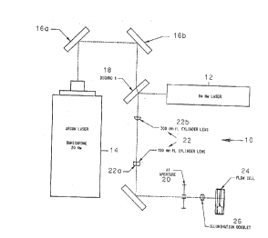
Referring first to FIG. lA, the illumination optics for a
blue excitable dye is generally identified by the reference
numeral 10, and incorporates two light sources.
A helium-neon (HeNe) laser 12 emits light at 633 nM and 2
mW, required for red cell scatter measurements for determination
of volume and hemoglobin concentration. An argon ion laser 14,
which emits light at 488 nm and 20 mW, is required for fluoro-
phore excitation. Three relay mirrors 16a, 16b and 16c, and a
dichroic beamsplitter 18 are needed for proper alignment of the
illumination system. The beams from the lasers are made
collinear at beamsplitter 18, which transmits the blue light
from the argon ion laser 14, and reflects the red light from
the HeNe laser 12. The resulting beam is then shaped into an
ellipse at a rectangular aperture (A-l) 20 by a pair of crossed
cylinder lenses 22. The focal length of the cylinder lens 22a
closest to the A-l aperture is 150 mm. The central axis of
this lens is parallel to the long dimension of the aperture.
The second cylinder lens 22b has a focal length of 300 mm. The
rectangular aperture, which is 89 ~m high by 653 ~m wide, is
imaged into the flow cell 24 using a diffraction-limited
achromatic lens 26 to define the measuring volume. The overall
-30-
MS-1716
20777~9
magnification is appro~imately 0.25. The aperture is under-
filled in the long dimension and overfilled in the short
dimension. The objectives here were to ma~imize the illumina-
tion intensity-and maintain a flat intensity distribution in
the short dimension of the image in order to minimize errors
due to intensity variation from cell to cell. The measuring
volume is essentially elliptical in shape and is 20 ~m high by
65 ~m wide at the 1/e2 points. The minor axis of the ellipse
is parallel to the direction of flow which is vertical with
respect to the horizon.
Cells which pass through the measuring volume scatter
incident radiation. Stained cells also absorb incident
radiation. The stained cells also fluoresce, emitting energy
at a frequency lower than that which was absorbed. These
optical signals are captured and detected in the capture optics
illustrated in FIG. lB.
The light scattered and fluoresced by cells traversing the
measuring volume is collimated by collection lens 28, and then
separated into four channels. The numerical aperture o~ the
collection lens is 0.34. At beamsplitter 30, light with a
wavelength below 500 nm is reflected. This light is filtered
again using a 488 nm narrow band interference filter 32. An
annular dark field stop 34 is used to transmit only blue light
MS-1716
- 2077789
scattered into the angular interval from 5-15 degrees. The
scattered light is then focused by detector lens 36 onto a
silicon photodiode 38 where electronic signals are generated.
The remaining light is equally split into two beams at beam-
splitter 40. Half of the energy is reflected to the red scatter
channels and half is tEansmitted to the red fluorescence
channel. (This beamsplitter 40 may be replaced with the approp-
riate dichroic beamsplitter to improve the signal-to-noise ratio
in the fluorescence channel.) The light reflected towards the
red scatter channels is filtered using a 633 nm narrow band
interference filter 42, and then split by the 50/50 (50%
reflection, 50% transmission) beamsplitter 44 into two beams.
Dark stops 46 and 48 are used in these two red scatter channels
to collect light in the two angular intervals required to
determine cell V and HC. The scattered light in each channel
is focused onto each of two silicon photodiodes 50 and 52 by
detector lenses 54 and 56, respectively, where electrical
signals are generated. Neutral density filters 58, 59 and 60
are used in each scatter channel, blue~and red, to adjust the
dynamic range of signals simply without modifying the
preamplifier circuit.
The light transmitted by beamsplitter 40 is filtered at 62
to allow only fluorescent energy to pass, and then focused by
MS-1716
~ ~a7778 ~
detector lens 64 onto a photomultiplier tube 66, through a 1 mm
diameter pinhole, where electrical signals proportional to the
magnitude of the incident energy are generated. The presence
of the pinhole minimizes shot noise produced by e~traneous
light. A 2 mm wide blocking bar 67 positioned before the
detection lens intercepts the main beam further reducing
bac~ground light noise. There is a sandwich of four filters 68
in this channel, comprising a 633 Notch Filter (X2~ 68a, a
Schott OG515 Color Glass 68b, a Schott OG530 Color Glass 68C,
and a Schott RG645 Color Glass 68d.
The net effect of this filter combination will be the
bloc~age of all light below 645 nm. The notcX and OG filters -
68a, 68b and 68c are re~uired since the RG645 filter 68d is not
efficient enough to block completeiy the scattered light at 633
nm and 488 nm.
The gain of the preamplifier circuit and optical density of
the neutral density filter in each scatter channel were chosen
to produce mean pulse signal levels of approsimately 2 volts at
the output of each channel when Technicon (TCN) optical test
material (OTM TCH T03-1704) was assayed. OTM consists of
sphered and hard-fised red blood cells. This material is
commercially available from the assignee hereof, and is adapted
for use on the TECHNICO~ H l system. This then allows fine
* Trade-mark - _33-
.~
. " .
,~
? ~ ~ 7 7 7 ~ ~
adjustment of the overall gain in each channel using a variablegain amplifier in the post-signal-detection-processing-hardware.
The overall gain in the fluorescence channel is controlled by
adjustment of the high voltage feeding the photomultiplier tube.
A functional bloc~ diagram of the post-detection signal
processing system is shown in FIG. lC. This system includes
preamplifiers 67, a variable gain amplifier 68, pulse height
analyzer 70, analog-to-digital converter 72, and data
acquisition hardware (computer) 74 and software.
The variable gain amplifier 68 contains four circuits which
permit inversion and amplitude conditioning of up to four input
signals.
The pulse-height analyzer 70, analog-to-digital converter
72 and data acquisition software are all components of the
4Cyte system which was purchased from Howard Shapiro, M.D.,
P.C. of Cambridge, Mass. The pulse-height analyzer is the
*
4Cyte Model FE Front End. This component produces held pulses,
representing the pulse heights, for up to four input signals,
and allows setting of the ~valid~ pulse height threshold
level. The 4Cyte Model I interface card is used in conjunction
- with the 4Cyte software for analog-to-digital conversion of up
to four input signals, and the capture of those values in the
* Trade-mark
-34-
,'~
~ ~ ~ 7 7 7 8 ~
RAM memory of the host computer. The digitized signals are
stored in list mode. There are five eight-bit bytes of
information for each cell, one for each of the four parameters
measured and one for flagging. The host computer for these
esperiments was an IBM PC/XT clone equipped with a color
monitor and a math co-processor. Data reduction can be
performed on any IBM compatible computer.
Turning now to FIG. lD, the illumination optical system for
a red e~citable dye is generally identified by the reference
numeral 110, and incorporates a helium-neon laser 112 that
emits a 2 mW beam of light at 633 nm. The beam is folded by
two reflecting mirrors 114 and 116 that pro~ide adjustment of
the laser beam position. The adjustment enables the beam asis
to coincide with the physical optical a~is of the illumination
optics. The beam is then shaped by the pair of cyliner lenses
118 and 120 into a 192 s 77 ~m elliptically shaped beam (at the
l/e ). The 192 ~m dimension is formed by the 150 nm focal
length cylinder lens 118, ?nd it illuminates the long asis of
the A-l aperture 122 ~which is parallel to the plane of the
page in FIG. lD). The 77 ~m dimension is formed by the 60 mm
focal length cylinder lens 120, and it illuminates the short
a~is of the A-l aperture. The A-l aperture is 653 s 89~um.
The illumination doublet 123 produces an elliptically shaped
Gaussian intensity distribution of 37.4 s 12.6 ~m in the
* Trade-mark
-35-
~ ~ ,i~
2077~9
flowcell 124. The minor azis of the ellipse is parallel to the
direction of flow, which is vertical, i.e. in the direction of
arrow 126.
-
All cells that pass through the measuring volume scatterand absorb the incident radiation. The light scattered and
fluoresced is captured and measured in the detection optics
illustrated schematically in FIG. lE. The light that is
scattered and fluoresced is collected by the high numerical
aperture (Hi-NA) lens 128 and collimated. The numerical aper-
ture of the collection lens is 0.34 nm. The beam is divided
into two parts by the dichroic beamsplitter 130, which allows
light at wavelengths less than 670 nm to pass. The beam 132 is
reflected onto a photomultiplier after filtering, and is used
for the fluorescence measurement, while the transmitted beam
134 is further split by the 50/50 (50% reflection, 50% trans-
mission) beamsplitter 136 to make the two scatter channels.
The reflected scatter channel 138 has a 5-15~ darkstop 140,
while the transmitted channel 142 has a 2-3~ darkstop 144.
The light passing through each of these darkstops 140, 144 is
then focused down through lenses 146 and 148 onto photodiodes
150 and 152, respectively. Neutral density filters 154 and 156
are used to reduce the light levels at each photodiode to a
level that is appropriate for the standard detectors and
preamplifiers.
-36-
MS-1716
- 2077789
The light reflected by the beamsplitter 130 is focused by
lens 158 through a narrow band (690 nm) filter 159, through a 1
mm diamter pinhole onto a photomultiplier tube 160, where
electrical signals, proportional to the magnitude of the
incident energy are generated. A 2 mm wide blocking bar 157
positioned before the detection lens 158 intercepts the main
beam further reducing background light noise. The signals
generated by the photodiodies 150, 152 and the photomultiplier
tube 160 are processed in a post-detection signal processing
system similar to that shown in FIG. lC. In this case,
however, only three signals, i.e. red low angle scatter, red
high angle scatter and red fluorescence, are processed (not the
four signals discussed with respect to FIG. lC).
The following esamples set forth reagent compositions and
methods incorporating the same for the identification of
reticulocytes and characterization of reticulocytes and red
blood cells using fluorescence flow cytometry techniques.
Standard commercially available reagent grade materials were
used whenever possible. It will be understood that the formula-
tions and the procedures which follow are provided for purpose
of illustration only, and that other ingredients, proportions
and procedures can be employed in accordance with the disclo-
sures of this invention.
-37-
MS-1716
-
~ ~ ~ 7 7 78 ~
E~amPle 1: Scatter and Fluorescence Measurements for
Distinguishing Reticulocytes and Erythrocytes
Within a Blood Sample Using Reagent Composition
Containing a Zwitterionic Surfactant
The dye 3,6-bis(dimethylamino)-10-2-hyrosyethyl acridinum
iodide tAOEOH) was stored in a 1 mg N,N-dimethylformamide/ml
stock solution. A working reagent was created by adding the
dye stock to give a final concentration of 6Jug/ml to 12Jug/ml
of dye. The final concentration of lauramidopropyl betaine was
from 12 ~g/ml to 87.5 ~g/ml. A buffer solution contained the
following components at the concentrations noted:
Calcium Chloride 0.4 mM
Potassium Chloride 4.0 mM
Magnesium Chloride 40.0 mM
Sodium Phosphate (Tribasic) 0.5 mM
Sodium Bicarbonate 20.0 mM
The final osmolality and pH of the working reagent used in
this study were 272 mmo Vkg and 8.1, respectively.
Samples were hand-mi~ed in a manner which simulated the
automated TECHNICON H l red cell sample processing scheme.
Glass test tubes were filled with 5 milliliters of the working
* Trade-mark
-38-
., .
23~77~9
reagent. Five microliters of a blood sample were then pipetted
into the reagent while the reagent was undergoing agitation on
a vortes miser. The 1:1000 dilution of blood was then fed
immediately into the sample line of the previously described
flow cytometric apparatus and the optical system of FIGs. lA
and lB. In approsimately two minutes, the sample passed
through the flow cell and was esposed to an argon-ion laser
source for red-cell and reticulocyte analysis. Each sample was
measured in duplicate if the sample volume permitted.
When viewed through a microscope, the mature red cells and
reticulocytes in a prepared sample were found to be sphered,
and the reticulocytes fluoresced in the red when escited by
blue light.
At the completion of the analysis, the raw data was
displayed in the form of a 81ue Scatter v. Red Fluorescence
cytogram, FIG. 2A(l), wherein the ordinate represents the
relative intensity of forward scattered light, and the abscissa
represents the relative intensity of red fluorescence. Each
point shown on the cytogram represents a cell. Distinct cell
populations were clearly observed based on their particular
scatter and fluorescence signals. The mature erythrocyte
population falls within Region A between the ordinate and the
vertical line X. These cells show high scatter signals and low
-39-
MS-1716
2û7~
cell fluorescence signals. The reticulocyte population falls
within the region to the right of X, Region B. These cells are
distinguishable from the mature erythrocytes due to the high
fluorescence signals from their AOEOH stained RNA. The platelet
population lies within Region C below line Y. Platelets have
relatively low scatter signals when compared to the
reticulocytes.
Based on the fluorescence separation between mature erythro-
cytes and reticulocytes, the reticulocyte count of a patient
sample may be determined by creating an electronic ~window~
which defines the ranges of scattered light and fluorescence
which identify reticulocytes and mature erythrocytes. The
number of reticulocytes and mature erythrocytes falling within
the ~window~ are determined so that the percentage of the
reticulocytes and erythrocytes present in the total cell
population is known. In FIG. 2A(l), the reticulocyte ~window~
is determined by Region B, and the mature erythrocyte ~window~
by Region A. Note in FIG. 2A(2) and in all following scatter/
scatter cytograms, the non-linear grid overlays indicate the
loci of constant volume and constant refractive inde~ for
perfect spheres according to the above-noted method of Tycko.
The reference percentage of reticulocytes in each sample
was determined using the manual microscopic procedure
-40-
MS-1716
20777~
recommended by the National Committee for Clinical Laboratory
Standards (NCCLS). In this procedure, a small volume of the
sample was vitally stained with New Methylene Blue. A
conventional dry wedge smear was then perepared, and the
percentage of reticulocytes in the sample was counted with the
aid of a microscope. The microscope was equipped with a lOOX
oil immersion objective and a lOX ocular. A minimum of 1000
cells were counted for each sample. A Miller disc was inserted
in the ocular of the microscope to improve counting precision.
Any red cell containing two or more particles of blue material
after staining was labeled a reticulocyte.
The reticulocyte count of the patient sample was measured
to be 1.7% by this flow cytometric technique. The same blood
sample was also analyzed by the NCCLS method. The result was a
reticulocyte count of 1.7%.
A second esperiment was conducted to demonstrate the high
degree of discrimination between reticulocyte and erythrocyte
populations when cells were stained with OYazine 750 and
measured by the previously described fluorescence flow
cytometer and optical system of of FIGs. lD and lE. O~azine
750 dye was stored in a 1 mg N,N-dimethylformamide/ml stock
solution. A working reagent was created by adding the dye
MS-1716
o 2 ~ 7 7 7 8 ~
stock to a final concentration of from 0.2 ~g/ml to 1.2 ~g/ml,
to the sphering agent and the buffer solution described above.
The sample preparation protocols as described above were
followed. FIG. 2B~l) displays the fluorescence vs. low angle
scatter cytogram of a normal human blood sample stained with
the Oxazine 750 containing reagent. Based on the fluorescence
separation between erythrocytes and reticulocytes, the
reticulocyte count of the sam~le was measured as 2.1%. When
analyzed by the NCCLS method, a reticulocyte count of 2.1~ was
obtained. FIG. 2C shows the reticulocyte as "+~ superimposed
on mature erythrocytes obtained from similar data as FIGs. 2A
and 2B.
X
-42-
20777~9
~xamPle 2: Correlation Study with the Reagent Compositions and
Methods of the Present Invention and the NCCLS
Method
A study was conducted to compare the performance of AOEOH
when used in a reagent composition in the previously described
fluorescence flow cytometer and the optical system of FIGs. lA
and lB and the-NCCLS manual method. Blood samples were obtained
from 39 Technicon employees and 23 hospital patients. The
hospital samples included three sickle cell, one thalassemia and
ten neo-natal blood samples. The 62 blood samples were stained
with the reagent composition described in Example 1, and assayed
for their reticulocyte content. Reticulocytes in the same set
of blood samples were also counted using the NCCLS method.
The sample preparation and analysis protocols as described
above with regard to E~ample 1 were followed.
The percentage reticulocyte counts obtained from these two
methods are compared in FIG. 3A. At a concentration of 6 ug
AOEOH/ml in the reagent composition, close correlation was
shown to exist between the reagent composition and measurements
obtained using the previously described fluorescence flow
cytometer and the optical system of FIGs. lA and lB and those
MS-1716
~ ~ 7 7 7 8 ~
obtained by the NCCLS reference method. The correlation
coefficient for the measurement was 0.95.
A second experiment was conducted to compare the performance
of Oxazine 750 reagent when used in the previously described
fluorescence flow cytometer and the optical system of FIGs. lD
and lE and the NCCLS method. The sample preparation and
analysis protocols of Ezample 1 were followed. Blood samples
were stained with the reagent composition and assayed for their
reticulocyte content. Reticulocytes in the same set of blood
samples were also counted using the NCCLS method. The
percentage reticulocyte counts obtained from these two methods
are compared in FIG. 3B. The correlation coefficient for the
measurement was 0.93.
~- -44-
o 2 ~ 7 7 7 8 ~
~~amPle 3: Correlation Study with the AOEOH Containing Reagent
Composition of the Present Invention and the
TECHNICON H l Jr. Reference Method
The same set of samples in Esample 2 were also measured
using the TECHNICON H l Jr. system for the red cell indices.
The TECHNICON H l Jr. automated system is a flow cytometer
that simultaneously measures the cell volume and hemoglobin
concentration of individual isovolumetrically sphered red blood
cells.
The indices, MCV and MCHC were separately determined on the
TECHNICON H l Jr. analyzer, and compared with the values
obtained using the AOEOH containing reagent composition of the
present invention. FIGs. 4A and 4B show the correlation data
for total red blood cell indices, MCV and MCHC, respectively.
The correlation coefficients for the measurement were 0.91 and
0.97, respectively.
* Trade-mark _45_
7 7 7 ~ g
~~amPle 4: Correlation Study with the Oxazine 750 Containing
Reagent Composition of the Present Invention and
the TECHNICON H 1 Reference Method
The erythrocyte and reticulocyte indices, MCY and MCHC were
separately determined and compared with the values obtained
using the Osazine 750 reagent composition of the present
invention. FIGs. 5A and 5B show the correlation data for total
red blood cell MCV and MCHC, respectively.
The correlation coefficients for the measurement-were 0.93
and 0.92, respectively.
* Trade-mark
~ ~7778 ~
E~amPle 5: Scatter and Fluorescence Measurements for
Distinguishing Reticulocytes and Erythrocytes
Within a Blood Sample Using the Reagent Composition
of Example 2 Containing AOEOH and a Buffer, Which
Fails to Distinguish Reticulocytes Within Blood
Not all buffers can be used to stain and sphere
reticulocytes simultaneously. This example demonstrates a poor
discrimination between reticulocytes and erythrocytes when
using AOEOH staining dye and phosphate buffer at pH 8.0 and
osmolality of 290 m Osm ~see FIG. 6A). In comparison, a good
separation between reticulocytes and erythrocytes is clearly
observed when using Barbital buffer (12 ug/ml LAB surfactant)
at pH 8.0 and osmolality 290 m Osm (see FIG. 6B).
-47-
-'.~,
~ 20~7789
Some advantages of the present invention evident from the
foregoing description include a reagent composition and method
for the identification of reticulocytes in a whole blood
sample, and for the simultaneous quantitation of the volume,
hemoglobin content and hemoglobin concentration of reticulo-
cytes and erythrocytes by fluorescence flow cytometric
techniques.
In view of the above, it will be seen that the several
objects of the invention are achieved, and other advantageous
results obtained.
As various changes can be made in the above constructions
and methods without departing from the scope of the invention,
it is intended that all matter contained in the above descrip-
tion, or shown on the accompanying drawings, shall be inter-
preted as illustrative, not in a limiting sense. For instance,
fractionated samples of blood can be processed in a similar way.
-48-
MS-1716