Note: Descriptions are shown in the official language in which they were submitted.
'~ V '~ ~ ~ 1 U
WO 91/13989 PCI/l)S91/01748
De~sT iptiOTI
METHODS AND COMPOSITIONS F{)R THE TRE~TMEN~ OF
MALIGNANCIES IN WHICH A PROTE~IN KINASE IS ASSOCL~TED
:
Technical Field
The present invention is generally directed toward compositions for
use within methods for the treatment of maligrlancies in which a protein tyrosine
10 kinase is associated. This invention is more particularly related to cancer
therapies using DNA sequences encoding protein tyrosine phosphatase or mutant
forms of the en~nne.
Back~ound of the Invention ;~
Despite enormaus investments of ~mancial and human resources,
cancer remains one of the major causes of death. A common charactenstic of
malignancies is uncontrolled cell growth. Cancer cells appear to have undergone
a process of transformation from the normal phenotype to a malignant pheno~pe
capable of autonomous growth. Mutation of somatic celI genes is considered to be20 a common prima~y event that results in the trar~sformation of normal cells tomalignant cells. The malignant pheno~pic characteristics encoded by the mutated ~ o
genes are passed on during cell division to the progeny of the trans~ormed cells.
Yarious genes involved with transformation have been designated as oncogenes.
Oncogenes were originally identified as components of the genetic material of
25 oncogenic viruses. The homologous genes on human chromosomes are commonly
termed oncogenes or proto-oncogenes.
Numerous oncogenic viruses appear to operate by encoding a
protein t~rosine kinase. This enz3rme ca~alyzes the phosphorylation OI ~rosyl
residues in proteins. Changes in the state of phosphorylation of tyrosyl residues in
30 proteins have been suggested to be involved in oncogenic transformation. The
identi~,r of the protein substrates and the mechar~ism by which their
phosphorylation mediates phenotypic resporlses remain to be elucida~ed.
Approaches to the development of cancer therapies have, in
general, centered on the use of characteristic differences between nonnal and
35 malignant cells. More specifically, comparisons of normal and cancer cells have
focused on transformation~dependent changes in the molecules residiIIg at the
surface of cell membranes. These molecules include glycolipids, proteins and
glycoproteins. Certain molecules have been found to disappear or at least
: . . , , : ~
.. . . .
, . . . . .
wo ~ 3g89 2 ~ 7 8 ~! ~ O PC~/US91/01748
decrease greatly upon onco~enic transformation, while other molecules increase.
Despite advances in this area, treatment of cancer cells by targeting tumor-
associated cell surface molecules has met with limited success.
Due to the dif~lculties in the current approaches to cancer therapy,
S there is a need in the art for improved methods and compositior~s. 'rhe present
invention ~Ills this need, and further provides other related advaIItages.
$umma of the lnverltion
Briefly stated, the present invention provides compositions whose
10 uses include within methods for treating a malignancy in a warrn-blooded animal,
~ wherein a protein tyrosine kinase is associated with the malignancy. In one aspect,
`- the composition comprises a gene transfer vehicle capable of infecting malignant
cells, wherein the gene transfer vehicle carries a DNA construct comprising a
DNA molecule encoding a non^receptor-lirlked protein tyrosine phosphatase. ln
15 one embodiment, the DNA molecule encodes a non-receptor-linked protein
.~ tyrosine phosphatase having a truncated carboxyl terminus. In another
embodiment, the DNA molecule ellcodes a non-receptor-linked protein ~rosine
phosphatase, wherein the pro~ein tyrosine phosphatase comprises aII amino acid
sequence of between 297 to 321 amino acid residues, and wherein the amino acid
20 sequence contains a portion of similar length to and having at leas~ approximately
80~ sequence similari~ with the amino acid sequence of ~igure 1 from
asparagine, amino acid 42, to glut~nic acid, ~amino acid 274. ln another aspect,the composition comprises a gene transfer vehicle capable of infecting malignantcells, wherein the gene transfer vehicle carries a DNA construct comprising a
DNA molecule encoding a receptor-linked protein tyrosine phosphatase. ln one
embodiment, the DNA molecule encodes a receptor-linked protein tyrosine
phosphatase having a truncated carboxyl terminus. Preferred gene transfer
vehicles for both aspects include a recombinant retrovirus or a recombinant
vaccinia virus.
In another aspect, the present invention provides a vanety of
isolated DNA molecules encoding non-receptor-linked protein tyrosine
phosphatases having tMncated carboxyl termlini. ~[n one embodiment, the DNA
molecule encodes a non-receptor-linked protein tyrosine pbosphatase, wherein
:~ the protein tyrosine phosphatase CollSiSts essentially of an amino acid sequence of
between 297 to 320 amino acid residues, and wherein the amino acid sequence
. contains a portion of similar length to and having at least appro~mately 80%
:. sequence sin~ilarity with the amino acid sequence of Figure 1 from asparagine,
';
;
,, , : .: , . . . . . . . . .
:~ " ` ' , ':~ ,
wo 91tl3989 2 n ~ 8 01 U Pcr/US91/0l7q8
amino acid 42, to glutamic acid, amino acid 274. In another embodiment, the
DNA molecule encodes a non-receptor-linked protein tyrosine phosphatase,
wherein the protein ~yrosine phosphatase consists essentially of the amino
sequence of Figure 1 from methionine, am~no acid 1, to an amino acid positioned
S between leucine, am~no acid 297, and ar~nine, am~no acid 317. In a related
embodiment, the DNA molecule encodes a non-receptor-linl;ed protein tyrosine
phosphatase, wherein the protein tyrosine phosphatase consists essentially of the
amino acid sequence of Figure 1 from methionine, amino acid 1, to arginine,
amino acid ~17. In another embocliment, the DNA molecule encodes a non-
10 receptor-linked protein tyrosine phosphatase, wherein the protein tyrosine
phosphatase consists essentially of the amino acid sequence of Figure 1 from
rnethionine, amino acid 1, to an amino acid positioned between glutamic acid,
amino acid 376, and serine, am~no acid 396.
'rhese and other aspects of the present invention will become
lS evident upon reference to the following detailed description and attached
drawings.
Brief Descripti~n of the Drawin~s
- Figure 1 depicts the sequencing strategy, nucleotide sequence, and
20 deduced amino acid sequence of huma~ T cell cDNA encoding protein ~rrosine
phosphatase (~Pase). The predicted amino acid sequence of the open reading
~ame is shown below the nucleotide sequence. The oligonucleotide sequences
used ~or screeni~ag the libr~y are indicated by dots [e.g., between nucleotides 425
and 479 (probe 1), and 689 and 737 (probe 2)]. ~he TAA stop codon is located at
25 nucleotide 1306 followed by a 3' untransL~ted end containing two possible
polyadenylylation sites AATAAA at 1521 alld 1677. The vertical arrows
demarcate a 236-residue core segment. The schematic diagram below the
nucleotide sequence depicts the sequencing strategy used. Open bar, open
reading ~ame; solid bar, 3' untranslated end. Horizontal arrows indicate the
30 length of sequence nbtained from different sequencing oligonucleotide primers.
E, EcoRI; H, HindIII, S, Sst I; X Xba I. The scale at the bottom represents 200
nucleotides (in kbp).
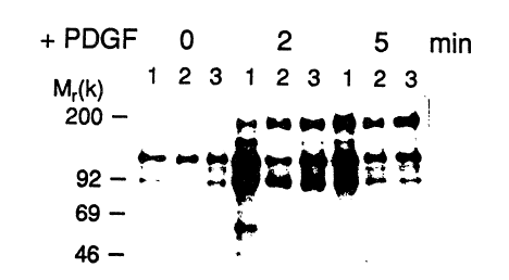
Figure 2 pictorially depicts a Western blot of phosphotyrosine-
immunoprecipitated proteins following PDGF stimulation. Phosphotyrosine-
35 containing proteins and PDGF-stimulated cells were immunoprecipitated with all
anti-phosphotyrosine antibody. A SDS/7.5% Laemmli gel was used to separate
the precipitated proteins followed by Western blot analysis with an anti-
~ .: , . .................... . .
., . ,. . ............................... ~
: : '' .
' '' '
wo 91/13989 2 0 7 ~ PCI/lJS91/Ot748
phosphotyrosine antibody as a probe in the blot. Detection of binding was with125I-labeled protein A followed by autoradiography at room temperature for S
days. Lanes: 1, control cells; 2, cells expressing the full-length 48-kDa T-cell~rPase; 3, cells expressing the truncated form. T~mes of PDGF stimulation
S follow~ng 48 hr of serum-deprivation are indicated (0, 2, or 5 min). Standard
molecular mass markers in kDa (200, myosin; 92, phosphorylase B; 69, bovine
serum alburnin; and 46, ovalburnill) are also shown.
Figure 3 graphically illustrates a hydrophobicity plot of the carboxyl-
ter~Linal region in the T-cell PI'Pa~se. The hydrophobicity calculations of arnino
10 acid residut~s 326~15 in the carboxyl-terrninal segment of the T-cell mase were
deterrnined as described by Kyte and Doolittle (J~Mol, B;Q1. 1~7:105-132, 1982).A computer-generated plot was obtaLined by using a default winclow of 7. The
numbers above and below ~he 0 line represent hydrophobic and hydrophilic amino
acids, respectively; the units in the y axis are defined as in Kyte and Doolittle. The
15 bar above the plot depicts a putative Arg-Lys-Arg-Lys-Arg nuclear recogmtion
signal (Van Etten et al., ~11 ~:669-678, 1989) between residues 377 and 381
(solid) and a hydrophobic terminal region (39S-415) (solid; 19 residues shown insingle-letter code) representing the ca~bo~yl-terminus of the molecule.
20 DetailedDescrip~iQn Q~ç InventiQn
As noted above, the present invention is directed towards
compositions whose uses include within methods for treating a malignancy in
which a protein tyrosine kinase is associated. The disclosure of the present
invention shows the reversion of malignant transformation by transfection with
25 human DNA encoding a protein tyrosine phosphatase.
Within one aspect of the present inverltion, a malignancy in which a
protein tyrosine kinase is associated may be treated with a ge~e transfe~ vehicle
capable of in~ectrng malignant cells, wherein the gene transfer vehicle carries a
lDNA co~struct comprising a DNA sequence encoding a protein tyrosine
30 phosphatase or mutant forms of the enzyme. This therapy is ~pplicable to a wide
- variety of malignancies found in warm-blooded animals such as humans.
Representative malignancies include breast cancer and leukernia. Within the
present invention~ it is not necessary for all malignancies to involve tyrosine
phosphorylation of the same type of protein substrate, provided that
35 dephosphorylation of tyrosyl residues by a protein tyrosine phospha~ase (whose
DNA sequence has been incorporated into malignant cells) results in the
reversion of the malignant phenotype.
,,
.-:
.
~- .. - . . ... .
-., . . . ~.
.. . . . . .
' , ' , : , . ~
) U ~ l~
wo 91/13989 PCT/US91/017~8
A variety of DNA sequences encoding a protein tyrosine
phosphatase ("PTPase") are suitable w~thin the present invention. The DNA
sequence may encode a non-receptor-linked ~TPase or a receptor-lirlked PTPase.
The latter type of mase h~s additional amino acid sequences that form a
5 domain bearing traits of a cell-surface receptor. Non-receptor-linlced mases
typic~lly have a molecular weight less than approximately 50,000. Receptor-linked
~Pases typically have a higher molecular weight, e.g., 150,000 250,000, and
display a wide variety of external domains.
In one embodiment of a DNA sequence useful within the present
10 inven~ion, the sequence encodes a wild type (i.e., a naturally occurring fonn)
mase. A representative example of wild type, non-receptor~lirlked mase
DNA is the sequence sho~ in Figure 1 for human T cell PIPase. Other
examples include a DNA sequence encoding a PI`Pase of human placenta. A
representative example of wild type, receptor-linked PI'Pase DNA ls the DNA
15 sequence encoding CD45 in human leUkOCYteS.
`. In another embodimer~t, the DNA sequence used within the present
invention encodes a ~Pase having a truncated carboxyl terrLunus, i.e., missing
amino acid residues normally found at or near the carboxyl terminus in the
sequence of a wild type mase. The disclosure of the present i~ve~ion provides
20 a representative example of a DNA sequence encoding a truncated mase. This
DNA was prepared by altering the nucleotide sequence of the cDNA encoding a T
cell PIPase (Figure 1) using standa~d mole~Nlar biology techniques. Briefly, a
- deletion of a few nucleotides by site directed n~utagenesis was introduced into wild
type cDN"~ This sequence alteration resillted in the placement of a premature
25 translation stop signal in the open reading frame of the cDN,~ The modiled
cDNA encodes a PIPase (molecular mass of about 37,000) with a truncated
ca~boxyl terminus relative to the 48,00Q molecular mass protein nonnally
~ produced in T cells. This truncated PIPase has the an~ino acid sequence of
; Figure 1 from methior~ine, an~ino acid 1, to arginine, amino acid 317. The
30 e~zymatic activity exhibited by trans,~ected cells expressing this truncated PI~ase
is higher than the activity in control cells.
; It will be evident to those skilled in the art that a variety of DNA
sequences encoding PIPases having truncated carboxyl termiDi may be employed
within the present invention. In genera!, any number of amino acid residues may
35 be deleted from the carboxyl terminus, provided tbat the resultaIIt protein does
not e,xhibit a significant reduction in Pl~ase activi~,~. The enzymatic activity of
', truncated PIPases may be measured, for example, using an assay based on the
.
.... , :,~: . . . ...
WO 91/13989 2 0 7 ~ ~10 PCI/US91/01748
measurement of a reporter g;roup released from a labeled substrate. A suitable
assay includes the one descr~bed below for the measurement of 3~P released from
reduced, carboxyamidomethylated maleylated-lysozy ne. As denoted in Figure 1,
there is a 236-am~no acid residue core identified by T cell P~Pase amino acid 42S (asparagine) to arnino acid 274 (glutam~c acid). T cell PrPase and placenta
P~Pase lB, for example, possess within this core about 65% sequence identity andapproximately 8û~o sequence sirnil3rity (after optin~iz~ng the alig~merlt between
; the two sequences). Truncated forms of PrPase rnay ~e prepared by the deletion
of amino acids starting at the carboxyl terrninus and moving back towards the 236~
10 residue core. Suitable DNA sequences include those encoding a PTPase
comprising an arnino acid sequence of between (including) 297 to 320 amino acid
residues where the sequence contains a portion of similar length to, and having at
least approximately 80% sequence sirnilarity with, this 236-residue core. This
portion of a sequence need not be of identical length and it may be necessary to15 shift the sequence and/or insert gaps (in it or the sequence of Figure 1) in order to
optimize the alignment between the two sequences. Sequence sirnilari~ is based
upon sequence identity plus conservative substitutions of amiIlo acids.
Conservative substitutions include interchanges of valine and isoleucine, leucine
and isoleucine, aspartic acid and glutamic acidp and others of a sim~lar nablre. A
20 prefelTed DNA sequence encodes a truncated ~Pase comprising the amino acid
sequence of Figure 1 from methionine, amino acid 1, to an amino acid positioned
between (including~ leucine, amino acid 297, and arginine, amino acid 317.
Following isolation or preparation of a suitable DNA sequence
encoding a wild type or tmncated PIPase, the sequence is inserted into a gene
25 traIlsfer vehicle. SuitablP gene transfer vehicles include a recombinant retrovirus
or a recombinant vaccinia ~1irus.
Retroviruses are RNA viruses that can replicate through a DNA
intermediate through the action of an RNA dependent DNA polymerase. Once in
DNA form, they may be stably integrated into the host cell genome and passed
,~ 30 down ~om a parent cell to its progeny. Thus, retrovinuses are suitable as transfer
vehicles for foreign DNA into a host cell (see Shimotohno and Temin, "Formation
of Infectious Progeny Virus After Ingestion of Herpes Simplex Thymidine Kinase
Gene into DNA of an Asian Retrov~rus," (:ell 26:67-77, 1981; see also,
Shimotohno and Temin, "Loss of Intervening Sequences in Genomic Mouse
35 Alpha-Globin DNA Inserted in an Infections Retrovirus Yector," ~ature 299:265-
268, 1982; see also; Miller and Rosman, "lmproved Retroviral Vectors for Gene
Transfer and Expression," BioTechnL~ues _ 98~990! 1989).
~,
..
. :.
-. .. -:
., , . . : -:
: , ~ ~ ' . , '
, . .
wo 91/l3989 2 ~ ~p~ ~lVs9~0l74R
Where the retrovirus transfer vehicle is replication competent, the
resultant active virus may not be appropriate for therapeutic purposes because it is
also replication competent, and represents a health hazard. Hovrever, various
methods have been util~zed to design a retrovirus transfer vehicle which can
5 produce a retrovirus containing foreign I)NA, yet which is nevertheless replication
in~ompetent (see U.S. Patent Nos. 4,650,754 (Tem~n et al.)7 4,861,719 (Miller),
4,405,712 (Van de Woude et al.), and 4,885,238 (Reddel et al.), which are
incorporated herein by reference; see also Jacob et al., European Patent
Application Publ. No. 0178220,E~uropean Patent Application Publ. No. 0243204,
10 PCI International Publ. No. WO 85/05629, PCI International Publ.
No. WO 87/03451, PCI` International Publ. No. 89/07150, and PCI` Internalional
Publ. No. W O 89/09271). Various retroviruses may be used within the present
invention including the Munne Sarcoma Virus (MSV) and the Moloney Murine
Leukemia Virus ~MoMLV). Furtherrnore, it will be evident to one of ordinary
15 skill in the art that envelope sequences may be derived from annphotropic viruses
such as vims 4070A.
Briefly, within one embodiment of the present invention, a mutant
of the Moloney murine leukemia vims is constxucted which contains a deletion of
about 350 nucleotides in the env region of the: retrovirus (see Shank and Linial,
20 J. Virol. 36(~2):4so-456~ 1980). This produces a virus (gag+,pol+,en~~) deficient in
encapsidation or packaging of viral RNA, which may, if cotransfected with an
encapsidation positive retroviral vector (gag,pol~,env+) containing a desired
foreign gene, result in the produc~ion of retrovirus which is replication
incompetent, yet which nevertheless produces the foreign gene (see Temin et al.
25 U.S. Patent No. 4,650,764).
There remains, however, the possibili~ of recombination betvveen
the gag~,pol~,env+ vector and the gag+,pol+,en~r~ vector. Such recombination
might produce an infectious retrovirus (gag+,pol+,env+). Therefore, it is
particularly preferred to design recombinant retrovirus vectors which are safe
30 transfer vehicles for foreign genes. Briefly, the retroviral genome, or provirus,
contains two Long Terminal Repeats (LTR) which encode sequences for initiation
and tern~ination of re~roviral transcIiption, including promoters, translation and
termination signals, as well as enhancers. The Ll'R has 3 regions: U3, R, and U5.
Within a preferred embodiment the U3 region (co~taining promoter a~d
35 enhancer se~nents) of the right side LTR is deleted resulting in a retroviral vertor
which is self-inactivating (see Yu etal., "Self-Inactivating Retroviral Vectors
Designed for Transfer of Whole Genes Into Mammalian Cells," Proc. N~tl~ Acad.
.
wo 91/13989 2 O 7 ~ O ~ U P~r/VS9~/01748
Sci. U~A 83:3194-3198, 1986; Markow~tz et al., "A Safe Packaging Line for Gene
Transfer: Separating Viral Genes on Two Different Plasm~ds," I Virol. (~:1120-
11241 1988). This vector (containing a promoter and the foreign gene, i.e., U3, R,
US----promoter, gene, ----U3(del.), R, U5) is used to transfect helper cells. Viral
S transcription begins on the lef~ side Ll~ beginn~ng at R, and transcribes a
sequence which does not contain either the left side or right side U3 reg~on (i.e.,
R, U5, ---- promoter, gene ---- U3(del), R). Thus, the retrosiral vector is self-
inactivating yet, nevertheless, because it contains an internal promoter allows
`` transcription of the foreign DN~
Vaccinia viruses are eukaryotic DNA viruses that reproduce entirely
vithin the cytoplasm of a host cell. The virus has been used for about 200 years in
vaccines for inoculation against smallpox. The virus is considered non-oncogenicand relatively benign. The naturally occurring vaccin~a genome can be modified
to produce recombinant vaccir~ia by rearrangement of the natural genome, by the
15 removal of DNA from the genome, cmd/or by the introduction of foreign DNA
into the genorne (e.g., U.S. Patent Nos. 4,769,330 and 4,886,782, which are
incorporated here~n by reference). ~bus, recornbinarlt vacciI~ia VilUS represents a
relatively ~nnocuous euka~yotic cloning vector useful within the present invention
for the introduction of a DNA sequence encoding a PrPase into a eukaryote.
The follo~ving examples are offere d by way of illustration and not by
way of limitation. ~ ~-
EXAMPLES
." '.
EXAMPLE 1
;: CONSTRUCrlON 0~ A HUMAN T C~LL PROTelN-TYROSINE-PHOSPHATASE
~' ' ' ' .
All restriction and modifying enzymes and in vl~ro transcrip~ion and
translation systems were purchased from Stratagene, La Jolla, California.
30 Oligonucleotides were synthesized with an Applied Biosystems 380A DNA
Synthesizer (Applied Biosys~ems, lE;oster City, Calif.). Radionucleotides were
` obtained from New England Nuclear (Boston, Mass.). Sequenase was obtained
from United States Biochemical (Cleveland, Ohio). Solutions such as Denhardt's
solution were prepared as described by Maniatis et al., Molecular Cloning: A
35 h~2[~, Cold Spring Harbor Lab., Cold Spring, N.Y., 1982.
,,
.,
,i
,
, :
,
wo 91/13989 PC~/us9l/al748
Screenin~ of a cDNA Librar~w~th Svnthetic Oli~onucleotides
A set of complementary overlapping oligonucleotides were
synthesized for each of the protein segments Lys-12~Asn-139 and Gly-209-Phe-
2~5 from the human placenta PrPase lB a~cording to the method of
~:~ S L. Charbonneau et al., Proc. Natl. ~c~sl Sc~. US~ 5252-56, 1989. Prediction of
the DNA sequence was based on the optimal codoll choice for human amino acid
sequence data (see R. Lathe, I~Mol ~QI 1~:1-12, 1985). Oligonucleotides in
set 1 (5'-AAGTGTGCACAGTACTGGCCGCAGAAGGAA-3' and
5'-GTTGGTATCCI CAAAGATCAT~CCCl'r~CITCCI'rC TGC-3') or in
10 set 2 (5'-GGTCC TGTGGTGGTGCACTGCAGTGCI'GGT-3' and
5'-AAGGl~CCAGTG CGCCAATACCAGCACTG-3') were armealed and
labeled using 32PdATP and 32Pd~' and the Klenow fragment of DNA
polymerase I, producing radioactive double-stranded DNA with a specific activityof 4 x 10⁷ cpm/pmol bp.
A ~gtlO recombinant cDNA phage library containing 500,000 phage
was prepared from human peripheral T cell poly(A)+ mRN.4 according to the
method of Iittman et al., S~ll 40:237-46, 1985, and plated at a density of 50,000
phage per plate. Duplicate nitrocellulose filter lif~s were taken from each plate
and bybridized with the oligonucleotide probes prepared above (2.5 x 10⁶ cpm per~llter) in 20% fonnamide/5 x $SC (1 x SSC is 150 mM NaCl/15 mM sodium
citrate)/2.5 x Denhardt's ~olution/1 imM sodium pyrophosphate/50 imM sodium
phosphate buffer, pH 6.8, at 37C for 18 ho~rs. The filters were washed in 2 x
SSC/0.2~o SDS at 42C and subjected to autoIadiography for 3 days at -70C with
- an intensi~Ser screen.
Although many recombinant phage hybridized to each probe, only
:3 one overlapping positive clone bound to both oligonucleotides.
.
. B. DN~ Se~u~ce ~alysis
Approximately 101 E. coli cells were infected with the recombinant
30 phage selected above, and Iysed by the alkali method (see Sambrook etal.,
Molecular Clonin~: _A Laboratorv Manual, 2 ed., Cold Spring Harbor Laboratory
- Press, pp. 1.25-1.28, 1989). The amplified phage were puri~ed by Cs(:l density
, centrifugation. DNA from the puriFIed phage was extracted and diges~ed with
EcoRI restriction endonuclease. The EcoRI fragrnents were analyzed by agarose
35 gel electrophoresis which resolved two vector DNA fragments (32 and 11 kbp) and
- a cloned cDNA fragment of 2.3 kilobase pairs (kbp) in length.
., , ,. , , ............................... ., ~ .
.
,
WO 91/13989 2 0 7 g 1~ t o PCr/US91/01748
One hundred nanograms of the 2.3 kbp EcoRI ~nsert and 100 ng of
the Bluesclipt cloning vector (Strategene, La Jolla, Calif.) were cleaved with
EcoRI (a site within the ~-galactosidase gene), ligated in ~ reaction m~xture
` containing 50 mM Tris (pH 7.4), lO mM MgC~, 10 mM dithiotheritol and 2 mM
ATP, and transformed into an E;schenchia coli host (~Blue, Stratagene~. The
transfonned cells were plated on agar plates contain~ng 100 ~g/ml ampicillin and50 ~g/ml X-gal (Bethesda Research L~b). White colonies (containing cDNA
inserts) were selected and a miniprep of plasIIIid DNA was prepared and run on
an agarose gel to ensure presence of an insert. A cell was selected which
10 contained the insert and grown in LB (Luria-Bertani) medium (see Sarnbrook
; .~ et al. supra, p.~1) for 12 hours. Cells were lysed by the alkali method, and the
closed circular plasrnid DNA extracted and purified by CsCI centrifugation.
The purified DNA was sequenced using the strategy illustrated in
~- ~ Figure 1 according to the chain-termination method of Sanger et al. (Proc. Natl.
15 Acad~ci. U~A 1~:5463-6467, 1977). Briefly, sequencing reactions contain a
mixture of deo:y (including 32PdATF'~ and dideoxy nucleotides. The DNA to be
sequenced is denatured by boiling and annealed with 18-mer single stranded
primers which recogI~ize a precise sequence in the cDNA or vector sequence
adjacent to the cloning site. The DNA is copied from the priming site utilizing the
20 Sequenase en:~yme provided in a sequencing lcit (United States Biocben~icals Co.,
Cleveland, Ohio). The exact ratios of deo~y and dideo~y nucleotides have been
determined by the supplier and were used accordingly.
-` Sequence analysis (Figure 1) shows that the T cell PrPase cDNA
contains an open reading ~ame of 1305 mlcleotides. A consensus sequence
25 CC(AG)CCAUG(G~ for eukaryotic initiation sites (described by Kozak,
Acid~Res, 12:X57-873, 1984) was found at nucleotides 56-64 encoding a putative
~nitiator methionine. The open reading frame terminates with a TAA stop codon
followed by 978 bp of 3' untran~slated end. However, neither a polyadenylylationsite nor a 3' poly(A)+ tail was observed. There are two possible AATAAA
~; 30 polyadenylylation signals (see Proudfoot and E~.rownlee, Nat~ 265:211-214, 1976)
at sites 213 and 369 bp past the stop codon (nucleotides 1521-1526 and 1677-1682,
respectively).
: ''
. C. In Ki~Q Tran~slation of T-~ell PlrP~se rnRNA
The Bluescript pla~smid containing the T cell cI)NA was made linear
by HindIII restriction endonuclease digestion. mRNA wa~s synthesized in vi~ro
frorn 1 ~g of plasmid DNA using the T7 polymerase promoter, which is positioned
,~
.. . .. ..
. .
~: ' . , '. ' . . ' '
~73o1 o
~0 ~1/13989 PCI/VS91/01748
S' to the T cell PTPase cDNA insert in the Bluescr~pt clorling vector. Synthesis of
mRNA was camed out ~n the presence of DNA, nucleotide triphosphates
required for RNA, buffer and T7 Polymerase (all reagents were provided by
Strategene). Following synthesis for 1 hour at 37C, the DNA was degraded w~th
5 DNAse I (Stratagene), and the proteins digested with Proteinase K (Stratagene)for 1 hour at 68C. RNA was then extrac~ecl with phenol:chlorofonn (1:1) and
ethanol precipitated. The mRNA (1 ~g) was added to 20 ~1 of a rabbit
reticulocyte translation system in the presence of 35S methionine and protein
synthesis was allowed to proceed for 30 mimltes. The control reaction mixture
10 contained mRNA produced from the linearized vector. The products were
analyzed on a :10~ SDS-polyacrylamide gel accordin~ to the method of Laen~nli
(Nature 227:6B~685, 1970) and subjected to autoradiography for 18 hours. A
protein product with an estimated Mr Of 48,000 was produced.
15 D. Northern Blot Analvsis
Total RNA was extracted according to the method of Cathala et al.,
` ~ DNA 2:329-335, 1983, from monkey brain, spleen, and thymus; human RNA was
extracted from placenta and T cells. Poly(A) t rnRNA was puriiied by oligo(dT)
column chromatography according to the method of Maniatis et al, supra, p. 197.
20 Poly(A)+ mRNA ~10 ~g) from brain, spleen, thymus, and placenta and 20 ,ug of
the total T cell mRNA were subjected to eleclxophoresis in a 0.8% formaldehyde
agarose gel and trarlsferred to nitrocellulose filter paper by the dif~usion method
described by Maniatis et al., supra, p. 203. The filter containing RNA (Northern; ~ blot) was hyblidized and denatured. 32P-labeled cDNA insert purif;ed from the
25 T cell clone was used as a probe. The hyblidization conditions were the sarne as
those descr~bed for the screening of the libra~y, except that the blot was washed in
0.1 x SSC/0.2% SDS at 50C. The gel was exposed to film for 3 days at -70C with. an intensifier screen.
Analysis of the blot revealed multiple bands of hyblidization. The
30 most abundant transcript (~2.3 kb) was found in all the above tissues, although the
level of expression in brain was quite low. Comparison of the thymus poly(A) +
mRNA with the T cell total mRNA showed a~ least a 2~fold enrichment of the
transcript. The predominant message, whose precise length ca~not be determined
in the agarose gel, appears to represent the Tcell PTPase cDNA since the
35 expected length of this transcript is at least 2.5 k~ including a 200~base poly(A)+
tail (see Pe2Ty, Annu. Rev. Biochem. ~:605-629, 1976).
.. ' ' .
' :
,,
.. . . .
;, . :
.
wo 91/l3gX9 2 i3 7 3 ~ :10 PCr/US91/01748
E. Southern Blo~
Human genomic DNA was cleaved with the restriction
endonucleases BamHI, EcoRI, and Hindm. The fragments were separated in a
0.8% agarose gel and transferred to nitrocellulose filter paper by diffusion (see
5 Man~atis, supra). The filter containing DNA (Southern blot) was hybridized to
the denatured 32P-labeled insert of the T cell cDNA and washed as described for
the Northern blot analysis and subjected to autoradiography for 3 days at -70C
with an intensifier screell. Autoradiography of the blot revealed several bands of
hybridization, indicating that either the gene is very large (>70 kbp with many
10 introns) or that there are multiple genes in this family. It was then reprobed with
the labeled cDNA using the same hybridizaeion conditions as above, but washed
under less stringent conditions, such as 2 x SSC/0.2~o SDS and 45C. No new
bands of hybridization were detected.
F. Insertion of the Human T Cell ~ ç C,lone Into a Plasmicl
An EcoRI-HindIII fragment (1.328 kbp) was isolated from the
human T cell cDNA PIPase clone obtained above. This fragment represents the
- entire coding region of the PI Pase cDNA; including 60 b.p. from the 5'
untranslated region and 22b.p. from the 3' untranslated region. The single
20 stranded ends produced by the restriction enzymes were removed with nuclease S1
O digestion.
;i Many plasrnid vectors are known to one of ordinary skill in the art
- which are suitable for expressing DNA inserts, including pMFG (Pharmacia L~
Biotechnology, Inc., Piscataway, N3.), and pNut (obtained from Richard Palmiter,25 IJniversity of Washington; see Palrniter et al., Cell ~Q:435 443, 1987). The pNut
expression vec~or was cleaved with SmaI and run on an agarose gel. A 5.5 kbp
fragment was isolated ~om the agarose gel by electroelution of the DNA into
buffer. ~e DNA was then precipitated with ethanol. The fragment encodes a
dihydrofolate reductase cDNA under the regulation of a simian virus 40 (SV40)
30 promoter and a Zn2+ metallothioneine I promoter required for in vivo
transcription of the newly inserted cDNA4 The plasmid pNlJT.TCPTP was
generated by ligating the 1.3 kbp T cell PrPase cDNA fragment with the SmaI
pNUT vector fragment.
.
-~ 35
': . , - ~ :
, . ' , ~ :
. . .
U ~
WO 91/1398~ PCr/US91/01748
EXAMPLE 2
CONSrRUCI10N OF A TRUNCA~D FORM
o~THeT-Ceu PrPAs~
S A mutation of the PlPase cDNA was performed according to the
method of Kunkel et al., "Rapid and Efficient Site Specific Mutagenesis Without
Phenotypic Selection," MethQ~inl~ nolQg~ 367-382, 1987. Br~efly, the 2.3
kbp cDNA EcoRI insert was ligated into the EcoRI site of an M13 mpl8 vector.
The ligated DNA was transformed into an E. coli host and M13 phage plaques
10 were produced on an a~arose plate. The DNA of these phage plaques is sin~le
stranded and can be used for DNA sequencing or for site directed mutagenesis.
In particular, the DNA was purified (see Sanger, supra) and hybridized to the
oligonucleotide 5'-GGG AAC AGA TAG AAG AAG-3'. The oligonucleotide
was synthesized with an Applied Biosystems 380A DNA Synthesizer, and
15 represents nucleotides 1004-102S with a seven base deletion in the wild type
cDNA resulting in the placement of a stop codon (TAG) into the translation open
reading frame. The primed, single stranded M13 DNA was used as a template to
genera~e double stranded DNA, one of which had the deletion. The newly
synthesized heterodimer double stranded DNA was transforrned into E. coli host
20 and plated on agarose plates to allow the growth of M13 recombinant phage. M13
phage carrying the deletion were selected by ut situ plaque filter hybridization(Wood etal., Proc. Natl. Aca~. S~i. I.J~ ~2:1585-15885 1985) with 32P-labeled
oligonucleotide, followed by washing 6 x SSC, at 48C. A 1.6 kbp fragment was
produced upon ThaI and SspI restriction enz~me digestion of the mutated M13
25 plasm~d DN~ The fragment was isolated and inserted into the SmaI site of the
pNUT expression vector. All plasmid constructs were veri~ed by DNA sequence
analysis using the chain termination method of Sanger et al., supra.
' :
~XAMP~E 3
SSAYS
j .
^ Cell extracts or purified enzymes were assayed for tyrosine
phosphatase using a method described by Tonks et al., in "Puri~lcation of the
35 Major Protein-tyrosine-phosphatases of Human Placenta," J.~iQI. Chem.
(2~?:6722-6730, 1988, based on measurement of the release Of 32p from labeled
substrate. Since phospholylation of reduced, carboxyamidomethylated,
.-
: , . ~
,, ' ,. , ~:
. .
: . . . . :
WO 91/13989 2 0 7 8 O 1. 0 P:~/US91/01748
14
maleylated (RCM)-lysozyme is not stoichiometric~ concentrations are expressed interrns of 32p phospho[yrosine. Bnefly, a m~xture of 0.02 ml of P~Pase (diluted to
less than 1 ulut/~ in Buffer A) and û.02 ml of Buffer A was warmed at 30C for 5rninutes. Buffer A is composed of 25 mM imidazole HCI (pH 7.2), 1 mg/ml BSA~
5 0.1~o (v/v) ~-mercaptoethanol. The reaction was initiated by addi~ion of 0.02 ml
of 32P-TyrRCM Iysozyme (final concentration of 5 ~M~ that had also been
preincubated to 30C. The reaction was term~nated after approximately 10
minutes by addition of 0.18 ml of 20% ~w/v~ trichloroacetic acid ~md 0.02 rnl of 25
mg/ml BSA~ added as a carrier protein. The suspension was vortexed, allowed to
10 stand on ice for 10 rlunutes, and centriuged at 12,500 x g for 3 rninutes. A 0.2-~nl
aliquot of the supernatant was added to 1 ml of Aquasol scintillant and counted in
a Beckman LS7000 scintillation counter. Dephosphorylation was linear with
respect to time and enzyTne concentration; up to 50% of the 32p was released.
Blank incubations were performed in which the PIPase was replaced by Buffer A,
15 and total 32p was detern~ined by counting 0.02 ml of the substrate. Radioactivity
in the blank was routinely less than 2% of total 32p in the assay. Released 32p
was confirmed to be inorganic phosphate, rather than radioactive peptides, by the
molybdate/isobutyl alcohol/benzene extractiorl procedure of Foulkes et al., FEBSLete. l~Q:197-200, 1981. C)ne unit of mase activi~ is defined as that amount
20 which releases 1 nmol of phosphate/min.
For those assays requiring tlypsin, 20 ~Ll of cell extract was diluted
1:2 in buffer and treated with 1 I~g of tIypsin for S min. at 30C. Trypsin digestion
was stopped by adding 6 ~sg of lima bean eIypsin inhibitor, ~ollowed ~mmediatelyby 20 ~l of substrate.
,~ ,~,,
EXAMPLE 4
P~PARATION OF ANn-P~PrlD~ SeRA
The peptide CNRNRYRDVSPFDHSRIK (identified by reference
to the single-letter amino acid code) was synthesi~ed on an Applied Biosystems
Peptide Synt}lesizer (Applied Biosystems, Foster City, Calif.~. The sequence wasderived from an amino terminal region (residues Asn 43 to Lys 60 of placenta
PrPase 1B en~yme (see Charbonneau et al., Proc. Natl. ~çad. Sci. USA 8~:5252-
5256, 1989). The peptide contains an additional cysteinyl residue to ~acilitate
cross-linking to rabbit serum albumin as a carrier protein. Polyclonal antibodies
were produced by immuI~ization of a rabbit using conventional techniques
' - - , , ~, , ~
wo 9l/13~89 PCr/uS9l/01748
followed by collec~ion of i~s sera. The se~um w~i affinity purified or w~i loaded
onto a cellulose Affi-Gel Blue/DE 52 column (rn~xed 1:1) and eluted in 20 mM
Tris pH 8.0, 20 mM NaCl. Fractions were collected and analyzed for protein. The
antibody still recogr~izes the T cell enzyme even though two amino acid
5 substitutions are found in this domain (tyrosine for Phe-52 and valine for Ile-57).
Specificity of the antibody for mase was verified by peptide competition
expenments.
'
EXAMPLe S
EXPRESSION OF WILD TYPE AND TRUNCA~ED T CELL ~l1PASE
:'
` A. ~HK~ells
Baby Hamster Kidney (BHK) cells were routinely grown in
15 Dulbecco's modified Eagle's medium containing 10~ (vol/vol) heat-inactivated
fetal calf serum. The cells were transfected with 10 l~g of ~he pNut plasmid
containing either the T cell PIPase DNA or tbe truncated T cell PrPase DNA
using the calcium phosphate precipi~ation method (Wigler et al., (~ell 16:772-785,
1~74) and after 24 hours were switched to selection media co~taining 25D ~M
20 methotrexate. Stable colonies were isolated about 14 days post tran~ifection.Co~luent stably transfected cells were treated with 80 ~M ZnS04
~; for 12 hours in order to induce tran(ic~iption of the mase in RNA through the
- metallothionine promoter. The plates were washed three times with phosphate-
buffered saline (PBS) and scraped in order $o remove the cells. The cells were
25 pelleted by low speed centrifugation (500 xg) for 5 min. and the pellet lysed in
either a low salt bufEer (LSB) ~25 mM Imidizole (pH 7.0), 2 rnM MgC12, 1 mM
EDTA, 1 mM EGT~, O.l~o ~-mercaptoetl~anol, 0.002~o PMSF, 0.1 mM
Benzamidine, 1 l~g/ml leupeptin, 250 mM sucrose), LSB-Tnton X-100 buffer,
(LSB and 0.5% Triton X-lOC) or KCl/CHAPS buffer (KCB) (same as LSB
30 without sucrose and including 0.6M KCl, 1.0% CHAPS). The homogenates were
dounced for 30 seconds with a Teflon~ homogenizer, followed by centrifugation at5,000xg for 5 min. to yield a low-speed cen~rifugation pellet ~"SP"). The
supernatant was recentrifuged at 100,000 xg at 4C for 30 minutes to yield a high-
speed cen~rifugatiorl supernate ("100S") and pellet ("100P").
. 35 PlPase assays (as described in Example 3) were can~ed out on the
various fractions (Table 1).
,:'. ,. :. ., .. ,.,
, . , -~ ,-: :
7 : ~ ,
,'. ' '' .~
: ~ ' , -:, ,, : , , ' : , : ,, ' , , .;'.. .
, ' . .
, .
w O 91~13989 2 0 7 ~ ~ ~ iJ pc~r~us91/o1748
16
~UBL~e 1
PrrPase Acti~tyin B H K Cells
S
Tot~l Uni~_ ~ils~ Fold
: Transfected trypsirl
` Fraction plasmid - + - ~ stimulation
:' 10
" I :
A Low-speedControl5.2 31.0 0.7 4.4 6.0 73 87
pellet (5 P)TC.PTPase 113 320.0 1.5 31.0 19.0 84 97
TC~C11.PTPase17.0 26.0 25 3.7 15 48 66
B High-speed Control 0.4 05 0.6 2.6 45 5 2
pellet (100 P) TC.PIPase 1.0 5.6 1.0 5.6 5.0 5 2
TC~C11.PTPase3.4 3.9 43 4.9 1.0 9 lU
C High-speedControl 1.6 4.0 0.4 1.} 3 0 22 11
supernatant TC~TPase 2.1 5.4 0.6 15 2.5 11
(100 S)T~C11.PTPase 125 75 3A 2.1 0.6 43 24
.~ BHK cells were fractionatet (SP, 100P, or 100S fractiDns) by ceDtri~gation as described. The
25 Pl Pase was determ~ed in cells expressing either the control plas~nid (control) or the cDNA of tbe
full-leDgth 48-l~DA T cell PllPase ~TCPrPase3 or the trullcated form (TC~C11.PIPase). Total
UDits of activity haYe beeu sta~dardized to a constant amou~t of protein in each fractio~. The sig~s
~-~ or ~+~ indicate assays without or with 1 /Ig of t~pSiD n the assay, respectively. ~% total units~
represents the percen~age of total cellular activity fou~d in each f~action.
` 30
Essentially all of the endogenous and expressed ~Pase activities
(of BHK cells expressing a full-length 48-kDa human T cell ~Pase) sedirnented
with the SP pellet from whicb they could be released by 0.5% TAton X-
100/0.6 m KCI. Triton alone was only partially effective, and sal~s alone at high
~`' 35 concentration were totally inefEective. The low levels of activ~ty and protein found
A~ in the 100P pellet was not considered further. Although the level of enzyme in the
trallsfected cells was found at first to be no greater than in the con~rols, it could be
~!~ increased considerably upon limited trypsinization (6- and 2~fold in the SP
fraction for the control and transfected cells, respectively). Under these
40 conditions, the total activity in the transfected cells was 10 times gr~ater than that
in the controls.
; A 0.S% Triton X-100 extract was subjected to Superose 12 fast
protein liquid chromatography (FPLC) gel fil~ration. In both control and
transfected cells, the PrPase activity emerged in a high molecular mass (~650
45 kDa) fraction. Western blot analysis of this mateAal following CCI3 COOH
,'~
-
,
:'' ' .. ,
.
~U ~O'JlU
wo ~I/13~89 Pcr/US91/01748
precipitation and SDS/PAGE revealed the presence of a 48-kDa in~nunoreactive
protein. Similar results were obtained when the cells were extracted in 1~ 3-[(3-
cholamidopropyl)dimethylammor~io]-1-propanesulfonate (CHAPS)/0.6 M KCI
and then freed of detergent by dialysis against a low-salt buffer prior to gel
S filtration, suggesting that the forrnation of the high molecular mass complex was
not due to the presence of detergents.
When the Triton-soluble extracts were treated with trypsin priDr to
gel filtration, the PTPase was eluted with an apparent mole~lar mass of
approximately 35 lsDa. Western blot analysis also revealed a band at
10 approximately 33 kDa, suggesting that cleavage had occu~ed at the carboxyl
terminus, since the antibody used recogl~ized a sequence near the arnino terminus
.~of the enzyme. Pretrypsin~zation of the extract from the control cells also resulted
in a new peak of activity that was eluted in fractions containing low molecular
mass proteins. The above data indicate that removal of a carboxyl-terrninal
15 segment from the enzyme by trypsin treatrnent results in the formation of a water-
soluble, low molecular mass, constitutively active en~4e.
When extracts of BHK cells expressing a tn4ncated PlPase in which
an 11-kDa segment was deleted from the carboxyl end (TC~C11.PTPase-
transfected cells) were fractionated as descr;bed above and assayed unthout prior
; ~20 ~ypsin treatment, only about 50% of the Pl~ase activity sedimented wi~h the SP
fraction, while the remainder was in the lOOS supernatant (Table 1). However,
the enzyme present in the SP pellet was filly active without trypsin trea~ment ar4d
could be extracted with either 0.5% Triton X-100 or 0.6 M KCI, indicating that it
was not as tightly associated with the particulate fraction as was the full-length
-~ 25 PirPase. Although the total phosphatase activit~ ~n the cells expressing the
. truncated enzyme was the same as that in the controls, it differed in that it was 8-
fold higher in the 100S fraction. When BHK cells expressing the truncated
- PIPase were extrac~ed with the Tnton buffer and subjected to Superose 12 gel
filtration, little of the activity distributed with the high molecular mass complex;
30 indeed, it was detected only in low molecular mass fractions of appro~mately 35
kDa as confirmed by Western blot analysis. BHK cells transfected with ~his
truncated form of the PrPase exhibited only about 50% of the growth rate of the
;~ control cells or cells transfected with the wild-type enzyme.
To determine the state of activity of the enzyme in vivo, cells
-35 expressing both forms of the T-cell PTPase were stimulated with PDGF, and
changes in protein-tyrosine phosphorylation were investigated. The BHK cells
'were treated with 80 ~M ZnSO4, followed by a 48-hour incubation in medium and
. .
' ,
.
, ' . ,' ~ ,,~ ,.. "....... '
, ,: , , ~ ~ ''
, . . .
wo 91/13989 Pcr/US91/01748
2 0 ~ 8 V 1 ~
18
~; in 0.1% heat-inactivated Fetal Calf Serum for 48 hours, which renders the cells
quiescent. Serum-deprived sells were treated with 40 ng of platelet-derived
:~ growth factor (PDGF) (~mgen Biologicals) per ml at 37C for various times and
then washed immediately uith ice-cold PBS. Lysis buffer (1 ml) containing
5 50 mM Hepes (pH 7.5~, 150 mM NaCI, 10% (vol/vol) glycerol, 1% Triton X-100,
1.5 mM MgC12, 1 mM EGTA, 10 ILg of aprotinin per ml, 2 ~g of leupeptin per rnl,
0.002~o phenylmethylsulfonyl chloride, 200 ~IM sodium orthovanadate, 10 mM
:~ sodium pyrophosphate, and 100 mM NaF was added to the plates, which were
then in~bated on ice for approximately 20 min. The lysates were ce~trifuged at
10 10,000 x g for 5 min. at 4C, and the protein concentrations were detennined as
described by Bradford (Anal~Biochem. æ:248-254, 1976). Thirty m~croliters of a
suspension of agarose-linked mouse monoclonal anti-phosphotyrosine antibody
beads (Oncogene Science, Manhasset, N.Y.) was added to equivalent amounts of
Iysate protein in each irnmunoprecipitation, and the n~L~ture was rotated overnight
15 at 4C. The beads were collected by centrifugation, washed twice in 20 mM
Hepes, pH 7.5/150 mM NaCl/0.1% Triton X-100/10% glycerol/200 ~M
orthovanadate, then ~wice again with the same buffer but with increasing salt
concentration to 0.5 M NaCl, and finally with buf~er containing 150 rnM NaCl.
The beads were boiled for 2 min. in 30 ~1 o~ Laemmli sample buffer (Laemmli,
20 Nature ~:68~685, 1970). The immunoprecipitated protein was subjected to
Western blot analysis (Ausubel, F.M. et al., eds., rrent ProtocQls in~lol~cular
Biolo~y, Wiley, N.Y., Vol. 2, 1988) with an a~ti-phosphotyrosine antibody. To
detect antibody binding, 125I-labeled protein A (New England Nuclear, Boston,
Mass.; 5~000 cpm/rnl in 10 r~f Tris, pH 7.4/150 mM NaCl/1~o bovine serum
25 alburnin) was added to the blot for 2 hr. and washed in 10 rnM Tris, pH 7.4/150
mM NaCl/0.05% Triton X-100. The blot was ~lhen subjected to autoradiography
for 2-5 days at room temperature.
As described in detail immediately above, serum-deprived BHK
cells were stimulated with PDGF and ex~racted at various times in the presence of
30 vanadate, and proteins phosphoIylated on ~rosyl residues were
.. irnmunoprecipitated with anti-phosphotyrosine antibody. The precipitated
- proteins were subjected to SDS/PAGE and then analyzed in a Western blot with a
second anti-phosphotyrosine antibody. Autoradiography of the blot (Figure 2)
revealed tha~ 2 rnin. after PDGF stimulation, there was a dramatic increase in
35 tyrosine phosphorylation in proteins of approximately 18Q 140, 116, 92, and 60
lcDa in the control cells. On the other hand, in cells overexpressing either thewild-type or truncated T cell PIPase, there was a considerably lower level of
,
,~
,::
..
.
:. . .
: ,, ~, , : , "
.
WO 91/13989 PC~ /O~ 14
19
phosphorylation in several protein bands, particularly those of 140, 116, and 60kDa but not that of the 1g~kDa protein assumed to be the PDGF receptor. The
116- and 6~kDa proteins appeared to undergo dephosphorylation in extracts from
control cells within this time per~od, suggesting that an endogenous PlPase was
S activated. Similar phosphoproteins were detected in phosphotyrosine
immlmoprecipitates from quiescent fibroblasts treated with PDGF or normal
fibroblasts pretreated with vanadate.
In summary, the results show that the overexposed full-length 48-
kI:)a T cell Pl'Pase localizes to a p~rticulate fraction that sediments at low speed.
10 The enzyme is inactive in vitro toward RCM-lyso~yme unless the fraction is
pretreated with t~psin. This behavior can be ascribed to the 11-kDa carboxyl-
tenninal segment of the molecule. The e}~ne is active in vivo. Despite this
activity, there was no effest on BHK growth rate or gross morphology (by phase-
contrast microscopy; however, there was an effect on the cytoskeleton~ as
15 compared with cells expressing the vector alone. It appears that the enzyme is
highly regulated within the eells and/or it may be partitioned into a
comp~ent(s) where its activity would be restricted to spe~ific substrates. The
results also show that the overe~pressed 37-lDa truncated form exhibits behavior~: different from the full-leng~h enzyme. This ~runcated forrn is constitutively active
20 a~d distributes evenly betv~een particulate and soluble fractions during
purification. I he BHK cells in which it was expressed grew at a reduced ra~e and
displayed gross morphological changes. l~herefore, it appears that the carboxyl-terrninal segment is involved in the localization and regulation of enzyme acti~ty.
A hydrophobicity plot (Figure 3) indicates that this segment is
25 essentially hydrophilic until approximately the last 20 residues, at which po~nt the
`-~ polypeptide chain becomes hydrophobic, with a hydrophobicity i~dex approaching
that of transmembraIle segments or signal peptides. A similar distribution of
-. hydrophobic and hydrophobic residues is also found in the carboxyl-terminal
-; segme~ts of low molecular mass human placenta and rat brain PIPases, even
30 though the prima~y structure of these segments is more variable th~ within their
conse~ved 236-residue core structure. Sucrose density centrifugation gave no
evidence for assoc~ation of the T-cell ~Pase enzyme with the plasma membrane.
However, this mase rnight be interacting with other cellular components
(mlcleus, Golg~, or endoplasmic reticulum) or the ~toskeleton, since both high
35 salt concentration and detergents are required for solubilization.
... .
~,
.` ' ` , ' ' ' ~ ' ,' '. ` ' , ' ' :
; ', " ' ',` ~ ' ~' .,,, '.
. ' ' '` ` ' ' ' ` ' `
'
w~ 91/13989 2 13 7 ~ Pcr/VSsl/017~X
~0
B. BaculovirusSystem
Wild type T cell cDNA and trun ated T cell cDNA were introduced
into Sf 9 (SpQdo~çra fru perd~) ~nsect cells according to the method of Summers
and Smith, Texas A~culture Experimen~l St~ion B~ n No. 1555, 1987.
5 :Briefly, the cDNA encoding Tcell PTPase and truncated Tcell PrPase was
inserted into the BamHI site that is 3' to the polyhedron promoter in the
Baculoviral expression plasmid. The recombiIlant baculoviral DNA was
~ cotransfected with the normal baculoviral genome into host Sf 9 insect cells (see
Summers et al., supra). DNA was taken up by the cells, the recombinant plasrnid
10 and baculoviral DNA underwent genetic recombination in vivo and recombinant
virus were generated. These viruses were selected by hybridization of the 32p
labeled T cell PTPase cDNA to insect cell Iysates. The mRNA encoding PrPase
~` protein was transcribed from the v~ral polyhedron promoter and the protein
synthesized by the infected Sf 9 host cells. Approximately 30% of the total protein
- 15 produced by the insect Sf 9 cells was due to the production of PIPase.
More specifically, open reading frames of the full-length 48-kDa T
cell mase (TC.PTPase) and the 37-kDa truncated T cell PTPase
.~ (TC~C11.PTPase) were isolated by a Thal/SspI digest (Examples 1 and 2, above)
and cloned into the unique BamHI site of the plasmid pVI, 941 (Lllekow and
20 Summers, VirolQ~ 170:31-39, 1989) after fillirlg in the sticky ends with Klenow
fragment and deoxynucleotides (Pbarmacia). Correct orieIItation of insertion was conf;~med by DNA sequence analysis (Sallger et ~1., Pro~ latl. Acad, Sci. Il~A
5463-5467, 1977).
Sf9 cells were maintained in monolayer cultures as described
25 (Summers and Sm~th, 1987). Cells were grown in Grace's Antheraea medium
(Grace, ~ature 195:788-789, 1962; C;ibco, Grand Island, N.Y.~ supplemented with
3.3 g/L yeastolate (Difco Labs, Detroit, MI), 3.3 g/L lactalbumin hydrolysate
- ~Difco Labs, De~roit, MI), 10% fetal calf serum (Hink, Natur~ 2~:446-467, 1970),
and lO0 U/mL penicillin, lO0 ,ug/mL streptomycin and 0.25 ILg/mL fungizone
30 (Fungibact antibiotic mix, Irving Scientific, Santa Ana, Calif.). Cells were
cotransfected with 1 ~g Ac-NPV DNA and 2,ug plasmid (pAc-Tc.mase) as
described by Summers and Smith, 1987. Purification of recombinant viruses was
achieved by five rounds of serial end-point dilution and dot hybridization using32P-labelled TC.PIPa~e cDNA as a probe. Puri~r of the final virus suspension
35 was checked by hybridization with a 32P-labelled oligonucleotide probe; its
sequence consists of nucleo~ides 37 to 66 of the Ac-NPV polyhedrin gene
(Iddekinge e~ al., Vir~log~ 131:561-565, 1983), a portion missing in the pVL 941
,' ' ,
':
: :, : :-
, : , , ~ . ~. ,
,
:
w~ s~ s~s Pcr/~ P4~
expression vector. Extraets of Sf9 cells infected with pure recombinant virus donot hybridize with ~his oligonucleotide.
Cells were infected at a high ~>3) multiplicity of infection and
grown at 27C. At different times post infection, cells were harveste~ by
5 centrifugation for 5 nun. at 5,000 x g. The follow~ng buffers were routinely used
for extraction: (a) low salt buffer consisting of 25 mM imidazole pH 7.2, 2 mM
EDTA, 0.1% 2 mercaptoethanol, 1 mM benzamidine, 0.002%
phenylrnethylsulfonyl fluoride, 2 ~g/rnL leupeptine, 1/ g/rnL pepstatin, S
kallikrein U/ml aprotinin; (b) low salt buffer containing 0.5% Triton X-100;
~; 10 (c~ high salt buffer containing 0.6 M KCI and 0.5% Triton X-100 or 1% CHAPS.
Cells were suspended in low salt buffer (3x10⁷ cells/ml~ and
disrupted by 30 strokes in a Dounce homogenizer on ice. After 10 rnin.
centrifugation at 10,000xg, the pellet was resuspended in half of the original
volume of low salt buffer containing 0.5% Triton X-100. The suspension was
15 homogenized as previously desclibed and centrifuged for 10 rnin. at 100,000 x g.
Finally, the pellet was resuspended in high salt buffer contairling 0.5% Triton X-
100 or 1% CHAPS. The homogenate was again centrifuged for 10 min. at
1ûO,000 x g; the supernatants were used for Western blot analysis, protein
punfication and ~l~ase assays. For Western blot analysis, proteiDs were
20 subjected to SDS-PAGE as described by Laemmli ~ 68~685, 1970) and
electrophoretically traDsferred to nitrocellulose. Rabbit antibody 8172 raised
against a synthetic peptide derived from the amino terminal reg~on of PlPase lB
(see Example 4 above) was used for detection in Western blots. Goat anti-rabbit
IgG conjugated to aLlcaline phosphatase (Bio-~Rad Laboratories, Richmond, Calif.)
25 was used as a secondaIy antibody according to the maIlufachlrer~s instructions.
For purification of TC.mase, the Triton/KCl extract was made
20% in ammonium sul~ate and cent&ged for 10 min. at 10,000xg. The
precipitate was resuspended in 200 ~L high salt bufEer containing 0.5% Iriton X-100 a~d applied to a Superose 12 FPLC column equilibrated in the same buffer.
30 For puriffcation of the C-telminal ~runcated en~yme, the low salt bufEer extract
was directly applied to a Sephadex G75 superfine column (2.6 cm x 82.5 cm, flow
rate ca. 10 rnL/h). Peak fractions were made 20% in glycerol and frozen at -70C.
mase activity was measured as described in Example 3 above,
except in some assays phospho~ylated myelin basic protein (MBP) was used in
35 saturating concentrations as substrate instead of tyrosyl phospho~ylated RCM-Iysozyme (RCML). T~ypsin treatment was c~med out by incubating the mase
(<0.25 U/r11) in 40 ~1 of assay buffer with 1 ~Lg of t~ypsin for S min. at 30C.
. .
. . .
. . .
''', "' '" ' '
', . ' ~', , ' , .
WO 91/139~9 2 0 ~ Pcrlus91/0l748
Trypsin was inhibited by addition of S ~g of lima bean trypsin inhibitor, and the
phosphatase reaction was started by addition of substrate.
The internal kinase doma~n of the epidermal growth factor receptor
(EGFR) as expressed in the baculovirus system (Hsu et al., Cell Growth Differ.
5 1:191200, 1990) was alltophosphorylated in 20 rnM HEPES pH 7.5, 0.1% 2-
mercaptoethanol, 5% glycerol, 1 ~g/mL pepstatin, S kallikrein U/mL aprotinin,
- 2 l~g/mL leupeptin, 5 mM rnanganese acetate and 0.1 rnM ATP (2.2 x 10⁵
cpm/pmol~ for 10 min. at 30C. The kinase reaction was stopped by adding
EDTA to a concentration of 10 mM. The autophosphoIylated receptor (80 ng)
10 was then incubated either w~th bu~er or with 100 ng of TC.mase or
` TC~Cll.mase, respectively, for 20 min. at 30C. S~mple buffer was added and
the reaction run onto a 7.5% SDS-PAGE. The gel was dried and
autoradiographed.
As described above, Sf9 cells were cotrans~ected with pAc-
-; 15 TC.PTPase plasmid DNA and Ac-NPV wild type DN~ At different times post
infectioD, cells were harvested and Iysed; the Iysates were subjected to SDS-PAGE
and immunoblot analysis. IJninfected cells or cells inferted with wild type (wt)~` Ac-NPV seIved as controls. The e~ression level (observed by Western blot
analysis) of both TC.mase and TC~(: 11.P~9ase increased steadily from day 2 to
20 5; after 5 days, the majoAty of cells had Iysed due to the viral infection. Additional
bands of higher Mr appeared after 3 days9 probably due to some post-translational
modification. Limited trypsinolysis of both proteins gave rise to a t~yptic fragment
of approxima~ely 33 kDa. SinGe antibody 8172 recognizes a sequence near the N-
terminus of the enzyme, the main tryptic cleavage must have occurred in the C-
25 terrninal region of the protein. No cross-reacting material was detected in
u~infected cells or in cells infected with the wt virus. The increase PIPase activity
in extracts from infected Sf9 cells paralleled the increased level of expression as
obsened by Western blot analysis. Substantial activity of the TC.l'TPase towardsRCML could be seen only following lunited t~psinolysis of the enzyme.
The cells were extracted first in low salt buffer and the suspension
centrifuged. The pellet was re-extracted with buffer containing 0.5~o Triton X-
100; this suspension was recentrifuged, and the second pellet was extracted withthe same Triton buffer but in the presence of 0.6 M KCl. The full-length enzyme
could be solubilized only by such a combination of salt and detergent; in contrast,
35 the truncated form was readily soluble in aqueous buffer.
The TC.mase from Triton/KCI extracts was precipitated by
adding ammonium sulfate to 20~o saturation. The suspension was centrifuged and
,:
": ~,. .. ,...................... . - ,
, , : . ; : ;,: ,. . .... ~
. , ,
. . . . . . .
, . ,
: ~ ',, ', '
r~ U ~ V V ~
WO 91/13~$9 PCr/U~i91/01748
~he pellet solubilized in Triton/KCI buf~r. Although the recoYery was only ca.
` 50~D, this step was necessary to concentrate the enzyrne. The protein solution was
applied to a Superose 12 FPLC column. TC.PI~Pase eluted as one major peak of
activity ~ith a trailing shoulder. When fractions from both (18-20 and 21-24) were
5 collected, ~he material in each pool resulted in the same band pattern on SDS-PAGE displaying a doublet at 48 IcDa and a faint band at 40 IcDa. Due to
contarninants, the speci~lc activity of the material in the trailing edge was
approximately half that of the forward peak. Both fractions eluted at molecular
weights (220 k and 160 k, respectively) higher than expected for the monomeric
10 molecule (48 k), implying aggregation, insertion into detergent micelles or
asymmetry of the molecule.
Aggregation of the protein could not be demonstrated directly.
Assurning that the activation of the er~ne by polyamines or MBP resulted from
disaggregation, the full-length mase was chromatographed on the sarne FPLC
lS Superose 12 column after equilibrating the column in 2 mM sperm~ne or after
preincubation with a ten-fold molar excess of unphosphorylated MBP. No shift in
the elution pattern was observed. Furthermore, attempts at cross-linking with
dimethylsuberimidate revealed no protein species with a Mr higher than 4 k on
SDS-PAGE, whereas cross-linking of hemoglobin as a control under the same
20 conditions was successful.
To test the possible influence of ~he detergent, chromatography was
also carried out in CHAPS which forms smaller micelles (ca. 5 kDa than Triton X-100 (ca. 90 kDa). However, here again, the enzyme eluted at a Mr higher than
expected (ca. 170 k).
All three band visible in SDS-PAGE correspond to dif~erent ~orms
of the TC.mase since they are recognized by antibody 8172 arld two other
antibodies directed against different segments of the T-cell ~Pase (residues 342-
357 and 369-381). Further characterization of the eDzyme was performed on
material obtained from peak fractions 19 and 20.
~, 30 Since 90% of TC~C11.PI~Pase distributed in the aqueous bu~fer, the
extract could be applied directly to a Sephadex G75 superfine column. In contrast
to the full-length protein, TC~C11.~Pase eluted at its expected molecular weight.
Table 2 summarizes the purification of both ~orms of the T-cell PIPase. These
could be stored for months in the presence of 20% glycerol at -70C without
35 significant loss of activity.
"
.
,"
.
:,
, . , .,, ' : , ' ' ,.
.
wo gl/13989 2 0 ~ 8 ~ 1 ~3 Pcr/US9l/~)1748
~4
TABLE 2
Pur~fication of expressed TC.PTPase (A) and
TC~Cll.~rPase ~B)
: A total sp~cific
vo~ astiYitya protcinas~ i* purificatioD yiGld
[ml] lunits] Imgllunits/mg] Ifold] 1%
`~ 10
-~ Triton/KCI extract 1.0 12,000 4.6 2.650 1 100
(NH4)2SO4 precipitate 0.2 6,400 2.3 2,850 1 53 ~:
Superose 12peak 0.5 3,250 0.3 10,700 4 27 `.
,,", n . . . ..... .
B total b spe~ificb
volume ac~ivity protein acti~ity purificadon yield
mll l~uts] Imgl lunits/mg~ Ifold] 1%
: :~ 20
extract 3.2 14,900 4.65 3,200 1 100
G75 Sephadex peak 9.0 7,200 0.24 30,200 9 48
a activitiPs determined with MBP as substrate in ~he presc~ce of 5 mM EDTA
25 b activities determined ~ith RCML as substrate in t~e prcseDce of S mM EDTA
:
Both forms of the T-cell Pl'.Pase were totally specific for
phospho~rosyl residues showing no acti~ty towards MBlE' or histones
phosphoryla~ed by the cAMP-dependeJIt protein Idnase. ~e truncated form of
30 the PI'Pase displayed a specihc activity of 26,000 U/mg toward tyrosyl
; phosphorylated RCML. By contrast, the full-length er~yme was far less active
(85û U/mg) suggesting that enzyme activity is repressed by the C-terminal
segment (Table 3) Both limited trypsinolysis amd truncation of the molecule by
introduction of a premature stop codon led to a 30-fold increase in activ~ty toward
- 35 RCML. The activity of the full-length enzyme depended greatly on the nature of
the substrate. In the presence of phosphorylated MBP, it displayed a specific
activi~ of 10,300 U/mg as compared to 4,700 U/mg only for the truncated form.
These data suggest that MBP interacts with the C-terminal segment, resulting in
an activation of the e~nne. Both fonns of the T-cell ~l~Pase readily
40 dephosphorylated the soluble kinase domain of the EGF~ following its
autophosphorylation.
:, `
,
; .
,..
, ~ ,
o v ~ v
wo ~ 39E~9 pcr/ussl/Dl748
TABLE 3
` Enzymatic Properties of Pl Pase ~o~ns
~: RCML MBP
specific specific
activitya Km activitya Krn
[units/mg~ [nM][un~ts/mg] [nM]
TC.PTPase 850 + 170 200 10,300 + 1,300500
after trypsinolysis 23,300+ 3,B00n.d. 3,600+ 1,000 n.d.
': : ,'
15 TC~ll.PrPase 26,000 +3,000 50 4,700 + S00 1250
after trysinolysis15,3Q0 +2,60~n.d. 1,900 + 200 n.d. :~
a a~/erage a~d staDda~d deviatiou fo~ t~see separatz pr~paratioDs measured under coDditions
.` of substrate saturation in the prese~lco of 5 r~M EDTA
~'( 20
Both fo~ms of the T cell enz~mle were inhibi~ed by mi~omolar
conceIltrations of the classical inhibitors vanadate (Swarup etal., Bioch~n,
~i~by5.B~s. ComIslun~ ~EZ~ 1100, 1982)t molybdate and Zn2+ (Brautigan
et al., 1. Biol. Gh~m. ~:6519-6522, 1981) (Ta~ble 4). Calcium and magnesium
- 25 were essentially w~thout effec~. Both forms of the T cell enzyme were inhibited by
., nanomolar concentratio~s of polyaDions; this e~ect was only observed with RCML
as substrate. The en~yme was activated by EDTA, but not as potently as PIPase :~`. lB. The full-length enzyme wa~s markedly actiYated by polycatioluc compounds
(up to sevenfold by spermine and up to threefold by unphosphorylated MBP) only
30 when negatively charged RCML was used as ~he substrate. By contrast, act*ation
. of ~he ~runcated form of the enzyme by these compounds was 30% at most, ~ .
suggesting ~hat the polyc~tionic molecules interact with the C-terminal region.
., :
.,. :
; .
. :
" ,
'~
, .
,. . . .
`
., , , ~, . ; ~ .
.. ,, .. . , . ~ . ;
..
'7 ~
WO ~1/13989 PCI/US91/0174X
26
TA13LE 4
Effectors of ~Pase Activ~ty
Activ~ty is expressed as the p~rcentage of phosphate released
rclative to control in which the effector was olmtted. All assays
were perfonned in duplicate in the presence of S ~M substrate.
En~yme form: TC.PTPase l'C~C11.PrPase
Substrate: RCML MBP RCML MBP
;: 10 , , ~
none 100 100 100 100
100 ~L~I vanadate 0 0 2
10 ~LM molybdate 0 1 2 2
100 /uM Zn+ + 33 101 15 66
,~
0.01 ~LM heparin 96 120 58 102
1 ~M heparin 8 108 8 75
10 ~M hepann 5 73 1 90
0.01 ~6M poly Glu:Tyr 4:1 66 119 70 1û2
- 1 I M poly Glu:Tyr 4:118 133 17 104
10 ~M poly Glu:Tyr 4:1 6 58 11 127
2S S mM EDTA 124 137 170 150
,,~ .
2 mM spermine 727 62 110 97
2 mM spermidine 249 43 132 108
1 ~M unphosphor. MBP 122 62 110 97
10 11M unphosphor. MBP297 43 132 108
50 ~M unphosphor. MBP 279 15 10 66
,
In addition to the method described above, U.S. Pa~ent
Nos. 4,879,236; 4,870,023; and 4,74S,~S1 (herein incorporated by reference)
disclose methods for producing recombirlant baculoviruses and their irJfectioll of
insect cells.
'` , .
' . .
, , .
;, :.
,
~ U (~UlV
wo 91/13989 Pcr/usg1/0l748
C. ~e~rovir~s
Wild type T cell ~Pase (48 kDa) cDNA or c-terminal truncated
PTPase cDNA are introduced into cells according to the method of Miller and
5 Rosman (BioTech~es 1:9~0-990, 1989). The cDNAs of interest are li~ated into
the vector LXSN ~kindly provided by Dr. Miller, Fred HutchiDson Cancer
Research Center) and transfected by the calcium phosphate precipitation method
into a retroviral packaging cell line (PESOl) (provided by Dr. Miller). The cells
which contain the DNA are resis~ant to the drug C;41B and thus are selected for
10 their ability to grow in ~he presence of 1.5 mg/ml of the drug in the media.
Individual colonies are isolated and their supernatants containing recombinant
retroviruses are saved.
A number of cell lines are appropriate to test whether the -
retroviruses are infectious and are producing the mases in vivo. These include
15 NIH 313, NIH Swiss D1, NIH 313 transformed by v-src, Rat 2, and Rat 2
transformed by v-f~ns. Approx~mately 105 cells are plated, in~ected with 100 ~1 of
retr~viral supernatant ~om each of the coloDies of retroviral producing cell lines.
Positive infection is tested by selection in G413 media. ~inally, any G418-resistant
cells are analyzed for expression of either the 48 kDa or the c-terminal eruncated
20 PlPase by immunopreapitation with a specific anti ~ T cell PIPase an~ibody (e.g.,
-the antisera of Example 4).
... . .
~'
EXAMPL1~6
25R~V~ION OP MALlGNANr TRANSFORMATION OP BHK CEL15 BY TRANS~CI10N
,
WrrH T C~LL cI)NA
DNA encoding a modified form of the 48 kDa form of the T cell
;~ mase was introduced into Baby Hamster Kidney (BHK) cells. The rnodi~lcation
30 of the cDNA is a deletion of an 11 kDa carboxy-terminal extension which renders
the enzyme very active with a specific activity of 40,000 units/mg in vi~ro. Theexpression of this modified mase is not toxic to the BHK cells. In addition,
BHK cells expressing this enzyme exhibited an altered phenotype in that they
were no longer trarlsforrned.
35The nature of the transformation of baby hamster kidney (BHK)
cells is unknown. Two standard techniques for determining oncogenicity are:
(1) to grow cells in soft agar demonstrating that the cells are no longer contact
,, . :
,' ~'~';
,, .: ~ , ,,: . ~
. . . . : .
W O 91/13989 2 0 7 8 ~ ~ a PC~r/US91/01748
inhibited, and (2) to inject cells into nude mice (i.e., m~ce which are lacking a
thymus and so cannot reject the cells) and look for tumor growth at the point ofinjection. These tests were positive for BHK cells, BHK cells transfected with an
expression vector, BHK cells transfected with the 48 kDa form of the T cell
5 PTPase, but negative for the cells expressing high levels of the c-term~nal
truncated form of the T cell ~Pase.
For sxample, the control cells showed colonies in soft agar in two
weeks which were much greater than 2 ~Lm in diameter whereas all the colonies
containing cells expressing the truncated Pl~ase were less than 2 llm. This was
10 true for cells grown up to one month in soft agar. The volumes of the tumors
formed after one month in nude rnice which had been injected with the control
cells, i.e., 2.5 x 10⁶ cells expressing either the vector or the 48 kDa enzyme, were
1.5 ml and 2.1 ml, respectively, and these large tumors undergo angiogenesis. The
volumes in nude mice injected with S x 106 cells expressing a c-terrninal truncated
15 form of the T cell Pl Pase ranged from 0.21-0.38 ml, and these small growths do
not undergo angiogenesis.
From the foregoing, it will be evident that, although specific
embodiments of the invention have been described herein for purposes of
illustration, various modiIScations may be made without deviating from ~he spirit
20 and scope of the invention.
, ~, .
.,
.
,
~,
.', . .
:. . . . ,.
,S
'' ~ , . .
. . , , : ~
~ , .