Note: Descriptions are shown in the official language in which they were submitted.
WO 91 / 16068 '~ ~ ~ ~ ~ PCT/US91 /02804
1
PHOSPHOLIPID-COATED I~IICROCRYSTALS:
INJECTABLE FORMULATIONS OE WATER-INSOLUBLE DRUGS
This invention relates to an injectable delivery
form enabling the injection of high concentrations of
water-insoluble drugs into a mammalian host and
affording sustained release of the injected drug,
The present invention shows that crystalline
water-insoluble drugs can be reduced to submicron
dimensions and suspended in aqueous media at high
concentration in a pharmaceutically elegant
injectable form by coating with a membrane-forming
lipid. The coating is generally a phospholipid but
can be made from any membrane-forming lipid. The
microcrystal is coated by a layer of membrane-forming
lipid tvhich stabilizes the microcrystal by both
hydrophobic and hydrophilic interaction. The fatty
acyl chains of the phospholipid stabilize the
microcrystal by hydrophobic interaction and the the
polar head groups of the phospholipid stabilize the
coated-microcrystal through their interaction with
solvent water. The coated microcrystal can be
further stabilized by envelopment by the lipid in
bilayer form, and by the inclusion of excess
membrane-forming lipid in the suspending medium in
the form of vesicles. The preparation is
tissue-compatible and gives sustained release upon
intra-muscular (IM), subcutaneous (Sub-Q),
intra-dermal injection or injections into other
confined tissues or spaces (intra-peritoneal,
intra-articular, epidural, etc.). The preparation is
capable of giving rapid release when injected into
Lrle L)lUVCl, d ldivjC dii'u' i~r'-u5 GOWfiT't2d compart~aer.; Iii
which it experiences rapid dilution. The preparation
can be injected intra-lesionally to produce high
SUBSTITUTE SNEET
WO 91 / 16068 ~ ~ '~ ,~~' ~~ ~ '~' PCT/ US91 /0280a
2
local doses without involving the rest of the
system. The invention is distinguished from existing
drug delivery systems by its injectability, its
tissue-compatibility, its small particle size, its
high payload, its syringability and stability in
storage, its use of phospholipid as the sole coating
material, and its non-antigenicity.
BACKGROUND OF INVENTION
Water-soluble drugs are readily injectable.
Water-insoluble drugs are not. For water-insoluble
(or oil-souble) drugs the creation of injectable
forms represents a substantial problem. The
Pharmacopea contains many examples of water-insoluble
drugs which must be taken orally because no adequate
injectable form exists for them. Present art is
limited in terms of the drug concentration and total
volume which can be injected. Its application is
limited by problems of local irritation, tissue
destruction, etc. (in IM injection) and
thrombophlebitis, thromboembolism, pulmonary
capillary blockage, etc. (in IV injection).
As a preliminary to discussing prior art, it is
useful to consider the criteria required of
injectable preparations. To meet the general
guidelines of clinical practice and the
pharmaceutical industry, an injectable product must
satisfy the following criteria:
A. The preparation and its vehicle must be
tissue-compatible: This requirement is equally
important for injection into tissues ataci iiitv
the circulation. Injection of a deleterious
agent into muscle can cause pain, irritation,
SUBSTITUTE SfiEE'L
~r0 91 / 16068 2 Q '~ ~ (~ (~ ~ PCT/ US91 /02804
3
tissue destruction, cellular reactions, fibrosis
or purulent reactions. Injection of a
deleterious agent into the circulation can
result in thrombophlebitis, including damage to
the artery or vein, clot formation in the artery
or vein and blockage of the circulation to the
tissue or the lungs. Currently-practiced
solubilization strategies involving the use of
organic solvents, extreme pH and detergents are
severely limited by these problems.
B. The formulation must not contain particles
of diameter > 10 um: Particles with dimensions
greater than 10 um will block blood
capillaries. If administered intra-arterially
(IA), they will lodge in the capillaries of the
tissue, causing local ischemia. If administered
IV, they will lodge in the lung capillaries and
cause respiratory distress. For reasons of
safety, the < 10 um criterion must be met for
other intended routes of injection (e. g.,
intra-muscular, IM) due to the danger of
inadvertent IV or IA injection. Most of the
available controlled release technology directed
at oral dosing is inapplicable to injectable
forms because it fails to meet this criterion.
C. The formulation must allow infection of
sufficient guantities of drug: The formulation
must carry the drug at high concentration. As
an example, if tza highest concentration
available for a drug is 2% (w/v) or 20 mg/ml and
the largsst praci.icdi volume for are IPR injection
into man is 5 ml, then a single injection can
supply only 100 mg of the drug. If the drug is
SUBSTITUTE SHEET
;~ y
WO 91/16068 PCT/US91/0280~
4
sufficiently potent, this will present no
problem. However, there are many examples in
which 1-2 gm of drug must be introduced into the
body. This would require either a 10- or
20-fold larger volume (impractical or
impossible) or 10-20 times the concentration
(heretofore unachievable).
D. The formulation must not rely on
constituents which may elicit an allergic
response: This is a particular problem for
injections into the skin and muscle. Repeated
injections of foreign proteins or macromolecules
can elicit an immune response. Much of the
present art in controlled release relies on
"plastics", crosslinked serum albumin, or
polymers such as poly (D, L) lactic acid.
E. To be generally useful, the delivery system
must haye a high "payload" Payload can be
defined as the ratio of weight drug delivered to
weight of carrier, or encapsulating substance.
For example, if a delivery system uses 10 gm of
wax or polymer to encapsulate 2 gm of drug, then
its payload is 0.2.. Delivery systems with low
payload will require large amounts of
encapsulating substance. The ability of the
tissue or vascular compartment to metabolize or
remove this substance, however benign, will
limit the amount of drug which can be given.
F. The formulation must be stabls, grossly
homogeneous, 5yri.ngable and pha~~maceutical~iy
elegant and must maintain these properties for a
reasonable shelf life
SUBSTITUTE SNIT
WO 91 / 16068 PCT/US91 /02804
The phospholipid-coated microcrystal disclosed
in this specification is unique in that it satisfies
all of these criteria. It is also unique in that,
while satisfying the above criteria, it enables
water-insoluble drugs to be injected at high
concentrations as high as 40% (w/v).
PRIOR ART
Survey of prior art, as represented in both
current pharmaceutical and clinical practice and in
terms of the patent and scientific literature, shows
that existing systems do not fulfills all of the
above 6 criteria for an injectable form of a
water-insoluble drug.
Commercially-available forms:
Descriptions of commercially-available forms are
available in compilations such as the Rote Liste
(Bundesverband der Pharmaceutischen Industrie, e.V.,
6000 Frankfurt, a.M., Germany) describing products
available in Germany and the Physicians' Desk
Reference (PDR, from Medical Economics Company, Inc.,
Oradell, N.J) describing products available in the
U.S.A.
One approach to the_problem of solublizing a
water-insoluble drug is to ionize it using
non-physiological pH. In one product, thiopental is
supplied as a sodium salt, which upon addition of
sterile water, makes an alkaline solution of the drug
at 0.2-5% which can be IV-injected. Listed adverse
reactions include venous thrombosis or phlebitis
extending from tine site of injection (Abbott, ia88
PDR, p. 556-559).
WO 91 / I 6068 PCT/ US91 /02804
6
Another approach, applicable to IM injection
only, has been to inject a solution of compound in
vegetable oil. Although the triglycerides in
vegetable oil are tissue compatible, bulk oil is not
readily absorbed or metabolized by the body. Oil
boluses become "walled off" by growth of
encapsulation tissue and can persist for months. Due
to problems with oil granuloma and serious risks
associated with inadvertent IV or IA injection, this
approache is largely superseded. A remaining example
is a 7.05% solution of haloperidol decanoate in
sesame oil (McNeil, PDR, pp. 1240-1241).
Another approach to the problem is to solublize
the drug in an organic solvent. As an example,
diazepam (ValiumR) is solublized to a concentration
of 5 mg/ml (0.5%) in a solution of 40% propylene
glycol, 10% ethyl alcohol, water and preservatives.
For IV use, warnings are given to reduce the
possibility of venous thrombosis, phlebitis, local
irritation, etc. which are reported under adverse
reaction. The preparation is not stable to dilution
in water (Roche, 1988 PDR, pp. 1764-1766.
Due to local reactions of organic solvents,
their human use is fairly restricted. This is not
the case in veterinary use in food animals in which
high drug concentrations.are required due to the
volume limitations of the syringe (20 ml) and other
practical considerations. An example is a commercial
10% (w/v) alkalinized solution of oxytetracycline in
propylene glycol for IM injection in cattle. This
produces pain on injection and local damage.
A different approach is to solublize or suspend
the 3rug witli nGli-ionic detargeut. nr, vxa:uple of
this is a cortisone acetate suspension, consisting of
25mg/ml or 50 mg/ml cortisone acetate, 4 mg/ml
SUBSTITUTE SHEET
~~~;~~~(~
WO 91 / 16068 PCT/ US91 /02804
7
polysorbate 80, 5mg/ml sodium carboxymethylcellulose
and preservatives in isotonic saline (Merck Sharp &
Dohme, 1988 PDR, pp. 1297-1299). Indications are for
intra-muscular use only, when oral therapy is not
feasible. In some cases an organic solvent and
detergent are combined. A pre-anesthetic sedative
product containing 2 mg/ml or 4 mg/ml lorazepam and
0.18 ml/ml polyethylene glycol 400 (non-ionic
detergent) in propylene glycol is available for IM
injection or IV injection after dilution. It is
reported to cause pain and burning with 17% incidence
with IM injection (Wyeth, 1988 PDR, pp. 2258-2259).
Forms described in the patent and scientific
literature:
Many of the systems in this category make use of
fatty acids, "fats", phospholipids, and non-ionic
surfactants, For the Reader's convenience the
chemical and physical nature of these substances will
be described briefly in the remainder of this
paragraph. Alkali cation salts of fatty acids are
"soaps" which tend to form micelles of < 5 nm
diameter when mixe3 with water. They are also
capable of coating larger hydrophobic structures.
Esterification of a fatty acid with glycerol
(CH20H-CHOH-CH2-OH) produces monoglycerides (e. g.
glycerol monooleate) are either oils or solids in the
pure state, depending on their fatty acid chain
lengths and temperature. Under some circumstances
they are capable of forming or participating in
membranous structures in the presence of excess
water. With esterification of two or three OH
posii.ions of the c~iyceioi (diglycerides and
triglycerides, respectively) this property is lost.
Triglycerides are major constituents of "fat" and
SUBSTITUTE SHEET
WO 91/16068 ~ ~ ~ ~ '~ ~' ~~
PCT/US91/02804
8
vegetable oil. They do not form membranes or
participate in membrane structures. Phospholipids
are 1,2 diacyl esters of glycerol, with the 3
position esterified with phosphate. They are the
major building block of biological membranes, and are
very tissue compatible. An important and abundant
example is lecithin (phosphatidylcholine), in which
the phosphate group is esterified with choline,
producing a zwitterionic polar head group. In the
presence of excess water, phospholipids form
membranes of bimolecular thickness. The polar head
groups are oriented to the water; the fatty acyl
chains form a palisade structure, with their ends
abutting in the center of the membrane. Non-ionic
detergents (or surfactants) used in drug formulations
are high molecular weight polymers of alternating
hydrophobic and hydrophilic segments. An often-used
and cited example is polyethylene glycol, which has
the structure H(O-CH2-CH2)nOH. Non-ionic detergents
are capable of coating and solublizing hydrophobic
oils and solids in aqueous media. They do not form
membranes. They lyse biological membranes and are
thus not tissue compatible.
Wretlind et al. (U. S. Patent 4,073,943, 1978)
described the use of fat.emulsions, of the type used
for intravenous feeding,.as a carrier for the
intra-venous administration of water-insoluble
drugs. The system has low payload (gm drug/gm fat)
which limits the amount of drug which can be
administered (cf. Criterion E, above).
Haynes (U. S. Patent 4,725,442, 1988) described
injectable aqueous suspensions of phospholipid coated
microdropiets of water-insoluble drugs. vhe drugs
were themselves oils (e.g. inhalation anesthetics) or
were dissolved in a pharmacologically-acceptable
SUBSTITUTE SNEET
_ .-.~.
WO 91 / 16068 PCT/ US91 /02804
9
oil. The present invention offers improved payload
for crystalline, water-insoluble drugs.
Liposomes, vesicles formed from membrane-forming
phospholipids such as lecithin, were first described
by Bangham, Standish & Watkins (in J. Mol. Biol.
13:238, 1965). Liposomes produced by homogenization
are multi-lamellar, with concentric bilayer
membranes. Liposomes produced by sonication are
small and unilamellar phospholipid vesicles as
described by Haung (in Biochem. 8:344, 1969).
Liposomes have the ability to entrap polar and
highly-charged molecules in their aqueous interiors.
The fact that liposomes are a non-antigenic delivery
system (Criterion D) is widely appreciated. There
are numerous patents directed to their properties of
entrapment and delivery of water-soluble drugs. In a
smaller number of patents, liposomes have been shown
capable of incorporating oil-soluble drugs, but the
payloads are low. Examples include Schrank (U. S.
Patent 4,411,894, 1983), Mezei & Nugent (U. S. Patent
4,485,054, 1984), Dingle et al. (U. S. Patent
4,427,649, 1984), or Abra & Szoka (U. S. Patent
4,766,046, 1988). Most of the drugs in the above
examples were membrane active agents which are
expected to incorporate into the bilayer structure.
Typical payloads were less than O.1 gm/gm. The upper
limit seems to be 0.2 gm/gm, a value which can not be
surpassed without disruption of the palisade
structure of the phospholipids in the bilayer. One
exception to this limit is when the drug itself has a
lipid-like structure and is able to undergo specific
and preferential interaction with phospholipids (cf.
U. S. Yacent 4, 973, 465 i.v 3aurair. s:~3 Trcu~t) .
The present invention makes use of phospholipid
to suspend water-insoluble drugs, but in a completely
SUBSTITUTE SH~EE
z
WO 91/16068 PCT/US91/02804
different way than described in the above liposome
patents. Rather than attempting to dissolve the drug
in the lipid bilayer, my invention retains the drug
in crystalline form and uses the phospholipid to coat
the crystal. The phospholipid vesicle is not an
integral part of the lecithin-coated microcrystal.
The patent literature gives examples of dru s
coated with wax or "lipoidal materials" However,
the examples are directed at oral administration, and
in some cases topical administration. For example
tristearin (triglyceride of glycerol and stearic
acid) is a common constituent of tablets. Forms
involving especially small crystals have been termed
microcapsules. Triglyceride or wax used in coating
is generally recognized to be water-repelling, and
waxy coatings do not lend themselves to aqueous
suspensions. Wax-coated microcapsules are primarily
directed towards oral use which does not require
stable suspensions in aqueous media, injectability
and compatibility with tissue, or small size
(Criteria A, B, and F, above).
The patent literature contains numerous examples
of preparations directed at topical use containing
crystalline drugs mixed with glycerol-lipids such as
glyceryl monooleate (Reller, U.S. Patent 4,219,548,
1988). The cosmetic literature also offers numerous
examples of creams of high water content (cf. Nieper
& Melsungen, U.S. Patent 3,274,063, 1966) to which
crystalline drugs can be mixed. These creams can not
be considered injectable because they constitute a
self-adherent mass which does not dissociate to give
particles small enough to pass through blood
capiiiaries (~riter.iou B). Additionally, the tissue-
and blood compatibility of the surfaces presented by
these topical preparations has not been demonstrated
SUBSTITUTE SNEET
WO 91 / 16068 ~' ~ '~ "
PCT/ US91 /02804
11
(Criterion A). Formulations containing free crystals
(cf. Mezei, U.S. Patent 4,761,228, 1988) will not be
tissue compatible and injectable.
The microsphere consisting of drug incorporated
into 1-200 um diameter spheres of heat-hardened serum
albumin, with the precipitated drug incorporated
therein, was described by Zolle (U. S. Patent
3,937,668, 1976). Variations on this have been
described by Widder and Senyei (U. S. Patent
4,345,588, 1982), by Mosier (U. S. Patent 4,492,720,
1985) and by Morris (U. S. Patent 4,331,654, 1982).
Some examples incorporate solidified mixture of fatty
acid and non-ionic detergent. Blood and tissue
compabilibity, resuspendability and stability of
these preparations have not been demonstrated.
The nanoparticle described by Oppenheim et al.
(U. S. Patent 4,107,288, 1978) consists of particles
of 120-660 nm diameter formed glutaraldehyde-fixed
gelatin incorporating drug at low payload
0.0067-0.0979 gm /gm. Couvreur et al. (U. S. Patent
4,329,332, 1982) described <500 nm diameter particles
of alkyl-cyano-acrylate incorporating drug at low
payload 0.0012-0.062, and apparently stabilized by
detergent. Information was not given on blood or
tissue compatibility, antigenicity and stability of
the suspensions.
BRIEF DESCRIPTION OF THE DRAWINGS
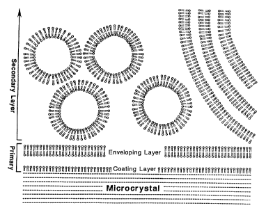
FIG. 1 is a schematic representation of the
phospholipid-coated microcrystal. The symbol O== is
a phospholipid (0 is polar head; -- is the pair of
fa-cty acyl chainsj. Diameter is O.5 um (range
0.05-10 um).
SiIBSTITUTE _ SHAT
~r . 'P s"' '( i
WO 91/16068 ~ ~~ ~ ~ ~:i ~' 4~ PCT/US91/02804
12
FIG. 2 presents drawings of a field observed
with a fluorescent microscope when the 20% (w/v)
oxytetracylcine, 20% (w/v) egg lecithin microcrystal
preparation, doped with Nile Red, was spread on a
slide. White indicates high fluorescence intensity.
The upper figure shows the pattern of oxytetracycline
fluorescence. The smaller particles are approx. 0.2
um diameter. The lower figure is of the identical
field excited at the Nile Red wavelength. This
fluorescence shows the distribution of the lecithin
in the preparation.
FIG. 3 shows the percent of oxytetracycline
remaining in the Ieg muscle of rats (n = 4) after
injection of 0.1 mI of 20% OTC in microcrystaline
form coated with 5% egg lecithin. The data for days
1-7 post-injection are compared with results for the
same quantity of OTC injected as a commercial
2-methyl-pyrolidone solution.
FIG. 4 shows the levels of OTG in central
arterial blood in the experiment of Fig. 3.
FIG. 5 shows the blood levels of OTC in calf
after intra-muscular injection of 20% (w/v) OTC (10%
(w/v) lecithin-coated) microcrystals.
FIG. 6 shows the time course of protection
against edema by 5 mg indomethacin injected
intra-muscularly as lecithin-coated microcrystals,
compared with an equal dose injected as an alkaline
solution.
FIG. 7 shows typical time courses of the level
anesthesia in rats measured as vocalization threshold
(mAmp) to intradermal electrical stimuli after
intra-venous injection of lecithin-coated alphaxalone
microcryscals.
FIG. 8 shows the time course of anesthesia in
the human skin (pin prick) achieved with
SUBSTfTUTE SHCC'.'
WO 91 / 16068 ~ F..~~ ~~ ~ fi ~;~ PCT/US91 /02804
13
lecithin-coated 20% (w/v) tetracaine hydroiodic acid
microcrystals (top panel), compared with
tetracaine-HC1 solutions (bottom panel).
DESCRIPTION OF T8E INVENTION
My invention provides a means for creating
injectable, tissue-compatible suspensions of
water-insoluble drugs at high concentrations. This
allows the parenteral (injection) administration of
drugs. It is generally applicable to any
water-insoluble drug which is in the crystalline
state at 37°C. Formulation as a phospholipid-coated
microcrystal enables the drug to be injected or
otherwise parenterally administered. The formulation
is unique in satisfying all of the 6 criteria
(tissue-compatibility, < 10 um size, injectable
quantity, non-antigenicity, payload, and physical
stability) for a maximally-useful injectable form.
The relationship between the microcrystal and the
coating phospholipid is depicted schematically in
Fig. 1. Central to my invention is the use of the
amphipathic or amphiphilic properties of
phospholipids in general and lecithin in particular.
Webster's Medical Desk Dictionary (Merrian-Webster
Inc., Springfield MA, 1986) defines
amphipathic/amphiphilic as "... consisting of
molecules having a polar water-soluble terminal group
attached to a water-insoluble hydrocarbon chain." In
Fig. 1, the polar head group of the phospholipid is
denoted by circles and the hydrophobic hydrocarbon
chains are denoted by "sticks". Many substances are
-amphipainic, including soaps, surfactants and
detergents. Unique to my invention is the use of
phospholipids to shield the hydrophobic surface of
SUBSTITU~'E ~Ei~T
~j '~ ~ ~ is ' i
1 l: v t? V~
WO 91 / 16068 PCT/ US91 /02804
14
the crystalline drug and to provide additional
membranous barriers against reassociation of the
crystals. Other amphipathic molecules such as soaps,
surfactants and detergents are unable to provide such
stable and tissue-compatible structures. Also unique
to my invention is the means of forming these stable
phospholipid-coated microcrystalline structures.
This is described below.
Size reduction and primary coatincl:
As described herein, the crystalline drug
substance is reduced to <10 um or submicron
dimensions in an aqueous medium by sonication or
other treatments involving high shear. Lecithin (or
other membrane forming lipid), present during the
sonication, is itself broken into highly reactive
fragments with exposed hydrophobic surfaces. These
fragments coat and envelop the submicron crystals
creating a primary coatin~c~. A requirement for this
process is that the lecithin and drug be present
together during the sonication or alternative
high-energy dispersing process. (Sonication of drug
crystals, followed by rapid mixing of pre-formed
phospholipid vesicles does not give stable sub-micron
aqueous suspensions of the drug.) The subsection
entitled "Methods of Preparation" specifies
alternative methods involving in-flight evaporative
coating and solvent dilution. The common aspect of
all of these preparative methods is that the fatty
aryl chains of the phospholipid must have direct
access to the microcrystal during the coating process.
In my invention, the amphipathic properties of
the'phospholipid satisfy both the hydrophilic
properties of water and the hydrophobic properties of
the crystal surface. Also, the phospholipid membrane
SUBSTITUTE SHEET
fJ
WO 91/16068 PCT/US91/02804
surface serves as a stationary barrier to reformation
of macroscopic crystals. A second useful property of
the primary coating is modification of the rate of
the dissolution process. Possible structural
features of the phospholipid-microcrystal interaction
are schematized in Fig. 1.
Secondarv coating: Peripheral phospholipid
In addition to making use of lecithin and other
membrane-forming lipids as a coating and enveloping
material, my invention makes novel use of membrane
-forming lipids as mechanical buffers, organizers of
aqueous volume and retardants of recrystallization of
the drug. This is achieved by excess phospholipid in
the form of unilamellar and multi-lamellar
phospholipid vesicles which form a secondary coating
of the suspended microcrystal. Predominantly
unilamellar vesicles are formed as a byproduct of the
sonication and primary coating process. Their
retention in the preparation was found to improve the
long-term stability of the formulation. Also,
preformed multi-lamellar vesicles (made by
homogenization) or uni-lamellar vesicles can be added
to the preparation to improve its stability or
pharmacokinetics (Example 5). The secondary coating
is loosely attached to the coated microcrystal.
Peripheral vesicles associate with and dissociate
continuously in the preparation. The secondary
coating can be removed by repeated centrifugation and
resuspension of the preparation (Examples 3 and 11).
Peripheral vesicles forming a secondary coating
stabilize the preparation. While not wishing to be
bound to Amy particular i.i:eor y O1 lWVU2 'v~ a~ ~iOn,
detailed consideration has suggested the following
mechanisms:
SUBSTITUTE SHEET
WO 91 / 16068 ~ ~ ~:~ a~ ~a a PCT/US91 /02804
16
> They act as volume buffers interposed
between the primary-coated microcrystals. The
crystalline and microcrystalline drugs are often
more dense than the phospholipid which is, in
turn, more dense than water. Thus they will
tend to settle under the influence of gravity
and will experience greater long-range
interactions (van der Waals attraction) than the
other two constituents. The secondary coating
increases the distance of closest approach of
the microcrystalline drug cores, thereby
decreasing the van der Waals attraction. It is
probable that part of the driving force for the
secondary coating is van der Waals attraction
between the primary-coated microcrystal and the
phospholipid vesicle. Phospholipids (notably
lecithin) are ideal as the primary and secondary
coating because they are strongly hydrated and
engage in well-documented short-range repulsive
interactions which make them very resistant to
aggregation and fusion.
> When peripheral phospholipid is present at
20% (w/v), the majority of the aqueous volume of
the preparation is enclosed within phospholipid
membranes. This serves as a topological barrier
to recrystallization of the drug in a
preparation during long-term storage. Re-formed
crystals can not be larger than diameter vesicle
or distance between them, both of which can be
kept small.
Pnysicai characteri5i.ic:a of formulation:
Sonication is most conveniently carried out with
SUBSTITUtE SHEET
i~ ~i
WO 91 / 16068 PCT/US91 /02804
17
the drug at concentrations of 5% (w/v) or less and
the membrane-forming lipid at 5% or greater. This
results in a syringable suspension of coated
microcrystals of predominantly sub-micron dimensions,
with the particles exhibiting Brownian motion
(Examples 2,3 and 11). Over a period of 1-2 days the
microcrystals settle creating a distinct zone in
which the drug concentration is 20-40% (w/v). The
final concentration and volume are dependent on the
choice of the drug and upon the peripheral
phospholipid concentration. In most preparations the
bottom zone is resuspendable with inversion to give a
homogeneous and syringable suspension, even after a
period of months. For preparations in which this was
not the case, resuspendability was obtained by
increasing the peripheral phospholipid concentration.
The slow sedimentation process can be used as a
means of concentrating the preparation. Removal of
the volume above the sedimentation zone after 1-2
days results in preparations in which the drug is at
20-40% (w/v). Long-term storage results in no
further settling. The preparations remain
homogeneous, syringable and pharmaceutically
acceptable for many months (cf. Examples).
Microscopic examination of these preparations reveals
separated micron and sub-micron diameter crystals of
the drug. The volume between these drug
microcrystals is almost completely filled with
phospholipid vesicles, visualized by Nile Red
staining (cf. Examples 2,3 and 11). In this
concentrated form, the drug microcrystals exhibit
only restricted Brownian Motion. Under microscopic
observation they are not observed to change posiiinn
in relation to eachother. They vibrate or "dance in
place" about their central position. This partial
SUBSTITUTE SHEET
a ~ L ;T i
J ~.~ 'V ~v'~ V
WO 91/16068 PCT/US91/02804
18
restriction of motion is probably an important factor
in the long-term stability of the preparation. When
stored, concentrated preparations are diluted many
thousand-fold into drug-saturated water, the
microcrystals retain their micron or sub-micron size.
Modes of Administration:
As noted above, the primary utility of the
coated microcrystal is its injectability. Applicable
injection sites are any tissue or body cavity. They
include but not limited to intra-venous (IV),
intra-arterial (IA), intra-muscular (IM),
intra-dermal, sub-cutaneous (Sub-Q), intra-articular,
cerebro-spinal, epi-dural, intra-costal,
intra-peritoneal, intra-tumor, intra-bladder,
intra-lesional, sub-conjunctival, etc. In addition,
the phospholipid coating and submicron size of the
preparation may prove to have advantages for oral
use, both as an aqueous suspension and as a
lyophilized product. Similarly, the aqueous
suspension may show advantages for topical
application, instillation into the eye. The
preparation can deliver drugs by the inhalation
route, in the form of either an aqueous suspension or
a lyophilized powder. It is also likely that the
preparation will be useful for administration of
pesticides and in creating high value biocompatible
products, as exemplified by suspension of drugs in
drinking water (Example 15).
Rate of release
The most important determinant of the rate of
releaSa OZ the drug IS i.llc c ioit.v~ of iiaj3 .tivia Site.
If the formulation is injected intravenously, it can
be released from the microcrystal quite rapidly. If
SUBSTITUTE S~iEET
~~'~~a
WO 91/16068 ~ ~_' ~. PCT/US91/02804
19
the formulation is injected at high volume into a
confined space such as muscle, the net rate of
release can be exceedingly slow. The intra-venous
case will be considered first.
The blood is a fluid medium which is capable of
diluting the preparation 1,000,000-fold within
approx. 1 min. When a concentrated lecithin-coated
microcrystal preparation is diluted in blood, the
individual microcrystals, initially in an~environment
consisting of other coated microcrystals, peripheral
lipid and drug-saturated water, are transferred to an
environment consisting of serum proteins, serum
lipoproteins and cellular blood elements. My in
vitro fractionation experiments (Examples 3 and 11)
suggest that the secondary coating will be rapidly
lost. All of the blood elements are capable of
binding lipophilic molecules and will do so as
rapidly as the microcrystal can dissolve. In cases
where the drug is sufficiently water soluble,
dissolution into the aqueous portion of the blood is
sufficient to distribute the drug. When
water-solubility is insufficient, a continuous
process of dissolution and binding of the drug to
blood elements serves to remove the drug from the
microcrystals. The rate~of dissolution of the
microcrystal will depend_upon the thickness and
stability of its primary coating, the
water-solubility of the drug and other
physico-chemical parameters. Example 10 shows that
the anesthetic alfaxalone can leave the microcrystal
and enter the brain within 10 sec of its IV
injection. It is possible to reduce the rate of
release after IV administration icy variation of the
thickness of the primary coating or by inclusion of
SUBSTITUTE SHEET
WO 91 / 16068 ~ ~~ ~ ~ a ~' °~ PCT/ US91 /02804
small quantities of water-insoluble oil (such as
vitamin E) in the preparation.
With injection into a tissue such as muscle, the
preparation does not undergo rapid dilution. It
generally remains in the initial elements of volume
created by the injection. These are generally
macroscopic and there is little flow or agitation.
Diffusion the drug out of this volume is slow because
of the relatively large distance involved and is
further slowed by the low water solubility of the
drug. The larger the injected volume and the lower
the water solubility, the slower will be the rate of
removal of the drug (Example 7). In the extreme, the
release process can require upwards of 14 days. For
high and fixed volumes and drug concentrations, the
rate of removal can be increased by incorporation of
hypertonic glucose or carboxycellulose in the
vesicles of the secondary coating. This resists the
mechanical pressure of the tissue which tends to
solidify the injected preparation (Example 5). IM
injection is useful to create a depot of drug and to
obtain sustained release to the blood over a period
of days. Injection directly into the target tissue
or lesion is useful because it achieves high and
sustained concentrations~of the drug at the site
where it is needed without involving the rest of the
system.
Methods of Preparation:
1. Sonication: The sonication process reduces
the size of supra-molecular structures by the
process of cavitation. The process creates
small empty volumes which collapse, propelling
material together at high speed, resulting in
shattering and sheer. This simultaneously
SUBSTITUTE SNEET
W0 91!16068
PCT/US91/02804
21
breaks up the drug crystals and phospholipid
lamellae into submicron fragments. The
phospholipid membranes are shattered in
directions both parallel and perpendicular to
their planes, yielding surfaces which can coat
the hydrophobic surface of the microcrystal, an
which can rejoin to envelope it, respectively.
Thus the phospholipid concentration must be
adequate for the rate of coating and enveloping
to exceed the rate of rejoining of broken
crystals. I have observed that sonication
usually works well when the drug concentration
is 5% (w/v) or lower and when the phospholipid
concentration is 5% or greater. The role of the
secondary coating of phospholipid vesicles has
been described above.
It does not work well if the drug and
lipid are sonicated separately and added
together. In fact, in the absence of a
membrane-forming phospholipid one seldom
succeeds in reducing the drug to sub-micron
size for even a short time. Sonicated or
homogenized lipid can be added to
already-prepared coated microcrystals to
increase or modify their peripheral lipid
content. As described above, the
preparation can be concentrated to 20% -
40% (w/v) by allowing it to settle.
The product can be put into dry form by
lyophilization to yield a powder which can
De i~tEr reCOIlSf.iviitEu (t'~''~iiauyric J) . TfaiS
is useful when the long-term chemical
SUBSTITUTE SHEET
1. ~i /:n
WO 91/16068 ~ ~ y ~ a ~ PCI'/US91/02804
22
stability of the to-be-encapsulated drug in
an aqueous environment is poor.
SUBSTITUTE SHEET
WO 91 / 16068 ~ ~ ~ ~ ~ ~ ~ ~ PCT/ US91 /02804
23
2. Methods involying high pressure and shear:
The crystalline drug and phospholipid are
pre-mixed by high-speed homogenization (as with
Waring Blender. Further size reduction and
coating can be accomplished by the process of
MicrofluidizationR (Microfluidics Corp., Newton
MA 02164). The process relies on high shear
created by collision of opposing jets of liquid.
The apparatus is described by Mayhew et al. in
Biochim. Biophys Acta 775:169-174, 1984. An
alternative is high pressure homogenization by
means of the French Pressure Cell or "French
Press" (SLM Instruments, Urbana IL). In this
process, the sample is forced at high pressure
and high shear through a narrow orifice and
undergoes rapid decompression to atmospheric
pressure. Other details are as in #1 above.
3. Sonication or high shear in volatile
organic solvents: Microcrystals can be
prepared by suspending the crystalline drug
and the membrane-forming lipid in a
volatile non-polar solvent in which the
drug is poorly soluble
(dichlorodifluoromethane or
dichlorotetrafluroethane or Freon (e. g.
trichlorotrifluroethane, c.f. Examples 6
and 8). The suspension is reduced in size
by sonication or high-speed shear by the
methods described above (#1 or #2). The
solvent is removed by evaporation. The
resulting powder can be stored for later
reconstitution with water or can be
reconstituted immediately.
4. Size reduction in air: Drug crystals can
SUBSTITUTE SHEET
WO 91/16068
PCT/ US91 /02804
24
also be reduced in size by high speed
impact in air. They can be subsequently
coated by wetting with a solution of
phospholipid or glycerol lipid in a
volatile solvent containing lecithin, with
the solvent removed by volatilization. The
powdered product can be suspended in
water. Alternatively, micronized crystals
can be wetted by a water-miscible liquid
dissolving lecithin and rapidly introduced
into an aqueous medium.
5. In-flight crystallization: A solutions of
the lipid and drug in a volatile solvent
can be sprayed, with the solvent removed by
evaporation while in flight. The
microcrystals are collected and dried on a
smooth surface. The microcrystals can
either be stored in the powdered form for
later reconstitution with water or can be
reconstituted immediately.
6. Solvent dilution: Solutions of lipid and
water-insoluble drug are made using a
water-miscible~organic solvent (e. g.
ethanol). The. solutions are expressed into
an aqueous medium with high agitation or
sonication. The solvent dissolves in the
water, leaving behind the drug in
microcrystalline form coated with the
lipid. The organic solvent can be
completely removed by filtration or by
sedimentation vi the vOatcu iTiiCiGi,i:y5tai5
and removal o~ the supernate.
SUBSTITUTE SHEET
wo 9 m bobs ~ ~ ~ ~ ~ ~ '~
PCT/US91/02804
Selection of the drug to be coated:
Any substantially water-insoluble drug which is
in the crystalline or solid state at temperatures of
37°C is applicable. The drug should generally have a
water solubility of < 5 mg/ml at physiological pH
(6.5-7.4). Use of drugs with higher water solubility
is not precluded if experimentation shows minimal
tendencies to reorganize into macroscopic crystals
during the desired shelf life. It is generally
desirable to choose a total drug concentration > 4x
the drug's water solubility, such that at least 80%
of the drug is in the microcrystalline form. This
choice takes advantage of the high payload and
sustained-release characteristics associated with the
coated microcrystal. Finally, it is preferred that
the drug be intrinsically non-irritating. It is also
desirable that the drug be chemically stable in a
humid environment. Otherwise it may be necessary to
produce lyophilized forms.
The most frequent examples are drugs which are
water-insoluble but have moderate-to-good oil
solubility. However, oil solublity per se is not a
requirement for incorporation into
phospholipid-coated microcrystals. Many drugs which
have tight crystal structures, high melting points
are not particularly water- or oil-soluble. These
drugs can also benefit from phospholipid-coated
microcrystal formulation. Similarly, it is not
necessary for the drug to be uncharged to be put into
microcrystalline form. It is only necessary that the
water solubility of the crystalline form of the drug
be low.
Rendering a Water-Soluble Drug Water-Insoluble
It is possible to use an intrinsically
SUUST1TUTE SNEET
4
WO 91 / i 6068 ~ ~ ~ ''~ " ~ '~ PCT/ US91 /02804
26
water-soluble drug in my invention providing that it
can be rendered water-insoluble by complexation. For
example an insoluble hydroiodic acid (HI) salt of the
local anesthetic tetracaine is used to extend its
duration of action S-fold (Example 12). If the drug
is charged at physiological pH, it can often be
rendered insoluble by substituting a more lipophilic
or structured counter-ion. Examples for rendering a
positively-charged drug less water soluble include
complexation with 2-naphthylenesulfonate (napsylate),
gluconate, 1,1' methylene bis
(2-hydroxy-3-naphthalene) carboxylic acid (pamoate),
tolylsulfonate (tosylate), methanesulfonate
(mersylate), glucoheptanoate (gluceptate),
bitartrate, polyglutamic acid, succinate, acetate, or
behenate (anionic form of waxy fatty acid). In
choosing fatty acyl anions it is advisable to select
species with either short chain lengths or very long
chain lengths, such that the tendency of towards
micellarization is minimized. In some cases
substitution with bromide, iodide, phosphate or
nitrate is sufficient to render the drug less
soluble. Examples for rendering a negatively-charged
drug less water soluble include complexation with
calcium, magnesium or their 1:1 fatty acid salts, and
with various amines, including
dibenzylethylenediamine (benzathine), rd, N'
(dihydroabietyl)ethylene diamine (hydrabamine) or
polymers such as polylysine. The choice of these
counterions is made largely on an empirical basis,
with stability of the derived crystals and their
compatibility with water being primary criteria.
since release oz the drug after dilution or injecciom
can,involve removal of both the charged and the
uncharged forms of both the drug and its counterion,
SUBSTITUTE SHEET
WO 91/16068 N ~~ ~ ;~j ~.~
PCT/US91/02804
27
these systems offer both complexity and diversity of
kinetics. With sufficient study of the in vitro
behavior of the phospholipid-coated microcrystals
made from a number of these binary salt systems, and
with judicious choice of the most promising examples,
the desired in viyo pharmacokinetics can be
approximated.
Also, it is possible in some applications, to
prepare microcrystals at more extreme pH (4.0 - 6.4
or 7.5 - 10.0) in order to suppress ionization and
thus decrease the solubility of the drug. The
allowable extremes of pH in each particular case will
be determined by the concentration of the drug, the
number of acid or base equivalents which it carries,
its rate of dissolution and the size of the injected
compartment and (in terms of shelf life) the
stability of the membrane-forming lipid.
Selection of the membrane-forming lipids for coating:
The primary requirement is that the coating
lipid be membrane-forming. This is satisfied by all
lipids which, in the presence of excess water, make
bilayer structures of the type which is
well-documented for phospholipid vesicles or
liposomes. This requirement is not satisfied by
fatty acids, detergents,. non-ionic surfactants (e. g.
polyethylene glycol) or triglycerides (vegetable
oils, tristearin, "fats"). A secondary requrement is
that the lipid not have a proclivity for converting
into micellar structures. This excludes
phospholipids of short chain length (6 or less) or
lyso-lecithin (containing a single fatty acyl
chain) . Higli stability ;,f the coati::y mwtcrial in
membrane form is necessary to keep the drug material
from rearranging into macroscoF~:c crystals. This is
WO 91/16068 ~ (~ r~ 4 w ~ PCT/US91/02804
28
one reason why non-ionic surfactants do not work well
for my intended purpose.
Useful examples of membrane-forming lipids are
given below:
CLASS A: Primary phospholipids (usable in pure
form) include the following:
Lecithin (phosphatidyl choline)
Sphingomyelin
Synthetic zwitterionic phospholipids or
phospholipid analogues
To this class belongs all phospholipids which
spontaneously form membranes when water is added.
These phospholipids can be used in pure form to
produce coated-microcrystals. Of all the
phospholipids, lecithin is the most useful example
because of its high availability and low cost.
CLASS B: Phospholipids capable of
calcium-dependent aggregation.
These phospholipids include the following:
Phosphatidic acid
Phosphatidyl serine
Phosphatidyl inositol
Cardiolipin (diphosphatidyl glycerol)
Phosphatidyl glycerol
These lipids carry a negative charge at neutral pH.
Preferably these phospholipids can be mixed with
lecithin to obtain negatively-charged surfaces which
will give repulsion between particles. When
introduced into a medium containing 2 mM calcium
(such as blood or interstitial), membranes containing
these phospholipids are expected to show elevated
aggregation and higher reactivity with cell
membranes. This cdm La usaiul in causing the
injected microcrystals to aggregate within the
tissue, giving slower release rates. The usefulness
Hr0 91 / 16068 ~ ~ ~ ~ ;~ ~ PGT/US91 /02804
29
of this class is limited by the high cost of these
phospholipids, relative to lecithin.
CLASS C: Phosphatidyl ethanolamine promotes
aggregation in a calcium-independent manner. It can
be used in the pure form to coat microcrystals at pH
9. When the pH is brought to 7, as upon injection
into blood or tissue the membranes become reactive,
causing the particles to aggregate and to attach to
cell membranes. This can have the useful property of
slowing the release rate.
CLASS D: Cholesterol and steroids. These can
not be used as a sole coating material: They do not
form membranes in the pure state. They can be added
to the lecithin or other coating material to change
its surface activity, the "microviscosity" or
distensibility of the coating. With a steroid
hormone (estrogen, androgen, mineralo- or
glucocorticoid), it is possible to influence the
local tissue response to the microcrystals as well as
influencing their physical disposition.
CLASS E: Semi-lipoidal molecules can be
incorporated into the phospholipid or glycerol lipid
membrane and change the surface activity of the
microdroplet. Molecules included in this class are
the following:
Stearylamine or other long-chained alkyl amines which
can be primary, secondary, tertiary or quaternary
substituted. These give the microcrystal coating a
positive charge and make them more reactive with cell
membranes. Benzalkonium chloride is an aromatic
example which is particularly useful because it also
functions as a preservative against microbioiogical
growth in the preparation.
suBSTiTU-rE sHE~-r-
WO 91 / 16068 ~ ~ '~ ~i ~ ~; ~ PCT/ US91 /02804
Fatty acids. These can be incorporated at low
concentrations (<0.02 gm/gm phospholipid) to alter
the phospholipid packing and reactivity.
CLASS F: Membrane-active agents, glycolipids
and glycoproteins to modify surface properties.
Examples of membrane-active agents include nystatin,
amphotericin B and gramicidin which are
surface-active antibiotics. These have been shown to
bind to the surfaces of phospholipid membranes and
change their permeability. Glycolipids or
glycoproteins could be included as a means of
modifying surface reactivity. Likewise, antibodies
can be coupled to membrane constitutents to direct or
retain the microcrystal association with targeted
cells or tissues. (Glycolipids, glycoproteins, and
anti-bodies are classified as "biologicals". They
would have to be screened for pyrogenicity,
antigenicity etc. before use, and the process of
gaining regulatory approval for such formulations
would be more complex.)
CLASS G: Mono-glycerides.
These are not phospholipids, but they have been shown
capable of forming oriented monolayers and bilayers
in the presence of decane (Benz et al. Biochim.
Biophys. Acta 394:323-334, 1975). They may thus
prove have some use in coating for microcrystals.
Examples of these lipids include, but are not limited
to, the following:
1-monopalmitoyl-(rac)-glycerol (Monopalmitin)
1-monocaprylol-(rac)-glycerol (Monocaprylin)
1-monooleoyl-(rac)-glycerol (C18:1, cis-9)
(Monoolein)
1-monostearyl-(rac)-glycerol (Monostearin)
~y0 91/16068
PCT/ US91 /02804
31
Commercially Available Membrane-Forming Lipids:
Several forms of lecithin are contemplated. As
an example, egg lecithin (Sigma Chemical Co.) is
used in all of the presented examples. It is
preferred for its low price and low degree of
unsaturation. Lecithin is also available from bovine
heart. Soy bean lecithin is less expensive. It has
a higher degree of unsaturation. Several synthetic
varieties of lecithin are available which differ in
chain length from 4 to 19 carbons (Supelco, Inc.).
It is believed that lecithins with chain lengths in
the biological range (10-18) are useful in various
applications. Unsaturated lecithins (dioleoyl,
dilinoleoyl; beta oleoyl; alpha-palmito beta oleoyl;
alpha palmitoyl beta linoleoyl and alpha oleoyl beta
palmitoyl) are also available. Diarachidonyl
lecithin (highly unsaturated and a prostaglandin
precursor) is also available.
Phosphatidic acid is available from egg or as
synthetic compounds (dimyristoyl, dipalmitoyl or
distearoyl, Galbiochem). Bovine phosphatidyl serine
is available (Supelco or Calbiochem).
Phosphatidyl inositol is available from plant
(Supelco) or bovine (Calbiochem) sources.
Cardiolipin is available.(Supelco) from bovine or
bacterial sources. Phosphatidyl glycerol is
available from bacterial (Supelco) cources or as
synthetic compounds (dimyristoyl or dipalmitoyl;
Calbiochem).
Phosphatidyl ethanolamine is available as egg,
bacterial, bovine or plasmalogen (Supelco) or as
synthetic compounds dioctadecanoyl and dioleoyl
amalague5 ama 3ihexadccyl, uilauryl, dimyristoyl anti
dipalmitoyl (Supelco and Calbiochem).
suBSrnurE sH~r
WO 91 / 16068 ~ ~ ~' (-'~~
PCT/ US91 /02804
32
Monoglycerides are available from Sigma Chemical
Co. (1-monopalmitoyl-(rac)-glycerol, monopalmitin;
1-monocaprylol-(rac)-glycerol, monocaprylin;
1-monooleoyl-(rac)-glycerol (C18:1, cis-9),
monoolein; 1-monostearyl-(rac)-glycerol, monostearin).
Other constituents:
It is possible to add other constituents to the
microcrystal to increase its stability or modify its
rate of release. For example,
pharmacologically-acceptable oils can be added at low
weight concentration to facilitate contact between
the microcrystal and the phospholipid or glycerol
lipid coating. It is necessary that the type of oil
and its weight concentration be chosen such that the
crystalline drug not be dissolved by the oil and that
the coating by the membrane-forming lipid not be
disrupted. These relationships can be determined
empirically. Useful oils include, but are not
limited to, vitamin E, isopropyl myristate, benzyl
benzoate, oleyl alchohol, mineral oil, squalene and
vegetable oil. Example 6 gives evidence that
incorporation of vitamin E in a lecithin-coated
microcrystal preparation of erythromycin decreases
the rate of dissolution of the drug, thereby reducing
tissue irritation by the. drug.
It is also possible to "precoat" the
microcrystals by phospholipid-compatible,
non-antigenic molecules which are solid at 37°C.
Examples include paraffin, tristearin, ethyl oleate,
cetostearyl alcohol, cetyl alcohol, myristyl alcohol,
stearyl alcohol and petrolatum. For example, these
materials can be incorporated into the primary
coating by sonication or shear at temperatures above
their melting points. Stabilization can be achieved
Hr0 91 / 16068 '~ ~ ~ ~ ~~ ~ PCT/ US91 /02804
33
by adding lecithin during the process as temperature
is allowed to return to the solidification point of
these materials. It is desirable to use low weight
concentrations (< 10%) of such that the payload is
not degraded, the rate of dissolution of the drug is
not unduly impeded. Also, biodegradability may
impose a further limitation. Example 13 describes
the lecithin-compatibility of paraffin in
micro-particulate form.
Suspending medium:
In the final preparation, the continuous phase
is generally water, buffered to a
physiologically-acceptable pH and containing an
iso-osmotic concentration of sodium chloride or
glucose. In certain applications involving
intra-muscular injection of large volumes of
microcrystals at high concentration, it is useful to
increase the osmolarity of the medium (e. g. glucose
concentration) to facilitate the spreading of the
material in the muscle. As noted above, this can
retard the process of compaction after intra-muscular
injection. Where permissible, viscosity-increasing
agents such as carboxycellulose can be useful to
alter the pharmacokineties following intra-muscular
injection and to decrease the rate of sedimentation
of the microcrystals upon storage.
In certain applications it is useful to
substitute a polar solvent for water, as in Example 7
where albendazole sulfoxide did not show sufficient
~long-term stability in the presence of water. (Also
see Example 5.) Examples of non-aqueous polar
SUIvents whiCi~ can bF used incluue, but ai8 irGt
limited to the following: glycerin (water-miscible
liquid with a dielectric constant of 42.5) and
gUBSTITUTE SHEET'
WO 91 / 16068 ~ ~~ ~ ;~ g~ (.:~ . . PCT/ US91 /02804
m« ~<i~
34
propylene glycol (water-miscible liquid with a
dielectric constant of 32). The coated
microcrystals can be made in these media, or can be
allowed to sediment into these media. The primary
requirement is that a substantial portion of the
phospholipid or coating material be in membranous
form in this solvent. An equally important
requirement is that the crystalline material not be
sufficiently soluble in the solvent that it will
recrystallize.
Preseryatiyes
Oil-soluble preservatives can be added in
process during the primary or coating phase. These
include, but are not limited to, benzalkonium
chloride, propylparabum, butylparaben, and
chlorobutanol. There are also numerous water- and
oil-soluble agents which can be added to the finished
product as preservatives, including, benzyl alcohol,
phenol, sodium benzoate, EDTA, etc.
Optional Lyophilization and Reconstitution
Aqueous microcrystal preparations can be
lyophilized to give a dry product which can be
reconstituted with water.(Example 6). This is
particularly useful for a drug which does not have
long-term stability in an aqueous environment. It is
also possible to conduct the sonication or shear
process in a volatile organic solvent in which the
crystalline drug is not substantially soluble, and to
prepare dry coated microcrystals by solvent
evaporation (Example 8). These procedures give
micrccrystai~ surroumieu by layers of phospholipid in
the anhydrous state. Such forms are suitable for
~r0 91 / 16068 ~ ~ ~ ~ ~ ~ ~ PCT/ US91 /02804
oral administration or for reconstitution with water
and injection.
Design of the Final Product
One skilled in the art following the
instructions provided herein will have no difficulty
in empirically determining the:
Most convenient method of vreparation:
Sonication vs. high pressure and shear vs.
methods involving organic solvents vs.
impact in air vs. in-flight crystallization
vs. solvent dilution
Most advantageous form of the drug:
Crystal of neutral drug vs. crystal of
charged drug vs. more complex solid forms
of the drug
Optimal membrane forming lipid:
Based on reactivity and stability of
membranes, blood and tissue compatibility
and price
Optimal conditions for manufacture, includin
Input drug and.phospholipid ratio;
incorporation of small amounts of oils or
waxes as modifying agents; duration of
sonication, shear, etc; use of
sedimentation as a means of size selection;
addition of more peripheral lipid in
unilamellar or multi-lamellar form;
addition of osmotic ~r viscosii:y-affectiy
agents.
SUBSTITUTE SHEET
WO 91 / 16068 ~ ~ ~ ~ ~'- ~~ ~ PCT/US91 /02804
36
Optimal particle size:
Which can be controlled to a certain extent
by the power supplied, the duration of
processing, the drug and phospholipid
concentrations,
And which can be selected between 50 nm and
um by sedimentation velocity
Obtimal compositions to achieve the desired
shelf life and pharmacokinetics:
Including study of the effect of the above
factors on the pharmacokinetics after
injection. Particularly important are the
particle size, primary and secondary
phospholipid content, and additives to
avoid compaction after injection into a
tissue.
Most advantageous mode of administration:
Including injection (IV, IA, IM, etc.),
oral, topical administration, inhalation,
etc.
Weights and measures
All parts and percentages reported herein are by
weight (w/w) or weight/volume (w/v) percentage, in
which the weight or volume in the denominator
represents the total weight or volume of the system.
Concentrations of water soluble constituents in
aqueous solution (e.g. glucose) are given in
millimolar concentration (mM = millimoles per liter)
referred tU Llle 4'GILiWE Of iwai.Cr iia t he ~liSveW. hil
temperatures are reported in degrees Celsius.
Diameters or dimensions are given in millimeters (mm
SUBSTITUTE SH~BT
~~ ~E~ i~
WO 91 / 16068 ~ ~ ~ ~ ~.~ a ~.~ PCT/US91 /02804
37
- 10-3 meters), micrometers (um = 10 6 meters),
nanometers (nm = 10 9 meters) or Angstrom units (_
0.1 nm). The compositions of the invention can
comprise, consist essentially of or consist of the
materials set forth and the process or method can
comprise, consist essentially of or consist of the
steps set forth with such materials.
DETAILED DESCRIPTION OF TFIE INVENTION
EXAMPLE 1
Lecithin-coated microcrystals of oxytetracycline
(OTC) were prepared by sonication in the following
manner:
Into a 150 ml glass beaker, 4.4 gm
oxytetracycline dehydrate (Sigma, 0-5750) and 16.0 gm
egg lecithin (L-alpha-phosphatidylcholine from egg,
Type XV-E, Sigma, P-9671) were added coarsely mixed
using a glass stirring rod. Next, an aqueous
solution of 300 mM glucose, 10 mM tris adjusted to pH
7.4, was added to give a final volume of 80 ml. The
1.0 cm diameter probe of a SonifierR Cell Disrupter,
Model W185D (Heat System and Ultrasonics, Plainview,
N.Y.) was immersed in the liquid and mixture was
sonicated for a total of-60 min at power stage 10
(nominally 100-150 watts). The temperature of the
mixture was controlled by water-jacketing and by
occasional interruptions of the sonication, and was
not allowed to exceed 60°C. A pH of 5.0 was
maintained by HC1 addition. Sonication resulted in
an opaque yellowish-biege suspension which was
covered and allowed to settle for 24 hrs. The botton
ii ml coni.aineci a visii~Ie p~Gcipitate of OTC at a
concentration of 40% (w/v). The supernatant
contained phospholipid vesicles. Bottom 22 ml were
SUB.. STITUTE . SNEET
WO 91/16068 , ~ ,~, ~y ,...)
PCT/ US91 /0280
l.i ri: Cu ''J
38
collected and the precipitate was resuspended with
gentle shaking. It contained lecithin-coated
microcrystals of OTC. It behaved as a somewhat
viscous but syringable liquid.
E'luorimetric analysis and high pressure liquid
chromatographic (HPLC) showed that the top phase
contained very little oxytetracycline. The bottom
phase sampled as the bottom 22 ml contained > 98% of
the added oxytetracycline. It was 20% (w/v) in OTC
and 20% (w/v) lecithin. Aliquots were taken from
both phases and were diluted into OTC-saturated
buffer and were analyzed for diameter (~ SD) using a
Coulter N4-MD Submicron Particle Analyzer. The top
phase was analyzed ~or particle size was found to
have diameters of 30.5+8 nm (77%) and >3,000 nm
corresponding to unilamellar and multilamellar
phospholipid vesicles, respectively. The top phase
was discarded. The lecithin-coated OTC microcrystal
fraction had the following weight-averaged particle
size distribution: 980+460 (SD) nm, 59%; 2,880+400
(SD) nm, 41%. Analysis of the preparation by
electron microscopy using negative staining
corroborated the above findings. Several
preparations were made as described above and were
filled into rubber-stoppered glass ampules and glass
bottles. With storage over a period of weeks some
settling was observed, but the preparation could be
rendered homogeneous with three inversions. The
preparation retained its properties, including size
distribution, OTC concentration, chemical integrity
and syringability (20 gauge or narrower) for over 9
months.
The importance of the lecithin coating was
demonstrated as follows: 4.4 gm OTC and 75.6 ml
glucose solution were sonicated for 60 min as
SUBSTITUTE SHEET
WO 91 / 16068 ~~ ~ '~~ ~ PCT/ US91 /02804
39
described above, but in absence of lecithin. A
coarse suspension was obtained with the following
characteristics: (a) Immediate sampling and
1,000-fold dilution into OTC-saturated water gave
particles visible to the naked eye. Analysis by the
Coulter Submicron Particle Analyzer reported that the
particles were "out of range" (> 3 um). The analysis
did not reveal any particles with diameters < 3 um.
(b) Within 10 minutes after sonication, all of the
OTC had settled to the bottom. The bottom phase was
not free-flowing and was not syringable. Within 1 hr
after settling, it became a solid mass which could
not be resuspended with shaking. Similarly, it was
impossible to stabilize this preparation by adding
pre-formed phospholipid vesicles to the sonicated OTC
immediately after cessation of sonication. Thus
sonication with lecithin (or other membrane-forming
lipid) is shown to be a critical step in the method
of preparation.
EXAMPLE 2
The preparation of Example 1 was repeated with
the following alterations: The preparation was
scaled to a total volume of 5.0 ml, the microtip
sonicator probe was used~and 0.15 mg Nile Red was
added at the same time as the lipid. This dye binds
to phospholipids and serves as a fluorescent marker
for the lecithin. A drop of the 20% (w/v)
preparation was spread on a glass slide and observed
with a fluorescent microscope (Leitz Wetzlar Dialux
20) at high power. Figure 2 is a black and white
drawing of a typical field. White indicates high
fluorescence. V~lti'1 ultra-ViviW ExCitati'via, 0TH
particles were observed by their intense yellow-green
fluorescence. The upper panel of Fig. 2 shows
SUBSTITUTE SHEET
L-~ ..~: i,: ,.. a
WO 91/16068 ~ ~ ~ 's ~~" ~'' '~
PCT/US91 /0280
depicts the fluorescent image of OTC. Discrete
particles with diameters ranging from approx. 0_2 um
and ca. 3 um were observed. The majority of the
particles were < 1 um diameter (number average).
The particles had clear boundries, but were
surrounded by diffuse yellow-green halo's. The
identical field was observed with near ultra-violet
excitation to give the Nile Red image associated with
the lecithin in the preparation (bottom planel). The
Nile Red image shows the lecithin to be surrounding
the OTC particles. A halo red fluorescence
surrounded the particles, extending 0.3 um to 3 um
beyond the boundry of the OTC microcrystal. Empty
spaces devoid of both OTC and lecithin, could be
readily discerned around alI particles situated near
the edge of the smear. Occasionally configurations
were observed suggesting that two OTC particles were
sharing a single phospholipid halo.
Brownian Motion was observed in the sample. The
larger (> 1 um diameter) particles which settled on
the glass slide showed no Brownian Motion, or highly
restricted motion. Particles of < 1 um diameter
showed Brownian Motion, moving between the larger
particles. Observations in regions of low
concentration showed that no particle could move a
distance greater than approx. 1/4 its diameter
without its halo experiencing a corresponding
movement. Furthermore, direct collisions of the
particles were never observed. These observations
explain the remarkable stability of the microcrystal
suspensions of my invention: The lecithin coats the
microcrystal, supplying both a hydrophobic surface
for contact with the crystaiiine surface and a
hydrophilic surface for contact with water. The
coated surface is enveloped by numerous layers of
SU3 TIiUiE SH
WO 91 / 16068 ~ ~ ~ ~ ~~ ~ PCT/US91 /02804
41
lecithin in membrane form, as revealed by the Nile
Red staining. The stability of the envelopment is
shown by the fact that the microcrystal always
remains within its lecithin halo, as revealed by
their respective fluorescence signals. The outer
lecithin layers guarantee that the coated
microcrystals do not approach closely enough to fuse.
The dissolution behavior of the lecithin-coated
microcrystals was observed under the fluorescent
microscope by placing it in contact a large quantity
of distilled water and applying the cover slip. The
smaller particles (approx. 0.2-1.0 um) moved with the
water flow. The Nile Red fluorescent halo moved
together with the fluorescent OTC particle, and the
brightness of the two fluorescent signals initially
remained in constant proportion. As the particle
moved into the distilled water, the OTC signal
dimmed, suggesting that the microcrystal was
dissolving. Only a small fraction of the Nile Red
image and intensity was lost, suggesting that the
lecithin coating was a persisting structure. The
dissolution behavior of the larger particles which
were generally more firmly attached to the slide was
somewhat different. The streaming caused them to
shed a large portion of their lipid. The larger
microcrystals were then observed to crack, splitting
off shards each of which carried away a portion of
the Nile Red halo with it.
Dissociation behavior was also studied using the
Coulter N4-Nm Submicron Particle Sizer. As stated in
Example 1, when the preparation is diluted 1,000 x
into isotonic glucose buffer saturated with OTC, the
preparation is stable and particles sizes of 980~4ti0
nm and 2,880~400 nm were observed. When the dilution
was made 1,000 x into distilled water, rapid
SUBSTITUTE SHEET
WO 91/16068
PCT/ US91 /02804
42
alteration of the preparation was observed.
Dissolution was expected since the final OTC
concentration becomes 0.2 mg/ml, which is lower than
the aqueous solubility of the drug (about 1 mg/ml).
Dissolution was observed, but it was accompanied by
the formation of some particles with diameters
greater than 3 um.
EXAMPLE 3
A Nile Red "doped" lecithin-coated microcrystal
preparation of oxytetracycline was made and the
following fractionation experiment was carried out to
delineate the relationship between the primary and
secondary phospholipid coatings for the
larger-diameter (1.0-1.9 um) coated microcrystals.
Oxytetracycline (2.0 gm), lecithin (8.0 gm) and
Nile Red (1.5 mg) were added to 40 ml of isotonic
glucose and sonicated for 30 min. The preparation
was allowed to concentrate to 20% (w/v) OTC by
sedimentation overnight. The volume of this
preparation was 10 ml. Small aliquots were taken for
fluorimetric assay of Nile Red concentration,
phospholipid analysis (by ammonium ferrothiocyanate
extraction) and for observation under a fluorescence
microscope. Then the preparation was centrifuged
with a clinical centrifuge at medium speed for 15
min. This resulted in a visible precipitate of 2.0
ml volume. The top phase was separated, and a
aliquots were analyzed (1st supernate). The bottom
phase was resuspended to a final volume of 10 ml by
addition of isotonic glucose. Aliquots of this were
analyzed (1st wash). This was repeated to give a
tO~ai Oi 5 wast~inga. Tile pr~~cuur8 r~Ti'v'v2u th G 5iuctii
(0.1-0.3 um) diameter coated microcrystals and
loosely attached phospholipid vesicles. This allowed
SUBSTITUTE SNEET
WO 91/1b068 ~ ~ ~ ~ ~ ~ ~,1~
PCT/ US91 /02804
43
the large (1-3 um diameter) coated microcrystals to
be isolated and enabled their phospholipid/drug ratio
to be assessed.
Table 1
Step fOTCI (Nile RedlILecithinlMicroscopic
Observations
Prep. 19.55r 100 units 1277 mg/mlObserved OTC crystals
0.1 - 1.5 um diameter.
All
crystals had bright
Nile
Red halo with
outer
diameter ca. 2x
that of
crystals. Small
crystals
and their halos
showed
Brownian Movement
between
larger crystals
and their
halos. The latter
were
largely stationary.
1st wash 19.557: 18.7 units203 mg/ml Observed OTC crystals
0.1 - 1.9 um diameter.
Smaller crystals
t0.1-0.3
un1 were in lesser
abundance. A substantial
fraction was lost
to
supernate of first
wash.
Brownian Movement
was as
described above.
Nile Red
halos were reduced
to ca.
1.5 x the crystal
diameter
and were dimmer.
2nd wash 18.897 2.6 units 24775 ug/mlSizes, composition
and
movement were
similar to
those observed
in 1st wash,
but the Nile Red
intensity
was much lower.
3rd wash 18.89r 0.7 units 9910 ug/mlSizes, composition
and
movement were
similar to
2nd wash, but
the Nile Red
intensity was
still lower.
4Ll wash io.40i< v.3 iiuit5JJ=J3 ug/mlSizes, composition
and
novement were
sinilar to
3rd wash, but
Nile Red
intensity was
very faint on
SUBSTITUTE SHEET
WO 91/ 16068 "~ ' ~ ~'~ ~ ~ ' y
~ .. ~° ~t -' PCT/US91/02804
44
large crystals, and not
visible on small crystals.
5th wash 17.OTX 0.4 units 160~50 uglml Same as 4th wash, but
crystals were grouped in
clusters of 5-10.
The observations show that as the larger
crystals are repeatedly washed they lose the greatest
fraction of their associated lecithin. The amount of
associated lipid stabilizes between at the 3rd-5th
wash at 108~80 ug/ml or approx. 0.4% of its input
value (Nile Red). The thickness of this coating can
be estimated from the volume relationships,
approximating the density of the OTC and the lecithin
as equal (ca. 1.4 gm/cm3). From the Nile Red data
the thickness of the layer 15 Angstrom units. This
is close to what is expected from a monolayer of
extended lecithin molecules. From the phospholipid
analysis the estimate is lower (ca. 3 Angstrom units)
but the experimental uncertainty is large and the
extraction efficiency for the 3rd to Sth samples may
have been considerably less than one. The above
procedure may underestimate the thickness of the
enveloping layer if the latter were stripped of~ by
the forces of centrifugation. For the small (0.1-0.3
um diameter) microcrystals, the Nile Red halo is
observed to be tightly associated with the the
microcrystal while undergoing Brownian Motion. This
suggests that its enveloping layer is quite stable.
EXAMPLE 4
The pharmacokinetics of lecithin-coated
oxytetracycline microcrystals were determined in
laboratory rats. The preparation was made
essentially as described in Example 1. It contained
24% (w/v) OTC and 20% lecithin. Samples (0.1 ml)
W091/16068 ~~~~~ ~w~
PCT/US91 /02804
were administered by deep intramuscular injections
into the hindlegs of laboratory rats. Injections
were made (distal to proximal) into the
gastrocnemius. Serving as a positive control were
0.1 ml injections of a commercial sample of
IM-injectable OTC (LiquamycinR 200, Pfizer),
consisting of 200 mg/ml OTC base as amphoteric OTC,
40% (w/v) 2-pyrrolidone, 5.0% povidone (w/v), 1.8%
(w/v) magnesium oxide, 0.2% (w/v) sodium formaldehyde
sulfoxylate and monoethanolamine and/or HC1 as
required to adjust the pH. At designated times
central arterial blood was taken, the animals were
sacrificed and the injected muscles were dissected
out and examined grossly and under ultra-violet light
for oxytetracycline fluorescence. The blood samples
and muscles were extracted with ethanol and the
oxytetracycline concentration was determined
fluorimetrically.
Figure 3 shows that the oxytetracycline is
released slowly from the muscle when injected in the
lecithin-coated microcrystal form, with approx. 20%
of the injected dose remaining in the muscle after 7
days. The release is substantially slower than the
commercial pyrrolidone solution. Figure 4 shows that
blood levels from 4 ug/ml to 1.5 ug/ml are sustained
over a 7-day period. This can be compared with the
commercial solution for which the blood levels drop
to 0.5 ug/ml or less within 3 days.
EXAMPLE 5
A large number of preparations of
lecithin-coated oxytetracycline microcrystals were
formulated ai: concentrations b~twePn ?,0 % lw/v) and
44% (w/v), as described above. The present Example
shows how the secondary coating (peripheral vesicles)
SUBSTITUTE SHEET
PCT/US91 /02804
WO 91 / 16068 ~ ~~ ~~ ~' ~*
46
can be added after thA initial sonication step, and
how hypertonic glucose and viscosity-increasing
agents can be included within its entrapped aqueous
volume. Twenty gm of egg lecithin and 5 gm OTC
dehydrate were placed in a beaker and an aqueous
solution of 12.5% (W/v) gl.ucose, and 10 mM tris
buffer, pH 7.4 was added to obtain a final volume of
100 ml. Sonication and pH adjustment were done as in
Example 1. Four separate batches were pooled and
stored overnight under refrigeration in at screw cap
container. After the preparation had settled, the
top 87.5% (350 ml) was drawn off leaving
lecithin-coated OTC microcrystals and a small portion
of. the top phase (phospholipid) in suspension.
Analysis showed that the lower phase contained 40%
(wjv) OTC. (Preparations of OTC cculd be
concentrated to 44% (w/v) by sedimentation.)
Peripheral phospholipid vesicles were prepared
separately and admixed with the concentrated
lecithin-coated microcrystals. Five gm egg lecithin
and 0.1 gm propylparaben (preservatii~e) were added to
45 ml of an aqueous solution of 12.5% glucose and 5%
carboxvmethylcellulose and the mixture was sonicated
at power level 8 for 15 min. This resulted in a
thick but syringable suspension of lecithin vesicles
ent-rapping the c3rboxymethylcell.ulose and hypertonic
glucose.
To complete the formulation, 33 ml of the 40%
OTC lecithin-coated microcrystal preparation were
mixed with 33 ml. of the above peripheral. lipid
preparation. The mixture was stored in sealed
ampules. The final concentrations were 2G% (w/v)
OTC, 15 % (w/v) lecithin, 0.1% (w/v) propylparaben,
with the aqueous phase consisting o.f. 12.5% (w/v)
glucose and 10 mM tris, pH 5Ø
SUBSTITUTE SHEET
WO 91!1606$ ~ ~ ~ .~~' ~ ~ ~~ PCT/US91/02804
47
Experimentation with IM injection in rat showed
that preparations made in hypertonic glucose or
carboxymethylcellulose, or admixed with peripheral
lipid sonicated in the presence of these agents had
faster removal of OTC from the injection site than
isotonic controls.
The above preparation (denoted Formulation F)
was injected intramuscularly into three approx. 300
lb calves at a dose of 9 mg OTC/Kg body weight.
Figure 5 shows the average (~ SE) blood OTC
concentrations as a function of time after
injection. The blood concentration vs. time curve is
flat, maintaining concentrations between 0.5 ug/ml
and 1.0 ug/ml in the time range between 2 and 120 hrs
(5 days). Sustained release of this type can be
useful, because with suitably high dosing, the animal
can receive therapeutic concentrations over a period
of 5+ days, without the need for repeated injection.
Results similar to those of Fig. 5 were obtained
with the following compositions:
Table 2
OTC jLecithinl Treatment of peripheral [glucose Polar phase
lecitin
20% 30% homogenized 21% water
20% 30% homogenized 12% water
20% 5% sonicated 0% 45%
propylene
gylcol;
30%
water
All of these samples showed good syringability
and physical stability. There were subtle
differences in their pharmacokinetics. They gave no
BussTrruTE sH~E~r
~~ 1i ~ S3
WO 91/16068 PCT/US91/02804
48
pain upon injection or swelling of the injection
site. The lack of pain on injection is a particular
advantage over commercial solutions.
EXAMPLE 6
Erythromycin, an antibiotic with poor aqueous
solubility, was also formulated as lecithin-coated
microdroplets in a manner similar to that of Example
4. The aqueous solubility of erythromycin is higher
than oxytetracycline at neutral pH (2 mg/ml vs 1.1
mg/ml). Erythromycin is known to be irritating to
tissue at high concentration. This was verified by
my experimentation, in which a slurry of erythromycin
crystals (20% w/v) suspended in propylene glycol was
injected intra-muscularly (rat). This resulted in
extensive pain on injection and damage, such that the
rat had to be sacrificed immediately. The following
example illustrates how use of lecithin coating
reduces the irritation intrinsic to the drug (also
see Example 6) and how the irritation can be further
reduced by incorporation of a water-insoluble
pharmacologically-acceptable oil in the
lecithin-coated microcrystal. Table 3 presents
results for two compositions prepared by sonication
and tested by IM injection in rats.
SUSSTf TUTS StIEET
WO 91/16068 ~ ~'~ ~ ~ ~ :~ PCTlUS91/02804
49
Table 3
Co~aposition Characteristics Obs. with 0.1 ml IM
injection
20% Erythromycin 0.7 um particlea~ Difficulty in walking,
apparent 15% Lecithin syringablel settles pains sacrificed at 4 houraf
Remainders in 2 days, resusp. obs. drug deposit and
5.4% glucose with inversion extensive hemato~na
20% Erythromycin 0.7 um particleas Ho abnormal behavior or
15% Lecithin syringablab settles pain on inj.f sacrificed
5% Vitamin E in 2 days, resuap. after 4 dayas little or
Remainders with inversion no drug depositions small
5.4% glucose hematoma.
48 The data show that when vitamin E is added, the
drug-induced damage is reduced to acceptable levels.
The extra protective effect of vitamin E is most
likely due to its ability to insert between
erythromycin microcrystal and the lecithin coating,
creating an additional buffer and barrier towards
erythromycin dissolution. This serves as an example
in which the lipid coating is modified to reduce the
availability of the incorporated drug.
My experimentation with erythromycin
formulations has also defined conditions under which
lyophilized forms of phospholipid-coated
microcrystals are useful.. A preparation of 20%
erythromycin, 15% lecithin was frozen and placed in a
lyophilization apparatus to yield a powder. Upon
mixing and swirling with 12% (w/v) glucose it
produced a suspension which was syringable and
physically stable. Particle sizing gave an average
diameter of 0.7 um, identical to the original
preparation.
Further experimentation showed that an
equivalent lyophilized product can be produced
without using water. Sonication was performed in the
SJBS ITUTE SHEET
~s i " i a"
WO 91/16468
PCT/US91 /0280
fluorocarbon Freon TF (trichlorotrifluroethane):
Into a sonication vessel, 2 gm erythromycin were
mixed with 0.5 gm egg lecithin using a glass stirring
rod. Next, 7.5 ml Freon TX was added to the
mixture. The mixture was then sonicated for 30 min,
placed in a 250 ml Erlemeyer flask and nitrogen gas
was blown over the sample. The flask was rotated to
distribute the drying material evenly on the bottom.
The remaining solid material was dried in~a Labconco
Bench Top Lyophilizer (Model 75034) overnight. The
resulting material was yellowish pellets that
resuspended with moderate shaking in a diluent
consisting of a presonicated aqueous suspension
containing 20% lecithin and 5.4% glucose. The
resuspended erythromycin was a syringable, physically
stable suspension of 20% (w/v) erythromycin in the
form of 0.7 um diameter particles (Coulter N4
Particle Sizer).
Additional experimentation showed that this
procedure requires sonication in a solvent such as
Freon, in which the solubility of the antibiotic is
minimal. The above procedure was repeated, with the
sonication taking place in chloroform or ethanol, in
which the erythromycin is soluble. In both cases a
powder was obtained which did not properly resuspend
when added to an aqueous.medium. This defines a
requirement for the drug to remain almost exclusively
in the crystalline state during dispersion and
evaporation processes.
EXAMPLE 7
The lecithin-coated microcrystal form has also
proven useful for the administration of anthelmintic
drugs. For these applications, it is desirable for
the drug to be absorbed from the IM injection site
SUBSTITUTE SHEET
WO 91/16068 ~ ~ ~ ~ ~~ ~' ~ PCT/US91102804
51
into the system within 2 days. A preparation of 10%
(w/v) albendazole (8%) lecithin-coated microcrystals
was found to give 470 nm diameter particles (Coulter
N4 Particle Sizer), to be syringable, to be grossly
stable for 60 weeks. Albendazole preparations could
be concentrated by sedimentation to 20% (w/v). Upon
IM injection of 25 ul into the hind legs of
laboratory rats resulted in the following
pharmacokinetic behavior:
Table 4
Time, post-ini. Drug in ini. site [Drug) in blood
4 hr. 2.00.7 mg (80%) 112.511.0 ug/ml
8 hr. 2.40.5 mg (97%) 62.57.8 ug/ml
24 hr. 1.70.5 mg (69%) <20 ug/ml
Gross observations of the muscles prior to extraction
showed drug deposits with no observable damage to the
surrounding tissue.
Injections of 10-20 ml of this preparation
intra-muscularly or sub-cutaneously into sheep were
well-tolerated. Necropsy at 7 days revealed deposits
representing a major fraction of the drug, with
little irritation of the.surrounding tissue. The
slow absorption of the drug into the circulation was
probably the result of large volume injected and the
low aqueous solubility of albendazole (< 0.1 mg/ml).
This limited the therapeutic usefulness of the
lecithin-coated microcrystal injection form for
albendazole, but portends usefulness for other drugs
of low water solubility fcr w~-~i~:i~ slori release ever
periods of weeks is desired.
SUBSTITUTE SHEET
WO 91/16068 ~ ~'~ ~ i ~ ~~ ;;~ PCT/US91/02804
52
The above experimentation was repeated with
albendazole sulfoxide, a more water-soluble analogue
of the drug. Faster rates of absorption into the
blood were obtained. Solubility data for albendazole
sulfoxide are: 0.42 mg/ml in water, 17.7 mg/ml in
propylene glycol and 179.0 mg/ml in ethanol.
Preparations were made with the following
compositions: (A) 20% albendazole sulfoxide, 20% egg
lecithin, in 5.4% buffered aqueous isotonic glucose;
(B) 20% albendazole sulfoxide, 5% lecithin, in
propylene glycol. Both preparations normally settled
to albendazole sulfoxide concentrations of 20%
(w/v). Samples (0.1 ml) of both preparations were
injected IM in rats (n = 6) and the muscles were
examined at necropsy, 2 days post-injection. Both
preparations showed small deposits of drug and no
irritation of the surrounding tissue. Propylene
glycol, which has two OH groups and a dielectric
constant of 32, is an example of a polar organic
compound which can be substituted for water. In
present case, this proved advantageous to the
long-term chemical stability of the drug and hence
the shelf-life of the preparation.
Experiments with calves showed that a
microcrystal preparation~of 15% (w/v) albendazole
sulfoxide, 3.75% (w/v) lecithin in propylene glycol
was particularly useful. Intramuscular injection of
ca. 6 ml of this preparation into calf (dose = 6
mg/kg) resulted in no pain, and barely noticable
swelling at 24 hrs. No drug residue and only slight
muscle discoloration were observed upon necropsy
after 7 days. Similar results were obtained with 15%
(w/v) microcryscallime ali~emdazule suifoxide plus 5%
egg lecithin suspended in a 70/30 mixture of
propylene glycol and water. Both of these
SUBSTITUTE SHEET
W091/16068 ~ ~ ~ ~ ~ '~ "~ PCT/US91/02804
53
preparations satisfy current criteria for the
usefulness of the preparation in veterinary
medicine. Control studies of these preparation
without added lecithin showed the drug to be
intrinsically irritating, as verified by the animal's
behavior, gross observations and histology. This
shows that the lecithin-coating helps to reduce the
local reaction to this drug.
EXAMPLE 8
Nitroscanate is a water-insoluble anthelmintic
compound. An IV- or IM injectable form would be
desirable. The compound is not chemically stable in
water or in the presence of high relative humidity.
It is also chemically reactive with amines.
Lecithin-coated microcrystals were made by sonication
in Ereon as described in Example 6 and were stored in
powder form. Reconstitution with aqueous vehicle
yields an injectable product. Reconstitution of this
product with 5.7 volumes of 20% (w/v) sonicated
lecithin containing 0.1% propylparaben in 12:5%
glucose yielded a 10% (w/v) suspension of
nitroscanate microcrystals. The suspension was
syringable and stable for several hours. The
particle size was approx. 500 nm, as estimated by
microscopic evaluation.
EXAMPLE 9
This example shows how the phospholipid-coated
microcrystal can be used as a delivery system for
non-steroidal anti-inflammatory drugs in the control
of inflammation. The preparation can be injected to
create an intra-muscular depot, or can be injected
into the tissue to be protected. Indomethacin was
taken as an example. The molecule is a carboxylic
SUBSiIiUTE SN~'r
WO 91/16068 PCT/US91/0280~
54
acid with a pKa of 4.5, but its aqueous solubility at
pH 7.0 is only 0.376 mg/ml. To produce a 3% (w/v)
solution it is necessary to raise the pH to 9.6.
Although its water solubilitiy is poor, the molecule,
in the diluted state, shows only moderate oil/water
partition coefficients: 55/1, olive oil/water; 85/1,
pentanol/water.
A lecithin-coated indomethacin microcrystal
preparation was made by the following procedure:
Indomethacin (500 mg) was mixed with egg lecithin
(2.0 gm) with a glass stirring rod, and an aqueous
solution of 300 mM glucose, 10 mM tris (pH 7.4) was
added to a final volume of 10 ml. Sonication for 30
min resulted in a homogeneous suspension of coated
microcrystals. The preparation was allowed to
concentrate by sedimentation to give a final
composition of 20% (w/v) indomethacin and 20% (w/v)
lecithin with an average particle size of 100 nm
(Coulter N-4 Particle Sizer). Long-term stability
was good. There was further settling but the
preparation could be resuspended with three
inversions.
The above preparation was tested in rat as an
intra-muscular depot for anti-inflammatory activity
using the Carrageenan-induced paw edema model. The
dose of was 5 mg indometY~acin given by IM injection
into the rear leg of 0.025 ml to the preparation or
0.0325 ml of a 15.4% solution at pH 10.5. Efficacy
of inhibition of the paw edema was evaluated after
Carrageenan challenges at 1, 24, 48 and 72 hr,
post-injection. There were 3 rats per time and
treatment group. The animals were sacrificed after
tBStlr'ag and gross ~bS2r'vatlOn Gi v he muSCieS 'vv~re
made at necropsy. Injections of the microcrystal
preparation were not painful as evidenced by lack of
SUBSTITUTE SHEET
WO 91/16068
PCT/US91/02804
appreciable vocalization or retraction of leg during
injection and by normal use after injection. In
contrast, injection of the alka~.ine solution resulted
in vocalization, retraction and limping on the
injected leg (12 of 12 animals). Figure 6 shows the
time course of the average percent (~ SD) protection
against paw edema (opposite leg) after Carrageenan
challenge at 72 hr for the two forms. The figure
shows that the microcrystal preparation gives 89%
while the alkaline solution gives only 38%
protection. The rats challenged at 72 hrs were
sacrificed and the injected muscles were examined at
necropsy. The indomethacin microcrystal-injected
muscles appeared normal (3 of 3 animals). In
contrast, two of the three muscles injected with the
alkaline solution showed damaged areas of ca. 3x6x1
mm dimension, with marked discoloration.
The above demonstrates the ability of the
microcrystal formulation to introduce high
concentrations of drug into the tissue with minimal
irritation. This indicates utility as a vehicle for
anti-inflammatory drugs, both in IM depot injections
and in injection into the inflammed tissue or space
(e. g. synovial fluid).
EXAMPLE 10
This example shows that phospholipid-coated
microcrystals are capable of rapidly releasing their
contents when injected intravenously. The
water-insoluble steroid anesthetic alfaxalone is
delivered by this mechanism, becoming available to
the brain within 10 sec of its intra-venous injection.
A lecithin-wate4 aifaxaloz~ microcrystal
preparation was made by cosonication of the two
constitutents in an aqueous solution 300 mM glucose,
SUBSTITUTE SHEET
WO 91/16068
PCT/ US91 /0280
56
mM tris buffer, pH 7.4 to give a preparation with
2% (w/v) alfaxalone, 2% (w/v) egg lecithin. The
microcrystals were 548~75 nm diameter (Coulter N4
Sub-Micron Particle Sizer). The preparation was
stable for upwards of 4 months. It separated to give
a free-flowing sediment which mixed with inversion,
and was completely resuspended with shaking.
The following data show that the preparation
gives rapid general anesthesia with intra-venous
injection: The above preparation (0.11 - 0.25 ml)
was injected intravenously (tail vein) into 200-250
gm female rats (Harlen S-D) to give a doses of 10, 15
or 20 mg/kg. A dose of 10 mg/Kg rendered 6 of 10
rats unconscious. This dose thus approximates the
EC50. Doses of 15 mg/Kg and 20 mg/Kg rendered all of
the animals unconscious (3/3 and 10/10,
respectively). The animals were rendered unconscious
within 10 sec of commencement of injection.
Anesthesia was as fast as the injection which itself
required 15 sec. Four types of qualitative and
quantitative data were recorded as a function of
dosing level: (a) Characteristic spontaneous
behavioral changes with emergence from anesthesia,
(b) threshold for vocalization with electrical
stimulation and (c) surgical anesthesia tested by
abdominal incision. .
(a) The first indication that the process of
emergence had begun was the onset of periodic
spasms. For dosing at 20 mg/Kg these occurred at
30.6~16.8 min (~SD, n~10). This was followed by
arousal and struggling to right which occurred at
50.6~22.0 min, righting at 57.9+22.4 min. The
animals regained normal responses and spontaneous
behavior at 84.0+25.5 min.
W0 91 / 16068 ~ '~ ~~ ~ ~ ~ PCT/US91 /02804
57
(b) The threshold for vocalization to
electrical stimulation via intra-dermal electrodes
was tested (needle electrodes in the skin of the
back, 3 mm apart; Grass S44 Stimulator and Stimulus
Isolation Unit; square waves of 1.5 msec width at 50
Hz; duration of stimulus = 2 sec; amplitude measured
in mAmp). Vocalization thresholds > 7.0 mAmp
represent very high anesthesia; thresholds in the >
2.0 mAmp range represent intermediate level
anesthesia. In unanesthetized human skin
(intradermal insertion in leg) a 7 mAmp stimulus
causes intolerable pain and a 2.0 mAmp stimulus
causes sharp pain. At all three doses, all rats
which were rendered unconscious showed immeasurably
high thresholds for vocalization: No vocalization
was obtained for a maximal 15 mAmp stimulus. Figure
7 shows typical results for individual rats. The
following summarizes the observations with the three
dosage groups (3 rats per group): Immeasurably high
anesthesia lasted for at least 5 min for all the
animals rendered unconscious at the 10 and 15 mg/Kg
doses, and for at least 15 min for all animals at the
20 mg/Kg dose. The duration of high level anesthesia
increased as a function of dose with times of 26.7+
20.8 min for 10 mg/Kg, 35.0~20.0 min for 15 mg/Kg,
and 41.7~7.6 min for 20 mg/Kg. The corresponding
times far intermediate level anesthesia were 35+15.0
min, 66.7~12.6 min and 63.3~15.3 min. Thresholds of
all treated rats returned to baseline levels within
two hours of injection.
(c) Surgical anesthesia was tested in a
separate group of rats at times approaching the
longest time at which the electi:iwl thresholu was >
15 mAmp. At 10 mg/Kg dose, none of three animals
tested responded to incisions opening the abdominal
SUBSTITUTE SH~~T
WO 9 i / 16068 > a PCT/ US9l /0280Q
58
cavity at t = 15 min post-injection. At 20 mg/Kg,
none of the three animals tested responded to opening
the abdominal cavity at t = 45 min post-injection.
(Immediately after this demonstration of surgical
anesthesia, the animals were sacrificed by C02
asphixiation.)
The above data show that lecithin-coated
alfaxalone microcrystals can dissolve and enter the
brain and produce general anesthesia within 10 sec of
their intra-venous injection. The data also show
that the preparation has utility as an induction
agent and as an injectable general anesthetic.
EXAMPLE 11
In this example, the alfaxalone preparation of
Example 10 is characterized with respect to structure
and dissolution behavior. A 2% (w/v) alfaxalone
preparation was made by sonicating the drug at 2%
(w/v) together with 2% (w/v) egg lecithin and 0.05%
Nile Red in a medium of 300 mM glucose, 10 mM tris
(pH 7.4) at high power for 20 min. As in Example 3,
Nile Red, a fluorescent dye which has affinity for
lipids and phospholipids, was used to visualize and
track the lecithin in the preparation. Analysis with
the Coulter N4 Sub-Micron Particle Analyzer
immediately after dilution into an
alfaxalone-saturated solution gave an average
particle diameter of 0.52~0.03 um. Visualization of
the preparation on a slide using a Leitz Wetzlar
Dialux 20 Fluorescent Microscope revealed particles
of this size. The particles consisted of colorless
birefringent crystals of spi~et~icai or rounded shape
surrounded by an intense halo of red fluorescence
with a diameter of approx. 1.2 to 1.7 times that of
~BBSTITUTE SNFFT
wo 9v1~
>_. J~ PCT1US91102804
59
the crystal. The fluorescence was intense near the
crystal surface and was progressively more diffuse as
a function of distance from the crystal surface.
The proportion of primary vs secondary lecithin
coating was determined in a fractionation experiment
of the type described in Example 3. The preparation
was sedimented in a clinical (blood) centrifuge at
medium speed for 15 min. The supernate was drawn
off. Aliquots (10 ul) of the original preparation
and its supernate were added to a cuvette containing
2.5 ml ethanol and Nile Red fluorescence (F1) was
measured in a fluorometer. The precipitate was
resuspended in the glucose/tris medium to
reconstitute the original volume, small portions were
removed for analysis with the particle sizer and
microscopic observation. This sequence was repeated
for a total of three times. Table 5 shows the
behavior of the particle size.
Table 5
Behavior of Alfaxalone Preparation with Repeated
Centrifugation and Resuspension
Nile (F1)
Red
Fluorescence
Preparation F1. Total F1. Supernate Particle Diameter
Original 292.0 310.0 0.520.03 um
1st Resusp. 24.0 13.5 2.4 0.7 um
2nd Resusp. 9.5 5.0 >3.0 um
3rd Resusp. 6.5 6.5 >3.0 um
Microscopic observation of the lst~ resuspension
showed that the microcrystals retained their Nile Red
halos, but that the halos were much thinner and less
SUBSTITUTE SHEET
WO 91/16068 ~ ~A ~ ;~ ~ ~~ '.~j PCT/US91/0280.
intense. Also, the microcrystals were clumped in
aggregates of ca. 2.5 um diameter. Thin layers of
Nile Red were visible between the microcrystals in
the aggregate. Observation of the 2nd and 3rd
resuspensions revealed still larger aggregates (of
diameter approx. 8x that of particles of the original
preparation). The aggregates showed a faint pink
fluorescence.
The fluorescence data in Table 5 show that 98%
of the lecithin in the preparation was peripheral
phospholipid (Fig. 1) which could be dissociated by
washing. The 2.22% of the lecithin, as reported by
Nile Red fluorescence, is tightly associated with the
alfaxalone microcrystals. This represents the
primary coating. Assuming equal densities of the
drug and lecithin, one can readily calculate that
distribution of this amount of lecithin on a 520 nm
diameter microcrystal would result in a layer 1.9 nm
or 19 Angstroms thick. This is very close to the
expected thickness of a monolayer of lecithin. The
experiment also demonstrates the role of the
peripheral lecithin in preventing aggregation of the
microcrystals. Its removal allows the microcrystals
to come into closer proximity and to be aggregated by
the action of long-range~forces.
EXAMPLE 12
The following example shows how the
phospholipid-coated microcrystal can be used as a
means of producing long-duration anesthesia of the
skin with a single injection. Cherney (U. S. Patent
2,803,582, 1957) described how the water-soluble
local anesthetic vetracaine car. ue rQ~der~d water
insoluble by formation of the hydroiodic acid (HI)
salt. Goodman and Gillman's "The Pharmacological
WO 91/16068 ~ ~ ~ ~ ~~ ~ ~ PCT/US91/02804
61
Basis of Therapeutics (7th Ed., MacMillan Publishing
Co., New York, 1985, p. 312) cite a study (Cherney,
L.S. Anesth. Analg. 42:477-481, 1963) showing that
crystals of this salt can be sprinkled into surgical
wounds to provide local anesthesia of 45 hr.
duration. However, reference to the 1988 PDR
indicates that hydroiodic acid salt of tetracaine is
not commercially available for clinical use in the
U.S. The present example shows how the utility of
Cherney's invention can be increased by making it
lecithin-coated microcrystals.
Insoluble tetracaine-H-I was prepared by adding
potassium iodide (KI) to a saturated aqueous solution
of tetracaine-H-Cl. The precipitate was resuspended
and washed several times with water and then dried.
For preparation A, 10 gm of tetracaine-H-I and 1 gm
egg lecithin were added to a test tube, and 5.4%
glucose, 10 mM tris, pH 7.0 was added to a final
volume of 10 ml. The material was sonicated (with
temperature control) for a total of 20 min to yield a
white suspension. This was allowed to settle
overnight, and the top half was discarded.
Preparation B was made in a similar manner.
Tetracaine-H-I (0.50 gm) and egg lecithin (1.0 gm)
were consonicated in 10 ml volume. The top 7.5 ml
were discarded and the bottom 2.5 ml were resuspended
to give the final preparation. Both preparations
were 20% (w/v) tetracaine-H-I and 10% (w/v) egg
lecithin. Both preparations showed the tendency to
sediment to give 30% (w/v) tetracaine-H-I. The
long-term stability of both preparations was good.
Preparation A (0.1 ml) was injected
intradermally in tree skin of the ::.acks of rats
raising a ca. 1.0 cm diameter wheal which was
demarcated with a felt tip pen. The degree of
SUBSTITUTE SHEET
n
L.~ v ~:'
WO 91 / 16068 PCT/ US91 /0280'
62
anesthesia in the injected skin was determined by the
shock vocalization test using indwelling intra-dermal
electrodes positioned in the center of the injected
area. The threshold for vocalization was
immeasurably high (>15 mAmp) during the first three
hours after injection. Four rats were tested during
the time-interval 22-25 hr. post-injection. This
group had an average (~SD) vocalization threshold of
6.6~2.6 mAmp, indicating good anesthesia. Retesting
of this group at 4I-44 hr. showed that the anesthesia
had subsided. For three of the four animals the skin
appeared normal. One animal showed an approx. 2 mm
diameter brownish spot in the middle of the injected
skin. As a control, two animals were injected with
0.1 ml of tetracaine hydrochloride solution. This
resulted in high levels of anesthesia (>15 mAmp) in
the initial, but the anesthetic solution caused
severe damage to the tissue with scabbing observed on
the second day such that measurement of anesthesia
was neither practical nor meaningful. This was
verified by the results in two additional rats with
two injections each. All four injected areas were
completely brown and scabbed at 24 hrs and craters
were observed at 48 hrs.
The Inventor carried out self-experimentation
with Preparation B. I made two intradermal
injections of 0.15 ml of Preparation B into the skin
of my calf, right leg, inside, at sites 12 cm and 22
cm below the knee. The injections raised weals
approx. 1.1 cm in diameter. There was no pain on
injection. The weals subsided within ca. 30 sec.
The injected sites were tested for pin prick and cold
stimulus anesthesia for the next 24 hr. Figure 8
shows pin prick anesthesia on a 5-point scale (4/4 =
full insensitivity, 0/4 = full sensitivity, to the
SUBSTITUTE SHEET
~~'~~a
WO 91!16068 PCT1US91l02804
63
sharpness of the pin). The figure also shows
observations with 2% and 5% tetracaine-H-C1
solutions. The lecithin-coated microcrystal
preparation showed complete anesthesia for 7-9 hrs
after the injection, with return to 50% sensitivity
at 12 1/2 - 14 1/2 hrs, and complete reversal at
16-21 hrs. The injection did not produce
irritation. At 11 min or 1 1/2 hr, a slight
erythrema was observed. The injected areas appeared
and felt completely normal at 24 hrs. The only
reliable means of differentiation of injected and
uninfected tissue was a greater sensitivity to
vigorous rubbing 1-5 days post-injection.
As a control for the above, I injected myself
with 0.15 ml volumes of solutions of 1%, 2% and 5%
tetracaine-H-Cl. In attempt to make the concentrated
solutions less damaging, the pH was adjusted from 5.3
to 6.5. Physiological tonicity was maintained by
including glucose (4.3%, 3.2% and 0%, respectively).
Figure 8 shows that these solutions produced full
anesthesia of not more than 2 hr. duration. The 1%
and 2% tetracaine-H-Cl solutions produced only mild
erthrema, with return to normal color when the
anesthesia subsided. The 5% tetracaine-H-C1 solution
produced a bright red spot (7 mm diameter) in the
center and hardness at 27 min. The presented
anesthesia values were taken at its periphery. The
site was sore after anesthesia had subsided. The red
spot resolved into a scab at 7 days which persisted
to 21 days. At 48 days the site has a 2 mm diameter
scab surrounded by a 1 cm diameter circle of pinkish
skin, sensitive to the touch and raised approx. 1
mm. This resolved imtc a scar whic~ hay persisted
for one year. This poor outcome with the 5% (w/v)
tetracaine solution is in stark contrast to the
SUBSTI1UTE SHEET
v A~
WO 91 / 16068 ~ ~,~' ~ ':;
PCT/US91 /02804
64
excellent results obtained with the 20% (w/v)
tetracaine-H-I microcrvstal.
The above data show that the use of the
lecithin-coated microcrystal method in conjunction
with the invention of Cherney allows the anesthetic
to be injected at over 4 times higher concentration,
producing safe, reversible anesthesia of 5 times
longer duration.
EXAMPLE 13
This example shows that particles of waxy
substances can be coated and stabilized with a layer
of lecithin. These phospholipid-coated
microparticles can be made from
phospholipid-compatible solid materials which melt
between physiological temperature (37°C) and 100oC.
The present example illustrates this using paraffin
wax. Paraffin (3.35 gm) was melted in a water bath
at 60°C. Egg lecithin (1.35 gm egg lecithin) was put
in a beaker and an aqueous solution of 300 mM
glucose, 10 mM tris (pH 7.0) was added a final volume
of 47 ml and homogenized. The liquid paraffin was
added to the homogenized lecithin and the mixture was
sonicated for 30 min to obtain a milky uniform
suspension. The beaker was covered and allowed to
cool to room temperature. The result was a
suspension of lecithin-coated submicron diameter
paraffin particles which was stable in excess of two
weeks. Repetition of the above in the absence of
phospholipid resulted in precipitation of solid
paraffin.
The above example shows that it is possible to
make lecithin-coai:ed microparticlcs from a material
with a melting point below the boiling point of water
and above that of the intended temperature of use
~r0 91/16068 ~ ~ ~ f ~ ra '~
~ ~ x ~.: -...~ ~? ~ PCT/US91 /02804
(37°C). Pharmaceutically acceptable waxes and
solids, of biological and synthetic origin, include
but not limited to hydrogenated castor oil,
cetostearyl alcohol, cetyl alcohol, cetyl esters wax,
myristyl alcohol, petrolatum, paraffin, and various
waxes (emulsifying, microcrystalline, white, yellow,
etc.), It is also possible to use these materials to
provide a waxy coating fox the drug microcrystals,
which is in turn coated with phospholipid. The waxy
coating will further slow the rate of release of the
drug, thus prolonging its duration of action.
Lecithin-coated microparticles of paraffin, or
alternatively tristearin as a biodegradable wax, will
have very long lifetimes in injected tissues. It is
likely that they will be useful for the fixation and
entrapment (cf. Example 16) of water-soluble antigens
or membrane fragments in muscle or skin to increase
the efficiency of vaccination (use as adjuvant).
EXAMPLE 14
The following example shows that lecithin-coated
microcrystals can be formed in the presence of a
water-immiscible organic solvent in which the
crystalline drug is not soluble. The example is
based on the muscle relaxant dantrolene. Its
physical form is bright orange crystals with a
melting point of 279-280°C and with low water
solubliity. Dantrolene (7.9 mg) was added to a test
tube, the following solvents were added
(cumulatively), and the drug did not dissolve: 0.3
ml mineral oil; +0.4 ml n-dibutyl ether; +0.4 ml
methoxyflurane; +0.3 ml methoxyflurane; +0.3 ml
methoxyflurane; +0.3 mi mineral oil. The above was
sonicated with the microtip for 15 min. This
resulted in a fine supension of dantrolene crystals
SUBSTITUTE SHOE ,~
WO 91 / 16068 ~~ , ~ ~ v ~ ''°
PCT/ US91 /02804
66
which sedimented in about 15 min. The mixture was
resonicated and 0.1 ml was removed and added to a
test-tube containing 19.8 mg dilauryl
phosphatidylcholine (lecithin). With swirling the
lecithin was wetted but did not dissolve. Isotonic
saline (1.5 ml) was added to the tube and the
contents were sonicated. This resulted in a
yellowish suspension of the consistency and
appearance of egg nog. After two days storage the
contents of the tube separated into three layers
which were removed individually from the tube. The
bottom layer had a volume of approx. 0.05 ml was a
yellow-reddish mass which was easily resuspended in
isotonic saline with gentle swirling. It contained
the bulk of the dantrolene. It consisted of
microcrystals of dantrolene wetted with the organic
solvents and coated with a layer of lecithin. The
middle layer, which represented the bulk of the
volume, was very turbid. The top layer was lighter
colored. The middle and top layers represented
lecithin-coated microdroplets, as described by me in
U.S. Patent 4,725,442 (1988). The microdroplets in
the middle layer were richer in methoxyflurane; the
microdroplets in the top layer were richer in mineral
oil. This example shows.that if a stable,
poorly-oil-soluble crystalline drug compound is
selected, lecithin-coated microcrystals will
spontaneously form during sonication, even when
organic solvent is present in large quantities. This
example provided insight into the physical
interactions involved in the stability of the
phospholipid-coated microcrystal.
EXAMPLE 15
This example shows how lecithin-coated
SUBSTITUTE SNEET
WO 9il16068 ~ ~ ~~ ~~ ~ ~~ PCTIUS91/02804
67
microcrystal preparations of the anthelmintic drug
albendazole can be diluted to give stable suspensions
suitable for administration in drinking water for
poultry and cattle. A concentrated preparation (20%
(w/v) albendazole, (w/v) 10% lecithin) Was made as
described in Example 7. An aliquot was diluted into
400 ml of tap water to give a 0.25 mg/ml suspension
which was stored without agitation in a capped 500 m1
sample bottle. Particle size analysis performed
immediately after dilution showed 10% of the material
in 254~200 nm particles, 85% in 2.7~0.5 um particles,
and 5% in > 3 um particles. After 64 hrs, only 45%
of the drug had settled to the bottom third of the
bottle. There was a thin translucent liquid film on
the bottom. This was readily resuspended with a
single inversion. Particle size analysis showed 54%
of the material in 17~11 nm particles, 13% in 3.0~0.3
um particles, and 32% in > 3 um particles. The test
shows that the lecithin-coated microcrystal dispersed
form is can be used in automatic dilution
(proportionator) systems, even in cases where flow is
interrupted for over 5 days.
EXAMPLE 16
This final example shows that the
lecithin-coated microcrystal is a useful means of
retarding the release of biomolecules after injection
into tissue. Utility includes the sustained release
of biologicals after depot injection and the
prolonged retention of viral or bacterial antigen in
the process of vaccination. Bovine serum albumin
(BSA, 14-C labelled) was taken as an example of a
water-soluble biomolecule. The BSA was admixed to a
final concentration of 27 ug/ml with preformed
oxytetracycline microcrystals (20% w/v OTC, 20% w/v)
SUB~Ui_ESHFFI
n~ i.>_3
WO 91 / 16061 ~ ~ ~ ~: ' '~
PCT/ US91 /02804
68
lecithin prepared as in Example 1. Laboratory rats
were injected with 0.1 ml of the admixture (a)
intradermally or (b) intramuscularly, and the skin
and muscle injection sites were analyzed for 14-C BSA
radioactivity remaining at sacrifice after two days.
Controls were the same concentration of BSA in
isotonic glucose solution and the same concentration
of BSA admixed with lecithin vesicles (20% w/v)
prepared by sonication. Table 6 shows that higher
levels of 14-C BSA activity are found in skin and
muscle sites for the lecithin-coated OTC microcrystal
admixture.
Table 6
14-C BSA Actiyity Remaining in Tissue 2 Days After Injection
Tissue/Trial Glucose Soln Lecithin Vesicles OTC Microcrystals
Skin 1 5.0% 3.0°~6 88.0%
Skin 2 4.4% 3.1°/ I7.5°~
Skin 3 4.2% 3.9% I2.6%
Muscle 1 5.9% 0.0% 27.8%
Muscle 2 8.6°,6 8.2% 6.2%
These data suggest that the phospholipid-coated
microcrystal can retain biologicals and antigens in
its interstitial aqueous. space, decreasing their
rates of release from the injection site and thus
prolonging their activity. The usefulness of the
coated microcrystal for administration of biologicals
or as a vaccine adjuvant (respectively) could be
increased by including an immunosuppressant or
immunostimulant drug (respectively) in the
n~icrocrystal.
SUBSTITUTE SH~~T