Note: Descriptions are shown in the official language in which they were submitted.
WO 91/17245 PCT/U591/03177
< .::;
L HI ha
-1-
UBI UITI~1-SPECIFIC PROTEASE
g_______________________
Back round ,
B_____
Ubiquitin (Ub) is a small polypeptide of approxi-
mately 8,500 daltons which was originally isolated from
calf thymus. Early studies of ubiquitin indicated that
this 76-residue protein is present in all eukaryotic
cells, and that its amino acid sequence is conserved to
an extent unparalleled among known proteins (fox a review
see Finley and Varshavsky, Trends_Biochem_ Sei_ 10:343
(1985)). While these observations clearly suggested that
ubiquitin mediates a basic cellular function) the '
identity of this function remained obscure until
relatively. reeently.
The first clue emerged in 1977 when ubiquitin was ,.
found to be a part of an unusual) branched protein
species, in which the earboxyl-terminal glycine of
ubiquitin was joined via an isopeptide bond to the
e-amino group of the internal lysine 119 in histone H2A
(Hunt, L.T. and M.O. Dayhoff) Bioehem~BiophYs. Res
_C_om_m__. _7_4:650-655 (1977)). This type of conjugate has
become known as a branched ubiquitin conjugate. w
Later biochemical and genetic studies indicated that
one function of ubiquitin is to serve as a signal for
protein degradation. Specifically) selective protein
degradation was shown to require a preliminary) ATP
WO 91 / 17245 1'CT/ US91 /031 '~'~
,:.
~s J
CNlii~~~hyG~~~
. 2 -
dependent step of ubiquitin conjugation to a targeted
proteolytic substrate. The coupling of ubiquitin to
other proteins is catalyzed by a family of ubiquitin~
conjugating enzymes, which form an isopeptide bond
between the carboxyl-.terminal glycine of ubiquitin and
the s-amino group of a lysine residue in an acceptor
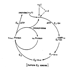
protein (see Figure 1). In a multiubiquitin chain)
ubiquitin itself serves as an acceptor, with several
ubiquitin moieties attached sequentially to an initial
acceptor protein to form a chain of branched ubiquitin-
ubiquitin conjugates. Formation of the multiubiquitin
ehain on a targeted protein has been shown to be
essential for the protein': subsequent degradation (Chau
et al., Science 24:1576-1583 (I989)).
A second, non-branched type of ubiquitin-protein
conjugate contains ubiquitin whose carboxyl-terminal
glycine residue is joined, via a peptidt bond) to the
a-amino group at the amino terminus of an acceptor
protein. The resulting conjugate is a linear fusion
between ubiquitin and a "downstream" protein. Although y
no enzymes have been found that can generate such linear
ubiquitin-protein fusions posttranslationally, these
ubiquitin fusion:, unlike the branched ubiquitin
conjugates, can be encoded by appropriately constructed
DNA molecules and synthesized on xibosomes as direct
products of mRNA translation.
Such DNA constructs were made and the proteins "
encoded by them were synthesized in vivo by Bachmair et
al. (Science 234:179-186 (1986)). In particular, a '
WO 91/17245 PCT/US91/03177
-3-
chimeric gene encoding a ubiquitin-~-galaetosidase
(Ub-gal) fusion protein was expressed in the yeast
Saccharomyces cerevisise. It was observed that the
ubiquitin moiety of this fusion was efficiently and
precisely cleaved off in vivo at the ubiquitin-gal
junction, yielding free ubiquitin and the gal protein
with its (natural) methionine residue at the amino
terminus. Using site-directed mutagenesis) the authors
replaced the methionine colon of gal at the Ub-~9ga1
junction With colons specifying each of the other 19
amino acids. The corresponding Ub-X-gal proteins (with
X denoting the junctional amino acid residue of gal)
were expressed in yeast) and the structure and metabolic
fate of the products were examined. It was found that,
in all cases) the ubiquitin moiety was cleaved off the
Ub-X-gal fusion protein in vivo by a ubiquitin-specific ,.
(Ub-specific) protease irrespective of the nature of the
zesfdue X at the Ub-gal junction (when X was proline)
the deubiquitination) while still occurring, was about an
order of magnitude slower than with the other 19
junctional residues) (Bachmair et al.) Science 234:179-
186 (1986); Bachmair and Varshavsky, Cell 56:1019-1032
(1989); Gonda et al.) ._1~_Biol,:_Chem_ 254:16700-16712
(1989)).
The resulting technique, the ubiquitin fusion
methodology, has provided, among other things, a
definitive solution to the so-called "methionine
problem". This fundamental problem stems from the fact
that, because of constraints imposed by the genetic code)
all newly synthesized proteins in all organisms start
WO 91/17245 PCT/US91/03177
2Q~~~~~
_a_
with methionine. The rules that govern subsequent fate
of the amino-terminal region of a newly made protein
(e. g.) whether the methionine will be retained,
acetylated, otherwise modified or removed) or whether
more extensive changes at the amino terminus would occur)
are poorly understood, and therefore cannot be used to .
produce in vivo a specific protein or polypeptide bearing
any desired (predetermined) amino-terminal residue. This
poses severe problems in many biotechnological appli-
cations, for instance, when medically important
eukaryotic proteins are produced,by recombinant DNA
methods in heterologous hosts sueh as yeast or bacteria.
Many such proteins, when produced under normal conditions
in their natural in v_iv__o environments, bear mature
amino-terminal residues that are different from those
that these proteins bear when overexpressed in hetero~
logous in vivo systems such as yeast or bacterial cells.
Possession of a correct (natural) amino-terminal residue
assumes particular importance in the case of recombinant
proteins produced for pharmaceutical applications. For
3
instance, incorrect (or extra) amino~terminal residues in
an intravenously administered protein may present w
antigenicity problems-(induction of immune response to a
protein), or result in too rapid clearance of the protein
from the bloodstream. Among the important clinical and
veterinary protein drugs which fall into these groups are
growth hormones) various interferons, fibroblast growth w
factors) and interleukins.
The invention of the ubiquitin fusion methodology '
has provided a definitive, generally applicable, solution
WO 91/17245 P(..°T/tJS91/03177
- 2 ~'~'~ ~. l c.~
-5-
to the problem of producing any desired (predetermined)
amino acid residue of the amino terminus of either a
protein) polypeptide or peptide (thane three terms are
often used interchangeably in the art, with "peptides"
usually) but not always) referring to relatively short
polypeptides) on the order of -SO residues or less).
The ability to generate any desired residue at the
amino terminus of a given protein) in addition to being
crucial for solving the above problems, is also useful in '.
IO a variety of other applications) from fashioning differ- w
ent amino termini of proteins or peptides for their
funetional studies to.manipulating the metabolic
stability (in vivo half-lives) of proteins by changing
their amino-terminal residues (Bachmair at al., S_cien_c_e
234:179-186 (1986}).
While the facile in vivo generation of desired
amino-terminal residues in specific proteins has been
achieved far the first time through the ubiquitin fusion
methodology) the analogous manipulation of proteins'
amino termini in vitro (in cell-free systems) has
previously been possible) to a limited degree) using a
variety of specific proteases) such as renin or Factor Xa
(Nagai and Thogersen) Meth__Enzymol_ 153:461-466 (1987)).
Unfortunately) all of these in vitro-used proteases have
severe drawbacks as reagents for generating the desired
amino termini fn specific proteins or peptides) either
because) like renin, they are not specific enough and
often cleave the target protein at inappropriate places
as well, or because, like Factor Xa) they are relatively
inefficient, requiring long reaction times and producing
low yields of the desired product.
WO 91/17245 PC1"llJS91/03177
~ )
2~'~i~;'~~)
-5-
For these reasons, from the time of the invention of
the ubiquitin fusion methodology in 1986, it has always
been desirable to isolate a gene for~the highly ,
efficient, exquisitely Ub-specific protease that under-
Iied the in vivo versian of the methodology and to use
this protease in vitro as an alternative to the flawed
proteolytic reagents that have previously been used for
the in vitro manipulation of proteins' amino termini.
The isolation of a gene encoding a yeast Ub-specific
protease, YUH1, and its (heterologous) expression in E_
cola has been reported by Miller et al. (Biotechnology
1:698-704 (1989)). However, a closer analysis of the
protease isolated by the above group has shown that it
cleaves only sufficiently short ubiquitin fusion
proteins) and does not cleave those fusions having a
non-ubiquitin portion exceeding -60 residues in length.
In particular) as Miller et al. stated in their above-
cited paper, the YUH1 protease is incapable of de-
ubiquitinating Ub-X-gal proteins, the very ubiquitin
fusions that have been used to establish the in vivo ':.
version of the ubiquitin fusion methodology by Bachmair
et al., Science 234:179-186 (1986). (The X-gal moiety
of Ub-X-gal is -1,000 residues long.)
Summaic of the Invention
Y___~____________
This inventian pertains to a new type of Ub-specific
protease (designated Ubiquitin-Protease 1 (UBP1)) and to -
isolated nucleic acid encoding UBP1 protease. The UBP1
protease deubiquitinates any fusion protein between ,
ubiquitin and a protein or peptide other than ubiquitin,
WO 91/17245 PCT/US91/03177
v~1,~~.
2~ ~ .~~~~~
-1-
without limitation on the size of a non~ubiquitin
component of the fusion. Thus, UBP1 has a qualitatively
different substrate specificity from that of the
previously isolated YUH1 protease. This invention also
pertains to recombinant vectors expressing the UBP1
protease) to eells,transformed with such vectors,. and to
specific versions of ubiquitin-protein fusions that
facilitate isolation and manipulation of non-ubiquitin
portions of these fusions using the UBP1 protease.
The compositions and methods of this invention
facilitate large-scale production of the UBPl protease.
The availability of this new type of Ub-specific protease
introduces an in vitro counterpart of the ubiquitin
fusion methodology. The UBP1 protease provides for more
IS efficient methods of isolation and purification of
various recombinant proteins or peptides.
Bzief Descri lion of the Fi ures
_ E______________8____
Figure 1 shows pathways of the ubiquitin system.
Figure 2 shows an outline of the sib'selection
strategy used to isolate the yeast UBP1 gene.
Figure 3 shows the map of the plasmid p3T60 that
encodes the UBPl protease.
Figure 4 shows the nucleotide sequence of the UBP1
gene and the amino acid sequence of the UBP1 protease.
Figure 5 shows the results of electrophoretic
analysis of Ubiquitin-Met-gal, Ubiquitin-Met-DHFR and
other ubiquitin fusions treated with Ub-specific protease
UBP1.
Figure 6 shows (A) a map of the expression vector
WO 91/17245 P(.'T/US91/03177
~~ t~
2J'~(''>~J
-g.
pJTUP which encodes a sandwich fusion protein DHFR-
Ubiquitin-Met-~gal; and (B) results of electrophoretic
analysis of DHFR-ubiquitin-Met-gal treated with the
ubiquitin-specific protease UBP1.
Detailed-Description-of_the_Invention
UBPI, the Ub-specific protease of this invention)
,.
specifically cleaves ubiquitin from any non-ubiquitin
protein or peptide to which the ubiquitin is joined.
Importantly, UBP1 cleaves any ubiquitin fusion (except
IO polyubiquitin) without upper or lower limits on the size
of the non-ubiquitin portion of a ubiquitin fusion. UBP1
cleaves at the junction between the ubiquitin and the
non-ubiquitin protein or peptide; i.e., it cleaves the
peptide bond in a ubiquitin fusion pratein between the
carboxy-terminal residue of a ubiquitin moiety and the
a-amino group of any non-ubiquitin protein or peptide to
which it is joined.
UBP1 also recognizes and cleaves "sandwich'
ubiquitin fusions in which the ubiquitin moiety is
located between a first and a second non-ubiquitin
moiety. As used herein, the first non-ubiquitin moiety
is a non-ubiquitin protein or peptide positioned upstream
of the ubiquitin moiety in the sandwich ubiquitin fusion.
The second non-ubiquitin moiety is a non-ubiquitin
protein or peptide positioned downstream of the ubiquitin
moiety in the sandwich fusion protein. UBP1 cleaves the
sandwich fusion protein between the carboxy-terminal
residue of the ubiquitin moiety and the a-amino group of
the second non-ubiquitin moiety.
WO 91 /17245 PCf/US91 /031 '~7
,r.": r
2!~ ~~L~'~~~
(9-
The first non-ubiquitin moiety in a sandwich
ubiquitin fusion can be any peptide or protein. The
sandwich ubiquitin fusion proteins can be generated, for
example) by ligating DNA fragments encoding the first and
second non-ubiquitin moieties to the 5' and 3' ends,
respectively, of a DNA sequence encoding ubiquitin.
These coding sequences must be joined in frame, in a
cantext appropriate for expression, such that no stop
codons are generated which would prematurely terminate
the translation of the mRNA encoding the sandwich fusion.
As described below in the Exemplification, a sandwich
ubiquitin fusion protein (DHFR-Ub-Met-gal), in Which the
ubiquitin moiety is located between a first and a second
non-ubiquitin moiety, has been constructed, expressed)
and shown to be cleaved efficiently and specifically by
UBP1.
Such a sandwich construct is particularly useful in
situations wherein the first non-ubiquitin moiety confers
some desirable property on the sandwich ubiquitin fusion.
For example, the first non-ubiquitin moiety may facili-
tate affinity purification of the ubiquitin fusion
protein. In such a case, the fusion protein can be
expressed in a cell (e.g.) E- cola) that lacks Ub-
specific proteases, and a cellular lysate can be passed
over an affinity column specific for the first non-
ubiquitin moiety. One example of a protein which is
useful for affinity purification is streptavi.din
(Sassenfeld, K.M., Trends,Biotech_ 8:88-93 <1990)).
Following affinity purification of the fusion protein)
the latter is contacted with the ubiqutin~specific
WO 91/17245 PGT/US91/03177
(~ ~ ) t'14) ')
Yy y ~~ r.~ r~
-10-
,
protease of this invention. Tha second non-ubiquitin
moiety is thereby liberated from the sandwich ubiquitin
fusion construct.
In contrast to UBP1) the previously isolated YUHl
enzyme cleaves ubiquitin off a ubiquitin fusion only if
the non-ubiquitin portion of a fusion is relatively short
(shorter than about 60 residues; see above). Since, for .
instance) many of the pharmaceutieally important proteins
are much longer than 60 residues, the YUH1 protease
cannot be used to deubiquitinate fusions of these
proteins with ubiquitin. In contrast) the UBPl protease
can be used for this purpose, thereby allowing the
generation of desired residues at the amino termini of
either large or small proteins, polypeptides or peptides
(as explained above) these terms are often used inter-
changeably in the art).
A variety of recombinant DNA approaches could) in
principle, be used to isolate the UBP1 gene. Typically)
such an isolation procedure involves the construction of
a cDNA or genomic DNA library from an organism known to
produce UBP1. Any euknryotic organism is an appropriate
source of nucleic acid for the construction of recombi-
pant libraries since ubiquitin is known to be produced in
every eukaryote tasted. Furthermore, ubiquitin is the
most highly conserved eukaryotic protein identified to
date. Protocols for the production and screening of cDNA
libraries or genomic DNA libraries are well known to
those skilled in the art.
The screening approach actually taken by the
inventors named in this application was a particular
WO 91/17245 PCT/U591/03177
. 20~~~~~
_Il-
version of the genetically based cloning strategy called
sib selection. This method exploits the fact that,
unlike eukaryotic organisms, bacteria cueh as E. coli
lack the cukaryotic ubiquitin systeo) and in particular,
Ub-specific proteases. It is this lack of Ub-specific
proteases in bacteria that has been exploited to isolate
a yeast (S. cerevisiae) gene encoding the desired Ub-
specific protease. Sfb selection is a method of~se-
quential fractionation of DNA clones which is particu-
larly useful in the absence of a selectable phenotype or
sequence information. This method is detailed in the
Exemplification section which follows.
In a preferred embodiment, the isolated DNA sequence
of the invention encodes the amino acid sequence set
forth in Figure 4, or modifications of this sequence in
which amine acids have bean deleted, inserted or substi-
tuted without essentially detracting from the activity
and substrate specificity of the encoded product. In
Figure 4, the UBP1 open reading frame starts at position
194 of the nucleotide sequence) and ends at position
2622, with a stop codon at position 2623. This reading
frame encodes a protein of 809 residues) indicated by
one-letter designations. This DNA can be isolated by the
methods outlined above or the DNA can be made in vitro by
conventional chemical DNA synthesis.
The isolated DNA of this invention can be used to
express UBP1 in Iarge quantities. For this purpose) the
DNA is inserted into a prokaryotic or eukaryotic expres-
sion vector) with appropriate regulatory signals, and
used to transform cells. A variety of appropriate
WO 91/17245 PCf/US91/03177
~f~r~(34~~~f~
'( c s~ i . ~ r c
-12-
vectors and regulatory signals have been previously
developed for this purpose and are well known to those
skilled in the art. UBP1 can be expressed in eukaryotic
or prokaryotic cells. Previous work hss indicated that
prokaryotes lack both ubiquitin and Ub-specific enzymes
(see) for example Finley and Varshavsky, Trends Biochem.
Sci. 10:343 (1985); and Ozkaynak et nl.) Nature 312:.663
(1984)). Large quantities of the protease can be pro-
duced and isolated from either bacterial or yeast
cultures using appropriate expression vectors well known
to those skilled in the art.
The Ub-specific protease can be used to cleave
ubiquitin off ubiquitin fusions in vitro. The UBP1
protease is contacted with the ubiquitin fusion under
conditions appropriate for proteolytic cleavage and the
cleaved adduct is recovered. In this procedure) UBP1 can
be used in free form or it can be immobilized on a solid
phase such as a bead. As mentioned, UBP1 cleaves
ubiquitin from large adducts as well as small. Thus,
proteins or peptides can be produced as ubiquitin fusions
in appropriate systems in vivo, and the ubiquitin moiety
can be removed in vitro using the Ub~specific protease.
In addition) prokaryotic cells harboring an expres-
sion vector encoding the protease can be transformed with
an expression vector encoding a ubiquitin fusion protein
or peptide of interest. These cells will then produce a
deubiquitinated product having a predetermined amino-
terminal amino.acid residue. There are many well known _
advantages to producing recombinant proteins in pro-
karyotic organisms such as E, cola.
WO 91 /17245 PCT/US91 /03177
- 2~~~'
-13-
In soma fusions of ubiquitin to a non-ubiquitin
protein or peptide) the presence of the ubiquitin moiety
may inhibit or modify the functional activity of the
non-ubiquitin protein or peptide. In this case,
ubiquitin can be used as a temporary inhibitor (or
modifier) of the functional activity of the non-ubiquitin
pzotein or peptide with the ability to restore the
original functional activity at any desired time) either
in _vitro or in vivo, by contacting the corresponding
ubiquitin fusion with the Ub-specific protease to cleave
the ubiquitfn moiety.
The invention is further illustrated by the followed
Exemplification.
EXEMPLIFICATION
Exam le 1
Q____
Escherichia coli (strain HB101) transformed with a
S_ac_ch__aromyces cerevisiae genomic library was used for a
sib selection strategy. The library) 88237, was produced
by partially digesting yeast genonic DNA with SauIIIA and
ligating the fragments into the Bnmtll site in the TatR
gene of the yeast/E. coli shuttle vector YCp50. Upon
initial analysis) the library contained insects with an
average size of -19 Kb.
E. coli, transformed with the above library, was
plated on agar containing Luria Broth (LB) and ampicillin
(amp) (100 pg/ml) at a density of about 40 viable cells
per plate. The plates were incubated at 36'C for 16
hours. The colonies ware then replicated onto LB/amp
WO 91/17245 PCT/US91/03177
~~ r) (~ 4) ~~ w~
f-, i.l 1-:
-14- -
plates. The original plates were stored at 4°C, and ,
their replicas were grown far 24 hours at 36'C. Each
replicate was eluted with 1 ml of LE/amp (50 pg/ml) by
repeated washing over the surface of the plate until all
of the calonies were loosened into the liquid. The
entire eluate was then added to 4 ml of LB/amp, and
incubated on a roller drum at 36'C overnight.
The E. cola cells in these overnight (stationary-
phase) cultures were then lysed. 1.7 ml of each culture
was placed in a microcentrifuge tube on ice, and then
centrifuged at 12,000 x g for 1 min at 4'C. The cell
pellet was resuspended, by vortexing at high speed, in 50
~l of 251 sucrose (w/v), 250 mH Tris-HC1 (pH 8.0). 10 pl
of freshly made lysozyme solution (10 mg/ml chicken
egg-white lysozyme (Sigma) in 0.25 H Tris-HC1, pH 8.0)
was then added, and mixed by light vortexing. The
suspension was incubated on ice fos 5 min. 150 pI of 75 ,
mM EDTA, 0.33 H Tris-HC1 (pH 8.0) wss added) mixed by
light vortexing, and the tuba was incubated on ice for 5
min) with occasional stirring. 1 ~1 of l0i Triton X~100
(Pierce) was then added to each tube, and mixed by
pipetting. The Bell lysate was centrifuged at 12,000 x g
for 15 min at 4'C. The supernatant was retained on ice)
and the pellet was discarded.
In an assay for the Ub-specific protease activity, 9
pl of the above supernatant was combined with 1 pl of .
35S~labeled ubiquitin-dihydrofolate reductase
(Ub-Met-DHFR) fusion in a 0.5 ml microcentrifuge tube, .
and incubated at 3b'C for 3 hr. 5 ~1 of a 3-fold
concentrated electrophoretie sample buffer (301 glycerol,
WO 91/17245 PCT/US91/03177
I,~V~" t
2~'~~~~~p
-ls-
3~ SDS (w/v), 15 mH EDTA, 0.2M 2-mereaptoethanol, 0.3
pg/ml bromophenol blue) 375 mM Tris-HCl, pH 6.8) was then
added, and each tube was placed in a boiling water bath
for 3 min. The samples were loaded onto a 121
polyacrylamide-SDS gel, and electrophoresed at 50 V until
the bromophenol dye reached the bottom of the gel.
Positions.of the radioactively labeled proteins 'in the
gel were visualized by fluorography. The gel was washed
in 108 acetic acid, 25! methanol far 15 min, rinsed in
H20 for 15 min, and incubated with Autofluor (National
Diagnostics) for 1 hr. The gel was then dried at 80'C
under vacuum) placed in a light-proof cassette against
Kodak XAR-5 film and stored at -85'C overnight.
35S-labeled Ub-Met-DHFR was prepared as follows.
Luria Broth (50 ml) supplemented with 50 pg/ml ampicillin
was inoculated with 1 ml of a saturated overnight culture
of E. _coli strain JH101 containing a plasmid expressing
the Ub-Met-DHFR fusion protein from an IPTG-inducible,
highly active derivative of the lac promoter. The cells
were grown with shaking at 37'C until they reached an
A600 of -0.9. The culture waa chilled on ice for 15 min)
then centrifuged at 3000 x g for 5 min and washed 2 times
with M9 salts at 0'C. The cells were resuspended after
the final wash in 25 ml M9 salts supplemented with 0.21
glucose, 1.8 ~ug/ml thiamine, GO pg/ml ampicillin) 1 mM
IPTG) 0.06251 (w/v) methionine assay medium (Difco). The
suspension was then shaken for 1 hr at 37'C and the cells
were labeled by the addition of 1 mCi of 35S-Translabel
(ICN)) followed by a 5-min incubation, with shaking.
Unlabeled L-methionine was then added to.a final con-
centration of 0.00321 (w/v), and the cells were shaken
WO 91/17245 PCT/US91/03177
r~ ~~ ~ ~ i~
z~,
-16- ,
for an additional 10 min. The cells were then harvested
(3000 x g for 5 min) and washed once in cold M9 salts.,
After the M9 wash) the cell pellet was resuspended in 0.5
ml 25s Sucrose, 50 mM Tris-HC1 (pH 8.0), and incubated on
ice for 5 min. During this time, chicken egg-white
lysozyme (Sigma) was dissolved freshly in 250 mM Tris-HC1
(pH 8.0) to a concentration of 10 mg/ml. 10 ~1 of the
lysozyme solution was added to the cell suspension,
mixed) and incubated for 5 min at 0'C. 5 pl of 0.5 M
EDTA (pH 8.0) was then added, and the suspension left at
0'C for 5 min, with intermittent mixing. The Bell
suspension was then added to a centrifuge tube. containing
0.975 ml of 65 mM EDTA (pH 8.0)) 50 mM Tris-HC1 (pH 8.0)
and protease inhibitors) antipain, chymostatin,
leupeptin) aprotinin and pepstatin) each at 25 ~g/ml. 10
pl l0i Triton X-100 (Pierce) was then added) and
dispersed by pipetting. The lysate was centrifuged at
39,000 x g for 30 min. The supernatant was retained) ,
quickly frozen in liquid nitrogen, and stored at -85'C.
To affinity-purify the 35S-labeled Ub-Met-DHFR) a
methotrexate (MTX)-agarose affinity matrix was prepared
according to the method of Kaufman (Kaufman, B.T., ?ieth_
Enzymol_ 34:272-281 (1974)). A 0.5 ml bed volume column ~.
was filled with the MTX-ngarose, and washed with 10 ml of
MTX column buffer (20 mM Hepes (pH 7.5), 1 mM EDTA 200 mM
NaCl, 0.2 mM dithiothreitol. The 35S-labeled supernatant
of the preceding step (see above) was thawed and applied
to the MTX~agarose column. The column was washed with 50
ml of MTX column buffer, 50 ml of MTX column buffer w
containing 2M urea, and again with 50 ml of MTX column
WO 91/17245 PGT/US91/03177
'.
-17-
buffer. The labeled Ub-Met-DHFR was eluted from the
column with folic acid elution buffer (0.2 M potassium
borate (pH 9.0)) 1 M KC1, 1 mM DTT, 1 mM EDTA, 10 mM
folic acid). Tha elution buffer was applied to the
column in 1 ml aliquots, and 1 ml fractions ware col-
lected. The fractions were assayed for 35S radioactivity
and those fractions that contained the major radioactive
peak were pooled. The pooled fractions were dialyzed for
-20 qtr against teao cE~anges o~ a storage buffer containing
IO 40 mM Tris-HCi (pH 7.5)) 1 mM MgCl2) 0.1 mM EDTA, 50%
glycerol. The purigied 35S-lgbeled Ub-hfee-DHFR vas
assayed by SDS-PAGE, followed by fluorography, and found
to be greatez than 95s pure.
The above deubiquitination assay was repeated with
lysates from different pools of E. cola transformants '
until the gel analysts revealed a lysate that displayed
proteolytic activity acting at the ubiquitin-DHFR
junction (Fig. 2). This result indicated that at least
one of the -40 E. cola. colonies on the original LB/amp
plate (from which the pooled lysate had been derived)
contained a YCp50-based plasmid having a yeast DNA insert
conferring Ub-specific proteolytic activity.
The next step of this sib selection approach to
cloning the UBP1 gene was to carry out a similar Ub-Met-
ZS DHFR cleavage aaray to determine which of the -40
colonies in a "positive" pool contained the desired
plasmid. To do so, a sample of each individual colony on
the plate of interest was inoculated into LB/amp and
grown overnight. The Ub-Met-DHFR cleavage assay was then
repeated exactly as above) but this time each lysate
wo 9W 7245 PCT/US91 /03177
f.
S..
'i,.~,x";.,b'~
v
a ,
-18~
sample was representative of a single clonal E, eoli
transformant rather than a mixture of -40 such trans-
formants. This analysis revealed a single colony that
contained a plasmid which conferred the ability to
specifically cleave at the Ub-DHFR junction, thereby
accomplishing the goal of cloning a S, cerevisiae gene
encoding the Ub-specific protease.
Analysis of the initially isolated plasmid (pJT55)
revealed a -15 kb insert of yeast genomic DNA in the '
YCp50 vector. SphI digestion of this plasmid yielded a
-14 kb fragment, which, upon subcloning into the vector
pUCl9, conferred the same proteolytic activity. This
plasmid was called pJT57. The -l4 kb fragment was
subcloned further by cutting with SphI and XhoI)
isolating the -5.5 kb fragment of the insert DNA and
subcloning it into the pUCl9 vector pre-cut with Sphl and
SalI. This resulted in the -8.1 kb plasmid pJT60
containing the -5.5 kb yeast DNA insert that conferred
the same Ub-speeific proteolytic activity as the original
Plasmid.
A map showing restriction endonuclease recognition
sites in plasmid pJT60 is shown in Figure 3. In the nap,
base pair positions ere indicated by a number in
parentheses following a restriction site) The yeast DNA v
insert in pJT60 contained a K~nI site near its center
that divided the insert into two smaller fragments A and
B (bases 423 to 5830). In this fragment) the open arrow
indicates the open reading frame (ORF) that codes for
UBP1. The entire ORF, and the thin lines bracketing it)
represent the extent of the Sequenced DNA shown in Figure '
4. Both fragments were subcloned into pUCl9) yielding ,
WO 91 /17245 1'CT/US91 /03177
r~ :~, ' ,
-19-
pJT60A and pJT60B. Fragment A was isolated from pJT57
after cutting with KpnI and SphI. This fragment was -
subcloned into pUCl9 that had been cut with the same
restriction endonucleases. Fragment 8 was isolated from
pJT57 that had been cut by KQnI and XhoI; it was sub-
cloned into pUCl9 that had been cut by KpnI and Sall.
Neither pJT60A nor pJT60B was able to confer Ub-specific .
~proteolytic activity) This result suggested that the
gene of interest straddled the KpnI site of the -5.5 kb
insert of pJT60.
To sequence the cloned gene, the inserts of pJT60A
and pJT60B were subcloned into the ti13mp19 phage vector.
Nucleotide sequence was determined (using the chain
termination method) in both directions from the internal
Kpnl site in pJT60. The KgnI site was found to be
ensconced within an open reading frame extending from
this site in both directions. Unidirectional deletions
were then made in the sequencing templates by the methods
of Dale _et _al. (Plasmid 13:31-40 (1989)) and the entire
open reading frame (ORF) was determined (Fig. 4). The 5'
end of the ORF was in fragment B and the termination
codon was in fragment A. The ORF was 2427 nucleotides
long) and encoded an 809-residue protein) with a
molecular mass of 93 kD. The sequenced ORF was then
isolated on a 2.8 kb fragment by cutting pJT60 with AccI,
filling in the 5' overhangs with Klenow Poll, and
ligating SalI linkers to the blunt ends. This construct
was digested with SalI and BamHI) the 2.8 kb fragment was
electrophoretically purified and ligated into pUCl9 that
had been digested with BamHI and Sall. The resulting
WO 91/17245 PCT/US91/03177
w
-20-
plasmid was called pJT70. This plasmid, when transformed
into E, coli, was able to confer the Ub-specific pro-
teolytic activity to the same extent as either the
original -15 kb insert in YCp50 or the -5.5 kb insert of
the pJT60 plasmid that includes the -2.8 kb fragment of
pJT70: The plasmid pJT60 has been deposited with the
American Type Culture Collection (Rockville, MD), and has
been assigned ATCC designation 68211. The 2.8 kb frag-
ment contained no other ORFs of significant size, indi-
eating that the sequenced ORF shown in Figure 4 encoded
the Ub-specific protease. .
This new gene has been named UBP1, for Ubiquitin-
specific protease. This designation conforms to the
existing convention for naming genes associated with the
ubiquitin pathway (Finley, Bartel and Varshavsky, Nature
338, 394-401 (1989)).
The substrate specificity of the UBP1 gene product
was examined) and results are shown in Figure 5. Figure
SA shows a fluorograph of a 121 polyacrylamide-SDS gel
used to detect deubiquitinating proteolytic activity;
with Ub-Met-DHFR as a substrate) and a set of subclones
of a yeast DNA fragment that confers Ub-specific
proteolytic activity upon E. ooli. Each lane corresponds
to a sample of the purified [35S]Ub-Met-DHFR treated with
an extract of E. cola and fractionated by gel electro-
phoresis. Lanes land 4 indicate a lack of Ub-specific
proteolytic activity and lanes 2,3 and 5-7 indicate the
presence of such an activity. In lane 1) the substrate
was treated with extract from untransformed (control)
JM101 E. coli. In lane 2) the treatment was with the
WO 91/17245 PCT/US91/03177
r_;,=..
,,;:<.;..~.
'° C
U 'I "t Ya I~J
-21-
extract from JM101 containing the initial plasmid pJT55.
Lanes 4-7 correspond to extracts from JM101 containing
plasmids that bear different subclones (in the vector
pUCl9) of the initial S, cerevisiae geno~ic DNA insert
present in pJT55. One plasmid that confered the Ub-
specific proteolytic activity (lane 6) was named pJT57,
and was used in the construction of pJT60 (as described
above). An arrowhead indicates a minor contaminant that
is present in the [35SjUb-Met-DHFR preparation.
Figure 5B shows a fluorograph of a 68 polyacryl-
amide-SDS gel demonstrating the ability of the UBP1
protease to deubiquitinate a ubiquitin-~-galactosidase
fusion. Lane 1 contains [35SjUb-Met~~gal treated) in a
moek reaetion) with the buffer alone. Lane 2 contains
the products of an otherwise identical reaction in which
E. eoli JM101 containing no plasmid was used as a source w .
of extract (no deubiquitination is observed). Lane 3
contains the products of a reaction in which E. coli
JM101 containing the plasmid, pJT60 was used as s source
of extract (note the -8 kD decrease in molecular mass
corresponding to the cleavage of the ubiquitin moiety off
the -115 kD Ub-M-~9ga1).
Figure SC represents a demonstration of in vitro
deubiquitination of natural ubiquitin fusions to yeast
ribosomal proteins (UBI2 and UBI3) by the yeast UBP1
protease. Lane 1 shows an extract from E. cola JM101 w
_-
containing a plasmid that expressed UBI2, a natural
ubiquitin-ribosomal protein fusion from S. cerevisiae
that had been subjected to electrophoresis in a 128
polyacrylamide-SDS gel) blotted onto polyvinylidene
WO 91/17245 PCT/US91/03177
' '~>.
~E~ir7~s~~)(
-22-
difluoride membrane, and detected using a rabbit anti-
ubiquitin antibody, with subsequent application of a
secondary goat anti-rabbit antibody linked to alkaline
phosphatase, and colorgenic substrates of alkaline
phosphatase. Lane 2 and lane 1 represent identieal
samples except that the UBI2-containing extract was
treated with extract from E. cola JM101 containing the
UBP1-expressing~plasmid pJT60. Lane 3 and lane 1
represent identical samples except that the UBI2~ " .
containing extract was treated with a whole cell yeast
extract. Lane 4 and lane 1 represent identical samples
except that an extract from E. coli JM101 contained a
plasmid that expressed UBI3) another natural ubiquitin
fusion (to a different yeast ribosomal protein). Lane 5
and lane 2 represent identical samples except that the
yeast UBI3 protein was used as substrate for the UBPl
protease. Lane 6 and lane 3 represent identical samples
except that the UBI3 protein as substrate. "ubi3,"
"ubi2," and "Ub" indicate the positions of the UBI3, UBI2
and free ubiquitin protein species. Bands in lane 4 that
migrate faster than the UBI3 band are the products of a
partial) nonspecific degradation of the yeast UBI3
protein in E. coli extract, with the proteolytic cleav- w w .
ages being confined to the non-ubiquitin portion of UBI3)
since the entire sample of lane 4, when treated with the
UBP1 protease) yields undegraded ubiquitin (lane 5).
Exam le 2
E____
To determine whether a sandwich-type ubiquitin
fusion protein in which the ubiquitin moiety had an ,
- , - . 9 , ~...:,.~: w... ;.. ~ . . .. . . , . .
WO 91 /17245 PCT/US91 /03177
F;,,
~~~~~~s~(~
T7 1.1 Y.7 ~:
-23-
amino- terminal extension was a substrate for UEPl, a
plasmid was constructed that encoded a triple fusion
protein consisting of an amino-terminal dihydrofolate
reductase (DHFR) moiety, a flexible linker region of
three glycine residues and a serine) followed by
ubiquitin and Met-gal moieties (Figure 6A). The mouse
DHFR gene was isolated on a BamHI/HindIII fragment from a
plasmid encoding Ub-Met-DHFR (Bachmair and Varshavsky,
Cell 56:1019-1032 (1989)). This fragment was treated
with Klenow Poll to fill in the ends, and KpnI linkers
were ligated. The fragment was then cut with KRnI to
yield a 678 by fragment which was cloned into the KpnI
site in a modified Ub~Met-gal expression vector in which
the second codon of the ubiquitin moiety was altered to
encode a KpnI site (Gonda et al.) J__Biol__Chem.
264:16700-16712 (1989)). This procedure yielded a
plasmid that.encoded DHFR) ubiquitin (without the initial
Met codon) and Met~~gal) with the open reading frames for
each moiety not yet aligned into a single open reading
frame. To effect the alignment of the open reading
frames and to position the initiator codon of DHFR
correctly with respect to the GAL promoter in the vectoz)
site-directed mutagenesis was performed at twa locations
in the plasmid.
The plasmid was cut with BamHI and HindIII, and the
-2.76 kb fzagment encoding DHFR, ubiquitin and the first
few residues of Met-gal was cloned into M13mp19 that had
been cut with the same enzymes. Oligonucleotide-
mediated) site-directed mutagenesis was performed using
the single-stranded M13 derivative and standard proto-
cols. The first oligodeoxynucleotide was designed to
WO 91/172d5 PCT/US91 /03177
~~(v~~()
tJ W i.) ~:
-24-
produce a 20 by deletion that would bring the initiator
codon of DHFR to a proper position relative to the GAL
promoter of~ the vector. The second oligodeoxynucleotide
was designed to bring together the reading frames of DHFR
and ubiquitin) and to introduce the 4~residue spacer
(-Gly-Gly-Gly-Ser-) between the DHFR and ubiquitin
moieties. After mutagenesis) DNA clones were tested for '
incorporation of both changes by direct nucleotide
sequencing using the chain termination method.
Double stranded, replicative form (RF) of the
desired M13 clone was isolated and digested with BamHI
and XhoI. The resulting -1.2 kb fragment was cloned into
the -9.87 kb fragment of a Ub-Met-gal expression vector
digested with the same enzymes, replacing the Ub-Met-
coding fragment with the DHFR-Ub-Met-coding fragment
produced by the site-directed mutagenesis. This last
step yielded an expression vector that encoded the triple
fusion DHFR~Ub-Met~~gal. The vector was named pJTUP
(Figure 6). ,
pJTUP was used to test whether a ubiquitin fusion in
which the ubiquitin moiety is located between two non-
ubiquitin moieties would be a substrate for cleavage by ,
UB.P1. In E. cola metabolically labelled with
~35S~methionine, the fate of expressed DHFR-Ub-Met-~9ga1
was determined in the presence or absence of UBP1 using
immunoprecipitation with a monoclonal antibody to ~B-
galactosidase, followed by polyacrylamide~SDS gel
electrophoresis and fluorography (Figure 6B).
In Figure 6B (a schematic representation of the
results), lane 1 shows the fluorogram of an electro
phoretieally fractionated sample produced as follows: an
WO 91/17245 PCT/US91/03177
~~~
-25-
aliquot of a stationary culture of E. cola carrying a
plasmid expressing Ub-Met-gal and a plasmid expressing
UBP1, was diluted 1:100 into fresh Luria Broth. The
culture was grown at 37'C with vigorous shaking to an
A600 of 0.3. 1 ml of the culture was spun at 12,000 x g
for 1 minute. The supernatant was discarded and the
pellet was resuspended in 50 pl of M9 medium supplemented
with 0.2! glucose. The cells were incubated at 37'C for.
minutes and then 20 ~Ci of (35SJmethionine was added. '
10 Incubation was continued for 2 more minutes and unlabeled
L-methionine was then added to a final concentration of
30 mM. The cells were then incubated for 5 minutes at
37'C) and subsequently lysed by the addition of 50 pl of
lysis buffer (4! SDS, 125 mM Tris-HC1 (pH 6.8)), followed y
immediately by heating at 100'C for 4 minutes.
Immunoprecipitation using a monoclonal antibody to
~-gal was then carried out. The lysate was diluted by
the addition of 1 ml immunoprecipitation buffer (IP
buffer) (1! Triton X-100, 0.5! Na-deoxycholate) 0.15 M
NaCl) 50 mM Tris-HC1 (pH 7.5), 20 mM NaN3, 5 mM EDTA) 1
mM phenylmethylsulfonyl fluoride). The sample was
centrifuged at 12,000 x g for 10 minutes at 4'C. The
upper 0.9 ml of the supernatant was collected in a fresh
tube to which 6 pl of a concentrated tissue culture
supernatant containing a monoclonal antibody to gal
(Bachmair et al., Science 234:179-186 (1986)) was added.
The tube was incubated on ice for 1 hour. 10 pl of a 50!
suspension of Protein A linked to Sepharose beads
(Repligen) was then added, and the tube was rotated
slowly for 30 mfnutes at 4'C., The tube was then centri-
fuged for 15 seconds at 12,000 x g, and the supernatant
WO 91/17245 PCT/US91/03177
. :~
r..
«~~ ~(1~')'~
...
-26~
was discarded. The beads were washed 3 times at 4'C with
1 ml of IP/SDS buffer (IP buffer plus O.ls SDS (w/v)))
with 15-second centrifugations at 12,000 x g to
precipitate the protein A-sepharose beads. The final
pellet was resuspended in 15 pl of a 3-fold concentrated
electrophoretic sample buffer (30s glycerol) 3t SDS
(w/v), 15 mM EDTA, 0.2 M 2-mercaptoethanol) 0.3 pg/ml
bromophenol blue, 375 mM Tris-HCI) (pH 6.8))) and
fractionated by polyacrylamide-SDS gel electrophoresis)
followed by fluorography. A fluorogram of the gel .-
(represented in lane 1 of Figure 6B) revealed that the
Ub-Het-gal was cleaved at the ubiquitin-gal junction by
the simultaneously expressed UBP1 to yield the expected
product, Met-gal.
The sample represented in lane 2 was identical to
that in lane 1 except that the triple fusion, DHFR-Ub-
Met-~gal, was expressed in E. coli that lacked UBP1.
Note that in addition to the full-length DHFR-Ub-Met-
~gal) this lane also contains bands representing shorter
proteins. These are the result of either alternative
initiation sites within the upstream (DHFR) moiety of the
triple fusion, or nonspecific endoproteolytic cuts within
that moiety. The smaller products are denoted by %- and
Y-Ub-Met-gal) respectively.
The sample represented in lane 3 was identical to
that fn lane 2 except that the triple fusion DHFR-Ub-
Met-~gal was expressed in the presence of UBP1. Note
that UBP1 efficiently cleaves all three triple fusion
proteins (DHFR-Ub-Mat-Sgal, X~Ub-Met-gal) and Y-Ub-
Met-gal) at the Ub-gal junction, yielding Met-gal.