Note: Descriptions are shown in the official language in which they were submitted.
CA 02081466 1998-04-21
WO92/14417 PCT/US92/01431
BONE ANCHOR
BACKGROUND OF THE I~v~NlION
Skeletal deformities become evident during the
growth of an individual, or may be acquired from trauma,
tumor resection, or systemic disease.
Correction of bone deformities requires either
surgical treatment to reposition the deformed bones into
a "normal" relationship, or by guided bone movements.
Currently pins or other transosseous devices are used in
conjunction with surgical procedures to anchor the bones
and to maintain bone position during treatment and
healing. These transosseous devices have limitations,
such as in small bones and in regions of the human
skeleton such as the face where other vital structures
exist preventing pins from being used. The correction of
facial deformities presents clinical challenges which
have led to this invention.
In patients with atrophic maxillary or mandibular
bone, prosthetic rehabilitation with conventional
2~ dentures is often not satisfactory since the patient has
very little bone to retain the dentures. In order to
rehabilitate these patients, bone grafting is often
required. However, many patients are not candidates for
bone grafting due to health reasons. Their
rehabilitation requires only an anchor for improved
retention of their prosthesis. This invention can be
placed either onto the bone or into a shallow, 3mm
depression and will provide anchorage for the patient's
dentures. Placement of the bioactively coated device
onto bone will allow bone deposition over the entire
WO92~1~17 ~ PCT/US92/01431
2081~66
...
-- 2 --
surface of the 2-3 mm thin device, increasing its ability
to withstand the forces of chewing.
Often the earliest signs of maxillary or mandibular
growth disharmony is dental malalignment. Once
recognized, it is possible to guide the growth of
segments of the cranio-facial skeleton in order to
minimize the need for surgical correction of the
deformity.
Maxillary hypoplasia exists in all three dimensions.
Transverse deficiency of the maxilla is often treated by
the orthodontist with orthopedic palatal expansion.
Deficiency in the maxillary in the vertical or anterior-
posterior direction has not been satisfactorily cured by
non-surgical guided movements because of a lack of a
stable or non-mobile anchorage source for orthopedic
movements.
Mandibular deficiency can be corrected by functional
appliances which position the mandible forward, and
presumably allow for posterior condylar appositional
growth which stabilizes the mandible in this forward
position. Orthodontists employ orthopedic traction in
all three dimensions to control or direct the development
of a bone to a favorable location.
Cleft palate patients often have transverse,
anterior-posterior, and vertical dysplasia.
Reconstruction of these patients often involves
orthodontic alignment of the segments prior to bone
grafting the defects. However, the defects can be large
and difficult to manage when the patient is young. The
deciduous dentition can also be difficult to manage in
regards to orthodontic anchoraae preventing definitive
alignment of the arches until the patient is in the early
teens.
All orthodontic and orthopedic forces adhere to
Newton's Law of Reciprocal ~orces. If a force is
applied to retract, or pull back on object such as a
WO92~14417 ~ PCT/US92/01431
2 0 ~ 6
-- 3
tooth, there exists an "equal and opposite" force to move
another tooth forward. The resistive value of the
posterior teeth is known as anchorage.
Orthodontists offset these reciprocal tendencies by
using an extraoral force known as a headgear to augment
the resistive value of the molar teeth. However, patient
compliance may be poor because many patients do not want
to wear the headgear, compromising orthodontic therapy
and often the final result.
The problem is that the retractive forces are
usually continuous, acting 24 hours a day. Realistically
most patients will not wear a headgear more than l0 - 12
hours a day. Therefore, the posterior anchorage is
typically fortified 40 - 50% of the time. All too often
inconsistent usage or overt non-compliance reduce this
effect even more.
Previous work in this field indicates that
endosseous implants can be used to anchor orthodontic
forces for tooth movement. All of the previously used
implants were cylindrical or screw shaped, from eight to
22 mm in length. These studies indicate that
osseointegrated implants have been used to anchor
realignment of teeth, without moving the implants. These
implants were placed deeply into the bone.
In the field of orthopedics, pins are routinely
placed through bones and connected to various supporting
frameworks to maintain bone position and also to act as
an anchor for guided bone movements. Morbidity is
associated with placing pins through the cortical and
cancellous bone, and if complications such as pin
loosening or infection occurs, loss of bone structure can
occur.
Clinically, hydroxylapatite coated cylindrical
implants have been used since July 1984. Solid blocks
of dense hydroxylapatite are available for
interpositional and onlay grafting of defects during
orthognathic surgery. The onlay grafts were used
exclusively for cosmetic augmentation of facial defects
without carrying loads.
A need exists for obtaining anchorage directly on
parts of the jaws in order to allow the orthodontist the
capability for moving teeth and bones in any direction.
A need also exists for obtaining anchorage directly
on parts of other bones to allow the orthopedic surgeon
the capability for moving the bones in any direction,
without the use of transosseous pins.
An anchorage device should be small, allow for
various parts to fit into it for versatility of use, and
be able to fit on bone and be applied to the bone
surface only. If the anchorage device requires
placement into or through bone, then it may be difficult
to place the device in children because of potential
damage to unerupted teeth. Also size limitations of
small bones prevents the use of transosseous pins.
In addition, for cranial bone movements for cases
of Crouzon's or Apert's syndrome for example, intra-bony
devices may interrupt vital structures such as dura or
sinusoids.
The objectives of this invention can be stated as
follows:
1. it must not enter the bone but should attach
to it;
2. it should be relatively thin to lay under soft
tissue against bone, without creating
significant inflammation;
3. it should have versatility of attachments in
order to assume a role for an orthodontic
anchor, a prosthetic anchor, an orthopedic
anchor, or to attach other devices to a bone
such as a pacemaker;
4. it must have sufficient shear strength to
absorb chewing forces and forces placed upon
it from orthodontic, occlusal, and orthopedic
loading.
SUMMARY OF THE INVENTION
As an orthodontic anchor system for treatment of
growth disharmony, bone deformity, bone atrophy, and
malalignment of teeth, a subperiosteal bone anchor is
surgically placed in a subperiosteal tunnel on or into a
shallow depression in the skeletal bone, allowing
osseointegration between the subperiosteal bone anchor
bone interface and the bone, after which a system is
attached to the subperiosteal bone anchor for treatment.
The system may consist of a palatal bar which is
attached to the anchor system, and the palatal bar is
also attached to bands around two teeth, holding them
non-mobile, permitting the orthodontist to treat the
malalignment of the teeth.
As an orthopedic anchor the onplant is surgically
placed in a subperiosteal tunnel on the skeletal bone,
allowing biointegration between the onplant bone
interface and the bone, after which a device is attached
to one or more anchors, with each onplant biointegrated
to the underlying bone, in order to guide movement of
the bones that the onplants are biointegrated to, to
bring bones closer or further apart, for the correction
of bone deformities.
The onplant may have a screw hole penetrating it
for the sole purpose of stabilizing it with a small
screw into the bone while the onplant is integrating.
The screw would serve no purpose once integration had
occurred and may be removed when the surgeon exposes the
onplant to attach the intended device.
1~
~ c~
- 6 -
The invention accordingly comprises the several
steps and the relation of one or more of such steps with
respect to each of the others, and the apparatus
embodying features of construction, combinations of
elements and arrangements of parts which are adapted to
effect such steps, all as exemplified in the following
detailed disclosure, and the scope of the invention will
be indicated in the claims.
Thus in accordance with the present invention,
there is provided a subperiosteal bone anchor having:
a) a first surface preformed to match the
cortical surface of a selected bone, said first
surface comprising a bone interface surface;
b) said first surface having an osseoactive
coating whereby said first surface osseointegrates
with the cortical surface of said bone;
c) said subperiosteal bone anchor having a
second surface opposite said first surface and said
second surface having means thereon to attach an
orthodontic or an orthopedic device;
d) said subperiosteal bone anchor being
substantially rigid and thin at its periphery;
e) said subperiosteal bone anchor adapted to
be located entirely on the surface of the bone;
wherein said subperiosteal bone anchor permits said
orthodontic or orthopedic device to apply or resist a
continuous force to an adjacent tooth or bone, is
applied in a simple one-step surgical procedure, does
not disturb the cortical surface or invade the medullary
contents of the bone, is designed for temporary
application and is easily retrievable.
WO92/1~17 ~ PCT/US92/01431
w~ 20~1~66
BRIEF DESCRIPTION O~ THE DRAWINGS
The invention will be better understood and the
objects other than those set forth above will become
apparent when consideration is given to the following
detailed description thereof. 5uch description makes
reference to the annexed drawings wherein:
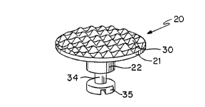
FIGURE l is a perspective view of the orthodontic
anchor system of the invention;
FIGURE 2 is an exploded side elevation view of the
invention of FIGURE l;
FIGURE 3 is a top view of the invention of FIGURE l;
FIGURE ~ is a bottom view of FIGURE l,
FIGURE 5 is a bottom view of an alternative
embodiment of the orthodontic anchor system of the
invention;
FIGURE 6 is a bottom view of an alternative
embodiment of the orthodontic anchor system of the
invention;
FIGURE 7 is a bottom view with the orthodontic
anchor system installed in the roof of a mouth with the
palatal wire connected to two banded teeth;
FIGURE 8 is a bottom view with another orthodontic
anchor system installed in the roof of a mouth with the
palatal bar connected to two banded teeth.
FIGURE 9 is a human skull with the prosthetic bone
anchor system of the invention;
FIGURE l0 is the mandible or lower jaw shown in
FIGURE 9;
FIGURE ll is a cross-sectional view taken on lines
ll-ll of FIGURE l0;
FIGURE 12 is a partial cross-sectional view of the
upper jaw of a livin~ person;
W092/1~17 ~ PCT/~S92/01431
2~146~
-
-- 8
FIGURE 13 is a cross-sectional of a tibia or leg
bone showing an orthopedic anchor system of the
invention;
FIGURE 14 is a fiDger bone with orthopedic anchor
system of the invention; and,
PIGURE 15 is a rib bone with orthopedic anchor
system of the invention.
wo92rl44l~ ~ PCT/U~92/01431
,",.,=~
9 2081~6
DESCRIPTION OP TffE PREFBRRED BMBOD~ S
The orthodontic anchor system 20 has two parts; the
onplant 21 and the abutment 22. These are connected to a
palatal bar 24 or palatal wire 28, which is attached to
bands 25 around the teeth to be held immobile.
As show in FIGURES 1-4 the onplant 21 has an
circular upper surface which is the onplant bone
interface 30. The circular shape is illustrative only.
The onplant may be an oval, a square, a rectangle, a
triangle, or other shape to resist the forces applied to
it. This onplant bone interface 30 is textured, which
both increases the surface area and presents surface area
which is better able to resist the shear forces imposed
by the orthodontic anchor system 20. Both the textured
onplant bone interface 30 and the surface 31 may be
covered with hydroxylapatite or other bioactive material.
The onplant 21 has a lower surface with a beveled
outer portion 31 and a central circular portion 36. The
ao outer portion 31 joins the outer periphery of the onplant
bone interface 30. The center of the lower surface 36
has at least one threaded aperture 32. There may be more
than one threaded aperture depending on the need to
resist rotational forces. When the onplant 21 is
initially installed the threaded aperture 32 has a
healing screw 26, not shown, installed to prevent tissue
from covering it and having to be removed.
The abutment 22 is circular, with an upper surface
37 matching the lower surface 36 of the onplant 21. The
upper surface 37 has a protruding threaded screw 33 which
cooperates with the threaded aperture 32. The abutment
22 has a neck 34 of reduced diameter and a head 35 of
increased diameter, compared with the neck 34. Surface
35 as shown is illustrative only. It may have a slot,
hexagonal, or threaded hole or other means of seatinq the
abutment or attaching the palatal bar.
WO92/1~17 ~ PCT/US92/01431
- lo - 2~8~'~6~
The dimensions of the onplant 21 may be 8 mm in
diameter and 2 mm thickness. The abutment 22 may be 4 mm
in overall height, with the neck 34 being 1 mm in height
and 1 mm in diameter. The device will vary in size
according to the shape and the designed force load.
The structure of both the onplant 21 and the
abutment 22 is a titanium alloy. The surface, except for
the onplant bone interface 30, is smooth and all corners
are beveled to prevent damage to soft tissue.
The test sample had a 50 micron coating of
hydroxylapatite. It was plasma sprayed on the metal.
The spray consists of a superheated solution of
hydroxylapatite applied to the roughened titanium alloy.
FIGURE 5 shows a onplant 21 which is similar to the
lS onplant 21 of FIGURE 1. This onplant 21 is generally
cylindrical in shape. It has the onplant bone interface
30 which is textured and coated by hydroxylapatite, and a
threaded aperture 32.
FIGURE 6 is similar to FIGURE 5, but is oval in
shape and has two threaded apertures 32. This embodiment
permits two orthodontic devices to be used.
As shown in FIGURE 7 the palatal wire 28 is soldered
to the two bands 25 of two molars or other teeth and
presses into the neck 34 of the abutment 22, preventing
the two teeth from moving forward. This palatal wire 28
may be fabricated ~rom 0.051 in. orthodontic wire or cast
from precious or non-precious metals.
FIGURE 8 shows the orthodontic anchor system 20
mounted between the two teeth to be held stable. The
palatal bar 24 is fabricated from thicker metal to resist
the shear forces of the teeth against the orthodont
anchor system 20. This palatal bar 24 is screwed on
abutment 22 which is screwed into the onplant 21.
The orthodontic anchor system 20 is able to resist
~5 both primary lateral and horizontal forces as well as a
vertical force.
The orthodontic anchor system 20 is not limited to
use with a palatal bar 24. It may alternatively be used
with any conventional orthodontic device, as will be
immediately apparent.
It is within the scope of the invention to use
other suitable materials for the orthodontic anchor
system 20. These will include inert metals, plastics
and composites. Likewise the bonding means can be any
mechanical means such as keylocks or miters or magnetic
or biodegradable polymer. The onplant bone interface 30
may have a different textured surface or a non-textured
surface which promotes adequate bonding strength. The
thickness and method of applying the hydroxylapatite
coating may be varied.
The orthodontic anchor system 20 is installed into
a patient's mouth in accordance with the following
procedures. These are generalized for an understanding
of the invention, and are not the detailed procedures
which would be actually followed by a surgeon.
Under local anaesthesia, an anterior palatal
incision will be made and a subperiosteal tunnel created
so that the tunnel will place the onplant at the
proposed location (most likely between the permanent
first molars). Conservative dissection will be used in
order that palatal reflection is minimal and restricted
to only the onplant site in order to prevent onplant
migration. One or two onplants will be placed depending
on the treatment needs for the patient. For orthopedic
applications, the anchor will be placed into a
subperiosteal tunnel and if placed on a curved surface
retained in position during biointegration by a small
screw or circumferential resorbable sutures.
- 12 -
Previous experience indicates that careful surgical
technique will result in secure positioning of these
onplants, without the need for retentive wires to
maintain bone contact on flat surfaces. However, on
curve surfaces a small screw or suture may be needed to
retain the onplant in the preferred position during
biointegration.
The onplant is usually provided sterile by the
manufacturer. It will be placed into the subperiosteal
tunnel taking great care to place it directly against
the palatal or other bone, or into the shallow
depression within the bone. The incision will be closed
using 4-0 polyglactin suture. The patients will be
given a prescription for antibiotics (typically
penicillin or doxyclycline) and analgesics. This small
surgical procedure should cause minimal pain to the
patient. The patient will be called at home by the
surgeon for follow up, and seen for suture removal one
week after the surgery. The patient will be followed
every two weeks for observation during the healing
period which is necessary to achieve integration of the
onplants hydroxylapatite surface with the underlying
bone.
Twelve weeks will be allowed for healing and
biointegration to occur. Twelve weeks is the expected
onplant biointegration time because that is the time
required for integration of hydroxylapatite coated
implants in humans. At twelve weeks, the patients will
be given local anaesthesia and a small incision will be
made directly over the onplant, exposing only the
healing screw 26 that was placed into the internal
thread of each device. An abutment 22 is then screwed
into the onplant. The overlying soft tissue thickness
may be thinned to 3 mm in order to allow for cleaning of
the attachment device. An impression will be taken in
order
W092~14417 ~ PCT/US92~01431
20~1~6~
- 13 -
to fabricate a palatal bar 24 which is secured to the
onplant and banded to the dentition, or to fabricate the
mechanical orthopedic or prosthetic anchorage devices
depending on patient needs.
The palatal wire 28 will be solid and minimally
pliable. The wire will be soldered to bands glued to the
anchor teeth. Approximately two weeks will be allowed
for fabrication of the bar or bending the wire on a
transferred study model. The wire will be fabricated of
O.OSl in. orthodontic wire.
Two weeks later, orthodontic devices will be
attached to the onplant and to the maxillary teeth, for
example the first molar, placed in such a way that the
wire attaching the onplant to the tooth acts to hold the
tooth in position, as an anchor. The onplant will serve
as the point of absolute anchorage, preventing the
anchored teeth from moving anteriorly.
The remaining dentition will be treated with
conventional orthodontic appliances. The location of the
teeth with respect to the onplants may be mea~ured and
recorded both by radiographs and actual physical
measurement with a Boley gauge.
At the conclusion of the treatment involving the
device, under local anesthesia, an incision will be made
exposing the entire onplant. Using a forcep designed for
this procedure, the hydroxylapatite coated device will be
removed. The prosthetic device will be left in place to
provide anchorage of the denture.
In a study investigating the difference between
diameter and length on the ultimate pull-out strength of
hydroxylapatite coated cylinders in the dog jaw, a
mechanically significant bonding was found with
hydroxylapatite coated implants. In the dog alveolus in
a cortical and cancellous bone environment, up to 4
pounds were required to pull hydroxylapatite coated
onplants from the dog jaw. Based on these mechanical
WO92~1~17 PCT/US92/01431
~ ~ 20~ 4 6 ~
- 14 -
studies of onplants, we are confident that the onplant's
hydroxylapatite bone bond can withstand continuously
applied forces.
Hydroxylapatite coated implants can be used for
restoration of partially and totally edentulous patients.
occlusal function has not resulted in loss of the
hydroxylapatite coating, thus continuous occlusal
function helps confirm our belief that a hydroxylapatite
coated device can function under continuous load.
To further verify this concept, clinical trials of
using hydroxylapatite coated dental implants as
orthodontic anchor systems 20 (Hoffman, Block personal
communication, 1989) demonstrate that one or two
hydroxylapatite coated implants placed within the bone of
the maxilla or mandible can be used as anchors for tooth
movement. Teeth attached to the implants did not move
whereas those teeth not attached to the implants moved
noticeably when subjected to a similar force. Both in
animal studies and in these clinical trials, these
implants did not mo~e, rather the teeth were moved when
constant forces in excess of ll ounces were continuously
placed on the implants.
The invention may be used as an anchor for dental
prostheses. FIGURE 9 shows a skull of an older male who
has lost his teeth, and whose jawbone has partially
atrophied. FIGURE 10 shows the mandible of the skull
with four onplants 21 attached. FIGURE ll shows the
onplant shaped to the surface contour of the mandible
bone. It discloses a integration stabilization screw 30
which may be used while biointegration occurs. On a
curved surface drifting or lack of intimate contact may
require this small retaing screw during the
biointegration process. The screw serves no other
purpose than to stabilize the onplant during this process
and may be removed after biointegration.
_~ - 15 -
FIGURE 12 shows the upper jawbone of a living
person between the gingiva or gum and the maxillary
sinus. The loss of teeth atrophies the bone, often
making the known implants impossible to use because of
the thinness of the upper jawbone. An onplant 21 is
shown biointegrated to the upper jawbone.
The surface of the onplant which will rest against
the bone may be textured, as shown, to increase the
surface and resist shear forces. This is not necessary
as a smooth surface which biointegrates with sufficient
strength may also be used. That surface of the onplant,
if an inactive metal, must be covered with a bioactive
material, such as hydroxylapatite, to promote
biointegration. If the onplant is itself bioactive,
such as a plastic or collagen, then the additional
coating with a bioactive material may be unnecessary.
For the prosthetic application, an incision is made
along the crest of the alveolar bone and the periosteum
is reflected. The surface of the onplant which will
rest against the bone is shaped to the contour of the
bone. The onplant may be held firmly against the bone
by the use of small sorbable screws.
The surface of the onplant opposite the surface
facing the bone will have means 25 to attach any of the
current conventional dental prostheses, including
dentures. This means may be, for instance, a ball, a
ring, or a magnet, which will cooperate with the
mounting on the prosthesis to stabilize the prosthesis.
The invention may be used as an anchor for an
orthopedic device in order to guide the movement of
bones to bring them either closer together or further
apart, for the correction of bone deformities or
injuries.
Onplants 21 are placed in subperiosteal tunnels in
two or more bones. The onplant 21 may have to be shaped
-
WO92/1~17 ~ PCT/US92/01431
- 2~81166
- 16 -
to either a convex shape to fit a tibia bone, see FIGURE
13, or a concave shape to fit a finger bone, see FIGURE
14. In certain cases, such as the finger bone, the
onplant could be held in place during biointegration by
S sutures or the aformational integration stabilization
screws.
After biointegration, orthopedic devices are
attached to two or more onplants on different bones in
tension or compression, to transmit attractive or
distractive forces for bone reconstruction.
Alternatively the onplant 21, such as that on a finger
bone, as in FIGURE 14, could be used as a prosthetic
anchor for an artificial finqer tip.
The invention may be used to attach a medical
device. FIGURE 15 shows an onplant 21 on a rib bone. A
pacemaker may be attached to it, which would provide a
more stable mounting than is conventionally used today.
This invention may also have similar application in
veterinary medicine.
It will thus be seen that the objects set forth
above,among those made apparent from the preceding
description, are efficiently attained and, since certain
changes may be made in carrying out the above method and
in the article set forth without departing from the
spirit and scope of the invention, it is intended that
all matter contained in the above description and shown
in the accompanying drawings shall be interpreted as
illustrative and not in a limiting sense.
It is also to be understood that the following
claims are intended to cover all of the generic and
specific features of the invention herein described, and
all statements of the scope of the invention which, as a
matter of language, might be said to fall therebetween.