Note: Descriptions are shown in the official language in which they were submitted.
2082410
SPECIFICATION
Luminal Stent, holding Structure Therefor and
Device for Attaching Luminal Stent
Technical Field
This invention relates to a stent introduced into a vessel,
such as a blood vessel, lymph vessel, bile duct or ureter far
maintaining the shape of the vessel. More particularly, it
relates to a luminal stent attached to a site of angioplasty
after the operation of percutaneous angioplasty of a stenotic
part of the blood vessel, such as artery (the operation of
introducing a balloon forming portion annexed to the end of a
catheter into a constricted portion of the blood vessel for
forming a ballooning for dilating the constricted portion for
improving blood flow) and a device for attaching the luminal
scent.
Background Technology
As this type of the luminal scent, there is known a tubular
stent constituted by wrapping a meshed structure formed by
intertwining longitudinal and transverse wires of e.g. stainless
steel. Such tubular stent is introduced into the site of
angioplasty and dilated there so as to be attached thereto.
This known type of the stent however suffers from the
problems that it is hard and tends to stress the vessel to
produce inflammation or hypertrophy in the vessel which may cause
reconstriction in the vessel, and that the stent is
semipermanently left as a foreign matter within the living body,
which is inherently not desirable to the living body.
If the metal stem, which is left in the vessel semi-
2082410
2
permanently or for a time longer than is necessary, is attached
within the vessel, it may occur that the stent turns out to be
a kind of a nucleus and the risk is high that stenosis be again
caused in the site of attachment of the stent. Besides, an
injury done to the vessel around the stent tends to cause
abnormal multiplication of living cells on the inner wall of the
vessel to contract the vessel.
It is therefore an object of the present invention to
provide a luminal stent free from these problems and a device for
attachment of the stent.
DISCLOSURE OF THE INVENTION
According to the present invention, the above object is
accomplished by a luminal stent consisting of a tubular member
produced by knitting a bioresorbable polymer fiber, and a luminal
stent attachment device comprising the luminal scent which is
fitted over a balloon forming portion in the vicinity of a distal
end of a catheter.
The bioresorbable polymers may be enumerated by polylactic
acid(PLA); polyglycol acid(PGA), polyglactin(PGA-PLA copolymer),
polydioxanone, polyglyconate(copoiymer of trimethylene carbonate
and glycolide) and a copolymer of polyglycol acid or polylactic
acid with E-caprolactone.
The bioresorbable polymer may be admixed with a variety of
materials, including pharmaceuticals. The materials may also be
deposited on the fiber surface.
The luminal stent of the present invention is introduced
i nto and attac hed to the s i to of ang i opl asty. by a cathete r f i tted
with a balloon and attached in place by dilating the balloon.
2082410
3
The luminal stm t may retain its shape for several weeks to
several months after attachment and disappears in several months
after attachment by being absorbed in the living tissue after
lapse of several months after attachment.
If an X-ray impermeable agent is admixed in the
bioresorbable polymer, the state of the luminal stent may be
observed after attachment by irradiation of X-rays from outside.
BRIEF DESCRIPTION OF THE DRAWINGS
The present invention will be explained in detail by
referring to the accompanying drawings in which:
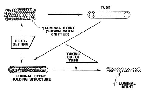
Fig.1 shows the process of producing a luminal stent
according to the present invention, in the diametrically
contracted state.
Fig.2 is a conceptual view showing the luminal stent of the
present invention as it is introduced into and attached to the
vessel.
Fig.3 shows an alternative method for contracting the
luminal stent, woven from yarns of PGA fibers of the present
invention, in the direction along its diameter.
Figs.4A and 4B are schematic views showing essential parts
of a devi ce for attachment of the 1 umi nal scent accordi ng to the
present invention, where Fig.4A shows the attachment device in
its entirety and Fig.4B shows a part thereof in cross-section.
Fig.S is an explanatory view showing the process of
attachment of the luminal scent by the attachment device of the
present invention.
Fig.6 shows another embodiment of the attachment device of
the luminal stent according to the present invention.
2082410
4
Figs.7A, 7B and 7C show the state of attachment between the
vessel and the luminal stent, where Fig.7A shows an illustrative
vessel, Fig.7B shows the state of attachment of the luminal stent
of the present invention, and Fig.7C shows an undesirable state
of attachment of a conventional luminal stent, for comparison
sake.
Fig.8 shows the possibility of attachment of the luminal
stent of the present invention in various vessel sites.
BEST MODE FOR CARRYING OUT THE INVENTION
Basically, the luminal stent of the present invention is
fabricated by knitting a sole yarn, so that a tubular product as
a luminal stent which is more homogeneous than a fabric formed
by weaving a so-called warp yarn and a weft yarn may be produced.
Besides, it is by far easier for the knitted luminal stent
of the present invention to pass through various meandering
vessels before reaching the target site. That is, the luminal
stent formed from a knitted cloth exhibits trackability with
respect to a variety of meandering passages, while it can be
introduced into and attached to a site of bend, because the
tubular knitted product tends to be dilated and is not likely to
mar the shape of the lumen. According to the present invention,
the tubular wove stent having a diameter of about 5 mm is heat-
treated and set so as to be contracted in diameter to about 2 mm
or less for being introduced into and attached to the inside of
the vessel of a lesser diameter in the living body than the
stent. This process is explained by referring to Fig. 1.
The process of attachment of the heat-set luminal stent to
the inside of the vessel is shown in a conceptual view of Fig.2.
208~4~0
An alternative method of contracting the luminal stent
knitted from PGA (polyglycol acid) polymer fiber is shown in
Fig.3. The method shown in Fig.3 has an advantage that, since
a tube formed of metal or a heat-resistant resin is not used, the
stent can be directly attached to a ballooning portion at the
distal end of the catheter.
The present invention provides a tubular luminal stent
formed by knitting a yarn of biologically resorbable polymer
fiber. The luminal stent is superior in pliability and shape
retention properties to other cloth forms, such as a non-woven
fabric, e.g. a felt, or a woven fabric formed by weaving weft and
warp yarns. The knitted luminal stent is additionally heat set
for exhibiting more prominent effects in pliability and shape
retention characteristics.
The tubular luminal stent knitted from yarns of a
bioresorbable polymer fiber has a diameter of an order of 4 to
mm and is heat set after it is introduced or as it is
introduced into a tube of heat-resistant resin or metal having
an inside diameter of about 1 to~3 mm, preferably 2 mm, to
produce a luminal stent having a set shape with a diameter of
about 2 mm, as shown in Fig. 1.
Besides, the heat setting has such a meaning that, by heat-
treating (heat-setting) the knitted tubular luminal stent while
it has a larger diameter, or of ter it is contracted in diameter,
the knitted fabric has terminal fibers, yarns or meshes which are
excellent in shape retention characteristics, such that the heat
setting affords superior shape retention characteristics while
minimizing the stress otherwise applied to the inner wall of the
2082410
vessel of the living body.
By using PLA + PGA as bioresorbable polymer fibers, and by
changing the mixing ratio, the half value period in strength of
the luminal stent of the present invention, that is the period
in which the bioresorbability disappears, may be freely
controlled within a time period of from three weeks to three
months.
Besides, by adding an X-ray impermeable agent at the time
of spi nni ng the f i tiers, the state of t;~e i ntroduced 1 umi nal stent
may be observed with X-rays. Thrombus lysing agents or anti-
thrombotic agents, such as heparin, urokinase or t-PA may also
be added, if so desired.
Besides, by taking advantage of the fact that the luminal
stent of the present invention, produced by knitting the yarns
of the bioresorbable polymer fibers, is vanished after a
predetermined time lapse from the site into which it has been
introduced, carcinostatics or anti--thrombotic agents may be mixed
into or attached to the fibers for concentrated administration
of these agents to the site of lesion.
In addition, the fibers used in knitting the luminal stent
of the present invention may be rendered variable in the cross-
sectional shape thereof more easily than if the luminal stent is
formed from metal. That is, affinity with the living body or
shape retention characteristic may be controlled by affording the
hollow or profiled cross-sectional shape to the filaments during
spinning or by using monofilament yarns or multifilament yarns.
Besides, the yarns of synthetic polymeric yarns may be
processed in many ways on its fiber surface. That is, using the
2~1~2410
7
yarns having substantially circular cross-section as usual and
which are not processed in any particular manner on its surface,
the yarns having the above-mentioned so-called profiled cross-
section, or the above-mentioned processed yarns, anti-thrombotic
materials, thrombus-lysing agents or cells of the living bodies
may be attached to these yarns for promoting multiplication of
the endothelial cells. Alternatively, X-ray non-transmitting
materials may also be attached to the yarns.
Meanwhile, if it is desired to dilate the stenotic site of
the vessel to the diameter of, for example, 4 mm, and to maintain
the diameter, the site is not dilated at a time. That is, for
avoidi ng an abrupt stress to the vessel or to the 1 ivi ng body per
se, the vessel is first dilated to a diameter of 3 mm by an
extender having a balloon-forming portion of a diameter of 0.8
to 1.2 mm. After the catheter fitted with a ballooning portion
is extracted, a catheter not fitted with a luminal stent and
fitted only with the balloon-forming portion is introduced into
the vessel for dilating the vessel to a diameter of 4 mm or more.
Finally, the knitted luminal stent is attached in place by a
luminal stent attachment device in which a luminal stmt
according to the present invention is attached to the balloon
forming portion of the device. However, it is not absolutely
necessary to dilate the vessel by steps in this manner, and the
luminal stent may be introduced into and attached to the target
site after the stenotic portion of the vessel is dilated at a
time to the desired diameter.
Alternatively, a luminal stent attachment device per se,
which is the catheter fitted with the ballooning device and with
2082410
8
the luminal stent of the present invention, may be used for
introducing and attaching the luminal stent into the vessel of
the living body simultaneously with vessel dilation.
The device for introducing and attaching the luminal stent
of the present invention in the stenotic portion of the vessel
of the living body is explained in detail. In the vicinity of
the distal end of the catheter, there exists a region capable of
forming a bal loon of a desi red diameter by a gas or a 1 iquid,
such as an X-ray contrast medium, which is injected via a hollow
part within the catheter under a liquid pressure of 8 to ;0
atmospheres. The above-mentioned heat-set luminal stent, having
the diameter of about 2 mm, is applied over the balloon forming
portion, which is about 20 mm long, with both ends of the luminal
stent being clamped by holders of silicone resin or the like
between the catheter and the outer periphery of the balloon-
forming thin film, as shown in Fig.4.
However, the length of the balloon forming portion or the
diameter of the luminal stent may be optionally set depending on
the types of the luminal stent or the specific nature of the
vessel.
Meanwhile, the distal end of the catheter is occasionally
provided with a guide wire which plays the role of a guide wire
when the catheter is introduced into the vessel.
For attachment of the luminal stent, a communication orifice
(see Fig.48) is formed at a mid part along the length of the
balloon forming portion of the catheter for permitting the fluid
injected for forming the balloon to exit from the hollow part of
the catheter to be charged between the hollow part of to catheter
2082410
9
and the balloon-forming thin film. A balloon is formed by being
dilated under a fluid pressure of 8 to 10 atmospheres via the
orifice and maintained for 30 to 60 seconds or for a longer time.
The ;tent undergoes a kind of plastic deformation at this time
under the force of dilation of the balloon so as to be
maintained in the dilated state. At this time, the polymer
itself is changed in the molecular level, or the knitted
structure, that is the mesh shape, is changed, that is, the stent
is contracted along its length and dilated along its radius so
as to be changed in shape to maintain the thus changed shape.
Fig.S shows the process of introducing and attaching the
luminal stent of the present invention within the vessel of a
living body. As shown therein, the luminal stent is contracted
in length with balloon dilation so that both ends of the stent
are detached from the holders 4. By the subsequent operation of
contracting the balloon, the catheter 2 may be removed in its
entirety.
Fig.6 shows another example of a luminal stent attachment
device according to the present invention. In this case, the
catheter fitted with a balloon is covered with a sheath 5 and
introduced in this state into the vessel of the living body.
Then, with the sheath 5 extracted slightly, the balloon is
dilated and maintained in the dilated state. The balloon is then
contracted and the sheath 5 is extracted simultaneously with the
catheter 2, while the luminal stent is left in the vessel.
Meanwhile, the thin film for balloon forming may be formed
of a variety of synthetic polymeric materials, such as
polyethylene terephthalate or polyethylene.
20~24i0
,o
It is noted that the luminal stent of the present invention
may be introduced into a bend in the vessel so as to adapt itself
to the bent shape of the vessel, as best shown in Fig.7B. On the
other hand, Fig.7C shows the state in which a metal stent
consisting in a tubular mesh or screen formed by weaving a weft
material and a warp material or a stent of a woven fabric is
introduced into a bend in the vessel. The metal stent or the
stent of the woven fabric is bent at a bend of the vessel so that
the shape of the vessel cannot be correctl y mai ntai ned i n the
site of the bend. Meanwhile, the luminal stent of i.he present
invention is superior in follow-up characteristics so that it can
reach the target site even if there exist branched parts in the
vessel, as discussed previously. Fig.7A shows an example of the
vessel of the living body in which it is assumed that a site
shown by an arrow a therein be the target site for attachment of
the luminal stent.
The luminal stent knitted from yarns of the bioresorbable
polymer fiber and heat-set according to the present invention may
cope with any thickness of the vessel with the use of the luminal
stent attachment device of the present invention. If, for
example, the luminal stent is loaded in an attachment device
which is dilated to a diameter of about 4 mm on dilating the
balloon, the luminal stent may be attached to the vessel site
having a diameter of 2.5 mm by controlling the degree of dilation
of the balloon. The luminal stent may similarly be attached to
the vessel site having a diameter of 3 or 4 mm. That is, to
luminal stent may be introduced and attached in any site shown
in Fig.B by using the same catheter fitted with the balloon. It
2482410
is because the inside diameter of the luminal stent may be
maintained at the thickness of the dilated balloon.
If re-constriction of the vessel should occur in several
monfi;hs after the luminal stent of the present invention is
decomposed and absorbed into a living body, the luminal stent may
again be introduced and attached in the same site. This is
rendered possible by using the bioresorbable polymer.
Meanwhile, if a thin sheet of a non-woven fabric of a
bioresorbable polymer, such as a felt, bent into a shape of a
tube, exhibits shape retention characteristics and flexibility
comparable to those of the luminal stent of the present
invention, such sheet may be used in place of the knitted
material.
With the above-described luminal ste m of the present
invention, such meritorious effects may be achieved that
inflammation or excess hypertrophy of the vessel may be prevented
and consequently reconstriction of the vessel may be inhibited.
The luminal stent of the present invention is absorbed in several
months into a living tissue, which is favorable for the living
body.
If an X-ray impermeable agent is applied to the
bioresorbable polymer fibers or yarns of the luminal stent of the
present invention, the state of attachment of the stent within
the vessel may be easily observed by X-ray irradiation from
outside.
Besides, the luminal stent may be applied over the balloon
forming portion of the catheter according to the present
i nventi on so that the stent may be easi 1 y attached i n the desi red
2082410
12
site within the vessel.
Experiment 1
Plural luminal stents formed by knitting a yarn of
polylactic acid fibers admixed with barium sulfate were
introduced and attached in the coronary of a test animal in a
tubular state of 4 mm in diameter and 20 mm in length by using
a cathete r f l tted wi th a bal 1 oon, and the state of attachment was
observed by irradiation of X-rays. It was seen that the stents
substantially maintained their shape until after about three to
six months. It was seen that the stents disappeared by being
absorbed into living tissue in about 6 to 12 months. During this
time, no abnormalities such as inflammation or hypertrophy of the
intima of the blood vessel were observed.
Exaeriment 2
Plural luminal stents formed by knitting a yarn of
polyglycol fibers admixed with barium sulfate were introduced
and attached in the femoral artery of a test animal in a tubular
state of 4 mm in diameter and 20 mm in length and the state of
attachment was observed by irradiation of X-rays. It was seen
that the stents substanti al 1 y mai ntai nod thei r shape unti 1 afte r
about two to three weeks and were absorbed into the living tissue
in about two to three months. The shape retention period and the
period of existence in the living body attained in Experiment 2
are thought to be more safe than the corresponding periods
attained in Experiment 1. Meanwhile, no inflammation or
hypertrophy of the intima of the blood vessel was observed during
these periods.