Note: Descriptions are shown in the official language in which they were submitted.
, ~VO91/18306 2 ~ 3 3 ~` ~ ~J PCT/E~1/00934
- 1 - i
OPTICAL PROBE
This invention relates to a method and apparatus
for making an optical fibre bundle and, more
particularly, but not exclusively, to a method and
apparatus for making an optical probe.
In 1985, David Costello, one of the co-inventors
named in this application, filed his application
entitled "Fibre Optic Probe For Quantification of
Colorimetric Reactions" which resulted in the issuance
of US Patent 4 682 895. This patent describes an optical
probe which can be inserted into an artery to monitor
the concentration of certain dissolved gases in the
blood. Previously, the concentration of such gases was
measured in a laboratory in blood samples taken from the
patient.
With the new optical probe, real-time determination
of a biochemical parameters became possible, providing
up-to-the-minute information to the physician. This can
be of critical importance with an unstable patient whose
biochemical parameters and blood gases may change
dramatically over a short period of time. Real-time
current and trend information is much more valuable to a
physician than the previously-available discrete
historical data.
Whilst the applicants optical probe is eminently
suitable for its original purpose the inventors felt
that it would be desirable to be able to simultaneously
monitor more than one functicn, for example the
concentration of oxygen and carbon dioxide in the
patent's bloodstream and the pH of the blood. Because of
the physical properties of the optical fibres and the
very small dimensions involved, many manufacturing
methods were conceived but found unsuitable for one or
other reason. A particular problem with many methods was
that they were totally unsuitable for even modest
JBS~ JTE SHE~
- . ... . .. . . .
~, r , , . ~, - ' , ' ,. : . -,
': ' ' ~ ' ' '~ . ' . : '
': ' ' ~, ' : . . , ' ' .' ' :
- . '. ' ;` ', ''' ' ' :: : ' , :-
' ' ' I ~: . ~ ' , ' ' ~ ' '
'' .' ,' ' ' ' :
WO91/18306 2 ~ ~ 3 ~ 2 - PCT/E~1/~0934
production runs resulting in a prohibitatively expensive
product bearing in mind the highly skilled labour
involved.
According to one aspect of the present invention
there is provided a method of making an optical fibre
bundle, characterized in that it comprises the steps
of:-
a) bending a plurality of optical fibres to form afibre bend in each;
b) suspending the plurality of optical fibres at
the fibre bends over at least one suspension member;
c) holding the fibres taut to mai~tain the fibres
bent;
d) applying potting material to the plurality of
optical fibres;
e) curing the potting material; and
f) removing the plurality of fibres from the
suspension member.
Other features are set out in Claims 2 to 48.
In a preferred embodiment, the mandrel has a
diameter equal to that of a fibre. The resulting bend
thus has a radius approximately equal to the diameter of
the optic fibress
An apparatus for making optical probes according to
the present invention, in one embodiment has a holding
jig or spider with an open centre and holes spaced
around the centre. Mandrels or spokes (e.g., of wire,
thread, suture, or of fibre optic material) are threaded
through pairs of holes to intersect at an open centre
area and divide the open centre area into wedges. The
ends of the optic fibres are inserted into the wedges,
preferably one per wedge. The fibre ends are clamped to
a fabrication stand which is flexible so that, by
bending the stand and readjusting the clamp, varying
degrees of tension are applied along the optic fibres. A
~;~J8S~T~E SHE~
- . , , ... -:. . - , - `: `
. ~ ,. ; ,.
, - . .`.
` . .. ~ `
WO91/18306 2 0 3 ~ PCT/E~1/00934
- 3 -
tip support coating, e.g., epoxy cement or other potting
material, is applied to the tip of the fibre bundle in
the area of the mandrels and extending down the fibre
bundle. This insures that the fibre geometry and
disposition will be maintained. After the epoxy has
cured the mandrels are removed by cutting them off close
to the intersecting point. In another embodiment the
mandrels are lubricated so they will slide away from the
fibres.
Under microscopic examination it has been observed
that in one embodiment one half of each fibre can be
made to tend toward the outside edge of the completed
probe by proper placement of the fibres. This gives the
probe in this embodiment a rounded triangular cross-
section and facilitates removing a slice from each
fibre, e.g., with a diamond edged wafering saw. In
another embodiment the use of cantilevered micro hooks
for fibre suspension permits a more rounded probe shape.
Tension may be applied to the fibres in a variety
of ways, including but not limited to: stretching the
fibres and clamping them in place; fixing the mandrels
in place on a frame and weighting the fibre ends; or
applying magnetic force to ferromagnetic elements
applied to fibre tips or mandrels. Tension can aIso be
applied to having a vacuum chuck receive and hold the
fibre ends distal from the fibre bend.
Turning now to the sample chambers, indicators may
be immobilized by chemically binding them to the surface
of solid particles, e.g., porous glass particles, latex
microspheres, or adsorbent polymers such as styrene or
polydivinyl benzene. These solid particles may be held
in place within the sample chamber by a gel, e.g. a
solution of hydroxy propyl-cellulose. A selectively
permeable membrane is applied over the indicator
containing sample chambers to prevent loss of the
STIT~3Ti~ S~IEE~
: ., .`..... .. : . .
. ~ , .
`
.
WO91/t8306 ~0~ 3 ~ PCT/E~1/00934
-- 4
indicator compound or contamination by undesired chemical
species in the environment.
In another embodiment of a process according to the
present invention, fibres are emplaced over cantilevered
micro hooks disposed above magnetic positioning clamps
secured to weights which provide tension to hold the
fibre bends in place on the cantilevered micro hooks. To
facilitate and simplify the process, the free fibre ends
are received in and held in small diameter tubes to
which a vacuum has been applied. The cantilevered micro
hooks are disposed on pivotable arms for ease of
disposition. After one or more fibres have been
positioned on the arms, the arms are moved together so
that the fibres, and particularly the fibre bends are
disposed with respect to each other as they will be in
the finished probe. Thus, a more rounded configuration
of multiple fibres can be achieved, rather than
triangular, which occupies less total space. In this
embodiment during the fabrication process the fibres
hang independently of each other and each fibre has its
own applied weight for tension and its own set of vacuum
tubes. Thus the fibres do not compete for disposition on
a mandrel or push against or down on each other. After
the fibres are removed from the cantilevered hooks and
vacuum tubes, the bends are also potted (in addition to
several inches of the fibres that are potted prior to
removal from the hooks). The potted tip is then
subjected to the ultraviolet lamp beam for curing.
3S~ITVTZE S~IE
. . . . .
- . .. . - . .
2~83~
5VO9l/18306 PCT/E~1/00934
-- 5
For a better understanding of the present invention
reference will now be made, by way of example, to the
accompanying drawings, in which:-
Fig. l is a cross-sectional view of an optical
fibre in tension around a mandrel;
Fi~. 2 is a top plan view of Fig. 1;
Fig. 3a is a side view of one arrangement of three
optical fibres on mandrels;
Fig. 3_ is a top plan view of Fig. 3a;
Fig. 4a is a side view of a second arrangement of
three optical fibres on mandrels;
Fig. 4_ is a top plan view of Fig. 4a;
Fig. 5a is a side view of a third arrangement of
three optical fibres on mandrels;
Fig. 5 is a top plan view of Fig. 5a;
Fig. 6 is a side view of an apparatus used in
fabricating an optical probe according to the present
invention;
Fig. 7 is a cross-sectional view of a vacuum chuck
similar to the vacuum chuck used in the apparatus of
Fig. 6a;
Fig. 8a is a top plan view of a "spider";
Fig. 8b is a side view of the "spider" shown in
Fig. 8_:
Fig. 9 is a perspective view of part of the tip of
one embodiment of an optical probe in accordance with
the present invention;
Fig. 10 is a cross-sectional view of a second
embodiment of an optical probe in accordance with the
present invention;
Fig. lla is a side view of the optical probe of
Fig. 10;
Fig. 11_ is a top view of the optical probe of
Fig. 10;
Fig. 12 is a cross-sectional view of a sample
3S~l~U~ SH~
. .
-
. , -
` ~ - :, . .
.. - ~ - ... ~ ..
WO91/18306 2 ~ ,~ c~ 3, ~ PCT/E~1/00934 ~
-- 6
:
chamber for measuring pH;
Fig. 13 is a cross-sectional view of a sample
chamber for measuring C02 levels;
Fig. 14 is a cross-sectional view of a sample
chamber for measuring 2 levels;
Fig. 15 is a side view of an optical probe mounted
in an intra-arterial cannula;
Fig. 16 is a schematic view of a sensor unit for
use with the optical probe shown in Fig. 10;
Fig. 17 is a side view in cross-section of a
protective housing for part of the optical probe;
Fig. 18 is a side view of the optical probe
emerging from part of the protective housing shown in
Fig. 17;
Fig. 19 is a side view of the tip of the optical
probe in Fig. 15 and part of the intra-arterial cannula
of Fig. 15;
Fig. 20_ is a side view of an apparatus for
producing optical probes according to the present
invention;
Fig. 20b is a front view of the apparatus of
Fig. 20a;
Fig. 20c is a view along line A-A of the apparatus
of Fig. 20a ;
Fig. 20d is a top view of the apparatus of
Fig. 20_;
Fig. 21 is a detailed side view of fibre holders of
the apparatus of Fig. 20a;
Fig. 22 is a cross-sectional view of an optical
probe made as shown in Figs. 21 and 20a;
Fig. 23 is a face view of the optical probe of
Fig. 22;
Fig. 24 is a side view of the optical probe of
Fig. 22;
Fig. 25 is a cross-sectional view of a third
~;UBS~Tl)TF S~E~:Y
. . . . .... . .................. .. . . . . ..
, . .. . .; .~,- ~ . -........... .. ..
~ VO91/18306 2 ~ ~ 3 IJ '~ ~ PCT/EP9l/00934
, .
- 7 -
embodiment of an optical probe according to the present
invention;
Fig. 26 is a cross-sectional view of a fourth
embodiment of an optical probe according to the present
invention;
Fig. 27 is a partial top view of a fibre guide of
the apparatus of Fig. 20a;
Fig. 28 is a partial top view of another fibre
guide of the apparatus of Fig. 20a.
Referring now to Figs. 1 and 2, an optical fibre 10
of diameter "a" is shown in tension around a mandrel 12,
producing a bend 14 in the fibre. The mean radius of the
bend 14 is approximately equal to the diameter ~a" of
the optical fibre 10. The partial geometry and
configuration of the optical fibre 10 is maintained by
applying a potting material 18 around the optical fibre
10. After the potting material 18 has been applied and
cured, the mandrel 12 may be removed by cutting it close
to the fibre optic bundle and carefully withdrawing it.
Another layer of coating can be applied to the fibre by
dipping or spraying on the appropriate potting material
; to seal the hole left by the mandrel.
A sample chamber 20 is provided in the optical
fibre 10 for containing and holding a checical-
indicating colorimetric material.
As shown in Figs. 3a and 3_, a common radialconfiguration of mandrels may be employed for different
arrangements of multiple optical fibres.
In Fig. 3a and 3_ three optical fibres 30, 31, 32
are stacked on top of a three-mandrel stack of mandrels
33, 34 and 35. In this configuration the optical fibres
contact each other and compete for position on the
mandrels. Half of each optical fibre resides slightly
recessed within the total cross-section of multi-fibre
arrangement. Each fibre is thus disposed so that it is
~;~8S~
2 ~ 8 ~
WO 91/18306 PCr/EP91/00934
-- 8
presented for convenient and effective formation of a
sample chamber therein.
As shown in Figs. 4_ and 4b, mandrels 43, 44 and 45
are stacked with a common radial pattern as viewed from
5 above (Fig. 4b) but the mandrels are spaced apart by
optical fibres 40, 41 and 42 disposed on the mandrels.
In this configuration the optical fibres do not bear
down directly on each other, particularly when tension
is applied.
A three channel probe was made of the configuration
of Fis~. 3_ using plastic fibre and wire mandrels of 2S0
micron diameters. The resulting probe had a maximum
diameter of 1.56mm.
Three probes were made with optical fibre mandrels
lS and optical fibres with 125 micron diameters. These
probes had maximum diameters of 630 microns, 800
microns, and 584 microns according to the tension
applied to the optical fibres. The maximum diameter of
the finished probe is inversely related to the tension
20 applied to the fibre bundle during fabrication.
Referring now to Figs. 5_ and 5_, optical fibres 50
and 51 are supported by a mandrel 53 and a mandrel 54
supports an optical fibre 52 spaced apart from the
optical fibres 50, 51 and the mandrel 53. The mandrels
25 53 and 54 may be spaced apart any desired and workable
distance. The optical fibres 50, 51 and 52 may have
different bend radiuses. These radiuses may be varied to
produce an arrangement that is of a desired overall
shape, e.g., circular or rectangular. These different
30 shapes of the fibre bundle may facilitate cutting of the
sample chamber or may enhance the flow profile of fluids
flowing past the optical probe in use.
As shown in Figs. 5_ and 5b, the optical fibres may
be positioned so that they are not in contact with each
35 other. This provides complete separation of sensor
~l~3E3ST~ S~EE~ ~
~WO91/~8306 2 d ~ 3 ~ PCT/E~l/oog34
channels and facilitates positioning and removal of
mandrels. It is, however, within the scope of this
invention to provide such an arrangement in which an
optical fibre does contact an adjacent optical fibre.
With respect to the arrangement shown in Figs. 3a,
4_ and 5_, thre fibres are shown. It is within the
scope of this invention to employ two or more fibres in
such arrangements. Of course using a greater number of
fibres will increase the overall size of the resulting
optical probe.
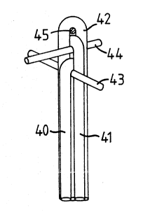
Referring now to Fig. 6 there is shown an apparatus
150 for manufacturing optical probes. The apparatus 150
comprises an upright member 152 to which are secured a
top spider 154, a mid-position spider 156 and a vacuum
chuck 158. These items may be semi-permanently secured
to the upright member 152 so that they can be moved and
positioned as desired.
The upright member 152 is secured to a top arm 160
and a bottom arm 162 which in turn are pivotally
20 connected to a wall 164 by pivots 166 and 168
respectively. The wall 164 is mounted on a base 174.
Radiation from an ultraviolet lamp 170 passes through a
quartz window 172 in the wall 164 to cure potting
material applied to a bundle 176 of one or more optical
25 fibres disposed between the spiders 154, 156 and the
vacuum chuck 158. The vacuum chuck is movably disposed
on the upright member 152 to facilitate the handling of
ends of the optical fibres in the bundle 176 and to
provide adjustability of the tension on the optical
30 fibres. A shield 178 movably connected to the wall 164
; can be moved into position to shield personnel from
radiation emitted by the ultraviolet lamp 170. An
electrostatic charge neutralizer 180 secured to the wall
164 eliminates electrostatic charges in the fibres.
Pulsed intermittent application of the radiation
~;~J8STIT~ E~:~
- .... . . . .. . .
- ... . ...
. . -
- , ~ . .. .
W O 91/18306 2 ~ 3 ~ 3 ~ ~ PC~r/EP91/00934 ~
- 10 -
from the ultraviolet lamp 170 can be achieved by
alternately turning the lamp on and off or by
periodically blocking the beam. As shown in Fig. 6, a
shutter 171 is disposed so that it may be moved to close
off the quartz window 172. The shutter 171 is connected
to a motor 173 which operates to move the shutter 171
toward and away from the quarts window to achieve the
desired pulsing of radiation for curing the potting
material. Pulses of 0.75 seconds duration are preferred
for preferred potting materials; pulses of longer
duration might cause a damaging temperature rise in the
fibres. A heat shield can be placed over the fibre bends
on the spider during this curing operation to protect
them from heat damage.
Referring now to Fig. 7, a vacuum chuck 194 (like
the vacuum chuck 158 of Fig. 6) has a chuck body 196
with a chuck funnel 198 having a concave funnel opening
200 for receiving the ends of optical fibres and a
funnel bore 202 through which the fibre ends pass into a
20 funnel chamber 204. The chuck body 196 is movably
mounted above a dash pot 206 which is mounted on a dash
pot base 210. When the chuck body 196 moves downwardly,
a ram 212 moves into a bottom bore 214 of the funnel
bore 202, clamping the ends of optical fibres between
the ram and an 0-ring 218 in the funnel bore. The rod is
sealed by a bottom seal 216. A vacuum is applied to the
chuck by a vacuum pump 217 (Fig. 6) through an opening
219. The vacuum chuck applies minimal tension to the
fibres.
As shown in Figs. 8a and 8_, a spider disc 182
(used, e.~., with the spiders 154 and 156 of the
apparatus 150) has six slots 184 and six notches 186 in
the middle ridge 188 surrounding a hole 190. Wires 192
pass through these notches and slots and secured to the
~5 disc serve to separate optical fibres held therebetween.
.
~;~3E3STIT~JlrE SO~E~:7
. ~
... . .. . . - . . ~ . . . . .
. .. .. . . . .. . .. .
~ ~091/18306 2 ~ $ 3 ~ 3 ~ PCT/E~1/00934
.~ :
-- 11 --
Dimensions indicated here are in inches.
Referring now to Fig. 9, an optical probe 550
according to the present invention with coatings and
membranes as previously described (and like the probe 60
described below) is shown containing three optical
fibres 551, 552, 553. Sample chamber 554 is shown as a
five-walled chamber. The sample chamber 554 is about 100
microns long and can be, preferably, 85 to 115 microns
long. The preferred depth ranges between 130 and 160
10 microns. The optical probe 550 is about 700 microns in
diameter. It is preferred that this sample chamber be
formed by using an excimer laser because use of su~h a
laser provides precise location, exact dimensions, and
an optically clear finish to cut fibre faces 253a and
15 253b.
As shown in Figs. 10, lla and llb, a second optical
probe 60 according to the present invention has three
optical fibres 61, 62 and 63 encapsulated in cured
potting material 67. Each optical fibre 61, 62 and 63 is
20 associated with a sample chamber 64, 65, 66
respectively. The distance _ is the outside diameter of
the fibre optic probe 60 and should be controlled as
appropriate for the intended use of the probe. As an
example, if the intended use is for insertion through an
arterial cannula the outside diameter should be small
enough to fit easily through the cannula. The distance _
is the thickness of encapsulant material lying over a
particular fibre, i.e., the cover. This distance affects
the volume of the sample chamber, the separation of the
light path from the outside environment, and the outside
diameter of the optical probe. The optical probe 60 h~s
been coated (undercoating, overcoating, e.g., as in
Fig. 12) and the sample chambers 64, 65, 66 are like the
chambers 70, 80, 90, respectivelv (Figs. 12, 13, 14).
Membranes have been applied over the sample chambers 64,
~J8~;T~TF St~lEF:7
. ~ ........................................................... .
-... .. ~ . ~ ~
W O 91/18306 2 ~ S 3 3 u~` J; PC~r/EP91/00934 ~ ~
- 12 -
65, 66 as for the chambers 70, 80, 90.
In the embodiment of Figs. 10, lla and 11_, the
longitudinal distances from sample chamber 64 to the tip
end of the optical probe is about 2540 microns and
preferably is in the range of 2240 to 2840 microns. The
longitudinal distance t between chambers 64 and 65 is
790 microns and is preferably in the range of 915 to 665
microns as is the distance v between the chambers 65 and
66. This preferred longitudinal spacing minimizes the
depth of intrusion into, i.e., a human blood vessel, yet
reduces structural weakness which might be caused by
chambers spaced closely together. This spacing also
facilitates loading of chemical indicators into sample
chambers as well as the application of membrane material
over sample chambers.
A pH sample chamber 70 through an optical fibre 79
is shown in Fig. 12. The chamber 70 has an undercoating
72 (preferably of a water impermeable polymer such as
commercially available under the Trade Marks Petrach SE,
Teflon, or Perylene), to protect the sample chamber from
changes in chemical concentration due to the diffusion
of water and an overcoating 74 of the sample material.
The undercoating 72 may be of multiple layers to
increase resistance to water transport.
A pH indicating material 76 (e.g., phenyl red,
phenolphthalein, buomocresol green, or other subtalein
indicators that react, e.g., change colour or fluoresce
upon a change in pH) is introduced and is covered by a
selective membrane 78 (e.g., nitro cellulose or porous
hydrophilic polymers). The membrane 78 selectively
permits hydronium ions to flow from outside the optical
probe 79 into the sample chamber 70. The coatings are
applied before sample chambers are formed.
As shown in Fig. 13, a carbon dioxide sample
chamber 80 through an optical fibre 89 has an
S~ 3T~ SI~EE7 ~ -
~ 091/18306 2 ~ g 3 3 ~ PCT/E~l/00934
- 13 -
undercoating 82 (like 72); an overcoating 84 (like 7g); a
C2 indicator 86 (e.g., phynol red combined with
bicarbonate or other pH indicator); and a selective
membrane 88 which selectively permits CO2 to pass from
outside the optical fibre 89 into the sample chamber 80.
The CO2 reacts with water present in the sample chamber
80 to create carbonic acid which in turn reacts to
change the colour-of the pH indicator present in the
chamber. Essentially any pH indicator may be used to
indicate C0 by combining the indicator with bicarbonate
and isolating the reaction from hydronium ions in the
environment while permitting access to C02.
Referring now to Fig. 14, an oxygen sample chamber
90 through an optical fibre 99 has: an undercoating 92;
an overcoating 94; an oxygen-indicating colorimetric
substance 96 [e.g., BASF Fluorol Green Gold (perylene
dibutyrate)] or other fluorescent chemicals which
respond to the presence of oxygen; and an oxygen semi-
permeable membrane 98 (e.g., silicone rubber, PTFE,
porous polypropylene or porous polycarbonate) which
selectively permits oxygen to pass from outside the
fibre 99 to the oxygen-indicating substance 96.
With phenol red material immobilized on porous
glass, the glass and membrane are supported by a
material such as hydroxy propyl cellulose or hydroxy
ethyl cellulose (plus bicarbonate for C02). An
immobilizer is used to inhibit phenyl red (indicator)
from migrating out of the sample chamber. The support
material can be applied manually by mixing it with
porous glass. Cellulose acetate is applied to the entire
tip of a sensor to separate the sample chamber from the
exterior environment to insure only the desired analyte
comes in and to hold the indicator in the sample
chamber .
A measurement of C02 or oxygen concentratior.
1E3STIT~E SH~E~
;, ... :.. . . . . . . . .
wo 91/18306 2 1~ ~ 3 ~ 3 ~' ~ PCT/E~1/00934 ~
- l g -
accomplished by the optical probe of Fig. 14 can be
expressed as a partial pressure of total gas.
As shown in Fig. 18 an optical probe 100 (like the
optical probe 60) has a bundle of optical fibres 232
which extend through a connector 230. As shown in
- Fig. 17 the bundle of fibres is glued into a male
fitting 234 with adhesive 235. (Bundle 232 is like
previously described bundle 60.) The connector 230 is
made preferably from polycarbonate plastic. A protective
tube 242, preferably made from polyvinylchloride tubing
is secured around the male fitting 234 and provides
; strain relief. The bundle 232 is surrounded by a black
inner tube 238, preferably made from polyethylene to
provide lubricity in the interior of the tubing to
facilitate the passing of fibres therethrough. The black
colour prevents ambient light from affecting the fibres.
A protective tube 240 surrounds the tube 238. The tube
240 is made preferably from polyethylene and extends
from the connector 230 to a junction box 231 (Fig. 16).
!; 20 The tube 242 surrounds a portion of the tube 240, as
does a tube 244 (like tube 242) adjacent the junction
box 231. In one embodiment the distance from the male
fitting 234 to the junction box 231 is about 305mm
(12 inches).
As shown in Fig. 15 the probe 100 has been disposed
in an intra-arterial cannula 101.
As shown in Fig. 16, the optical probe 100 may then
be connected to a sensor interface unit 120 (shown as
separate from a base unit but which could be
incorporated therein) which is connected to a base unit
122. The sensor interface unit 120 provides light input
to the probe and detects and measures light coming out
of the probe. Signals from the unit 120 are then fed
into the base unit where they are processed for display
or recordation or both.
~;Ug~S~T~lT F: SHE~7
;
(~
~VO9t/18306 PCT/EP91/00934
. j
- 15 -
As shown in Fig. 19, a tip 103 of the optical probe
100 extends into the intra-arterial cannula 101. The
cannula 101 is suitable for introduction into and
disposition within a human blood vessel. The tip 103 is
disposable in the cannula (e.g., when the cannula is
emplaced in an artery) by connecting the connector 102
to the cannula and then inserting the probe 100 into the
cannula through one branch 107 of the connector 102. It
is preferred that the distance k from the end of the
cannula to the tip of the optical probe be such that
there is good "washability" or fluid flow over the
sample chambers; e.g., in certain preferred embodiments
this is about 4910 microns. It is preferred that the
distance from the end of the cannula to the first
adjacent sample chamber be such that fluid injected
through the connector does not diffuse into the blood in
the region of the sample chambers producing erroneous
readings; e.g., in preferred embodiments this is about
790 microns. Whatever fluid was being introduced into or
withdrawn from the cannula may be introduced or
withdrawn from a branch 105 of the connector through
which the probe 100 does not extend.
Figs. 20a - 20d illustrate another apparatus 250
for fabricating an optical probe according to the
present invention. The apparatus 250 has an upright
member 252 to which are pivotably connected two arms 254
and 256. A weight holder 258 is secured to the arm 254
and a weight holder 260 is secured to the arm 256. A
micro pin mount 262 is secured to the top of the arm 254
and a micro pin mount 264 is secured to the top of the
arm 256.
A fibre guide 266 is secured to a bottom portion of
the arm 254 and a fibre guide 268 is secured to a bottom
portion of the arm 256. The micro pin mount 262 has two
micro pins, each for independently holding an optical
~;~E3S~ St~E~
., . ~ - ~
,.: .
WO91/18306 2 ~ g 3 ~ ~ ~ PCT/E~1/00934 ~
- 16 -
fibre. The weight holder 258 has two weights, 270 and
; Z72, one each for each of two optical fibres 274 and 276
supported from the micro pin mount 262. The weight
holder 260 has a weight 278 for a fibre 280 supported
from the micro pin mount 264. The weights 270, 272 and
278 are freely movable up and down on a wire extending
through the weights; wire 282 extending through weights
270, 272 and secured to a top arm 284 and a bottom arm
286 of the weight holder 258; and wire 288 extending
throu~h the weight 278 and secured to a top arm 290 and
a bottom arm 292 of the weight holder 260. Each weight
has a sheet magnet 294 secured thereto. Optical fibres
are clamped between the fixed magnets 294 and a free
sheet magnet 296 which provides a sufficient clamping
effect to hold the fibres and hang the weights from them
to provide the desired tension. Each weight weighs about
18 grams. Notches are provided in the fibre guides 266,
268 and in a top shoulder 298 (e.g., Fig. 20_ of each
weight so that the fibres are held separately and
independently in the apparatus 250. As shown in Fig. 27
the fibre guide 68 (shown partially) has a recess 269
for receiving and holding the fibre ends of the fibre
280. The fibre guide 266 (shown partially in Fig. 28)
has a recess 267 for receiving and holding the fibre
ends of fibres 274 and 276. (Dimensions in Figs. 26, 27
are in inches.) This insures formation of an optical
probe of desired configuration. Three pairs of vacuum
tubes 300, 302, 304 receive and hold the ends of each of
the optical fibres 274, 276 and 280 respectively. Each
vacuum tube receives and holds one fibre end. ~y thus
holding the fibre ends, the use of the apparatus and
handling of the fibres is facilitated and some minimal
tension is applied to the fibres.
Since the arms 254 and 256 are pivotable with
respect to the upright member 252 one or more optical
~3UBS~ S~E~
.. . ............ . - . . . ..
. ... . . . -.. . . ....... . ........ . - . .- - ..
r ~
~V091/l8306 PCT/E~1/00934
- 17 -
fibres can easily be emplaced on each arm's micro hooks
prior to closing of the arms 254, 256. Closing the arms
into proximity with each other moves the fibres into a
desired relationship with each other so that an ultimate
configuration and size for an optical probe is
achieved.
Potting material (e.g., commercially available ELC
4481 of Electro-Lite Corp.) can be applied manually to
the exposed portion of the optical fibres between the
top portions and bottom portions of the arms 254 and
256.
As with the apparatus 150 an ultraviolet lamp may
be used with the apparatus 250 for curing the potting
material.
Internal stresses in the fibre bends around the
micro hooks can be relieved by applying heat to the
micro hook holders, the micro hooks, and the fibre bends
while the fibres are still emplaced on the apparatus 250
before the application of potting material. For example,
hot air at about 80C is blown at the micro hook
holders. The holders and hooks heat up gradually (e.g.,
in about 90 seconds) so that the temperature of the
fibre bends comes up to about 80C. This helps to make
the bends more permanent and reduces the tendency of the
fibres to spring back. Auxiliary weights (e.g., double
the weight of the weights 270, 272, 278) are temporarily
applied to the fibres (e.g., by hanging onto the weights
270, 272 and 278) during the heat treatment to achieve
optimum bending stress while the temperature is
elevated.
The potted assembly is removed from the apparatus.
Potting material is applied to the tip of the assembly -
the portions hanging over the micro hooks including the
fibre bends. This material is then cured. Thus the fibre
bends and a portion of the fibres adjacent the fibre
S~ E S~E 7
.
WO9l/l8306 ~''~ 18 - PCT/E~I/00934
bends is covered with cured potting material and the
fibres (and thermocouple or other device) in these
portions are not in contact. The harness (tubing,
connector, junction box) is assembled and then sample
chambers are formed in the fibres. Coatings are applied;
indicators are emplaced in the sample chambers; and
membranes are applied over the sample chambers by
placing membrane material dissolved in a suitable
solvent over the sample chambers: e.g., cellulose
acetate in acetone as a membrane over a pH indicator,
polydimethyl siloxane in methylene chloride as a
membrane over a C02 or 2 chamber or polycarbonate in
chloroform. The resulting assembly is useful as an
optical probe.
Fig. 21 shows the micro pin mounts 262, 264 in more
detail. These micro pin mounts are configured so that
the fibres to be worked with and treated hang
independently of each other. Micro pin mount 262 has a
cantilevered pin 310 from which hangs a thermocouple 312
20 (not shown in Figs. 20_, _) and a cantilever pin 314
from which hangs an optical fibre 316. The pins are
preferably disposed at an angle to maintain the fibres
thereon. Micro pin mount 264 has a cantilevered pin 318
from which hangs an optical fibre 320 and cantilevered
25 pin 322 from which hangs a fibre 324. The pins 310, 314
and 318 are emplaced in grooves that are about 0.13mm (5
mils wide). The pin 322 is emplaced in a groove that is
about 0.33mm (13 mils) wide. Thus an optical fibre
hanging over pin 322 hangs with its ends spaced further
apart than the ends of a fibre hanging over one of the
other pins.
Figs. 22, 23 and 24 show an optical probe 332 (like
the optical probe 60 previously described regarding
sample chambers, coatings and membranes) corresponding
to the fabrication layout of Fig. 21. Dimensions given
SlJ!~3ST~ S~E~lr
. .......... - ...... .. - . - , ~ . .
. -
~091/18306 PCT/E~1/00934
- 19 -- .
in Figs. 22 and 23 are in inches. The distance between
the strands of the thermocouple fibre is .005 inches.
The size of sample chambers 326, 328 and 330 may
preferably range in width between about 130 to about 160
microns with 145 microns preferred. The overall diameter
of the probe 332 as shown is 650 microns (.0256 inches).
This diameter preferably ranges from about 600 microns
to about 730 microns with preferred diameter being 650
microns. As shown there is a .0014 inches thick cured
potting layer between the fibres and the exterior
surface of the probe so it will fit easily through a 20
gauge cannula and permit blood to be withdrawn
therefrom. This also separates the membrane from the
optical path. As shown in Figs. 22 and 24 the fibres
15 316, 320 and 324 and thermocouple 312 do not contact
each other in the tip end of the probe. The thermocouple
312 has a small (e.g., 10 mil) metal bead 325 to which
are connected two 2 mil diameter metal leads 327.
Commercially available Type E thermocouples can be used
or a thermister. As shown in Fig. 23 the length from
line P (line P represents the extent of the potting
material applied to the probe tip) to the exterior of
the first fibre bend is about .026 inches. A preferable
range for this distance is .020 to .031 inches. The
distance from point P to the thermocouple is about .044
inches ~and ranged preferably between .040 and .051
inches). The distance from point P to the end of fibre
324 is shown as about .070 inches (preferably ranging
between .066 and .075 inches). The thermocouple is
located preferably interiorly of all the fibres because
it needs no tangential access to fluids and it can
occupy interior space not occupied by optical fibres.
Fig. 25 illustrates a probe 334 in which all fibre
bends (of fibres 336, 338 and 340) are of substantially
the same diameter. A thermocouple 342 is present in the
;HE~
,
.. .. .. ..... . . . . . . ..
:' .'.
.
WO9l/18306 ~v~,J~, PCT/E~1/00934
- 20 -
centre of the probe 344.
Fig. 26 presents an optical probe 346 produced with
apparatus 150, 160 using a spider as shown in Fig. 4a.
Since the optical fibres in the apparatus 150 contact
and compete with each other for position, the generally
triangular disposition achieved requires more potting to
effect a desired cover of potting material over the
optical fibres. In this sense a circular configuration
of the optical fibres of the probe (e.g., as shown in
Fig. lO) is more efficient.
Various modifications to the method of construction
described are envisaged. For example, the sample
chambers could be formed by placing the ends of two
fibres facing one another.
It should be appreciated that whilst the method
described is primarily intended for the production of
optical probes with a plurality of sensors it is also
applicable to the production of an optical probe with a
single optical fibre and a single sample chamber.
;T~T~E Sl~7
.. . . . ..
- . . ' '. ' ' ': '
.
. . , , . - , - . . . . .: . -
. . . .. . ., ~ , ~ . . . . .