Note: Descriptions are shown in the official language in which they were submitted.
BIOCHE~ICAL OXYGEN DEMAND ANALYZER, METI IODS OF
ANALYSIS, MICROORGANISMS USED FOR ANALYSIS
1. Introduction
The present invention relates to a biochemical oxygell demancl
(BOD) analyzer, methods of analysis, and a novel bacterial strain used for
analysis. Specifically, the present invention relatcs lO a BOD analy%er
comprising a microbe sensor containing an oxygen electrode and a
microbe membrane. Tlle microbe membrane is made by immobilizing
1 0 microorganisms belonging to the genus Klebsiella in a membrane.
2. Background of the Invention
BOD analysis is presently carried out in accordance with Japanese
Industrial Standard Method (JIS Industrial Wastewater Test Method K-
1 5 0102-1972). Since it takes 5 days to analyze BOD, various attempts have
been made for quick BOD analysis by utilizing microbe sensors. Among
the microorganisms, Trichosporon cutaneum and activated sludge have
been used as microbes immobilized on the microbe sensol s (Japanese
Patent Application KOKOKU No.7258/1986; Suzuki. S., ed., Biosensor
ppl35-136,140-142 Kodansha Publication (1989)). In addition, an
apparatus comprising a flow cell equipped with a microbe sensor has been
known as a BOD analyzer (Japanese Patent Application KOKAI Nos.
47895/1978, 123851/1991).
The problem of the microbe sensor is that the BOD result of lhe
2 5 microbe sensor has a low correlation to that of the JIS method. The
problem is partly because of microorganisms used for microbe sensors.
For example, Trichosporo)l cutaneum has a narrow assimilation
spectrum on various organic substances, i.e.no response to disaccharides
but specific high response to particular substances such as ethylalcohols.
z~
In addition, the microbe sensor has been impractical bccause the sensor
has to be activated 1-3 days berore BOD analysis in or(lel to h~ve
microbes normally respond to samples. When activatecl ~ludge is Llsed,
activated sludge immobilizecl on a membrane has to be constanlly
5 eontrolled and the immobilization procedure is requile(l every time the
membrane is changed. BOD analysis is unable lo cally oul using any Or
the above microor~anisms if high concentration o~ bactericidal substances
such as arsenic contained in a sample.
In a BOD analyzer using a flow cell, bubbles remained in samples
may affect analysis. In addition, it is necessary to have a more than 30-
minute interval from one analysis to another to avoid an affect of a
previous sample and it takes a long time to activate microbe membranes
stored in a dried condition. Furthermore, maintenancc and control of the
15 BOD analyzer are cumbersome; various solutions have to be preparer~ in
a large volume for analysis, which are easily decomposed and are
frequently changed.
The present inventors have studied the problems described above
2 0 and found that microorganisms belonging to the genus Klebsiella have
the ability to assimilate a variety of organic subslances and are activated
in a short period of time by an activation procedure, the properties that
are suitable for BOD analysis.
Although BOD analysis using a batch processing can be utilizecl for
2 5 the present invention, the present inventors found lhat a micro-flow cell
is useful for the BOD analyzer of the present invention because a micro-
flow cell is advantageous to short residence time of samples, changes of
solutions, and temperature control in a flow cell, the features that make
2~ 3~
the most of the properties of the microorganism and enable operators to
carry out BOD analys;s quickly and precisely.
In addition, the present inventors have developed a method of
immobilizing microorganisms in a membrane~ a BOD analyzel thal
S shorten the time for analysis, a method of aetivating dried microbe
membranes stored for a long period of time, and a melho(l of m~lintaining
the activation level of microorganisms in the microbe membrane using a
minimum amount of solutions. These methods have overcome the
disadvantages of the method of prior art, resulting in a practical BOD
1 0 analyzer.
3. Summary of the Invention
The present invention is characterized by the following description.
(1). A BOD analyzer comprising a microbe sensor containing an oxygen
15 electrode and a microbe membrane wherein microorganisms belonging to
the genus Klebsiella are immobilized in a membrane.
(2). The BOD analyzer of (I) in which the microorganisms belonging to
the genus Klebsiella eomprise Klebsiella oxytoca.
(3). The BOD analyzer of (I) in which the microorganisms belonging to
the genus Klebsiella eomprise Klebsiella oxytoca 12092.
(4). The BOD analyzer Or (I) in which the mierobe membrane comprises
2 5 mieroorganisms belonging to the genus Klebsiella and immobilize(l in a
porous hydrophilie membrane having an average pore size of 0.~5-3 llm
in diameter by using a gelating agent.
3~ ~r~LO
(5). The BOD analyzer of (4) in which the microorganisms belonging to
the genus Klebsiella comprises Klebsiella oYytoca.
(6). The BOD analyzer of (4) in which ~he microorgani~m~ belonging to
S the genus Klebsiella comprises Klebsiella o,Yytoca 12092.
(7). The BOD cmalyzer of (4) in which the gelating agen~ comprises at
least one agent selected from the group consisting of arginic acid or salts
thereof, agar, gellan gum, xanthane gum, gelatine, carageenan, locust
1 0 bean gum, methylcellulose, pectin, and pullulan.
(8). The BOD analyzer of (I) comprising a flow cell equipped with a
microbe sensor containing an oxygen electrode and a microbe membrane
wherein microorganisms belonging to the genus Klebsi~lla are
15 immobilized in a membrane.
(9). The BOD analyzer of (8) in which a liquid passage connected to the
entrance of the flow cell equipped with a microbe sensor is equipped with
an outlet.
(10). A BOD analyzer comprising a flow cell equipped with a microbe
sensor containing an oxygen electrode and a microbe membrane wherein
Klebsiella o~Yytoca 12092 is immobilized in a porous hydrophilic
membrane having an average pore size of 0.65-3 ~m in diameter by using
2 5 at least one gelating agent selected from the group consisting of arginic
acid or salts thereof, agar, gellan gum~ xanthane gum, gelatine,
carageenan, locust bean gum, methylcellulose, pectin, and pullulan; and
a liquid passage which is connecled to the entrance of the flow cell
equipped with the microbe sensor and which is equipped with an outlet.
2~
(ll).A BOD analysis method of using any one Or ~he BOD analy%er of
(1)-(10) wherein, before the use of the BOD analyzer, a nutrition solution
is supplied to the microbe membrane, which is then washed ror analysis.
5 (12).A BOD analysis method of using any one of the BOD analyzer of
(1)-(10) wherein a washing solution or a substrate solution is
intermittently supplied to the microbe membrane when the BOD analyzer
is not used for a long period of time.
10 (13).A BOD analysis method of using any one of the BOD analy%er Or
(1)-(10) wherein boric acid or sorbic acid or salts thereof are added to a
BOD sample.
(14).A BOD analysis method of using the BOD analyzer of (10) wherein,
15 before the use of the BOD analyzer, a nutrition solution is supplied to the
microbe membrane, which is then washed for analysis, and wherein a
washing solution or a substrate solution is intermittently supplied to the
microbe membrane when the BOD analyzer is not used ror a long period
of time, and wherein boric acid or sorbic acid or salts thereof are added
2 0 to a BOD sample.
(15). A novel strain Klebsiella oxytoca 12092 belonging to Klebsiella
oxytoca which has a broad assimilation spectrum and is resistant to
arsenic.
The present invention provides a BOD analyzer that is useful for
quick and precise BOD analysis and easy for maintenance and control.
~3~ 0
4. Description of the Fi~ures
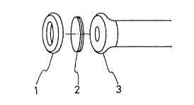
Fig. I shows a microbe sensor of the presen~ invelltion. The
microbe sensor comprises an oxygen electrode 3 having contact wi~h a
microbe membrane 2 and cap 1 capping the electrode from Ihe top.
S Fig. 2 shows a diagram of the BOD analyzel of the present
invention. A BOD sample is sent from a sample container 4 via a
magnetic valve 5 and a liquid-feeder pump 6 to a llow ccll 7. In the flow
cell 7, the sample is stirred by a stirring rod 9 actuated by a motor 10.
Changes are detected by a microbe sensor 8 comprising an oxygen
l 0 electrodes 3 with a microbe membrane 2t both of which are firmly
pressed by a cap 1, and are then amplified by an amplifier 13, and are
recorded by a recorder 14. The sample flows to a drainage tank 11 after
analysis. The magnetic valve 5 is a switch to open a washing solution
tank 12 to wash the BOD analyzer.
l 5 Fig. 3 shows a flow diagram showing the BOD analyzer of the
present invention in which a liquid passage connected to the entrance of
the ilow cell equipped with a microbe sensor is equipped with an outlet.
Fig. 4 is analysis of various concentrations of BOD standards using
a BOD analyzer in Fig. 2.
2 0 Fig. 5 is analysis of various concentrations of BOD standards in the
presence of arsenic using a BOD analyzer in Fig. 2.
Fig. 6 is analysis of various concentrations of BOD standards using
a BOD analyzer schematically shown in Fig. 2 wherein microbes are
immobilized by a gelating agent.
2 5 Fig. 7 is analysis of various concentrations of BOD standards using
a BOD analyzer schematically shown in Fig. 2 wherein microbe
membranes are porous membranes having various average pore sizes in
diameter.
2~
Fig. 8 is analysis of various concentrations of BOD standards using
a BOD analyzer schematically shown in Fig. 2 whercin microbe
membranes are porous mcmbranes having various average pore sizes in
diameter.
Fig. 9 shows a comparison of the sensitivity of lhe microbe
membrane of the present invention and the sensi~ivily ol` a
polyacrylamide/microbe membrane.
Fig. 10 is analysis of various concentrations of BOD standards
using a BOD analyzer wherein the membrane is an asymmetric ultla-
filtration membrane.
Fig. 11 shows a fluctuation of output of an oxygen electrode with
or without an outlet.
Fig. 12 shows activation of dried microbe membranes using
nutrition solutions.
Fig. 13 shows a timetable of intermittent supply Or solutions.
Fig. 14 is a correlation between the rnicrobe sensor method of the
invention and the S-day method.
Fig. 15 is continuous BOD analysis by the microbe sensor method
of the invention and the S-day method.
Definitions and Abbreviations
1 ; cap
2; microbe membrane
3; oxygen electrode
2 5 4; sample container
5; magnetic valve
6; liquid feeder purnp
7; flow cell
8; microbe-sensor
9; stirring rod 2~3~LO
10; motor
1 1; drainage tank
12; washing solution tank
13; amplifier
14; recorder
15, 16, 17, 18, l9; switch valve
AP; air pump
P; liquid-feeder pump
l O
5. Detailed Description of the Invention
Microorganisms used in the present invention are any
microorganisms belonging to the genus Klebsiella. These
microorganisms include Klebsiella oxytoca JCM1665, Klehsiell~
15 planticola JCM7251, Klebsiella ozaenae JCM1663, and Klebsiella
terrigena JCM1687. The present inventors have searched
microorganisms in nature suitable for BOD analysis and found a
bacterium isolated from soil in Kagoshima prefecture. The bacterium has
a broad assimilation spectrum and is able to analyze BOD in a short
0 period of time.
The properties of the bacterium are described below.
A. Morphologv
(1) Shape and size of the cell: bacilliform,
2 5 size: 0.8 - 1.2 ,um X 3 -6 ~lm.
(2) Polymorphism:
(3) Motility:
(4) Sporulation:
(5)Gram staining: -
(6) Acid -fast: -
B. Culture medium
(l) Agar plate containing meat extract: grow well, rorm slightly blue
5 gray colonies.
(2) Slant agar culture containing meat extract: grow well
(3) Liquid medium containing meat extract: grow well
(4) Stab culture containing gelatin: no liquefaction
(5) Litmus milk culture medium: acidification and solidirication
l O
C. PhvsiologY
(1) Reduction of nitrate: +
(2) Denitrification: +
(3) Methyl red test:
(4) Voges-Proskauer test: +
(5) Indole test: +
(6) Hydrogen sulfide production:
(7) Hydrolysis of starch:
(8) Citric acid utilization:
2 0 (9) Inorganic nitrogen utilization: +
(10) Pigmentation:
(11) Urease: +
(12) Oxidase:
(13) Catalase: +
2 5 (14) Growth condition: temperature 5 - 40 C, pH 4- 9.5
(15) Respiration: facultative anaerobe
(16) Oxidation-fermentation test: fermenter
D. Acid or ~as ~eneration
Medium Acid Gas
( 1 ) L-arabinose + +
(2) D-xylose + +
(3) D-glucose + +
(4) D-mannose + +
(5) D-fructose + +
(6)D-galactose + +
(7) Maltose + +
(8) Sucrose + +
(9) Lactose + +
(10) Trehalose + +
(11) D-sorbitol + +
(12) D-mannitol + +
(13) Inositol + +
(14) Glycerine + +
(15) Starch
E. Other phvsiolo~ical properties
(1) ~-galactosidase: +
(2) DNase: -
(3) Tryptophan deaminase:
(4) Decomposition of esculin: +
(5) Decomposition of arginine:
2 5 (6) Decarboxylation of Iysine: +
(7) Decarboxylation of ornithine:
(8) Arsenic resistance: viable in the presence of l 0,000 ppm arsenic
] O
2~
The bacterium was classified by Bergey's Ma~ al of Systematic
Bacteriology Volume 1, pp 415-416, pp 461-465, 1984, basecl on the
properties described above. The bacterium was iclenliried as a baclelium
belonging to Klebsiella o~ytoc~l and was designatecl as Kk~l~si~ o~-ytoc~
12092. Klebsiella o,rytoca 12092 was deposited with Fellllentalio
Research Institute, Agency Or Industrial Science and Techllology un(lel
the Budapest Treaty on October 22, I 991, and was assigned Ihe acccssion
number FERM BP-3616.
1 0 Klebsiella oxytoca 12092 can be cultured by any Or the typical
culture method for bacteria. Carbon sources of culture include any one
of glucose, maltose, sucrose, and molasses or a combination thereof.
Nitrogen sources of culture include any one of organic nitrogen
containing substances, such as various amino acids, corn steep liquor,
1 5 malt extract, peptone, yeast extract, meat extract, and urea, and inorganic
nitrogen containing substances, such as ammonium chloride, ammonium
sulfate, ammonium nitrate, or a combination thereof.
Vitamins and minerals are also included in a cul~ure medium.
Suitable culture temperature may be 20- 40 C, preferably 28-37 `'C.
2 0 Suitable medium pH may be 4.5- 9.0, preferably 5.5-8Ø Growth culture
may be liquid or solid. Suitable bacterial cells for a microbe sensor
material can be obtained at a log phase of growth. Suitable incubation
time in a liquid culture may be 10-72 hours, preferably 12 - 48 hours
under aeration conditions. Suitable incubation time in a solid culture may
2 5 be 12-96 hours, preferably 24 - 72 hours. After growth, bacterial cells
can be harvested by the method known in the art, e.g., centrifugation.
Bacterial cells are washed and then immobili%ed. For
immobilization, the immobilization method known in the art can be used
in which bacterial cells are placed between two permeable membranes
2~ $~)
and then two membranes are stuck to trap the bacterial cclls. However,
these immobilization methods are liable to detachment in a long period of
repeated use. The immobilization method of the invention is preferable
in which bacterial cells are immobilized by a gelating agent on a porou~s
hydrophilic membrane having an average pore size of 0.65 11m-3 llm in
diameter.
A membrane, a support for immobilization of bacterial cells, used
in the present invention includes any gas or liquid permeable, porous
hydrophilic membranes. The membrane is required to freely pass
oxygen, various organic compounds, and various inorganic compounds.
For example, membrane filters and asymmetric ultra-filteration
membranes can be used. Membrane materials include cellulose ester
compounds such as nitrocellulose and acetylcellulose, and hydrophilic
polyfluoride vinylidene and polyether sulfone. Porous hydrophilic
membranes used in the invention are needed to be flexible and moldable
to fit the form of the surface of an oxygen electrode because membranes
are used as a part of a biosensor element and must be fully contact with
the surface of the oxygen electrode to have good sensitivity.
2 0 Porous membranes used in the invention are prererably an average
pore size of 0.65 - 3 ~lm in diameter so as to maintain bacterial cells
trapped between the pore structure. When asymmetric ultra-filteration
membranes are used, the average pore size of 0.65 -3 Ilm in diameter in
the top side of the membrane, that is, a larger pore size side of the
2 5 membrane, is sufficient to be used in the invention. The thickness of
membranes is selected by uses, and is preferably 50 !1m-200 !lm, the
thickness that gives good manipulativeness, flexibility, and strength.
A gelating agent used to immobilize bacterial cells in the membrane
includes any hydrophilic substances that form gel. For example, arginic
acid or salts thereof, gellan gum, xanthane gum, gelalille~cl~rageenan,
locust bean gum, methylcellulose, pectin and pullulan are suitable as a
gelating agent.
The method to immobilize bacterial cells in a membrane is
5 described below.
Microorganisms grown in a suitable culture medium are harvested,
washed, and then immobilized. Microbial cells are combined with an
appropriate amount of an gelating agent such as an arginic acid solution
to give a microbe/gelating-agent mixture. The mixturc is dripped on a
10 porous membrane such as acetylcellulose membrane filters. Suction is
applied from the bottom side of the membrane and pressure is applied
from the top side of the membrane to have the mixture enter the pore,
thereby immobilizing microbes with the gelating agent in the pore.
Suction and pressure are kept applying to the membrane so as to ensure
15 that all of the microbe/gelating-agent mixture on the surface of the
membrane is trapped in the pore and are firmly retained in the pore.
Strength of suction and pressure is such that porous membranes are not
destroyed.
Microbes coated with a gelating agent are trapped in the pore of the
2 0 membrane and the gelating agent is then solidified using a suitable agent
to fix the microbes in the pore. The microbe immobilized in the
membrane are not easily detached from the membrane. Agents to
solidify gelating agents include inorganic salts such as calcium and
polymerization agents. Alternatively, refrigeration can be used to
2 5 solidify a microbe/gelating-agent mixture.
In the immobilization method described above, a trace amount of a
gelating agent is sufficient. For gelation, it takes only a short time at
room temperature, the mild condition which can help keep the activation
of microorganisms at a high level without damaging the microbes.
13
The microbe membrane is permeable to oxygen, various inorganic
and organic compounds and, if part of the microbe membrane is
damaged, only a very small amount of microbes is lost. In addition, the
microbe membrane is durable compared to those known in the prior art.
S The microbe sensor of the BOD analyzer of the present invention
comprises a microbe membrane and an oxygen electrode. The microbe
membrane is firmly contact with the oxygen electrode by a removable
cap, which makes the operator easier to change the microbe membrane.
The microbe membrane has a significant sensitivity for cletection even in
10 a small amount of a sample. To shorten the time of analysis, a sample
volume added to the flow cell may be I ml or less, preferably reduced to
0.3-0.6 ml. In addition, the flow cell can be vertically positioned. A
sample is supplied from the bottom side of the flow cell with stirring the
sample in the flow cell by the stirring rod, and is drained the sample
15 -from the top side of the flow cell. By vertically positioning the flow cell,reliable BOD analysis can be carried out because bubbles do not attach to
the microbe membrane and bubbles are eliminated from the flow cell.
For example, one sample can be analyzed in about S minutes at 30 C
when the sample is applied at 4 ml/min. on the apparatus schematically
2 0 shown in Fig. 2.
In the BOD analyzer of the present invention, it is desirable to set
up an outlet at the liquid passage connected to the entrance of the flow
cell equipped with the microbe sensor. In the BOD analyzer of the prior
art, each inlet of washing solutions, buffers, BOD standard, and a sample
2 5 is directly connected with the passage to the flow cell, the design that
makes quick analysis impossible. For example, when a first sample
analyzed is high BOD, the sample remains in the passage. If a second
sample to be analyzed is low BOD, the operator has to wait until the first
sample does not affect the BOD analysis of the second sample. The
1 4
present inventors have solved this problem by setling up an outlet al the
liquid passage connected to the entrance of the tlOw cell.
Fig. 3 shows the design of the BOD analyzer Or lhe invention. In
Fig. 3, the numbers (15)-(19) are magnetic switch valves. P is a liquid-
S feeder pump, which concurrelltly sends washing solutions, BODstandards, or a sample and phosphate bufrer to the rlow cell. Washing
solutions include tap water, distilled water, and deionizecl water. Burfers
include a phosphate buffer comprising potassium dihydrogen phosphate
(KH2PO4) and dipotassium hydrogen phosphate (K2HPO2). The
10 concentration of the buffer is typically 50-300 mM, prel`erably 130-260
mM. The pH of the buffer is adjusted to pH 5.0-8.0, preferably 6.0-7.0,
depending on microorganisms to be utilized. Other buffers containing
potassium dihydrogen phosphate and disodium hydrogen phosphate
(Na2HPO4) can be used.
BOD standard is a mixture prepared using 150 mg/L glucose, 150
mg/L glutamic acid and is adjusted to 220 ppm. 220 ppm BOD standard
is diluted to have a desirable concentration.
After passing the liquid-feeder pump, washing solutions, BOD
standards, phosphate buf~er and a sample are mixed. AP is an air pump
2 0 supplying air to the mixture passed the valve 19 to allow BOD standards
and the sample to contain the same amount of dissolved oxygen, and also
serves to remove the effect of oversaturated oxygen. The number (3) is
an oxygen electrode and its tip (2) is a microbe membrane. The number
(7) is a flow cell having the entry and exit of solutions.
2 5 The advantages of the BOD analyzer of the present invention are
described below. For instance, time required a sample llowing from the
entrance of the sample container to the valve 18 is assumed to be X.
Time required a sample flowing l`rom the valve 18 to the valve 19 is
assumed to be Y. When analysis of a first sample is completed and a
3~
second sample is placed in the sample feeder, the valve 19 opens for X or
more minutes and, at the same time, the valve 18 opens. Subseqllenlly,
the valve 18 closes for Y minLItes and the valve 15 opens lor Y or more
minutes to flush washing solutions. The mechanism Or the BOD analy7er
S of the present invention prevents the l`irst sample flom flowing to the
flow cell. Instead, the first sample is drained from the valve 19 and the
second sample fills the passage from the sample reedel lo the valve 18.
The second sample is ready for analysis.
When the valve 19 opens, air is supplied lo the llow cell from the
1 0 air pump (AP), which makes the baseline of output unstable. But, after Y
minutes, the valve 19 closes and washing solutions are sent to the flow
cell, making the baseline stable.
In the BOD analysis of the present invention, it is prefel able to
supply nutrition sources to the microbe membrane and then wash the
15 membrane prior to use. These procedures before analysis shorten lhe
activation time of the microbe membrane. The microbe membrane is
stored in a dried form to maintain the activation level of the microbes for
a long period of time. To activate dried microbe membranes to be used
for analysis, the microbe membranes must be changed lrom a dried form
2 0 to a wet form. In the method known in the prior art, microbe
membranes are soaked in buffer and are then placed in an oxygen
electrode, followed by continuous supply of BOD standard until stable
output is obtained. It takes two days to be used for analysis in the prior
method.
The present inventors have studied methods that shorten the
activation time of microbe membranes. In the method of the present
invention, microbe membranes are soaked in water or buffer that is used
for analysis and then placed in the oxygen electrode. Then, solutions
1 6
containing nutrition source.s (hereinafter referred to as "nutrition
solutions") are poured in any one of the inlets ol the BOD standards I, 2,
3, or the sample feecler. After a certain period of time, washing solutions
and nutrition solutions are alternately flushed.
S The flushing time of washing solutions or nutl ilion solutions is 30
seconds to 30 minutes, preferably about ] 0 minu~es. The composition of
the nutr;tion solution is one typically used for cultul~ g nlicroorganisms
or one suitable for growth of microorganisms in membranes. Carbon
sources include monosaccharides such as glucose and fructose, and
disaccharides such as sucrose and maltose. Nitrogen sources include
ammonium salts such as ammonium chloride and ammonium sulfate,
various amino acids and polypeptones. Vitamins include yeast extract and
trace elements include metal salts such as magnesium sulfate and iron
sulfate.
Alternatively, nutrition solutions are a BOD standard solution
containing yeast extract as a vitamin source, and metal salts can be added
to the solution, if necessary. These nutrition solutions comprising a BOD
standard solution and yeast extract contain glucose, glutamic acid and
yeast extract. In the nutrition solution, the concentration of glucose or
2 0 glutamic acid is 5-1,000 ppm, preferably 300-600 ppm, and the
concentration of yeast extract is 10-10,000 ppm, preferably 200-400
ppm. Phosphate necessary for the growth of microorganisms is supplied
by a phosphate buffer.
2 5 In the BOD analysis method of the present invention, it is
preferable to intermittently supply washing solutions or substrate
solutions to the microbe membrane when the microbe membrane is not
used for a long period of time. By supplying the solutions, the microbe
membrane is kept active for a long period of time in a small amount of
solutions. Generally, nutritions are believed to be supplied from organic
materials in a sample when microbe membranes are usecl lor analysis.
Nutritions are not supplied to microbe membranes, lhereby reducing the
activation level of microbes when microbe membranes are not used ror
S analysis. To maintain the activation level of microbe membranes,
nutrition sources are kept supplying to microbe memblanes when
microbe membranes are not used for analysis. One Or thc method Or
supplying nutritions to microbe membranes is one where washing
solutions and BOD standard solutions are continuously supplied to
microbe membranes, as is done when BOD standard is analyzed. In this
method, there are two alternatives: the nutritional supply is all of the
BOD standard used for analysis, or the nutritional supply is a single
concentration of BOD standard. But this method requires a plenty of
BOD standard solutions, washing solutions, and buffers, even if one of
the alternatives is taken. For example, approximately S liters of a
combined volume (washing solutions, BOD standards, buffers) are
consumed for 16-hour operation at a flow rate of 5 ml/min. Thus,
frequent preparation of the solutions as well as time for the preparation
are required so that BOD analysis becomes cumbersome in this method.
2 0 The present inventors have found that intermittent supply of
nutrients in a minimum amount that keeps the activation level of microbe
membranes is sufficient for maintaining the activation level of microbe
membranes, rather than supplying nutrients continuously In the
intermittent nutrient supply method, a pump is operated for a certain
2 5 period of time and then stopped for a certain period of ~ime. Washing
and substrate solutions are alternately supplied in a certain cycle during
operation. Pumping time is 10 seconds to 4 minutes, preferably 30
seconds to 2 minutes, depending on the properties of microorganisms.
Pumping stops for 30 seconds to 3 minutes, preferably 1-2 minutes.
1 8
s Substrate solutions supplied to microbe menrlbranes may include BOD
standards and nutrition solutions containing the nutrients described above.
The method of supplying washing and substrate solutions is such that
alternate supply of washing solutions and substrate solutions, or 10 series
of supply of washing solutions followed by one supply of substrate
solutions, or various combinations of supply of these solutions. This
method does not require frequent preparation of vario~ls .solutions, which
makes the control of the BOD analyzer of the invention easier.
In the BOD analysis method of the invention, il is preferable to add
boric acid, sorbic acid or salts thereof to solutions used for BOD analysis.
Generally, hydrochloric acid or acetic acid is added to a solution used for
BOD analysis to reduce pH to prevent the solution from putrefaction.
Alternatively, sodium hypochlorite or other chemicals are added to
reduce the pH of the solution and chloramphenicol is then added.
Preservatives should be those that have the least effect on microorganisms
in the membrane.
The present inventor have found that, among various preservatives,
boric acid (H3BO3) or sorbic acid or salts thereof are suitable for
microbe membranes. Salts of boric acid or sorbic acid include sodium
2 0 borate, potassium sorbic acid. Boric acid and potassium sorbic acid are
preferable. The concentration of the preservative varies: The
concentration of boric acid is 0.1- 1.0%, preferably 0.3-0.5%, and the
concentration of sorbic acid is 0.1-1.0%, preferably 0.25-0.5%.
In addition, boric acid and low pH is a good combination for the
2 5 prevention of putrefaction. The pH is adjusted to 2-3 with inorganic
acids such as hydrochloric acid and the like or organic acids that are not
metabolized by microorganisms in the microbe membrane to improve
putrefaction prevention.
l 9
This method does not require frequent preparation Or various
solutions, which makes the control of the BOD analy%er of the inven~ion
easier.
6. Example
The present invention will be understood mole re~ldily with
reference to the following examples; however these examples are
intended to illustrate the invention but are not construed to limit the scope
of the invention.
Example 1: Culture of Klebsiella orytoca 12092 (FERM BP-3616)
Klebsiella oxytoca 12092 (FERM BP-3616) was aseptically
inoculated into 100 ml of a sterilized liquid medium/pH 6.5 (1%
polypeptone, 0.1% yeast extract~ 0.5% sodium chloride) in a 500-ml
erlenmeyer flask and was incubated with shaking under aerobic
conditions at 30 C for 24 hours. After growth, bacterial cells were
harvested by centrifugation at 6,000 rpm for 20 minutes. The bacterial
cells were suspended in a small amount of sterilized water and the
suspension was centrifuged at 6,000 rpm for 20 minutes (washing). The
2 0 washing procedure was repeated three times and 150 mg bacterial cells
(dry weight) were obtained.
Example 2: Oxygen consumption rate of Klebsiella o~ytaca 12092
(FERM BP-3616) and various other bacteria
2 5 An oxygen consumption rate of Klebsiella orytl)ea 12092 (FERM
BP-3616) obtained in Example I and of various other bacteria cultured
by the same manner as described in Example 1 was analyzed using a BOD
standard solution. 250 ppm BOD standard solution was prepared using
150 mg/l glucose and 150 mg/l glutamic acid as is described in JIS K0102
and was diluted when necessary. This diluted BOD solution is hereinafter
referred to as "BOD standard".
Table I
Strain of bacterium Oxygen consumption rate k
(m~O_/mh~ in clry wei~ht
Klebsiella oxytoca 12092 4.3
Klebsiella oxytoca JCM 1665 4.0
Klebsiella ozaenae JCM1663 3.8
1 0 Klebsiella planticola JCM7251 3.2
Klebsiella terrigena JCM 1687 3.5
Trichosporon cutaneun1 IFO 10466 2.1
* Oxygen consumption rate was analyzed at 30 `'C using 37 ppm BOD
standard as a substrate.
l S
As is shown in Table 1, microorganisms belonging to Klehsiella
have a high oxygen consumption rate on BOD standard. Among the
microorganisms, Klebsiella oxytoca 12092 (FERM BP-3616) has the
2 0 highest oxygen consumption rate.
Example 3: Apparatus, Analysis, and Standard curve
0.4 mg (dry weight) bacterial cells of Klebsiella o~ytoca 12092
strain (FERM BP-3616) obtained in Example 1 was placed between two
2 5 nitrocellulose membranes [membrane filter HAWPO2500 (pore size: 0.45
!lm in diameter), Millipore Co.,]. The two membranes were firmly stuck
together so as not ~o give any other spaces but bacterial cells (hereinafter
the membrane containing bacteria is referred to as "microbe
membrane"). The microbe membrane was immersed in 100 mM
3 0 phosphate buffer/pH7.0 and aerated at 100 ml/minute for three hours
2s~ ?~
using an air pump to activate the microbe membrane. Various
concentrations of BOD standards were tested on the appalatu~ equipped
with the microbe membrane shown in Fig. 2. As is shown in Fig. 4,
there is a linear correlation between a change in a BOD stanclard
5 concentration and the corresponding change in vollagc measuled by ~he
oxygen electrode.
BOD standard was tested by varying its volume in the reaction
vessel. The results are shown in Table 2, in which a BOD standard
concentration analyzed by the apparatus is one calculated from a challge
1 0 in voltage based on the standard curve shown in Fig. 4. It was found that
less than 1 ml of BOD standard gives an appropriate measurement. It
takes more time for measurement and washing when more than I ml of
BOD standard is used. In contrast, measurement is umeliable when less
than 0.3 ml of BOD standard is used.
Table 2
BOD standard Volume in reaction vessel (ml)
concentration
(ppm) 0.1 0.2 0.3 0.50.6 1.0 2.0 5.0
2 0 BOD 22 10 18 21 2122 23 21 22
found 44 20 35 44 4443 45 44 46
(ppm) 66 50 58 65 6667 66 67 66
122 98 105124 123 123122 125 123
Time for analysis*
2 5 (min.! 2 3 4 4 5 5 15 20
*Time for analysis includes time for washing.
Example 4: Comparison of activation time
Various bacterial cells obtained in Example l werc immobilized in
a nitrocellulose membrane as described in Example 3 to give a microbe
membrane. Immediately after immobilization, the microbe membrane
was immersed in a 100 mM phosphate buffer/pH7.() and aeratecl ~ l00
ml/minutes using an air pump to activate the microbe lnembrane. The
activated microbe membrane was taken out perioclically and inserled in
the device schematically shown in Fig. 2 to test various concentrations of
BOD standards. As is shown in Table 3, it takes 1-2 days ror
Trichosporon cutaneum IFOI0466 to be activated while it takes only
three hour for microorganisms belonging to the genus Klebsiella,
Klebsiella oxytoca 12092 to be activated.
Table 3
Aeration (hr)
0 1 3 5 l 0 24 36 48
Klebsiella oxytoca 12092 (ppm) 3038 65 67 66 67 65 66
Trichosporon cutaneum
IFO10466(ppm) 48 12 11 46 58 60 64
2 0 Values found are those analyzed using 66 ppm BOD standard.
Example 5: BOD analysis of various compounds (comparison of
Klebsiella oxytoca 12092 membrane and Trichosporon cllfanellm
2 5 membrane)
The microbe membranes of Klebsiella o,r~toca 12092 in Example 4
and of Trichosporon cutaneum were fully activated and tested to analyze
BOD of various compounds. The results were compared to those
analyzed by the 5-day BOD method, a method described in JIS K0102.
3 0 The microbe membranes were tested on the apparatus described in
Example 3 and the calibration curve was drawn USillg BOD standard.
23
As is shown in Table 4, BOD obtained flom tlle Kle/7.~iella o.rytoea
12092 membrane is similar to that obtained from 1he 5-(lay BOD method,
an officially accepted method. The coefficient Or corr~l~tion (r~) is
0.993. Trichosporon cutal1ellm IFO 10466 does not responcl to
5 disaccharides while Klebsiella oxytc)ca 12092 responcls lo them. When
ethyl alcohol is measured, Trichospo10)7 clltanellm IFO 10466 has highe
BOD than the 5-day BOD methocl while Klebsiell~l o.ryto( a 12092 has
almost the same BOD as the 5-day BOD method.
I 0 Table 4
BOD (g/g)
Test
Microbe sensor 5-day method Microbe-sensor of
sampleof the invention method T/ieho~po)on clltanewm
1 5 IFO 10466
Glucose 0.77 0.78 0.72
Fruetose 0.74 0.71 0.54
Sucrose 0.45 0.45 0.36
Laetose 0.45 0.45 0.06
2 0 Maltose 0.53 0.50 0.03
Glutamie aeid0.56 0.56 0.70
Glyeine 0.15 0.10 0.45
Ethanol 0.95 0.93 2.90
Aeetic acid0.85 0.88 1.77
Example 6: Analysis of BOD standards in the presence ol arsenic
Various concentrations of BOD standards were analyzed in the
presence of arsenic by a similar method described in Example 3. As is
3 0 shown in Fig. 5, there is a good linear correlation between a BOD
standard concentration and the corresponding change in voltage on the
24
o
oxygen electrode. As is evident from the results described above, various
BOD standards can be precisely analyzed by the presen~ invention in a
short period of time.
S Example 7: Analysis of wastewater (comparison Or the microbc sensor
method of the invention and the 5-day BOD melhod)
The activated microbe membrane of Klebsiella orytoc a 12092
obtained in Example 3 was inserted in the device shown in Fig. 2.
Various wastewaters were tested for BOD using the Klebsiella o rytoca
1 0 membrane and the 5-day BOD method. The comparison of the microbe
sensor method and the 5-day BOD method is shown in Table 5. A high
correlation is found between the results obtained by the methods.
Table 5
1 5 Wastewater BOD (ppm)
5-day method Microbe sensor
sample method
Swine housing 1940 1880
Sewage disposal plant 24.2 20.0
2 0 Sewage purifier 25.7 28.2
Car repair shop 52.6 49.0
Poultry laboratory 30.3 33.8
Food plant 49.3 39.4
Marine product
2 5 Processing plant 45.6 45.2
Balneotherapy clinic 60.9 60.0
2 5
5~
Example 8: Immobilization of Klebsiella o.~toc a 12092 in membranes
0.4 mg (dry weight) baclerial cells of Kle~7.si(~11a o~lto~l 12092
strain obtained in Example I and 50 ~1 of a sterilized 3% soclium arginic
acid solution were combined.
S To immobilize the bacterial cells, the mixture was dripped On an
acetylcellulose membrane (membrane filter type ~-IA, average pore si;~e;
0.8 ~m in diameter, ~lillipore Co.,). Suction was applied from the
bottom side of the membrane until all the mixture was absorbed by the
membrane. The membrane was then immersed in 50 ml of a 5% calcium
chloride solution at room temperature for 10 minutes to solidif`y the
arginic acid to immobilize the bacterial cells in the membrane.
Example 9: BOD analysis using the apparatus of the present invention.
BOD standard (JIS-KO102) was analyzed using the microbe
membrane obtained in Example ~. As is shown in Fig. 1, the microbe
sensor is a device comprising the microbe membrane 2 inserted between
the oxygen electrode 3 and the cap 1. Analysis was carried out using the
apparatus shown in Fig.2. 10 ml of the sample was analyzed at 30 `'C at a
flow rate of 3 minutes (3 minutes for analysis). Samples and washing
2 0 solutions can be aerated, if necessary, or can be aeratecl on the way to the
flow cell 7. As a result of the analysis using the appara~us of the present
invention, a good linear relationship between a BOD standard
concentration and the corresponding change in voltage of the oxygen
electrode was obtained (see Fig. 6). Wastewater collected from a food
2 5 plant in Kagoshima prefecture was tested for BOD under the same
condition as described above by the apparatus of the present invention and
the S-day method. The resulting BOD did not differ one another: BOD
was 42 ppm in the apparatus of the present invention and 45 ppm in the
26
0
5-day method. The comparison indicate that the apparalus of ~he present
invention can work in the field.
Example 10: Comparison of a pore size of a membrane
Commercially availablc porous membrancs (avel clge pore size is
different from the one described above) were used ~o immobilize
bacterial cells obtained itl Example I by a similar methocl described in
Example 8. The membrane was inserted in the microbe sensor device
described in ~xample 9 and BOD standard was analyzed under the same
condition described in Example 9. The analysis of various concentrations
of BOD standards are shown in Fig. 7. The repeated analyses of 66 ppm
BOD standard are shown in Fig. 8. Microbe membranes made from
porous membranes in an average pore size of 0.45 llm in diameter are
less responsive. Microbe membranes made from porous membranes in
1 5 an average pore size of 5 ,um in diameter become less responsive when
repeatedly used and which are unable to use for another analysis.
Microbe membranes made from porous membranes in an average pore
size of 0.65 ,um or more in diameter provide a good linear relationship
between a BOD standard concentration and the corresponding change in
2 0 voltage of the oxygen electrode. Microbe membranes made from porous
membranes in an average pore size of 3 llm or less in diameter provide a
stable response when repeatedly used. Talcen altogether, microbe
membranes made from porous membranes in an average pore size of
0.65-3 !lm in diameter provide stable and responsive microbe sensors.
Example 1 1: Comparison of porous membranes
Various porous membranes having different average pore sizes in
diameter but having 150 llm in thickness were used to make microbe
membranes. Bacteriai cells free of a gelating agent were dripped onto the
~s ~ o
porous membranes. Suction was applied from the bottom side of the
membrane to trap the bacterial cells in the membrane.
The microbe membrane of the present invention was al~so made as
described in Example 8. Analysis was carried out under the same
5 condition described in Example 10 using the both membranes. As is
shown in Table 6, the microbe membrane free of a gelating agent do not
have any response compared to the gelated microbe membl anc of the
present invention. The microbe membrane free of a gelating agent was
believed to trap an insufficient volume Or bacterial cells so that porous
10 membranes were changed thickness from 150 llm to 600 llm. In
addition, a volume of bacterial cells to be trapped was increased to 4 mg
(dry weight), 10 times the volume used for the microbe membrane of the
present invention. The increased volume of bacterial cells was
immobilized by the same method described above.
1 5
Table 6
Average pore
size in diameter(llm) 0.45 0.65 0.8 3.0 5.0 0.8*_
Response to 66 ppm
BOD standard (~mV! 20 60 40 0 0 600
*Present invention
Table 7 shows the results. Microbe membranes made from porous
2 5 membranes in an average pore size of 0.8 ~m or less in diameter are
unable to use for analysis. This is due to the following reason; When
analyzing BOD, washing water is flushed to the reaction vessel. Then, a
baseline voltage on the microbe electrode, which indicates an initial
oxygen concentration, is measured. The baseline voltage measured on the
28
oxygen electrode was 20 mV (see Table 6), indicating almosl no oxygen.
With almost no oxygen, it is impossible to measule changes of dissolved
oxygen concentration that occur when microorganisllls assimilate various
organic materials.
s
Table 7 _
Flow rate ` Average pore size in diametel (~m)
of sample 0.45 0.65 0.8 3.0 5.0 0.8t
3 min. (~mV) - - - 0 0 600
30 min. (~mV) - - - 80 150 NA*
Baseline voltage 20 30 40 200 300 1000
(mV)
tPresent invention
1 5 *Not analyzed
No change in oxygen concentration indicates that oxygen in
washing water and/or samples can not pass the microbe membrane freely.
2 0 The baseline voltage of the microbe membrane having an average pore
size of 3 ~m or more in diameter was 200-300 mV, a fairly high voltage
(see Table 7). There was no response to 3-minute flow samples, and little
response to 30-minute flow samples. In contrast, the microbe membrane
of the present invention was as much as 1000 mV in a baseline voltage
2 5 and responded to 3-minute flow samples.
The microbe sensor of the present invention gives a baseline
voltage as much as l,000 mV and quick response to 3 minute-flow
samples. The microbe sensor of the present invention therefore enables
quick BOD analysis.
29
~r~ c~
O.
Example 12: Comparison of immobilization me~ho(ls of baclerial cells
Two types of microbe sensor were made: One is tl1e same type of
microbe sensor made in Example 8 and the other is one that was m~lde by
mixing 0.4 mg (dry weight) of bacterial cells obtained in Example l with
S a 10 weight percentage acrylamide solution and subscquently gelating the
mixture. These microbe sensors were inserted in the device described in
Example 9 and were used to analyze BOD standard. Fig. 9 shows that, in
3-minute flow samples an~l a 66 ppm BOD standard concentration, the
microbe sensor of the present invention produced 600 mV response (A)
10 while the acrylamide/microbe sensor produced no response (B). In 30-
minute flow samples and a 66 ppm BOD standard concentration, the
acrylamide/microbe sensor produced 150 mV response. The microbe
sensor of the present invention responds faster and is able to analyze BOD
in a short period of time. Another disadvantage of the
15 acrylamide/microbe sensor is that the sensor is susceptible to damagewhen inserted into the oxygen electrode with a slightly slrong pressure.
Example 13: Immobilization using ultra-filtration membranes
Bacterial cells obtained in Example 1 were immobilized on
asymmetric ultra-filtration membranes (Filton ultra-riltration membrane,
2 0 Omega-Membrane; Fuji Filter Co., ) by the method described in Example
8. The microbe sensor was inserted in the device described in Example 9
and was used to analyze BOD standard. As is shown in Fig. 10, a good
relationship between a BOD standard concentration and the
corresponding change in voltage of the oxygen electrode was obtained.
2 5 The microbe sensor was also found to respond well.
Example 14: Preparation of microbe membranes
Klebsiella oxytoca 12092 strain (FERM BP-3616) was inoclllatecl
into 100 ml of a liquid medium/pH6.5 (1% polypeptone, 0.1% yeast
z~
extract, 0.5% sodium chloride) in a 500-ml shaking cullule flask and was
incubated by shaking under aeration conditions at ~0 C for l 7 hours.
After incubation, the culture was centrifuged al 6000 rpm for 20
minutes. Bacterial cells thus obtained were suspended in a small amount
S of sterilized water. The suspensioll was centrifuged. The washing
procedure was repeated three times. The bacterial cells wcre suspended
to a final cell density, D660 of 0.58 (bacterial concentrale). ~2 !11 of the
concentrate was suspended in 2 g o~ a mixture (1.7% lc-carageenan, 0.8%
locust bean gum). The mixture was dripped onto acetylcellulose
10 membranes (Membrane filter type HA, an average pore size of 0.8 !lm in
diameter, Millipore Co.,). Suction was applied from the bottom side of
the membrane until all the mixture was absorbed. The filtrate, a K-
carageenan solution, was removed. The microbe membrane was cooled
on ice and then immersed in 100 ml of a 40 mM phosphate burfer at
15 room temperature for 5 minutes. K-carageenan and locust bean gum
were solidified to give a microbe membrane.
Example 15: Comparison of an apparatus having or without having an
outlet
BOD standard was analyzed using the microbe membrane obtained
20 in Example 14. 220 ppm BOD standard prepared from a mixture (150
mg/L glucose and 150 mg/L glutamic acid) was diluted to a BOD
concentration of 100 ppm and 5 ppm. The dilutions were used as
samples.
Two apparatuses were used for comparison: One apparatus of Fig.
2 5 3 had an outlet 19, and the other did not have an outlet. A flow rate of
air of the air pump was 1,000 ml/min., and a flow rate of washing
solutions, BOD standard, and buffers was 4 ml/min., 4 ml/min., and I
ml/min., respectively. These solutions were sent by a liquid-feeder
pump.
~3~0
A washing solution (water) was llushed ror 10 millutes prior to
BOD standard samples in order to stabilize a baselinc. 100 ppm BOD
standard was then flushed continuously. The initial poin~ Or a constant
line of output was clesignated as an output value. A washing solution was
S flushed for 10 minutes, and then 5 ppm BOD standard sample was ted
into a sampling inlet and flushed until an output value becomes constant.
Fig. 11 shows the fluctuation of output of the oxygen electrode.
The apparatus shown in Fig. 3 has a long passage between the BOD
standard feeder and the flow cell. To prevent the passage from clogging
10 with solid materials, the diameter of the passage was made larger so that
i~ takes more time in analysis than the apparatus shown in Fig. 2, a
simpler structure apparatus. The apparatus with an outlet have a peak
output at 15 minutes while it takes 30 minutes for the apparatus without
an outlet to stabilize output by an action of 100 ppm BOD standard.
1 5
Example 16: Activation of dry microbe membranes by nutrition solutions
Nutrition solutions contained 426 ppm glucose, 426 ppm glutamic
acid, and 300 ppm yeast extract.
Microbe membranes were used the same ones prepared in Example
2 0 15. An apparatus was one having the outlet 19 described in Example 15.
Other conditions were the same as those described in Example 15.
Nutrition solutions and buffers were supplied for 10 minutes each.
The output is shown in Fig. 12. High output values indicate when
dissolved oxygen concentration is high while low output values indicate
2 5 when dissolved oxygen concentration is low.
As is shown in Fig. 12, once nutrition solutions are supplied to
microbe membranes, substrates in nutrition solutions ~re consumed by
microorganism in the membranes and dissolved oxygen concentration is
reduced. Supply of nutrition solutions therefore reduces an output value.
Subsequently, when washing solutions are supplied to microbe
membranes, an output value is increased because no oxygen is consumed
by the microorganism in the membrane due to lhe absence Or substrales.
Again, when nutrition solutions are supplied to the membrane, oxygen
5 consumption is increased more than that resulted Irom the rirst supply of
nutrition solutions because of the activation and growth of the
microorganism by the first supply of nutritions, and an output value is
further reduced. A cycle of supply of nutritions and washing water
reduces an output value, which is measured when nutrition solutions are
10 applied, due to further activation of microorganisms. At the same time,
an output value is slow to increase when washing water is supplied, and
an output baseline is gradually reduced. A reduced output baseline is
believed to be the result from the increased activation of microorganisms.
A constant output baseline suggests that the activation of microorganisms
15 have reached a suf~lcient level for analysis.
The activation level of the microbe membrane increases by supply
of nutrition solutions. However, microbe membranes having an elevated
level of an output baseline are not suitable for analysis. To correct such
microbe membranes, the microbe membranes have to be washed to
2 0 stabilize an output baseline. Hence, supply of nutrition solutions should
be stopped to the microbe membrane at the time when a certain level of
activation was obtained, and washing solutions should be started flushing
to stabilize an output baseline.
An output baseline that was considered the successful activation of
2 5 microbe membranes was 1,000, 900, 800, and 700 mV, which was
measured when nutrition solutions were supplied. When an output
baseline has reached to the voltage, washing solutions were supplied to the
microbe membranes. Analysis was carried out after a constant output
was obtained. When an output has reached 800 mV or less, microbe
33
2~3~
membranes were washed, the procedure which produce(l the same output
as that of microbe membranes before being dried (Table 8).
Table 8
Activation of microbe membranes before drying 100%
Activation of microbe membranes after drying 50%
Activation of microbe membranes after drying 55%
(1,000 mV)
Activation of microbe membranes after drying 73%
(900 mY)
Activation of microbe membranes after drying 90%
(800 mV)
Activation of microbe membranes after drying 90%
(700 mV
l S
The same activation level of microbe membranes was observed in
an output baseline of 700 mV and 800 mV. It took approximately 26
hours to reach the activation level in an output baseline of 700 mV while
2 0 it took approximately 21 hours to reach the activation level in an output
baseline of 800 mV. Washing was started at approximately 21 hours
after activation started, and was carried out until the baseline was
stabilized. Five hours washing was found to be suitable for stabilization.
It took totally 26 hours to complete activation and stabilization of
2 5 microbe membranes. The activation level of the activated microbe
membrane is shown in Table 9.
Table 9
Activation of microbe membranes before drying 100%
3 0 Activation of microbe membranes after drying 50%
Activation of microbe membranes after dryin~ 90%
34
As is evident from the above description, once dried microbe
membranes activated by nutrition solutions are able ~o regain lhe
sufficient activation level for analysis, they show the same level Or output
as that of original wet microbe membranes. Time requiled for ac~ivation
S is only 26 hours. In the methods of prior arts, it took two days ror dried
microbe membranes to be used for analysis: Dried microbe membranes
are immersed in a buffer solution for one day, inserled in the device and
alternately flushed with BOD standard and washing solutiol1s. In the
methods of prior art, BOD standard is used insteacl of nutrition solutions.
10 The component of BOD standard is glucose and glutamic acid but no
vitamins and metal salts. It takes one more day to activate clried microbe
membranes so as to regain sufficient activation for analysis. Therefore,
the activation method of the present invention shortens 22 hours for the
activation of dried microbe membranes.
Example 17: Effect of intermittent and continuous supply of solutions on
the retention of the activation level of microbe membranes
In the following, microbe membranes prepared as in Example 14
were used. A flow rate of solutions was 5 ml/min., and the operation
2 0 time was 16 hours. The same apparatus described in Example 16 was
used.
Condition 1: Intermittent supply of washing solutions and BOD standard
2 5 Time of pumping; 30 seconds
No pumping; 90 seconds
As is shown in Fig. 13, washing solutions wcre fl~lshed lrom the
first pumping to the fifth pumping and BOD standard was flushed al the
sixth pumping. This cycle was repeated.
5 Condition 2: Continuous supply of washing solutions and BOD standard
Washing solutions; 11 minutes
50 ppm BOD standard; 4 minutes
This cycle was repeated.
The results are shown in Table 10. In the intermittent pumping7
consumed washing solutions, BOD standardt and buffer solutions were
about 1.2 liters, which was 1/4 of continuous pumping. However7 the
O 15 activation level of microbe membranes in the intermittent pumping is
equivalent to that of the continuous pumping.
Table 10
Activation of microbe membranes
after 16 hrs.
Activation of microbe Activation of microbe Activation of microbe
membranes membranes membranes
before activating after activating after activating
bv condition 1 bv condition 2
2 5 100% 93% 93%
Example 18:Effect of preservatives on the activation of microbe
membranes
3 0 (1) Activation (output) of microbe membranes was compared: one
is that various preservatives were added to washing solutions, buffer
36
solutions, and BOD standard, and the other is that no preservative was
added to these solutions.
The first activation (output) analysis was compared to the avelage
of 10 activation analyses, using 50 ppm BOD standard. The results are
shown in Table 11.
Table 1 1
Preservatives Activation level of microbe membranes*
Phenol (0.1 %) 68%
Sodium salicylate (0.75%) 77%
Sodium hypochlorite (0.01%) 74%
Boric acid (0.5%) 93%
Potassium sorbic acid (-s%! _ 93%
*Initial activation level of microbe membranes is taken as 100%.
1 5
As is shown in Table l l, boric acid and potassium sorbic acid are
the best preservatives for the activation of microbe membranes.
(2) The presence and absence of microorganism clusters on the
2 0 surface of microbe membranes were visually observed. Boric-acid-addecl
washing solutions, buffers, and BOD standard were added to the microbe
membrane of the present invention. Wastewater from a rood plant was
analyzed using the membrane. When wastewater was not analyzed, the
microbe membrane was exposed to the condition 2 in Example 17 to
2 5 maintain its activated level. Microoganism clusters were then measured.
The results are shown in Table 12.
Table 12
Concentration of Bacterial cluster
added boric acid At 0 day At 10 day
0% None Present
0.3% None None
0.5% None None
1.0% None None
* When 1.0% boric acid was added, microbe membranes were affected.
Although the microbe membranes were able to be used for analysis, it
10 took more than 24 hours to activate the membranes.
As is shown in Table 12, a preferable boric acid concentration is
0.3-0.5%.
(3) The number of colonies was counted on the microbe membrane
by placing the membrane in 50 ppm BOD standard (pH2.0, adjusted with
hydrochloric acid) under the condition shown in Table 12, at 30 C for 10
days. The results are shown in Table 13.
20 Table 13
Preservative Number of colony
At 0 dav At 10 day
Not added 0/ml 107/ml
0.3% Boric acid 0/ml 103/ml
0.3% Boric acid/pH2.0
adjusted 0/ml 10 or less/ml
with hvdrochloric acid
38
Example 19: Correlation between the 5-day method (JIS method) and the
method of the present invention (microbe sensor method)
Conditions: Microbe membranes were prepared as is described in
Example 14.
The microbe membrane was activated as described in Example 16.
Boric acid was added to washing soluiions, buffers, BOD standard to a
final concentration of 0.3%. The pH of these solutions was adjusted with
hydrochloric acid to 2Ø
The activation level of the microbe membrane was maintained as
described in Condition 1 of Example 17.
Methods: Samples were taken from wastewater from a food plant
once a day and analyzed by the 5-day method and the microbe sensor
method. The correlation between these methods was analyzed. The
1 5 correlation coefficient is as high as 0.95 or more, suggesting a high
correlation between these two methods (see Fig. 14).
Example 20: Continuous use of the microbe sensor
To investigate the correlation between the 5-day method and the
2 0 microbe sensor method as well as retention time of the activation level of
the microbe membrane, the microbe membrane Or the present invention
was used for continuous BOD analysis under the same condition as in
Example 19
There is a high correlation between these two methods even in
2 5 continuous use. In addition, the microbe membrane can be used for 3
months (see Fig. 15).
As is evident from the above description, it tal~es S days for the JIS
method to analyze BOD. In contrast, the present inven~ion provides a
39
;~3~
quick BOD analysis and an easy monitoring system ror the routine testing
of wastewater.