Note: Descriptions are shown in the official language in which they were submitted.
~""191/19465 ~ ~ ~ ~ ~~ PCT/US91/03834
- 1 -
MITRAL HEART VALVE REPLACEMENTS
This invention relates to mitral heart valve
replacements.
Heart valve replacement and in particular mitral valve
replacement devices fall into two broad categories, mechanical
and bioprosthetic. Both kinds of device are obstructive to
flow compared with the normal natural valve.
Mechanical valves of all kinds must be used with
coumadin type anticoagulants: without this treatment there is
a prohibitive risk of the formation of clots which will either
obstruct the valve or break away to block vital arteries
(e. g., arteries in the brain, leading to a stroke). Even
with anticoagulants the risk of clotting and its complications
remains. The durability of most mechanical valves is
excellent.
Bioprosthetic valves (of treated biological tissue
supported by a frame or stent) are less likely to be affected
by clot formation but have less durability, inevitably
developing calcification and/or tears with time, with this
process being prohibitively accelerated in children.
The haemodynamics of the natural mitral valve are
dependent on the absence of a stent, the absence of a rigid
ring, the relatively unrestrained opening of the flaps and the
flexibility of the tissues. The durability is dependent on
the fact that it is living tissue capable of self
regeneration. The absence of clotting problems is related to
the living endothelium which is composed of special cells with
properties that prevent local clot formation.
SUBSTITUTE SHEET
WO 91/19465 ~ PGT/US91/03F-''-~
"~0~ ~~~. . _ 2 _
One object of the present invention is to provide an
entirely flexible non-elastic bioincorporable unstented
replacement mitral valve-that has no rigid parts to project
into the inner cavity of the ventricle.
Another object is to avoid the use of any metal or
plastics stiffener in the circumferential part of the
replacement valve that is to be sewn to the orifice of the
ventricle.
A further object is to provide a replacement mitral
heart valve in which the part to be fixed to the inlet orifice
of the ventricle is an integral part of and is in continutity
with the valve mechanism proper, and therefore:
a) has no bulk to detract from the effective orifice area of
the valve;
b) has a smooth inflow into the orifice Without any potential
nooks and crannies or excrescence that can produce local
stasis that leads to thrombus formation;
c) has no interface between bioincorporable and non-
bioincorporable material that when it exists is always
accompanied by clot formation;
d) allows healing to take place directly between the
bioincorporable implant and the native tissue, thereby,
ensuring a permanent secure bond between body and implanted
valve that is not dependent on the continued integrity of the
sutures used for insertion.
Yet another object of the invention is to provide a
form of valve whereby one size fits more than one size of
patient heart, and which has the parts to be stitched to the
~U~t3TITUTE SrtEET
"'O 91/19465 2 ~ ~ ~ ~ ~ PCT/US91/03834
- 3 -
heart marked to indicate the correct placement of sutures for
a particular heart size.
A still further object is to provide a combined sizing
and suture placement guiding device which indicates the
correct placement of sutures to ensure optimal opening and
closing of the respective valve.
According to the principal aspect of the present
invention, a mitral heart valve replacement of essentially
flexible bioincorporable material comprises a generally D-
shaped sewing ring having an opening with at least one long
straight side portion, an anterior cusp hinged contiguously
from that straight side portion, a posterior cusp hinged
contiguously from a shorter side portion of the opening
opposite the anterior cusp, and two lateral cusps hinged
contiguously one from each of the remaining side portions of
the opening extending between adjacent ends of the side
portions from which the anterior and posterior cusps are
hinged, together with chordae of bioincorporable material
extending from edge portions of the cusps for connection to
the papillary muscles in the cavity of the ventricle when the
sewing ring is sutured to the atrio-ventricular junction of
the host or patient heart, the aggregate area of the cusps
exceeding the area bounded by the sewing ring so that when the
valve is caused to open, by the papillary muscles pulling the
chordae, the cusps simply deflect away from each other, and
when the papillary muscles allow the valve to close, the edge
portions of the cusps meet out of the plane of the sewing ring
so as to be capable of meeting in the cavity of the ventricle
SUBSTITUTE SHEET
N
WO 91119465 ~~,~~ ~~ PCT/US91/038.~
- 4 -
of the patient heart.
The anterior cusp is preferably contiguous with one-
third of the perimeter of the sewing ring opening and
preferably has a generally semicircular edge portion to enable
it to project deeply into the cavity of the ventricle; and
the posterior and lateral cusps - which are, therefore,
together contiguous with two-thirds of the perimeter of the
sewing ring opening - preferably also have generally
semicircular edges portions.
The sewing ring opening may be truly D-shaped, and may
be matched by a correspondingly larger D-shaped external
profile to suit the atrio-ventricular junction in a patient
heart. Preferably, however, the opening is trapezium-shaped,
to provide longer and shorter parallel straight side portions
from which are hinged the anterior and posterior cusps
respectively, and two even shorter non-parallel side portions
from which are hinged the lateral cusps, while the sewing ring
has a more truly D-shaped external profile to suit the atrio-
ventricular junction in a patient heart. Making the sides of
the sewing ring opening straight ensures minimal bending of
the cusps and an absence of undesirable folds or creases
during movement back and forth between open and closed
positions.
The sewing ring may be formed by a combination of
integral flange portions of the anterior, posterior and
lateral cusps stitched to a flat basic ring element cut from
the bioincorporable material, preferably with a similar flat
reinforcing or stiffening element of biocompatible material
SUBSTITUTE SHEET
2084~I7
°' -1 91 / 19465 PCT/US91 /03834
- 5 -
between which and the basic ring element the flange portions
of the cusps are interposed. The posterior cusp and the
lateral cusps and their flange portions may be formed from a
flat strip of the bioincorgorable material, with spacer
portions between adjacent cusp portions, which spacer portions
are partially cut through and/or pleated and stitched so that
the integral flange portion can conform to the arcuate portion
of a D-shaped opening, and to bring the adjacent cusp portions
into contiguous disposition with each other.
The chordae may be separate chords attached by sewing to
the edge portions of the cusps and to attachment portions
adapted to be eventually sewn to the papillary muscles, but
are preferably formed integral with the cusps and are provided
with integral attachment portions at the ends remote from the
cusps for suturing to the papillary muscles. The cusps,
chordae and attachment portions may be provided with non-
elastic reinforcing strands running from the cusps to the
attachment portions. Similar strands may also run from side
to side of the cusps adjacent their free edges or across the
middle. The attachment portions may be attached to the
papillary muscles with their adjacent edges together or apart,
depending on the shape and form of the papillary muscles.
When the host anatomy allows the adjacent edges to be spaced
apart, this arrangement allows for the largest possible area
for flow of blood. .
The bioincorporable material may be biological
material, such as auto, homo or xenograft pericardial tissue
treated with glycerol (possibly reinforced by bio-incorporable
SUBSTITUTE SHEET
WO 91/19465 ~ PCT/LJS91/03p'~1
Ar
~ ~~ ,~ ~y~ _ 6 _
non-elastic sutures, such as extruded polytetrafluoroethylene
sutures to guard against stretching), so as to allow the host
to cover the valve with living_'self-repairing fibrous tissue
and on this a growth of new endothelium presenting a surface
to the blood that does not clot. Alternatively, the valve
may be made of a non-biological material that allows the host
to provide a cover of fibrous tissue and endothelium leaving
the implanted material completely covered by natural host
tissue.
Because of the great flexibility of the valve in
accordance with the invention one size may be used for several
sizes of patient heart; thus the sewing ring may be provided
with a central marker line (e.g., a broken line, formed by an
interwoven suture) to indicate inner and outer stitching zones
for different ranges of heart size. Two sizes of valve in
accordance with the invention may thus suffice for the full
range of patient heart sizes.
A rigid holder is preferably temporarily secured to
the sewing ring and has a generally U-shaped bar extending
between the chordae of the anterior and posterior cusps as far
as the attachment portions, so as to prevent collapsing of the
valve, particularly during placement in the patient heart.
The temporary securing of the holder to the sewing ring may be
effected by tacking stitches which are cut and removed when
the valve has been secured in place. The holder is
preferably provided with notches or slots for location of
sutures through the sewing ring and the holder preferably has
a detachable handle used during placement of the valve in the
SUBSTITUTE SHEET
CA 02084517 2002-02-25
7
patient heart.
Preferably, according to the present invention
there is provided a combined sizing and suture placement
guiding device comprising an elongate handle carrying an
arcuate sizing member having a circumferential shallow groove
nor fitting within the corresponding part of the remnant of
the eYCised natural valve o~ the patient heart at the atrio-
ventricular junction, with notches or slots, one adjacent each
end of the sizing member and one intermediate thereof, for
indicating the positions for placement sutures to be passed
also through corresponding positions on the sewing ring of a
replacement valve in accordance with the first aspect of the
ir.=.~en~ion, and with a depth guaga dependinc from the sizing
member f~~r c~ntCct with the papillary muscles to indicate ins
positions of placement sutures in the papillary muscles for
securing the attachment portions of the chordae.
A number of embodiments of the invention and their
manner of use will new be described, by way of example only,
with reference to the accompanying drawings, in which:
Figures 1 and 2 are diagrammatic plan and posterior
views respectively of an embodiment cf the invention, the
valve being shown closed;
Figures 3 and 4 correspond to Figures 1 and 2
respectively but with the valve shown open;
Figures 5 and 6 correspond to Figures 2 and 4
respectively but show another embodiment of the invention;
Figures 7 to 12 are templates of the component parts
of a preferrred embodiment of the invention, a perspective
WO 91/19465 ~~'~ PCT/US91/03s~~.
_ 8 _
view of which from below forms Figure 13;
Figure 14 is a fragmentary diagrammatic section
through a patient heart showing the preferred embodiment of
valve in position at the atrio-ventricular junction;
Figure 15 is a fragmentary perspective view showing
the remnant of the excised natural valve of the patient heart
at the atrio-ventricular awaiting fitting of a valve in
accordance with the invention;
Figure 16 is a perspective view from above of a
combined sizing and suture placement guiding device in
accordance with the invention;
Figure 17 is a fragmentary perspective view showing
the device of Figure 16 in position;
Figure 18 is a vertical medial section through Figure
17 showing sutures being positioned;
Figure 19 is a perspective view from above of the
preferred embodiment of valve temporarily fitted With a
holder;
Figure 20 correpsponds to Figure 19 but shows the
holder being used to position sutures through the sewing ring
of the valve, and with other sutures through the remnant of
the excised natural valve; and
Figure 21 is a fragmentary perspective view showing
the valve secured in position after detaching the temporary
holder.
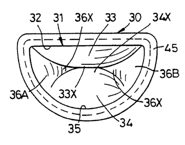
' In Figures 1 to 4, a mitral heart valve replacement is
formed of essentially flexible bioincorporable material and
comprises a generally D-shaped sewing ring 30 having an
~'~3 E3ST: ~ t~ : ~ ~~ ~.- ~
'"'191!19465 PCT/US91/03834
208451
opening 31 with one straight side portion 32, an anterior cusp
33 hinged contiguously from that straight side portion, a
posterior cusp 34 hinged contiguously from a shorter portion
of the arcuate side 35 of the opening opposite the anterior
cusp, and two lateral cusps 36A and 36B hinged contiguously
one from each of the remaining portions of the arcuate side 35
of the opening extending between adjacent ends of the side
portions from which the anterior and posterior cusps are
hinged, together with chordae 37 of bioincorporable material
extending from edge portions 33X, 34X and 36X of the cusps 33,
34, 36A and 36B for connection through attachment portions 38
to the papillary muscles 39 (see Figure 14) in the ventricular
cavity 40 when the sewing ring 30 is sutured to the atrio-
ventricular junction 41 of the host or patient heart 42, the
aggregate area of the cusps 33, 34, 36A and 36B exceeding the
area bounded by the sewing ring 30 so that when the valve is
caused to open (see Figures 3 and 4), by the papillary muscles
39 pulling the chordae 37, the cusps simply deflect away from
each other (seQ particularly Figure 3), and when the papillary
muscles allow the valve to close, the edge portions 33X, 34X
and 36X of the cusps meet out of the plane of the sewing ring
30 so as to be capable of meeting in the ventricular cavity 40
of the patient heart.
It should be noted that the chordae 37 from the
anterior cusp 33 have been Qmitted from Figures 2 and 4 for
the sake of clarity.
The anterior cusp 33 is contiguous with one third of
the perimeter of the sewing ring opening 31 and has a
SUBSTITUTE SHEET'
WO 91/19465 r '~~ PCT/U~91/03i~-'.
~~'~~ f~ - 10 -
generally, semicircular edge portion 33X, to enable it to
project deeply into the ventricular cavity 40; and the
posterior cusp 34 and lateral cusps 36A and 36B - which are,
therefore, together contiguous with two-thirds of the
perimeter of the sewing-ring opening - also have generally
semicircular edge portions 34X and 36X.
The sewing ring 30 is formed by a combination of
integral flange portions 43 of the anterior, posterior and
lateral cusps stitched to a flat basic ring element 44 cut
from bioincorporable material, with a similar flat reinforcing
or stiffening ring 45 of bioincorporable material between
which and the basic ring element the flange portions of the
cusps are interposed (see also Figures 7 to 13).
The posterior cusp 34 and the lateral cusps 36A and
36B in the embodiment of Figures 1 to 4 are formed from a flat
strip of the bioincorporable material, with spacer portions
(not shown) between adjacent cusp portions, which spacer
portion may be partially cut through and/or pleated and
stitched so that the integral flange portion can conform to
the arcuate portion 35 of the D-shaped opening, and to bring
the adjacent cusp portions into contiguous disposition with
each other.
zn the embodiment of Figures 1 to 4, the chordae 37
are separate chords attached by sewing to the edge portions
33X, 34X and 36X of the, cusps and to the attachment portions
38, while in the embodiments of Figures 5 and 6 (which
otherwise is similar to that of Figures 1 to 4) the chordae 37
are formed integral with the cusps and are provided with
SU~STtTU'~'~ ~.°:~rT
.:"~ 91/19465 ~ ~ ~ ~ ~ ~, ~ PCT/US91/03834
- 11 -
integral attachment portions 38, those attachment portions
integral with the lateral cusps being interposed between
attachment portions of the anterior and posterior cusps
respectively and stitched together therewith.
In the preferred embodiment of Figures 7 to 13, and 19
to 21, the chordae are also formed integral with the cusps and
the attachment portions, but all four cusps 'and their
respective chordae 37, attachment portions 38 and flange
portions 43 are formed as separate components for fitting to a
basic ring element 44 (Figure 7 and 13) having a trapezoidal
opening 31X, and a reinforcing or stiffening ring element 45
(Figure 12) is similarly shaped with a trapezoidal opening
31Y. Fold lines between the cusps and their flange portions
43 are indicated by chain dotted lines 46, while ordinary
broken or dotted lines distinguish the chordae 37 from the
cusps and the attachment portions 38. The chordae of the
anterior and posterior cusps 33 and 34 respectively have
longitudinal slits 47 which, in use, can open to assist blood
flow, while the chordae of the lateral cusps 36A and 36B have
slot-like openings 48 for the same purpose, and the slits and
slots also facilitate the flexing of the cusps to enable their
respective edge portions to meet when the valve closes. Tabs
49 on respective adjacent edges of the cusps adjacent the
flange portions 43 are stitched together (Figure 13) in
respective pairs after. the flange portions have been stitched
on to the basic ring element 44, and the attachment portions
38 of the lateral cusps 36A and 36B are stitched to respective
attachment portions of the anterior and posterior cusps 33 and
SUEST~TUTE SHEET
WO 91/19465 d ~~ PCT/US91/03P~st
_ 12 -
34 respectively in readiness for suturing to the papillary
muscles 39; and, between the tabs 49 and the attachment
portions 38 the respective pairs of adjacent edges of the
chordae are free to gape (.ashcan be seen in Figure 13) to
assist blood flow.
The surgical procedure involved in inserting the
preferred embodiment of the invention will be described with
reference to Figures 15 to 21, which procedure also involves
the use of two other devices in accordance with the invention.
The first device is shown in Figures 16 to 18 and is a
combined sizing and suture placement guiding device 50
comprising an elongate handle 51 carrying an arcuate member 52
having a circumferential shallow groove 53 for fitting within
the corresponding part of the remnant 54 (see Figure 15) of
the excised natural valve, with slots 55, one adjacent each
end of the arcuate member and one intermediate thereof for
indicating the positions of placement sutures 56 (see Figures
15 and 20) to be passed also through corresponding positions
on the sewing ring 30 of the replacement valve, and With a
depth gauge 57 consisting of a generally U-shaped bar having a
rectilinear base portion 58 to lie across the papillary
muscles 39 to indicate the positions (below that base portion
58) for placement sutures 59 in the papillary muscles for
securing the attachment portions 38 of the chordae 37.
It will be evident that when the placement sutures 56
and 59 have been inserted the sizing and guiding device 50 is
removed to enable the replacement valve to be inserted, which
step in the procedure is assisted by a rigid holder 60 which
SUBSTiTtJT~ ~ ;~~T
PCT/US91/03834
~-'7 91/19465 - 13 _ 2 p
is shown in Figures 19 and 20 temporarily secured by tacking
stitches 61 to the sewing ring 30. The holder 60 has a
generally U-shaped bar 62 extending between the chordae 37 of
the anterior and posterior cusps 33 and 34 respectively as far
as the attachment portions 38, so as to prevent collapsing of
the valve. The holder 60 is also provided with notches 63
for location of the placement sutures 56 previously inserted
in the remnant 54 of the excised natural valve. A detachable
elongate handle 64 (Figure 20 only) is attached to the holder
60 to facilitate insertion of the valve as the placement
sutures 56 and 59 are drawn through the sewing ring 30 and the
attachment portions 38 respectively.
When the placement sutures have been secured, further
suturing 65 can be effected all round the sewing ring 30 to
complete the procedure.
It will be seen in Figures 1 and 3 that the sewing
ring 30 has a central broken marker line, which - as shown in
Figures 19 to 21 - can be provided by a suture 66 interwoven
into the reinforcing or stiffening ring 45, to indicate inner
and outer stitching zones for different ranges of heart~sizes,
the inner zone being utilized when a smaller sizing device
than the device 50 in Figures 16 to 18 is found to fit the
remnant 54 of the excised natural valve.
SUBSTITUTE SHEET
,.