Note: Descriptions are shown in the official language in which they were submitted.
DETECTIC)N OF ELECTRODE/PATIENT MOTION
AND FAST RESTORE LIMITS
F eld of the Invention
This invention relates generally to limits detection and, more particularly,
S to the detection of limits involved in the monitoring of electrode/patient motion
and fast restore systems in medical instruments.
Background of the Invention -~
A variety of medical instruments have been developed for use in monitoring
and treating patients. Many of these instruments are designed to be electrically10 coupled to the patient via one or more electrodes. The electrodes receive electrical
signals from, or transmit electrical energy to, some portion of the patient's body.
In that regard, a defibrillator/monitor typically includes two or more
monitoring electrodes that receive electrical signals from the patient's heart. These
signals are then commonly displayed by the monitor, allowing the attending
15 physician to evaluate the heart's operation. In addition, a pair of defibrillation
electrodes are used to transmit electrical energy from the de~lbrillator to the patient
to, for example, terminate undesired fibrillation of the heart.
The monitoring and defibrillation electrodes used with the
defibrillator/monitor are often applied externally to the patient's chest and/or20 limbs. As will be appreciated, the impedance of the electrodes, the transthoracic
impedance of the patient, and the impedance of the electrode/patient interfaces, all
influence the signals received by the monitor and the energy delivered to the
patient. Typically, the electrodes are designed to reduce the influence of
impedance on the instrument's operation as much as possible.
In that regard, external electrodes are made relatively large to reduce the
impedance of the electrode/patient interface. Also, a conductive gel is often
PHYS\6177AP.DOC
,
., . , . : :
,''',', ' ~ ~ '
. ': .
. .
-2- ~ fi ~ r3 ~j ~, 7
applied to the surface of each electrode before the electrode is attached to thepatient to further limit the interface impeclance. Despite such precautions, theimpedance of the electrode/patient interface may still have undesired influences on
the instrument's operation.
S One of the most common problems involving electrode/patient impedance isrelated to motion. For example, with monitoring elec~rodes applied to a patient's
chest, movement of the patient or the electrodes may disturb the patient/electrode
interface. The resultant variations in interface impedance introduce corresponding
variations in the electrical signals received at the monitor, independent of theoperation of the heart. This "motion artifact" in the monitored signal can, in turn,
cause the instrument or operator to erroneously interpret the condition of the heart.
Relative motion between the patient and defibrillation electrodes may
similarly be of interest. For example, patient motion may indicate that the patient
is conscious or is being moved by a health care provider. In either instance, it may
be undesirable to discharge energy to the patient. Further, motion-induced
variations in the impedance of the electrode/patient interface may result in
corresponding variations in energy losses at the interface. Thus, the energy
actually delivered to the patient to terminate ~lbrillation may differ considerably
from that selected by the operator.
Prior art systems have been developed to address these limitations. In that
regard, some systems monitor the impedance at the electrode/patient interface todetermine when motion is occurring. In the event the monitored impedance
suggests that motion is occurring, operation of the instrument is then inhibited.
By way of illustration, U.S. Patent No. 4,919,145 (Marriott), assigned to
Physio-~ontrol, reviews a number of different techniques used to sense lead
impedance and/or transthorac;c impedance (TTI). In that regard, the background
section of the Marriott patent indicates that a small DC signal can be applied to the
leads, with the resulting DC voltage across the leads then being representative of
impedance. Another approach described in the background section of the Marriott
patent involves the application of a high-frequency, constant current signal to the
leads. The Marriott patent then goes on to disclose an arrangement in which two
carrier signals are used to detect a lead impedance related voltage and an
impedance respiration related voltage.
U.S. Patent No. 4,619,265 (Morgan et al.), also assigned to Physio-
Control, discloses an arrangement in which a patient's TTI is evalua~ed to detect
motion. More particularly, ~l'I signals are compared a~ainst some predetermined
PIIYS\61~7AP.I~OC
-3- ~, ~j & s~ ~s ~, ~
threshold level. If the last two measurements of rrI exceed the threshold, a
display is generated prompting the operator to stop all motion. If motion is
detected for more than fifteen seconds, the operator is also prompted to performcardiopulmonary resuscitation.
S With only one or two impedance measurements use{l to detect motion,
temporary aberrations in the measurements due, for example, to noise are likely to
influence the detection of motion. In that regard, noise in the measured impedance
signal may cause the signal to be erroneously high or low at any given time.
Although the resultant signal variations may average out over time, with only one
or two measurements used, the measurements are likely to be inaccurate. As will
be appreciated, it would be desirable to allow motion to be detected in a mannerthat is relatively free from the influence of noise.
As disclosed by Morgan et al., the use of limits detection plays an
important role in conventional motion detection schemes, allowing an impedance
measurement to be compared against some predetermined threshold level
associated with motion. In accordance with the present invention, limits detection
plays roles both in the processing of impedance data used in the detection of
motion and in the processing of monitored cardiac signals used to evaluate the
condition of the patient's heart.
In that regard, the signals used to monitor cardiac activity and electrode
impedance are conventionally f1ltered by a preprocessing circuit prior to analysis.
Filtering is performed to remove select portions of the signals, preserving onlythose portions that have a high information content. The removed portions may beattributable to, for example, some baseline signal contributor or noise.
The conventional filter circuits used often employ capacitive elements, as
well as resistive and inductive elements. When the signal applied to such a filter
circuit undergoes large deviations, the capacitors may become fully charged,
rendering the filter inoperative until the charge stored on the capacitors has time to
decay. As will be appreciated, it would be desirable to determine when the inputto such a filter undergoes a large deviation, so that some form of corrective action
can be taken to lirslit the inoperability of the filter circuit.
In view of the preceding comments, it would be desirable to develop a
method of detecting limits associated with electrode/patient motion, free from the
disruptive influence of, for example, noise. It would further be desirable to
develop a method of detecting limits associated with the inoperability of filtercircuits conventionally used in medical instruments. To reduce the complexity of
PIIYS\6177AY.DOC
the overall processing performed by the instrument, it would further be desirable
for the same general method to be used in detecting both types of limits.
Summa~h~.nvention
In accordance with this invention, a method is disclosed of hysteretically
S detecting the limits of a physiological signal processed by a rnedical instrument.
The method includes the steps of comparing the signal to a first range of values.
An inside time, representative of the time during which the signal is within therange of values, is then stored along with an outside time, representative of the
time during which the signal is outside the range of values. An inside action signal
10 is produced when the inside time exceeds a first inside time limit and an outside
action signal is produced when the outside time exceeds a first outside time limit.
The preceding steps are then repeated for a second range of values, with the
repetition of steps for the first and second ranges of values introducing a hysteretic
aspect to the method.
In one application of interest, the method is used to detect relative motion
between an electrode and a patient. The electrode is coupled to the patient and to a
medical instrument which provides a signal related to the impedance of the
electrode/patient interface. The method includes the step of comparing the signal
to a first range of values. A first inside time, representative of the time during
20 which the signal is within the ~1rst range of values, is then stored along with a first
outside time, representative of the time during which the signal is outside the first
range of values. The first inside and outside times are set to zero when the first
inside time exceeds a first inside time limit. A motion detection output, indicative
of relative motion between the electrode and the patient, is produced when the first
25 outside time exceeds a first outside time limit.
In accordance with yet another aspect of the invention, a method of
restoring a filter circuit used to process the physiological input to a medical
instrument is disclosed. The method includes the step of comparing the signal to a
first range of values. A first inside time, representative of the time during which
30 the signal is within the first range of values, is stored along with a first outside
time, representative of the time during which the signal is outside the first range of
values. The first inside and outside times are set to zero when the first inside time
exceeds a first inside time limit. A filter restoration olltput is produced when the
first outside time exceeds a first outside time limit.
PIIYS\6177AP.DOC
C~ f; ~ / l
Brief Description of the Drawing
The invention will generally be described in greater detail, by way of
example, with reference to the accompanying drawings wherein:
FIGURE 1 is an illustration of a defibrillator/monitor constructed in
5 accordance with the present invention and attachable to a patient via a pair of
electrodes;
FIGURE 2 is a block diagram of a defibrillator/monitor of the type shown
in FIGURE 1, illustrating the int~rrelationship of the various components of theinstrument;
FIGURE 3 is a more detailed block diagram of a motion detection circuit
included in the defibrillator/monitor of FI(:;URE 2;
FIGURE 4 is a more detailed block diagram of a control and processing
circuit included in the de~lbrillator/monitor of FIGURE 2;
FIGURE S is a flow chart illustrating the way in which the
defibrillator/monitor processes an impedance signal to detect motion at the patient-
electrode interface;
FIGURE 6 is a more detailed flow chart, illustrating a high impedance
threshold limit subroutine included in the flow chart of FIGURE 5;
FIGURE 7 is a more detailed flow chart, illustrating a low impedance
threshold limit subroutine included in the flow chart of FIGURE 5;
FIGURE 8 is a graph depicting a time-varying signal processed by the
defibrillator/monitor in accordance with the subroutine shown in FIGURE 6,
illustrating a pair of upper and lower limits used in the ~Irst part of a hysteretic
motion detection operation performed by the instrument;
FIGURE 9 is a graph depicting a time-varying signal processed by the
defibrillator/monitor in accordance with the subroutine shown in FIGURE 7,
illustrating a pair of upper and lower limits used in the second part of a hysteretic
motion detection operation performed by the instrument;
FIGURE 10 is a flôw chart illustrating a motion clear subroutine included
in the flow chart of FIGURE 5 and used by the defibrillator/monitor to determinewhen motion is no longer present;
FIGURF, 11 is a graph depicting a time-varying signal processed by the
defibrillator/monitor in accordance with the subroutine shown in FIGURP. lO;
FIGURE 12 is a flow chart illustrating the way in which the
defibrillator/monitor processes impedance signals to activate a fast-restorationsystem included in the motion detection circuit of FIGURE 3.
I'IIYS\6177AI'.I)OC
- 6-
Detailed Description of the Preferred E~mbodiment
Referring now to FIGURE 1, a defibrillator/monitor 10, constructed in
accordance with this invention, is shown The de~lbrillator/monitor 1~ performs avariety of different functions. For example, the defibrillator/monitor lO receives
5 electrocardiographic (ECG) signals from the patient for use by an operator in
monitoring the patient's heart. The defibrillator/monitor 10 is also conventionally
designed to allow relatively large pulses of energy to be applied to the patient's
heart to, for example, terminate fibrillation of the heart. Alternatively, smaller,
periodic pulses of energy may be applied to stimulate a desired heart rate
Each of these various functions requires the defibrillator/monitor 10 to be
electrically coupled to the patient Usually, three separate sets of monitoring,
defibrillation, and pacing electrodes are employed While conventional
defibrillation and pacing electrode sets typically include two electrodes each, a
variety of different monitoring electrode sets have been developed, including, for
15 example, two, three, four, and ten electrodes
Motion-induced impedance variations at the different electrode/patient
interfaces may cause the ECG signals received from the patient to be
misinterpreted by the defibrillator/monitor 10 Similarly, defibrillation and pacing
pulses applied to the patient may be attenuated by the impedance fluctuations to an
20 unknown degree. Further it may be undesirable to defibrillate a patient when
motion is occurring. To overcome these limitations, the defibrillator/monitor 10 is
designed to detect motion and, for example, to inhibit further operation until
motion is no longer present.
In one preferred arrangement, the limits detection scheme implemented
25 involves a hysteretic analysis that provides greater immunity from noise thanconventional motion detection systems. The broad limits detection scheme also has
applicability to the restoration of certain filtering circuits used in the detection of
motion and the general processing of ECG signals.
Tun~ing now to a more detailed review of the construction of the
30 defibrillator/monitor 10, reference is had to the block diagram of FIGURE 2. As
shown, the defibrillator/monitor 10 includes a monitoring circuit 12, defibrillation
circuit 14, pacing circuit 16, and motion detection circuit 1~, all regulated by a
control and processing circuit~0. An input/output (I/O) circuit 22 allows the
operator to apply inputs to circuit 20 and provides the operator with the various
35 instrument outputs. With the exception of the motion detection circuit 18 and the
PIIYS\6177AP.DOC
,
',~J',,' ~',',~. '? I
related operation of the control and processing circuit 20, the various components
of defibrillator/monitor 10 are conventional in nature and are only briefly discussed
herein.
The monitoring circuit 12 is typically coupled to the patient via two or
5 more conventional ECG monitoring electrodes. As will be discussed in greater
detail below, the monitoring circuit 1~ includes the conventional processing
circuitry required to sample, filter, and amplify electrical signals received from the
different electrodes. The monitoring circuit 12 may further be constructed to
produce, for example, any of the standard vectorcardiographic leads of ECG
10 information from the received signals. The monitoring circuit 12 also typically
includes some form of isolation circuitry designed to restrict the passage of
potentially harmful currents between monitor circuit 12 and the patient.
The defibrillation circuit 14 conventionally includes some one or more
capacitors used to store energy for discharge to the patient via the de~lbrillation
15 paddles or electrodes. The amount of energy stored on the capacitor is controlled
in response to inputs from the control and processing circuit 20. The energy is
discharged by depressing discharge switches included on the defibrillation paddles
or instrument. In a synchronized cardioversion mode of operation, the control and
processing circuit 20 times the discharge to coincide with a particular portion of
20 the cardiac cycle, identified using ECG information from the monitoring
circuit 12.
Pacing circuit 16 is coupled to the patient via a pair of conventional pacing
electrodes. The pacing circuit 16 is constructed to produce a periodic pulse of
relatively low current used to initiate a desired heart rate in the patient. The25 magnitude and repetition rate of the pacing pulses are controlled by pacing
circuit 16 in response to inputs t`rom the control and processing circuit 20.
Turning now to a discussion of the motion detection circuit 18, and the
related operation of control and processing circuit 20, reference is had to
FIGURE 3. As shown, the motion detection circuit 18 includes a number of
30 different components. In that regard, an impedance measurement circuit 24 is
coupled to at least one pair of the various electrodes used with instrument 10. In
one currently preferred arrangement, motion of the patient relative to two
monitoring electrodes is detected and used to indicate electrode/patient motion in
general. As a result, the impedance measurement circuit 24 is coupled directly to
35 two of the monitoring electrodes.
PIIYS\6177AP.DOC
- 8 ~ "~
~ f desired, the impedance of one or more alternative sets of monitoring,
defibrillation, or pacing electrodes can be evaluated to detect motion. In that
regard, the control and processing circuit 20 could alternatively switch the
connection of the impedance measurement circuit 24 to more than one electrode
set, allowing a single circuit 24 to measure the ;mpedance of various ECG, pacing,
and/or defibrillation electrode pairs. As another option, a separate motion
detection circuit 18 and, hence, impedance measurement circuit 24 could be
connected to each of the different electrode pairs whose impedance is to be
monitored.
The impedance measurement circuit 24 is of conventional construction, and
generally involves the passage of a known current between a particular pair of
electrodes of interest. The resultant voltage drop across the electrode pair is then
representative of the collective impedance of the electrode pair, the patient, and the
electrode/patient interfaces. Additional details regarding suitable impedance
15 measurement circuits 24 can be obtained from the Marriott and Morgan et al.
patents discussed above, the disclosures of which are incorporated by reference.As previously suggested, the output of the impedance measurement
circuit 24 is a time-varying voltage, measured using a 16 kiloHertz square wave.The magnitude of this voltage is proportional to the impedance of the electrodes,
20 patient and electrode/patient interfaces. If the patient moves, the impedance of the
electrode/patient interface will typically vary, causing the output of impedancemeasurement circuit 24 to vary accordingly.
As shown in FIGURE 3, the output of the impedance measurement
circuit24 is applied to a differentiator26. The differentiator26 produces an
25 output that is proportional to the derivative of, or change in, the impedance signal.
As a result, although the output of differentiator 26 is still an impedance-based
signal, the magnitude of the differentiated output is proportional to the motiondetected, at least over short intervals. In the currently preferred embodiment, the
differentiator26 is, for exarnple; a capacitive coupler between the impedance
30 measurement circuit 24 and a low-pass filter 28.
As noted, the output of differentiator 26 is applied to a low-pass filter 2~.
The low-pass filter 28 may be of a Bwtterworth, or modified Butterworth,
construction, which makes use of the capacitive nature of differentiator 26 and has
a cut-off frequency of from one-to-ten Hertz. As w;ll be appreciated, filter 28 thus
35 removes extraneous high frequency components from the motion signal output bydifferentiator 26. These high frequency components may be attributable to, for
l'IIYS\6111AP.DOC
'
example, radio frequency interference (rfi), static discharge interference, and cross
talk within the electronics of instrument 10.
The next component of the motion detection circuit 18 shown in FIGURE 3
is a sample and hold circuit 30. The sample and hold circuit 30 is used to
5 repetitively sample and store ~he processed motion data from fllter 28 ~or further
processing. Circuit 30 may be, for example, a single-slope analog-to-digital (A/D)
converter operated at 480 Hertz.
The information sampled by circuit30 is then applied to a low-pass
filter 32. Filter 32 is included to remove noise introduced into the conditioned10 motion signal by the sampling process. The low pass filter 32 may also be of the
Butterworth type and has a cut-off frequency of roughly five Hertz.
In the present arrangement, the preceding components of circuit 18 have
been described as being implemented with hardware. As described below, the
remaining components are, in contrast, implemented in software. Alternative
15 implementations can, of course, be employed.
The output from low-pass filter 32 is applied to a calibration circuit 34.
The function of the calibration circuit 34 is to calibrate the filtered motion output
so that it exhibits a predetermined or calibrated magnitude when no motion is
present. In that regard, the calibration circuit 34 includes a differential
20 amplifier 36 and variable gain amplifier 38.
The differential amplifier 36 has two inputs. One of these inputs is the
filtered motion signal output by filter 32. The other input of amplifier 36 is a
calibrated offset generated by the control and processing circuit 20. As a result,
the output of differential amplifier 36 is effectively equal to the output of low pass
25 filter 32 minus the offset. In the preferred arrangement, the appropriate offset is
empirically determined during initialization of software used by the control andprocessing circuit 20. More particularly, with the electrodes coupled to a patient
that is not moving, an automated calibration process executed by circuit 20 adjusts
the offset until the output of amplifier 36 is equal to zero. In the currently
preferred arrangement, amplifier 36 is implemented as a software difference
operation.
The adjustable gain amplifier 38 next amplifies the output of amplifier 36 to
ensure that the signal representative of motion has an amplitude that is sufficiently
large to allow motion to be detected relatively easily and with the desired accuracy.
In that regard, the amplifier 38 receives both the output of amplifier 36 and a gain
input from the control and processing circuit 20. Like the offset, the magnitude of
P~IYS\6177J~P.DOC
' '
-10~
the gain input is empirically determined as part of an initialization process in which
an automated calibration process executed by circuit 20 adjusts the gain to provide
the desired performance over the range of expected motion. In the currently
preferred arrangement, amplif1er 38 is effectively provided by a controllable
S software gain factor.
Finally, the output of amplifier 38 is applied to a motion detection block 40
included in the motion detection circuit 18. Block 18 represents a sequence of
operations performed by software included in the control and processing circuit 20.
To illustrate the operation of the motion detection block 40, reference will be had
10 to FIGUR~S 4 and 5, which further describe the construction and operation of the
control and processing circuit 20.
In that regard, FIGURE 4 is a block diagram of the control and processing
circuit 20. As shown, the control and processing circuit 20 includes a
microprocessor 42, which performs a variety of control and analysis operations
lS determined by various software routines stored in a read only memory (ROM) 44.
The microprocessor 42 stores information used in the control and analysis routines
in a random access memory (RAM) 46. The microprocessor 42 is linked ~o the
other components of the defibrillator/monitor 10 by an input/output (I/O) circuit 48
and a preprocessing ciFsuit 50 which provide the necessary buffers and signal
20 conversions required to allow microprocessor 42 to effectively interface with the
remainder of the system.
Of the various software routines stored in ROM 44, one routine of
particular interest is the motion detection routine 52 represented by the flow charts
of FIGUl~E~ 5, 6, 7, and 10. As will be described in greater detail below,
25 routine 52 monitors the processed motion signal from calibration circuit 34 and
indicates that motion is occurring if that signal is outside a relatively large range
for a short time or outside a smaller range for a longer time. The relationship of
these two ranges and times gives motion detection routine 52 a hysteretic operation
that reduces the influence of, for example, noise. As will be described in greater
30 detail below, the relationship of the ranges and times used to determine the
presence and, then, absence of motion can also be described as hysteretic in nature.
Once motion has been detected, routine 52 continues to indicate the
presence of motion until the signal remains inside a relatively small range for a
relatively long interval of time. Having briefly summarized the operation of
35 motion detection routine 52, the routine will now be discussed in greater detail.
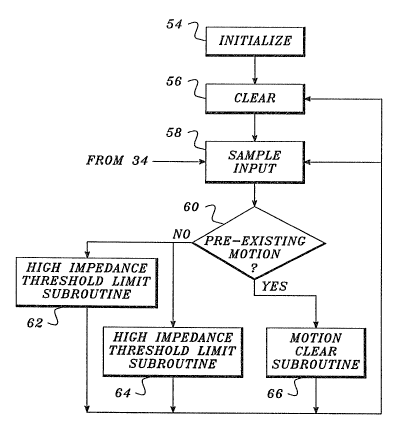
As shown in FIGURE S, the routine 52 begins with an initialization step 54, in
P~WS\6177~P.I)()C
)7'; 5~ J I
which the following parameters are initialized by microprocessor42 to, for
example, the following levels:
(1) upper limit l = ~293 milliohms,
(2) lower limit l =-293 milliohms,
(3) inside time limit 1=33 milliseconds,
(4~ outside time limit 1 = 100 milliseconds,
(5) inside procedure 1=reset inside timer 1 and outside timer 1,
(6) outside procedure 1=set motion flag,
(7) upper limit 2= + 117 milliohms,
(8) lower limit 2=-117 milliohms,
(9) inside time limit 2=333 milliseconds,
(10) outside time limit 2=333 milliseconds,
(11) inside procedure 2=reset inside timer 2 and outside ~imer 2,
(12) outside procedure 2=set motion flag,
(13) upper limit 3=+117 milliohm,
(14) lower limit 3=-117 milliohm,
(15) inside time limit 3 = 1.50 seconds,
(16) outside time limit 3=750 milliseconds,
(17) inside procedure 3=clear motion flag, and
(18~ outside procedure 3=reset inside timer 3 and outside timer 3.
For the purposes of the ensuing discussion, items (1~ - (6) will be
collectively referred to as condition group A, items (7) - (12) will be referred to as
condition group B, and items (13) - (18) will be referred to as condition group C.
These three groups of initialized parameters, which include both amplitude and
time constraints, are used by routine 52 to hysteretically detect the presence of
motion and the subsequent absence of motion.
After initialization, three inside timers 1, 2, and 3 and three outside
timers 1, 2, and 3, employed by the routine, are cleared or set to zero at block 56.
These timers are used to determine the length of time the signal from circuit 34 is
within the various ranges of interest. Prepared in this manner, the routine 52 is
now ready to start processing the output of the calibration circuit 34.
In that regard, the motion or impedance signal from circuit 34 is polled at
block 5~. At block 60, a test is performed to determine whether motion was
detected during the last iteration of routine 52. In the event that motion was not
detected, further operation of the motion detection routine 52 proceeds along a
high impedance threshold limit subroutine 62 and a parallel low impedance
PIIYS\6177AP.DOC
~ 12- ~ r ~
threshold limit subroutine 64. Altcrnatively, if motion was previously detected,the operation of routine 52 proceeds via a motion clear subroutine 66.
Reviewing these different sllbroutines individually, the relationship between
the high impedance threshold limit subroutine 62 and the time-varying output of
calibration circuit 34 is depicted graphically in FIGURE 8. The subroutine 62
begins at block 68 where the impedance represented by the impedance-based
motion signal is compared to the upper limit 1 and the lower limit 1. If the signal
is between these limits, the inside timer is incremented at block 70. On the other
hand, if the motion signal is outside those limits, the outside timer 1 is incremented
at block 72.
If the inside timer 1 has been incremented at block 70, the present count on
the inside timer 1 (representing the number of times the motion signal has fallen
between upper limit 1 and lower limit 1) is compared to the inside time limit 1 at
block 72. If the inside time limit 1 is exceeded, the inside and outside timers 1 are
reset at block 56 and the cycle will then be repeated when the next input sample is
sequentially received at block 58. On the other hand, if the inside time limit is not
exceeded at block 70, operation is returned to block 58 for the receipt of the next
input sample, without clearing of the timers.
Alternatively, if the outside timer 1 has been incremented at block 72, the
present count on the outside timer 1 (representing the number of times the motion
signal has fallen outside the range defined by upper limit 1 and lower limit 1) is
compared ~o the outside time limit 1 at block 74. If the outside time limit 1 has
been exceeded, a motion flag is set at block 76. On the other hand, if the outside
time limit 1 has not been exceeded, the next input sample is obtained at block 58.
As will be appreciated, the basic flow of the high impedance subroutine 62
is repeated for each new sample obtained at block 58 until the motion flag is ~Inally
set at block 76 by either the high impedance subroutine 62, or the low impedancesubroutine 64 described next.
In that regard, the low impedance subroutine 64 is shown in FIGURE 7 and
its relationship to the output of circuit 34 is graphically depicted in FIGURE 9. At
the same time an input sample is applied to block 68 of subroutine 62, the sample
is also applied to another block 78 in subroutine 64. At block 78, the motion
signal is compared to an upper limit 2 and lower limit 2. In the event a particular
input sample falls between these limits, the inside timer2 is incremented at
block 80 and a test is performed at block 82 to determine whether the inside time
limit 2 has been exceeded. In the event that it has, the inside and outside timers 2
PIIYS\6177AP.DOC
- 1 3- c~
are cleared at block 56. If inside time limit 2 has not been exceeded, a new input
sample is obtained at block 58.
On the other hand, if the input sample is outside the range defined by upper
limit 2 and lower limit 2, the outside timer 2 is incremented at block 84. A test is
then performed at block 86 to determine whether the outside time limit 2 has been
exceeded. In the event the outside time limit 2 has been exceeded, a motion flag is
set at block 76. Alternatively, if the outside time limit 2 has not been exceeded,
the inside and outside timers 2 are cleared at block 56.
The high and low impedance subroutines 62 and 64 cooperatively check the
input for relatively large variations over short times and smaller variations over
longer times. This hysteretic analysis is relatively unsusceptible to the influence
of, for example, noise because it ;s unlikely that (1) the magnitude of the noise
would be sufficient to cause the input to exceed the larger range limits, or (2) the
recurrence of the noise would be sufficient to cause the input to exceed the smaller
range limits for the longer time. As will be appreciated, if subroutine 62 were
used by itself, a signal representative of moderate but continuous motion might not
exceed the range limits for the short duration involved. Similarly, it subroutine 64
were used by itself, a signal representative of substantial but brief motion might
not exceed the lower range limits for a suf~lciently long time.
Expressed in another way, the subroutines 62 and 64 rely upon different
condition groups A and B to detect motion. Condition group A is used to reduce
the influence of channel saturation on the detection of motion, while condition
group B is used to reduce the influence of low level motion noise on the detection
of motion. The combined use of the condition groups with their different
amplitude and time constraints gives the routine52 a characteristic operation
referred to herein as hysteretic.
With the motion flag set at block 76 by either subroutine 62 or 64, an
output would normally be applied to the defibrillation circuit 14 to inhibit thedischarge of energy to the patient. As a result, a moving patient can not be
defibrillated, protecting both the patient and any attending health care provider that
might be moving the patient. If desired, the motion flag may also be used to, for
example, alert the operator to potential errors in the information collected by
monitoring circuit 12.
Once the motion flag has been set at block 76, the motion detection
routine 52 continues via the motion clear subroutine 66 depicted in FIGUR~ 10.
PIIYS\6177AP.I)OC
-14- ~J~", 3 j ~,
The relationship of the processed motion signal to the various parameters employec',
by this portion of routine 52 is depicted graphically in Fl(;~URE 11.
As shown in FIGUR~ 10, with block 60 having determined that the motion
flag is set, the input sample is compared to upper limit 3 and lower limit 3 at
5 block 90. In the event that the sample is between upper limit 3 and lower limit 3,
the inside timer 3 is incremented at block 92. Alternatively, if the sample obtainec',
at block 88 is outside the range defme~, by upper limit 3 and lower limit 3, theoutside timer 3 is incremented at block 94.
At blocks 96 and 98 tests are performed to determine whether the inside
10 time limit3 and outside time limit3 have been exceeded, respectively. In the
event the inside time limit 3 has been exceeded, the motion flag is clearec', atblock 100. On the other hand, if inside time limit 3 has not been exceeded at
block 96, the routine returns to block 58 to obtain the next sample of the motion
signal. If block 98 determines that the outside time limit 3 has been exceec,ed, the
15 inside timer 3 and outside timer 3 are reset at block 56 and the next input sample is
obtained at block 58. On the other hand, if the outside time limit 3 has not been
exceedecd at block 98, the next input sample is obtained without clearing the timers.
As notec, above, the combined use of subroutines 62 and 64 (employing
condition groups A and B) in the detection of motion, causes the motion detection
20 routine 52 to operate in a hysteretic manner while detecting motion. Sir~,ilarly, the
combinecd use of subroutine 62 (employing condition group A) to detect motion and
subroutine 66 (employing condition group C) to clear the motion fl,ag, or the
combined use of subroutine 6~ (employing conc,ition group B) to detect motion and
subroutine 66 (employing condition group C) to clear the motion f,ag, causes the25 motion detection routine 52 to operate in a hysteretic manner while setting and
clearing a rnotion f,ag. In ~oth cases, the operations involve separate amplitude
and time limits, which may be related in substantially any manner desired.
As will a',so be appreciated, any one of the three different subroutines of the
motion detection routine 52 represent a basic protocol whose applicability in the
30 instrument is not limited solely to motion detection. For example, it may be
helpful to compare a variety of different physiological signals processed by theinstrument to some range defined by upper and lower limits. If the input is within
the range, an inside timer is incremented and, when the inside timer exceeds theinside time limit, some inside proceclure is performed. Alternatively, if the input
35 is outside the range, an outside timer is incremented and an outside procedure
perforrned when the outside timer is greater than the outside time limit. If this
PliYS\6177AP.DOC
,
-15- ~J ~ ,J I
process is performed for different ranges and/or times a hysteretic aspect is
introduced into the analysis.
One alternative application for such a protocol is a fast restore routine 102,
shown in FIGURE 12. The fast restore routine 102 can be used in the motion
S detection circuit 18 of FIGURE 3, where it is represented by block 104. The
routine 102 is used to determine when deviations in the input signal are so great as
to fully charge the capacitive coupling of the differentiator 26 connected to
filter 28, rendering the filter inoperative. In that case, an output from the fast
restore block 104 is applied to a switch circuit 106 to temporarily close a switch or
10 switches coupled in parallel to the differentiator capacitance. As a result, the
energy stored by the differentiator 26 is quickly discharged and, when the switches
in circuit 106 are again opened, the filter and motion detection circuit 18 are
restored to operability.
Reviewing the OpeMtiOn of the fast restore routine 102 in greater detail, at
15 block 108, the upper and lower limits are established at, for example, +one ohm
and -one ohm. The inside and outside times are established at, for example,
83 milliseconds and 330 rnilliseconds, respectively. At block 110, the inside timer
and outside timer are cleared.
Next, the signal from calibration circuit 34 is sampled at block 112. At
20 block 114, the impedance represented by the input signal sample is compared to
the upper and lower limits. In the event that this impedance is inside the rangedefined by the upper and lower limits, the inside timer is incremented at 116.
Then a test is performed at block 118 to determine whether the inside time limithas been exceeded. If the inside time limit has been exceeded, the timers are reset
25 at block 110 prior to the collection of the next signal sample at block 112.
Alternatively, if the inside timer limit has not been exceeded, the next signal
sample is obtained at block 112 without resetting the timers.
On the other hand, if block 114 determines that the impedance of the signal
sample is outside the range defined by the upper and lower limits, the outside timer
30 is incremented at block 120. A test is then performed at block 122 to determine
whether the outside time limit has been exceeded. If the outside time limit has not
been exceeded, the next input sample is obtained at block 112.
On the other hand, if the outside time limit has been exceeded, an
impedance channel fast restore flag is set at block 124. With the upper and lower
35 limits and outside tirne limit set appropriately, the fast restore flag will thus be set
PIIYS\6177AP.DOC
-16- ~ r~
when an unacceptably severe deviation has occurred in the input signal, fully
charging the differentiator capacitance and rendering filter 28 inoperative.
As noted previously, the impedance channel fast restore output from
block 124 is applied to a shunting switch or switches included in switch
5 circuit 106. These switches are connected in parallel with the capacitance
associated with differentiator 26 and filter 28 of the motion detcction circuit 18
shown in FIGURE 3. The fast restore output initially closes the switches,
discharging the energy stored by the capacitances to ground. Once the energy hasbeen discharged, the switches are opened, having restored filter 28 and circuit 18
10 to their operative condition.
Although not described in the same level of detail, it will be appreciated
that the basic fast restore routine 102 of FIGURE 12 can also be used
advantageously in other filter circuits, including those in the ECG monitor
circuit 12 of FIGURE 2. In that regard, an ECG processing section of the monitor15 circuit 12 can be constructed to closely parallel the motion detection circuit 18 of
FIGURE 3. The primary di~ferences between the two circuits are as follows.
As will be appreciated, the ECG processing circuit includes an ECG
measurement circuit in place of the impedance measurement circuit 24. The outputof the ECG measurement circuit is proportional to one lead of the ECG
20 information obtained from the patient. In additis)n, as will be appreciated, the
differentiator 26 and motion detection block 40 of motion detection circuit 18 are
not required and are, therefore, absent from the ECG processing circuit.
Otherwise, the processing circuit and motion detection circuit 18 are the same.
Regarding the use of fast restore routine 102 with monitor circuit 12, as
25 might be expected, the various parameters initialized at block 108 will be different
than thoss previously discussed in connection with the motion detection circuit 18.
More particularly, the upper and lower limits are initialized at +5.5 and -5.5
millivolts, the outside time limit is set at 67 milliseconds, and the inside time limit
is set at 33 milliseconds.
The analysis performed by routine 102 using these various limits then
follows that discussed above. It should be noted, however, that the test performed
at block 114 involves a comparison of the ECG-based, rather than impedance-
based, output of calibration circuit 34 and the flag set at block 124 is an ECG
channel fast restore, rather than impedance channel fast restore. Ultimately, the
35 ECG channel fast restore flag is used to close the switches in switch circuit 106
and restore filter 2~ to operability.
P~IYS\6177AP.IX~C
' '
.
.
-1 7- '~
Those skilled in the art will recogni~e that the embodiments of the invention
disclosed herein are exemplary in nature and that various changes can be made
therein without departing from the scope and the spirit of the invention. In that
regard, as was suggested above, various combinations of range and/or tirne limits
5 can be used. For example, while one or two evaluations involving both range and
time limits may be employed, alternative evaluations involving only range or time
limits may be used. Because of the above and numerous other variations and
modifications that will occur to those skilled in the art, the following claims should
not be limited to the embodiments illustrated and discussed herein.
PHYS\6177AP.DOC