Note: Descriptions are shown in the official language in which they were submitted.
6
-1-
~'~'TIJS FflR G.Tl<r~T~~I~ t9F lg~~D VESS~L~
Technical Field of the Invention
The present invention relates generally to the cannulation of arteries and
veins
through the use of ultrasonic techniques.
8~~,~o_und of the Invention
It is well established that the insertion of ~u~teerial and venous catheters
for various
purposes such as for angiogzaphy can be responsible for patient discomfort.
Locating and
penetrating arteries and veins can be especially difficult when dealing with
patients who are
obese or present unusual anatomy.
Arterial and venous catheters are particularly useful for cardiac
catheterization and
other radiologic pr~edures such as cerebral angiograms.
The potential utility of Doppler ultrasound for accurately guiding the needle
into a.
vessel has been recognized. Most applications utalize the transmission of
ultrasonic waves
through the needle and reception of ultrasonic echoes by a separate transducer
located on the
body of the patient separated from the syringe and needle. Such applications
obviously have
limited accuracy. For example, U.S. Patent N'o. 3,556,079 dir~ted to a "Method
of
Puncturing a M~ical Instrument Under Guidance of Ultrasound" discloses in one
embodiment the placement of both the transmitting and receiving transducers in
the neaile
CA 02085912 2003-02-28
75655-3
2
and syringe. Such an embodiment:, however, requires a
special catheter construction and can give an erroneous
signal when the needle engages the blood vessel before
penetrating the vessel.
A major advance was made to this technology by
virtue of U.S. Patent No. 4,887,606 directed to "Apparatus
For Use in Cannulation of Blood Vessels." This patent
teaches the use of a transducer insert positioned within a
hollow needle including an ultrasonic transducer at one end
for transmitting and receiving ultrasonic waves through the
sharpened end of the needle. Upon location and penetration
of a blood vessel, the transducer insert is removable from
the needle for implementation of the known Seldinger
technique for placing a catheter in a blood vessel.
:15 FIG.. 1 depict:, in cross section, a device which
is the subject: of U.S. hatent No. 4,887,606. In referring
to FIG. 1, needle 10 is shown as having sharpened end 11
and, located therein, ultrasonic flow sensing assembly 12.
The assembly 12 include:; a plastic support member 13 through
which a first conductoz:~ 14 extends intc contact with an
electrode 15 on the back surface of transducer 16.
Transducer 16 is affixed to support member 13 by means of a
low impedance epoxy 17 which is :filled with glass
microballoons (not shown). A second conductor 1F3 is formed
2,5 on the exterior surface of suppo=t rod 40 by means of metal
deposition and extends into contact with electrode 15 on the
front surface of transducer 16. The conductors 14 and 18
form a coaxial cable and the outer shield conductor 18 can
be grounded during use.
CA 02085912 2003-02-28
75655-3
2a
An insulative material. 19 such as an epoxy is
formed around the periphery of t:he transducer 16 to
electrically isolate electrode 1.4 from conductor 18 which is
in turn connected to electrode 15. Transducer 16 is
positioned near the distal sharpened end 11 of needle 10 for
the transmission and recepta.on c~f energy through the opening
in the distal sharpened end of the needle.
Although the device disclosed in U.S. Patent No.
4,887,606 represents a superior apparatus for cannulation of
~~~~~12
blood vessels, such apparatus is difficult to manufacture and at times
provides a device
whose sensitivity is somewhat low and, ideally, could be improve upon.
The present invention provides an apparatus for the cannulation of blood
vessels
S which is not only easier to manufacture but is also of higher sensitivity
than the device
shown in IJ.S. Patent N~. 4,887,6t3~.
These and other advantages of the invention will biome more apparent when
considering the following description of the invention and the accompanying
exemplary
drawings.
Summary ode Invention
The present invention involves an ultrasonic flow sensing assembly for use in
an
apparatus to be ernployed in the cannulation of blood vessels. The apparatus
comprises a
hollow needle having a longitudinal axis and sharpened distal end for
penetration of tissue
and a proximal end having means for detachably connecting a syringe to the
needle.
The ultrasonic flow sensing assembly comprises an elongated electrically
conducting first tubular member which has a longitudinal axis, a distal end
and lumen
extending therein. An elongated electrically conducting second tubular member
is provided
which has a longitudinal axis and lumen preferable coincident with the
longitudinal axis and
lumen of the first tubular member. 'The second tubular member resides
substantially within
the first tubular member.
2S
An electrically insulating means such as a polymer tube is bated between the
first
and second tubular members. A piezoelectric transducer capable of generating
or receiving
ultrasonic waves is located proximate the distal ends of the first and second
tubular members
and is electrically connected to these tubular members. Means are further
provided for
~0 connecting an electrical power source to the tubular members electrically
connected to the
piezoelectric transducer for the generation and reception of ultrasonic waves.
_3_
CA 02085912 2003-02-28
75655-3
3a
The invention may be summarized as an ultrasonic
flow sensing assembly comprising: a. an elongated
electrically conducting outer tubular member having a
longitudinal. axis, a distal end and a lumen extending
therein; b. an elongated electrically c~onducting~ inner
tubular member disposed within the outer tubular member
having a longitudinal axis and lumen coincident with the
longitudinal axis and lumen of said outer tubular member; c.
electrical insulating means located between the inner and
outer tubular members; d. a piezoelectric means capable of
generating and receiving ultrasonic waves which is located
proximate the distal ends of the inner and outer tubular
members and being electrically connected to the tubular
members, and wherein the piezoelectric means is attached to
the distal end of the inner tubular member providing a
closure for the lumen thereof; and e. means for connecting
an electrical power source to the piezoelectric means far
the generation and rece~otion of ultrasonic waves through the
tubular members.
According to another aspect the invention may be
summarized in an apparatus for cannulation of a blood vessel
comprising a hollow needle a longitudinal axis, a sharpened
distal end for penetration of tissue and a syringe portion
detachably connected to the proximal end of. the needle, the
improvement comprising an ultrasonic flow sensing assembly
located within the hollow needle which includes: a. an
elongated electrically conducting outer tubular rnember
having a longitudinal axis, a distal end and an inner lumen
extending therein; b. an elongated electrically conducting
inner tubular member disposed within the outer tubular
member having a longitudinal axis and an inner lumen
coincident with the longitudinal axis and the inner lumen of
the outer tubular member; c. electrical insulating means
CA 02085912 2003-02-28
75655-3
3b
located between the inner and outer tubular members; d. a
piezoelectric means capable of generating and receiving
ultrasonic waves which is located proximate the distal ends
of the inner and outer tubular members and bei.na
electrically connected to the tubular members; and e. means
for connecting an electrical power source to the
piezoelectric means for the generation and reception of the
ultrasonic waves through the tubular members.
Brief De,~cri 'on f the Drs
PIG. 1 represents a cross-sectional view of the prior are device depict~i in
U.~.
Patent hTo. 4, 887, f~6.
PIG. 2 is a schematic ration of a needle being inserted into tissue. fw
cannulation of a vessel.
FIG. 3 is a plot of Doppler signal intensity versus distance in tissue of a
needle in
FIG. 2.
FIG. 4 is a perspective view illustrating cannulation apparatus in accordance
with
the present invention.
FIG. 5 is a longitudinal cross-sectional view of an embodiment of the
ultrasonic
flow sensing assembly of the present invention.
FIG. 6 is a longitudinal cross-sectional view of an another embodiment of the
ultrasonic flow sensing assembly of the present invention.
Detailed Due' txon of t~l ~ Invention
With reference to FIG. 2, a schematic illustration of a syringe assembly is
shown
generally at 20 which includes needle 21 and a cant~~tirter portion or syringe
22 with
ultrasonic transducer means 23 within necdie 21 as will be described herein
below. i%lire
conductozs 24 are el~trically connects with the transducer means 23 for the
transnussion
and reception of electrical signals. In the illustrated schematic, needle 21
is inser~i through
tissue 25 toward blood vessel 26.
As noted in U.S. Patent Ielo. 4,887,606, the insertion of arterial and venous
catheters can be a major source of discomfort. According to the teachings of
U.S. Patent
No. 4,887,606 as well as the present invention, the piezoelectric transducer
containing
assembly 23 is employs to more accurately dirt the ne~le 21 to vessel 26 and
facilitate
its penetration. As the needle 21 is passed through tissue 25, the sharpen~i
distal tip of the
needle is moved transversely, e.g. in a slight arc, for directing ultrasound
energy transmitted
through the n~dle to the vessel 26. The return or mho signal r~eived by the
transducer 23
is use for accu_~ately guiding the ne~le 21 to the vessel 26 and provides an
indication of
when the needle penetrates vessel.
FIG. 3 is a plot of intensity of the Doppler signal versus depth within tissue
25.
When the needle 21 is first inserted into the tissue 25, the response is small
and relatively
flat as indicated. Urn directing the needle toward a vein, an increased
generally uniform
signal is detected. As the n~le is advanced toward the artery or vein, the
intensity of the
reflected wave increases and upon penetration of the vessel a stepped increase
in the intensity
of the reflected signal is indicated. Actual penetration of the vessel will be
further indicated
1~ by the back flow of blood when the vessel is penetrated by maintaining a
negative pressure in
the ne~lle by pulling back the syringe plunger while the needle is being
advanced. A plot of
intensity of the Doppler or reflected signal verses depth within the tissue
with respect to the
advancement toward an artery and the penetration thereof is similar to FIG. 3
except for the
undulations from the heart beat. Once the vessel is penetrated, a brisk back
flow of blond in
the n~clle indicates safe penetration of the vessel and can cause a stepped
increase in
neflect~ wave intensity thereby indicating a safe location for injection of
medications or for
the safe passage of an introducer shaft or a guidewire into the vessel.
FIG. 4 is a perspective view of apparatus for the cannulation of blood vessels
in
accordance with the present invention. The apparatus includes a needle portion
21, shown in
sectioned view to illustrate the ultrasonic assembly 23 therein. The needle 21
and assembly
23 are connected to syringe 27 by means of connector 28. Electrical wires 30
and 31 are
interconnected through the assembly with an ultrasonic transducer 23 at one
end thereof.
Transducer 23 is positioned at a sharpened distal end 32 of ne~lle 21 for the
transmission
and reception of ultrasonic energy through the ogee end of the needle.
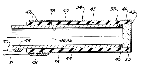
The present invention employs an ultrasonic flow sensing assembly 34 as shown
in
FI(i. 5 which includes an elongated electric conducting inner tubular member
35
charactex5zed by having a longitudinal axis 36, a distal end 37 and an inner
lumen 3g
extending therein.
The ultrasonic flow sensing assembly 34 also includes an elongated
electrically
conductaaag outer tubular member 40 which has a distal end 41, an inner lumen
43, and a
longitudinal saris 42 coincident with the longitudinal axis 36 of the inner
tubular member.
Elongate electrically conductive outer tubular member 40 is separated from
inner tubular
member 35 by the thickness of insulating means 44 which is preferably an
insulating tube
formed of a polyimide. The inner tubular member 35 can be formed of stainless
steel. The
outer tubular member 40 of this emb~iiment is a layer of conducive material,
such as gold,
on the exterior of the insulating golyimide tube 44.
Piezoelectric transducer means 23 is capable of generating and receiving
ultrasonic
waves, and is located at the distal ends 37 and 43 of inner and outer tubular
members 35 and
40, respectively, and is electrically connected to the tubular members as
shown. As a
preferred embodiment, piezoelectric means 23 can be connect~i to tubular
member 35 by an
electrical conducting silver epoxy 45.
The inner lumen 3g of the inner tubular member 35 is closed by the transducer
23
which is'secured to the distal end thereof, The closexi inner lumen 3g forms a
chambez°
behind the ~ansducer which is filled with air or other gas and which greatly
enhances the
sensitivity of the transducer 23.
Electrical conductors 30 and 31 are shown in FTG. 5 connected to inner and
outer
tubular members 35 and 40, respectively. Conductor 30 is joined to the inner
conductive
tubular member 35 by means of a solder joint 46, whereas conductor 31 is
Connects to
outer tube 40 via tungsten bands 47 and solder joint 4g. An electrical coating
49, i.e. gold,
is provide on the exterior of the transducer 23 to electrically connect the
outer tubular
member 40 with the transducer.
The entire assembly 34 shown in FIG. 5 can be placed within needle 21 as shown
schematir;ally in FIG. 4 for cannulation of blood vessels which can be
utilized for the
carrying out of a Seldinger t~hnique. After the needle 21 is inserted and
guide to a blood
vessel, as described in conjunction with the discussion of FIG. 3, the blood
vessel
~netradon is indicated by the back flow of blood through the needle past
assembly 34.
~nce this is accomplished, assembly 34 can be removed from the needle 21 and a
guidewire
can be placed through the needle into the blood vessel and the ne~le itself
then removed.
1~inally, prothesis can be guided into position in the blood vessel over the
guidewire~
Reference is made to FIG. 6 which illustrates another preferred embodiment of
an
ultrasonic flow sensing assembly 50 in accordance with the invention. The
assembly 50
includes an outer, electrically conductive tubular member 51 having a distal
end 52-and an
inner lumen 53 extending within the outer tubular member, and an inner,
electaically
conductive tubular member 54 having a distal end 55 and an inner lumen 56
extending within
the inner tubular member. A piezoelectric transducer 57 is adjacent proximate
and
el~trically connected to the distal ends 52 and 55 of the tubular members 51
and 54
resp~tively. An electrically insulating riabular member 5S is disposed between
the outer and
a
inner tubular members 51 and 54. An electrically conductive coating or layer
59 is providers
on the exterior of the piezoelectric transducer 57 which extends to and
electrically contacts
the distal end 52 of the outer tubular member 51. An electrically conductive
adhesive 60
bonds and electrically connebts the distal end of the inner tubular member 54
to the backside
of the piezoelectric transducer 57. Conductors 61 arid 62 are secured to the
proximal ends of
the inner and outer tubular members 51 and 54 respectively by suitable means
such as solder
63 and 64. 1fie proximal end 65 of the outer conductive tubular member 51 is
dispose a
short distance from the distal end of tlhe inner conductive tubular member 54
to provide
access to the inner tubular member in order to join the conductor 66 to the
innez tubular
member by means of solder 63.
The operation of the ultrasonic flow sensing assembly 50 of this embodiment is
essentially the same as the operation of the previously described assembly as
shown in FIGS.
2-5.
_7_
Typical dimensions of the components which form the assembly SO include an
outer tubular member 51 with an OD of about 0.038 inch (0.965 mm) and an TAD
of about
0.034 inch (0.864 mm). The insulating tube 58 has an OD of about 0.034 inch
(0.804 mm)
an Ip of about 0.03 inch (0.76 mm). The inner conducting tubular member 54 has
an OD of
S about 0.03 inch (0.76 mm) and an ID of about 0.20 inch (0.S1 mm). The
overall l~gth of
assembly SO is about 3.75 inches (9.53 cm) The inner and outer el~trically
condu
tubular members 51 and 54 may be made of stainless steel and the iruier
insulative tubular
member 58 may be formed of polyimide. Other conducting and insulating
materials may also
be employed. The transverse crossssectional shape of the ultrasonic flow
sensing assembly is
circular to readily fit within the inner lumen of a needle and is dimensioned
to leave a space
between the outer surface of the assembly and the inner surface of the needle
so that blood
may r~dily flow there between. ''JVhen the needle penetrates a blood vessel,
blood will flow
through the annular area between the assembly and the ne~le when a vacuum is
pulp by
the syringe (not shown) which is releasably soured to the proximal end of the
needle.
The ultrasonic transducer is preferably formed of a lead zirconium titanate
ceramic
material which is sold by the Vernitrun Company of Bedford, OH. It is sold by
the
designation 5H. Ceramic material SA is also suitable.
Various modifications and improvements may be made to the invention without
departing from the scope thereof.
_8_