Note: Descriptions are shown in the official language in which they were submitted.
2085939
`_
BLOOD SAMPLING DEVICE
BACKGROUND OF THE INVENTION
;. Field o~ the Invention
The present invention rela-tes to a simple blood
sampiing device.
2. Description of the Rela~ed Ar'~
In recent years, the numbers of persons
suffe~ir.g fro~. various diseases derived f~o~ rich
~ietary patterns and increased stress fGr example,
diabetes, have been soaring. Trips to -the hospital pose a
major incon-~enience to patients in their ~2ily acti~ y,
so as examinations of blood sugar etc. over t,he course of
th~e .regular day become paxt of the daily routine, thc
me~-hod of sampling blood has come under attention as a
large problem. The problem of the pain involved in the
blcod sal:lplil.lg becomes an even greater probl2m T~hen it
h2s to be repeated. In particular, this is bcccming ,~
fllrther serious problem in the case of insu'in-dep~ndent
patients, many of whom are small children. Fu--t~r, .~
~ece~t years, Qiseases transmitted through ~he blood `nave
become social issues. To prevent AIDS, hepatitus, and
other esp.?cially serious diseases, some sort of dev ce
wni ch enables patients themselves to s~mple their G~
2~ blood without problem is needed. However, no device has
yet been p~oposed which enables blood ~o be samrled
painlessly and simply.
SU~ARY OF THE INVE~ITION
Accordingly, the objects o the present inv2ntior
~0 ~le to eliminate t.l,e above-mentioned disadvantages in the
prior art and to provide
Other objects and advantages of the present
invention will be apparent from the following
description.
In accordance with the present invention, there is
provided a simple blood sampling device comprising a
208s939
-2-
vacuum chamber, a skin suction portion, and a piercing and cutting means.
The present invention provides skin-adhesive blood sampling device
S comprising-a sealed vacuum chamber in a state of preexisting reduced pressure;
a support member for the sealed vacuum chamber, the support member defining
a suction portion adjacent the sealed vacuum chamber, the suction portion, in
cooperation with the sealed vacuum chamber subjecting an area of skin of a
patient to a reduced pressure state when the device is actuated; means arranged
within the suction portion for slightly rupturing a portion of the area of skin of
the patient exposed to the reduced pressure state; and the support member havingadhesive means for securely fixing the device to a surface of the body of the
patient so as to m~int~in the reduced pressure state during suction and collection
of blood through the slightly ruptured portion of the area of skin of the patient.
In the aforesaid the support member further comprises a stopper material
arranged around an outer periphery of the suction portion so as to cause friction
with skin thereunder.
The device may have the rupturing means disposed along an inner
periphery of the suction portion so as to painlessly cut epidermis sliding thereto.
The aforesaid skin rupturing means may be a cutting means.
Also, the skin rupturing means may be a piercing means.
The present device is of simple construction and small in size and light in
weight and does not use special parts, so that the present device is of low costand may be used as a disposable device.
BRIEF DESCRIPTION OF THE DRAWrNGS
The present invention will be better understood from the description set
forth below with reference to the accompanying drawings; wherein:
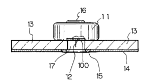
Fig. 1 is a side elevational, partly sectioned view showing an example of
a device according to the present invention;
B
2085939
-2a-
Figs. 2 and 3 are views showing the device of Fig. 1 when in use, for
explaining the operation of the device;
Fig. 4 is a view corresponding to Fig. 1 showing another embodiment of
the present device;
Fig. 5 is a bottom view of the Fig. 4 embodiment;
Fig. 6 is a side elevational view in diametrical section showing another
embodiment of the present invention;
Fig. 7 is a side elevational, diametrical section showing another
embodiment of the present invention;
Fig. 8 is a view showing the Fig. 7 embodiment in use, for explaining the
operation of the device; and
Fig. 9 is a perspective view showing the device of Fig. 7 applied to the
arm of a user.
DESCRIPTION OF THE PREFERRED EM13ODIMENTS
The characteristics of the present device will now be explained.
A vacuum suction action is applied to a local portion of the surface of the
skin of the subject (i.e., the living body). Along with this local vacuum suction,
the area inside the subject's skin becomes engorged with blood and therefore theepidermis expands and rises up. The raised portion of the skin comes in contact
with a piercing means provided at a predetermined position.
2085939
Since the raised portion of the skin is sufficiently taut
in state, the piercing means pierces the skin (epidermis)
easily. When the piercing means pierces the skin, the
blood engorged inside it flows out and is collected.
Since the skin is locally drawn up, even though the
piercing means pierces the skin, it does so
instantaneously and the sensation of this is cancelled
out by the stimulus caused by the suction action, so no
pain is felt. Further, the piercing and cutting means
pierce and cut in an engorged state, so the blood can be
collected painlessly and reliably.
The vacuum chamber shown in the present device is a
means for drawing up the surface of the subject's skin.
Ones which perform the vacuum action mechanically and
chemically may be mentioned. While not particularly
limited to them, ampules, cassette devices, etc., which
are formed in a vacuum state in advance by an air-tight
material may also be mentioned. Further, the piercing and
cutting means may be one or more solid needles, hollow
needles, needles with sawtooth sides, acupuncture type
needles, etc.
The length of the piercing and cutting means is
preferably about several 100 micrometers to several
millimeters, but is not particularly limited. Further,
regarding the disposition and construction of the same,
the means may be disposed at the center of the suction
portion or the periphery of the same. It is sufficient if
the means can make use of the stretching action of the
epidermis by the vacuum suction to painlessly and
effectively pierce or cut through the epidermis.
EXAMP~ES
The present invention will now be further
illustrated by, but is by no means limited to, the
following Examples.
Figure 1 is a view showing one example of the
present device.
Reference numeral (11) is a vacuum chamber, which
2~85939
houses a vacuum drive portion. At the top of the vacuum
chamber (11) is provided a switch (16). By pressing this
switch (16), the vacuum operation is performed. Reference
numeral (100) is a hole portion, one or more of which are
provided through the bottom of the vacuum chamber.
Reference numeral (12) is a piercing means, which is
formed by, for example, a fine needle and is provided at
the bottom of the vacuum chamber (11) near the hole
portions (100). Reference numeral (13) is a support
member, which is formed as a concentric cylindrical film
by plastic, rubber, paper, or another material having
flexibility. The vacuum chamber (11) is joined to the
support member (13) on the top of the same at the
periphery near the approximate center of the concentric
portion. Reference numeral (14) is an adhesive. Use is
made of a material which does not react with the body,
such as a material used for adhesive plaster. The
adhesive (14) is provided at the bottom of the support
member (13) at the outer periphery of the same. Examples
of such an adhesive are rubber-based adhesives, acryl-
based adhesives, silicone-based adhesives.
Reference numeral (15) is a stopper, which keeps
down the elongation and contraction of the support member
(13) at the time of application of vacuum and assists the
skin in being pulled up. The stopper (15) is provided at
the bottom of the periphery of the approximate center of
the concentric portion of the bottom of the support
member (13). The material for the stopper is desirably
rubber, plastic, etc. so as to cause large friction with
the skin. Reference numeral (17) is a suction portion,
which portion is formed to cover the inside surface of
the concentric portion of the support member (13) and the
bottom portion of the vacuum chamber, including the
piercing means (12) and the hole portions (100).
Next, the operation of the example shown in Eig. 1
mentioned above will be explained in detail with
reference to Fig. 2 and Fig. 3.
2085939
- 5 -
First, the above-mentioned example of the present
invention is placed, with the adhesive portion (14) down,
on a position of the subject's skin (MMA) suitable for
sampling blood. The adhesive portion (14) adheres to the
subject's skin (MMA), so the device is affixed to the
surface of the subject's skin and the suction portion
(17) is sealed. At this time, the piercing means does not
contact the subject's skin.
The switch (16) is depressed. The vacuum chamber
thereby begins the vacuum operation. By this vacuum
operation, the suction portion (17) enters a vacuum state
through the holes (100) and a suction action is applied
to the subject's skin under the suction portion (17).
By this suction action, the body fluid, including
the blood, inside the subject's skin (MMA) begins to
engorge, forming the engorged position (MMB).
Gradually, as shown in Fig. 3, the subject's skin
under the suction portion (17) begins to rise up and
contacts the piercing means (12). The subject's skin at
this portion, in the pulled up state, is locally taut, so
the piercing means (12) easily pierces the epidermis at
the subject's skin (MMA) and the vacuum reaches the
engorged portion (MMB). At this time, the stopper (15)
prevents the movement of the skin under the stopper (15)
by a suction action and assists the surface of the
subject's skin being raised up. The blood and other body
fluid travel along the piercing means (12) and are sucked
out to the surface of the subject's skin where they are
collected. In accordance with need, further, the blood
sucked out to the surface of the subject's skin is taken
into the inside of the vacuum chamber (11) through the
holes (100).
Finally, the device of this example is taken off the
subject's skin surface (MMA).
Further, Fig. 4 shows a cutting means (121) disposed
at the periphery of the suction portion. When the
epidermis is pulled up and stretched by the vacuum, the
2085939
- 6 -
surface of the epidermis contacts and slides against the
fine sawtooth edge (122) of the cutting means and is
painlessly cut. The cutting means is formed to have a
sawtooth edge construction over all or part of its edge.
Figure 5 is a view looking at Fig. 4 from the bottom.
Reference numeral (100) is a hole portion, which
communicates the vacuum chamber (11) with the suction
portion (17). The rest of the construction is the same as
in Fig. 1, so the same reference numerals are attached
and the explanations are omitted.
Next, another example will be shown in Fig. 6. In
Fig. 6, a vacuum drive portion (not shown) is provided
outside and the piercing means is made a hollow needle.
Reference numeral (11) is a vacuum chamber in the
same way as in Fig. 1 and has a cylindrical shape.
Reference numeral (41) is a valve, which moves up and
down in the cylindrical vacuum chamber. Reference numeral
(42) is a friction portion, which is formed at the bottom
of the cylindrically shaped inside of the vacuum chamber
(11). Reference numeral (43) is an opening, which serves
as an interface between the inside of the vacuum chamber
and the outside vacuum drive means. The interface of the
outside vacuum drive means is shown by reference
numeral (440).
Reference numeral (12) is a piercing means, which is
formed by a hollow needle. The hollow needle reaches into
the inside of the vacuum chamber. Reference numeral (100)
is a hole portion, which connects the inside of the
vacuum chamber and the suction portion (17). The rest of
the construction is the same as in the example of Fig. 1
and will therefore not be explained.
Next, the operation of the example shown in Fig. 6
will be explained.
The device according to this example is placed on
the surface of the body. The adhesive portion (14) is
joined to the surface of the body. The interface (440) of
the outside vacuum drive means is connected to the top of
2085~39
the vacuum chamber. The outside vacuum drive means is
driven. The valve (41) begins to move upward. Since the
valve (41) contacts the friction portion (42), it
gradually moves upward. When the valve begins to move
upward, the gas in the suction portion (17) moves upward
through the hole portion (100). At the same time, the
subject's skin under the suction portion (17) rises up
and engorges with blood.
Along with the valve (41) moving upward, the skin
under the suction portion (17) rises up. When it passes
the friction portion (42), the valve (42) moves up all at
once and the skin under the suction portion (17) rises up
to the m~;mum extent possible, contacts the piercing
member (12), and is pierced.
When pierced, the piercing means (12) reaches the
engorged portion under the skin. The blood is taken into
the vacuum chamber (11) through the piercing means (12).
After the blood is collected, the interface (440) is
removed. The valve (41) falls, but stops at the top of
the friction portion (42), preventing leakage of the
collected blood from the hole portion (100).
Next, a further example will be shown in Fig. 7 and
explained.
The device according to this example shown in Fig. 7
is provided with a vacuum drive portion inside and
further has a plurality of piercing means.
Reference numeral (51) is a holding piece which is
provided so that a sliding member A (52) and a sliding
member (B) are held at predetermined positions so as not
to separate.
The sliding member A (52) and the sliding member B
(53) slide left and right and are connected by a spring
(54). The portion over which the sliding member A (52)
and the sliding member B (53) face each other constitutes
a vacuum space (55).
Reference numeral (12) is a needle, a plurality of
which are provided at the bottom of the vacuum chamber
2085~39
- 8 -
(11). Reference numeral (100) is a hole portion, of which
a plurality are made and which connect the vacuum space
(55) and the suction portion (17). Reference numeral (56)
is a peeling member, which prevents the drying and
reduction of tackiness of the adhesive (14) and which is
peeled off at the time of use.
The rest of the construction is the same as in the
example shown in Fig. 1 and thus will not be explained.
Next, an explanation will be made of the operation
of the example shown in Fig. 7, including Fig. 8.
At the time of use, the peeling member (56) is
peeled off and the device is placed on the position of
the body for drawing the blood. The sliding member A (52)
and the sliding member B (53) are affixed at
predetermined positions by a holding piece (51). At this
time, a spring (54) maintains the compressed state.
Next, the holding piece (51) is removed, as shown in
Fig. 6. The sliding member A (52) and the sliding member
B (53) are pushed outward by the force of the release of
the spring (54) and the vacuum space (55) grows in
volume. At the time of adhesion, the suction portion (17)
and the vacuum space (55) were sealed by the subject's
skin, so when the vacuum space (55) grows, the skin under
the suction portion (17) is pulled up.
Blood engorges under the skin and the surface rises
up. The piercing means (12) contacts the skin, then
pierces through it. When the piercing means (12) reaches
the engorged portion, the blood comes out along the
surface of the piercing means (12) and is thus extracted.
The time waiting for blood to engorge after the skin
is drawn up and the time until the piercing means pierces
the skin also may be suitably selected. Further, it is
not that particularly necessary to wait for the blood to
engorge. So long as the skin is pierced by the piercing
means in a state after suction when there is tautness in
the skin due to its being drawn up, the present device
operates sufficiently.
2085939
Next, the state of one of the examples shown in Fig.
7 adhered to the upper arm of the body is shown in Fig.
9. Reference numeral tll) shows a vacuum chamber, and
(13) a support member. Since the device is small in size
and light in weight, it may be used adhered in the manner
shown in Fig. 9 as well. Also, the adhesive portion is
suitably used. It is also possible to use the device with
no adhesive portion, i.e., held by the hand.
As explained above in detail, the present device is
small in size, light in weight, and low in price and
therefore has the effects that it is suited as a
disposable implement and further enables blood to be
drawn reliably etc.
