Note: Descriptions are shown in the official language in which they were submitted.
WO 92/00327 PCr/US91t04482
2a~a~
K~ IACROPHAGE l~FLA~ TORY P.~OTEI~ 2rt
BACKGROUND OF THE IN~rENTION
Macrophage Inflammatory Proteins (MIPs) represent a class of
proteins that are produced by certain cells (for example, macrophages
or lymphocytes) in response to invasive stimuli such as gram negative
bacterial lipopolysaccharide. Thus, they may be important partici-
pants in the response of the cell (host organism) to such stimuli. As
such, these molecules may have therapeutic potential in the treat- -
ment of infections, cancer, myelopoietic dysfunction and auto-
immune diseases. Two distinct forms of MIP have been found in cul-
tures of macrophage tumor cells from the mouse: MIP-l and MIP-2.
Murine ~IIP-l
Murine (m)MIP-1 is a major secreted protein from
lipopolysaccharide (LPS) - stimulated RAW 264.7 cells, a murine
macrophage tumor cell line. It has been purified and found to consist
Ol' two related proteins, m~IIP-l~ and m~lIP-lB (~olpe, et al., 1987, J.
Exp. Med., 16~':570; Sherry, et al., 1988, J. EXD. Med., 168:2251).
The cDNA's for both mMlP-l~ and mMIP-13 have been cloned
and sequenced (Davatelis, et al., 1988, J. EXD. Med., 167':1939; Sherry,
et al., OD. cit.~ The cloning and sequencing of cD~;As corresponding
to mMlP-l~ and m~lIP-lB have been reported as well by Brown, et al.
(1989, J. Immunol., 142:679); Kwon and Weissman (1989, Proc. Natl.
Acad. Sci. USA, 86:1963) and by Brown, et al., OD. cit., respectively.
Both groups isolated homologs ol' m~IlP-1~ and/or mMIP-13 from
cDNA libraries prepared l'rom R~'A of murine helper T-cells that had
been activated by treatment with conconavalin A. These results sug-
gest that ~IIP-1t~ and MIP-lB may play a role in T-cell activation.
.... ... - : -: .
: - : . ..
..
.
,:
.
wo 92/003? PC~/US91/04482
2 ~ 0 9 1 - 2 -
Human ~lIP-l Homolo~s
Several groups ha-e cloned ma~ be the human homologs of
m~llP-1~ and m~llP-13. In all cases, cD~Aa were isolated from librar-
ies based on R!.'A from activated human T-cells. Thua O~a~u et al.,
(1986, J. Biochem., 99:885) and Zipfel, et al. (1989, J. Imm~lnol.,
142:1582) have bnth reported cloning of a cD.~A that predicta a pro-
tein with high homology to m~IlP~ 6-','o). Similarly, Bro~n, et al.,
oP. cit., Zipfel, et al., OD. cit., Lipes, et al. (1988, Proc. Natl. Acdd.
Sci. USA, _:9704~ and Miller, et al. (1989, J. Immunol., 143:290?) have
reported the cloning and sequencing of human cD.`I'As, which predict
a protein with high homology to mMIP-13 (~5%). ;~IIP-l~ and ~IIP-ls
belong to a newly described family of related proteins which have
immunomodulatory activities (see Sherry, et al., OD. cit. for a review).
Murine MIP-2
Murine MIP-2 (mMIP-2) is an inducible protein that has also
been purified from the conditioned medium of LPS-stimulated RA~
264.~ cells (~olpe, et al., 1989, Proc. Natl. Acad. Sci. USA, 86:3121).
The cDNA encoding murine MIP-2 has been cloned and sequenced.
mMlP-2 also is a member of a homologous multigene family.
Members of this family include gro/MGS.~, platelet factor 4, platelet
basic protein, the precursor of connective t~ssue activating peptide
(CTAP-III) and B-thromboglobulin, a gamma inter~eron inducible pro-
tein, IP-10, and interleukin 8 (IL-8), also known as 3-lOC, MD~CF,
NAP-l and NAF. Members of this family that have highest homology
in protein sequence (generally predicted from cloned cD~~A) include
MGSA and KC. MCSA (Richmond, et al., 1988, EMBO J., 7:202J) is an
autocrine growth factor with mitogenic activity secreted by human
melanoma cells and is the product of the human ~ gene (Anisowicz,
et al., 1987, Proc. Natl. Acad. Sci. USA, 84:7188). MGSA has 57.9%
identity in amino acid sequence to murine MIP-2; the predicted pro-
tein sequence of the putative hamster homolog of MGSA (ibid.) has
68.3% identity to mMIP-2. The murine KC gene product (Oquendo, et
al., 1989, J. Biol. Chem., 264:4233) is induced by platelet-derived
growth factor ~PDGF) and ia thought to be the murine homolog of the
human MGSA/gro gene (63.0~b amino acid identity to m~llP-2).
-
- ~
- ~
Wo 92/00327 P~r/US9l/04482
203~091
_combinant E:cpre~ion of :~IP
There is no prior art on the expre;~,ion of recombinant MIP-2,
or human ~IIP-2~ and ;~/lIP-2a although members of the MIP-2 gene
family as well as members of the related MIP-l gene family hd~,e been
expre~ed. The pertinence of these results to the expression of
murine or human MlP-2 is question3ble, given the idio~n^ratic nature
of expre~lon in heterologous s~tem,. The literature ~ill be summa-
rized nonetheless.
m~llP-l~ and mMlp-lB have been independently exp~essed in
COS cells (Graham, et al., 1990, Nature, 344:442). LD78 cD~A
(Obaru, et al., ou. cit.) which encodes a protein that is likely to be the
human homolog of murine MIP-l~ has been expressed in E. coli as a
carboxyl terminal fusion to human interleukin-2, as well as in COS
cells (Yamamura, et al., 1989, J. Clin. Invest., 84:1707). Human I-309,
a cD~A that encodes a protein with homology to members of the
MIP-1 family of proteins, has been expressed in COS-l cells in order
to confirm that it encodes a secreted protein (Miller, et al., OD. cit.).
Lipes, et al., (oP. cit.) described baculovirus expression of Act-2
cDNA, the human homolog of murine MIP-13, to show that the protein
encoded was secreted and to identify the mature N-terminal
sequence. JE, a murine cDYA that encodes a protein with homology
to MIP-1~ and MIP-lB, has been expressed in COS-l cells to confirm
that it encodes a polypeptide core of about 12 kDa (Rollins, et al.,
1988, Proc. Natl. Acad. Sci. USA, 85:3738).
KC, a murine cDNA that encodes a protein with homology to
mMIP-2, has been expressed in COS-1 cells to show that it encode~ a
secreted protein (Oquendo, et al., OD. cit.) Connective tissue activat-
ing peptide-lII (CTAP, Mullenbach, et al., 1986, J. Biol Chem.,
261:719) and IP-10, (Luster and Ravetch (1987, J. ExP. Med., 166:1084)
both members of the MIP-2 gene family, have been expressed in yeast
as an ~-factor fusion and in E. coli, respectively. Maione, et al. (1990,
Science, 247:77) expressed human platelet factor 4, (MIP-2 family) in
E. coli as a peptide fused to a 35 amino acid peptide fragmen~ of E.
coli B-glucuronidaSe. The insoluble fusion protein must be cleaved
with cyanogen bromide in order to generate bioactive material.
. - . . ~ - -- . ,, ., , . - . .
. . ;. : -
: . : ,
.- . :~
Wo 92/00327 PCr/US91/04482
9 ~ - 4 -
Lindley, et al., (1988, Proc. N~atl. ~cad._Sci. IiS~, 85:9199) ha~!e
e.Ypressed NAF (IL-8), a member of the ~IIP-2 famil~-, in E. coli. Af ~er
purification and renaturation, this recombinant p~otein was found tO
ha-e the same bioactivity as that identified for the native molec~le.
Furut~, et al. (1989, J. Biochem., 106:~.3~) ha~,e a!so exp~essed IL-8
(;~IDNCF) in E. coli. Finally, Gimbrone, et al. (1989 Science, 2~5:1~?L)
ha~ e expressed end~.helial IL-8 in human 2~3 cells and sho~n tha~ tr.e
recombinant and natural material have the same bioactivity.
B activitv of MIP
Studies on the bioactivities of ;\,IIP-l and -2 are in progress and
have utilized native murine MIP-1 or ~IIP-2 and very recently recom-
binant murine (rm) MIP-1~, rm~lIP-1a and rm~lIP-2. Among the
bioactivities defined for both recombinant and native mMlP-l and
m~lIP-2 is colony-stimulating-factor promoting activity as well as
roles in inflammation.
Murine MIP-2 has been shown to elicit a localized inflammatory
response when injected subcutaneously into the footpads of C3H/HeJ
mice, to have potent chemotactic activity for human
polymorphonuclear leukocytes (PM~), and to induce P~t~i
degranulation of Iysozyme but not of 3-glucuronidase (~olpe, et al.,
1989). In addition, m~lIP-2 has been shown to ha~e myelopoietic
enhancing activities for CFU-G~I (Broxmeyer, et al., 1989, J. E~cp
Med., 1~0:1583). Given the various biological activities o~ these fac-
tors, it is desirable to isolate their human homologs. Due to the
necessity for quantities of purified factors to pursue definition of
bioactivities and the expense of isolating these factors from cell cul-
ture fluids it is desirable to produce these materials by employing
recombinant DNA technology.
SUhlMARY OF THE INVENTION
An attemp~ to identify the human counterpart o~ m~lIP-2
unexpectedly resulted in not one, but two candidate sequences. These
two sequences were designated hu-~lP-2~ and hu-MlP-2B. The more
abundant of the two homologs is hu-MlP-2~.
, , ~ , ,, ~,,
.- : - - - .
.
;
~ ~ ,
. .
wo 9~/00327 PCr/US9~ 82
2 ~
It is an object of this in~ent.on to provide a human MIP-2~
(hu-~llP-2~) polypeptide su~stantially free from other human proteins
and to pro~ide a nuc!eic acid coding sequence for hu-:~llP-2~.
It is ~et another object of this invention to provlde antibodies
reacti~e ~ith hu-MIP-2~.
It is still another object of this invention to provi~e diagnostic
methods for detection of hu-~llP-2~ or of nucleic acid coding
sequences for hu-~5lP-2~.
In accordance with these and other objects, which will become
apparent from the description herein, one aspect of this invention
contemplates a protein composition comprising hu-MlP-2~ -
polypeptides wherein the composition is substantially free of human
tissue and preEerably is substantially free of other human proteins.
In another aspect, the invention contemplates a DNA molecule
comprising a heterologous region that encodes hu-MIP-2 polypeptide.
In yet another aspect, the invention contemplates a recombinant pro-
duction system comprising cells transformed with a D~A molecule
comprising a heterologous region that encodes hu-;~lIP-2~ polypeptide.
The invention also contemplates antibodies which are reactive
with hu-MIP-2~ polypeptides. Still further aspects of the invention
relate to immunoassay methods for determination of hu-MIP-2~ in
tissues or fluids and nucleic acid probe methods for detecting
polynucleotide sequences coding for hu-MIP-2 in biological samples.
Another aspect of the invention contemplates polypeptides and
small organic molecules that interact with the hu-MIP-2~ receptor(s)
and p~ssess agonistic or antagonistic activities.
Yet another aspect of the invention contemplates hu-MIP-2c~
polypeptides that exhibit agonist and antagonist activities to any of
the members of the homologous multigene family.
Further, the invention contemplates a method of inducing
wound healing, a method of modulating myelopoeisis, and a method of
inducing adjuvant activity, by administering an effective amount of
hu-MlP-2 polypeptides.
The invention also contemplates a me~hod of treating malig-
nancy, autolmmune and inflammatory diseases, such as, pathological
-,
.
.
. . . ' , ' .; :, ,
;. . , ' ~ : ',
.
WO 92/00327 PCr/US91/04482
2~ ~9~. - 6 -
intiltration of neutrophils including psoriasis, rheumatold arthriti~,
and diseases of the lungs. b~ administering an effecti~le amount of
hu-~llP-2~ antagonist or hu-~lIP-2~ polypeptides with antagonist..
activity to the members the homologous multigene family.
By using the D.~.~ sequences and the recombinant s~stem pro-
vided by this invention, it is possible to produ~e large quantities of
one of the two human ho~a~~,logs of mMIP-2 subjtdntially free of othe.
human protein, because only one human protein is e~pressed in the
recombinant system. Compositions such as this, containing only one
MIP-2 homolog, would be difficult to obtain by purifi~ation from natu-
ral sources.
BRIEF DESCRIPTION OF THE DRAWINGS
Figure 1. Nucleotide sequence and predicted protein
sequence of murine MIP-2.
Figure 2. Nucleotide sequence and predicted protein
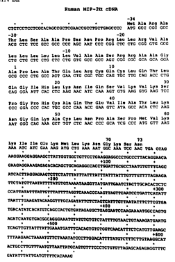
sequence of hu-~qIP-2c~.
Figure 3. Nucleotide sequence and predicted protein
sequence of hu-~IlP-2B.
Figure 4. Nucleotide sequence homology of MIP-2
homologs. Percentages of nucleotide sequence identity between
mMIP-2, hu-MIP-2~, hu-MIP-2a, hu-gro/MGSA and murine KC in the
cDNA (A), coding region ~B) and 3~ untranslated region (C).
Figure 5. Amino acid homology and alignment of MIP-2
homologs and human IL-8. (A) Percentages of identity bet~een the
predicted amino acid sequences of MIP-2 homologs as well as human
IL-8. (B) The aligned amino acid sequences.
Figure 6. Southern analysis of genomic DNA with murine
and human MIP-2 cDNA digested wi~h BamHl, ~B~; EcoRl, 'E~ or
E~oRV, 'R~. (A) Murine DNA hybridized with mMIP-2 cDNA. A blot of
restricted human DNA was hybridized first with labelled hu-~IP-23
cDNA (C) then the filter was stripped and rehybridized with labelled
hu-MIP-2~ cDNA (B).
~ - - . . . . . . . .
, ,, . . . , -
wo 92~0()32- PcrtUS91/04482
~g~
DETAILED DESCRIPTION
The practice o~ the prejent invention emplo~s, unle~ oth-
er~ ise indicated, conventional molecular biolog~, microbiol--g!, and
recombinant DNA techniques within the skill of the art. Such tech-
niques are exp'ained fully in the lite~ ature. See, e.g., ~lal~iatis,
Fritsch ~ Samb~oo~ Iolecular Cloning: A Laborator~ :~13nual~
(1982); "D~A Cloning: A Practical Approach,~ Volumea I and II (D.N.
Glover, ed., 1985); ~Oligonucleotide 5~nthesis" (M.J. Gait, ed., 198~);
"Nucleic Acid H~bridization" (B.D. Hames ~; S.J. Higgins, eds., 1985);
"Transcription and Translation" (B.D. Hames ~c S.J. Higgins, eds.,
1981); ~Animal Cell Culture~ (R.I. Freshney, ed., 1986); ~Immobilized
Cells and Enzymes" (IRL Press, 1986); B. Perbal, "A Practical Guide to
Molecular Cloning" (198~).
Definitions
In describing the present invention, the following terminology
is used in accordance with the definitions set out below.
A ~replicon" is any genetic element (e.g., plasmid, chromo-
some, virus) that functions as an autonomous unit of DNA replication
in vivo; i.e., capable of replication under its own control.
A ~vector ' is a replicon, such as plasmid, phage or cosmid, to
which another DN.~ segment may be attached so as to bring about the
replication of the attached segment.
A ~double-stranded DNA molecule ' refers to the polymeric
form of deoxyribonucleotides (adenine, guanine, th~mine, or cytosine)
in its normal, double-stranded helix. This term refers only to the pri-
mary and secondary structure of the molecule, and does not limit it to
any particular tertiary forms. Thus, this term includes double--
stranded DNA found, inter alia, in linear D.~.~ molecules (e.g., restric-
tion fragments), viruses, plasmids, and chromosomes. In discussing
the structure of particular double-stranded D:`,A molecules, sequences
may be described herein according to the normal convention of giving
only the sequence in the 5I to 3~ direction along the nontranscribed
stand of D~A (i.e., the strand having a sequence homologous to the
mR~'A).
.
.
wo 92/0032, Pcr/us91/04482
2i~6~9~ - 8-
A D~ lcoding sequence is a D~.~ sequence which is tran-
scribed and translated into a pol~peptide in vb,o when placed under
the control of appropriate regulatory sequences. The boundaries of
the coding sequence are determined by a start codon at the 51 (amino)
termin la and a translation stop codon at the 31 (carbo.Y~) terminus. A
codlr.g sequence can incl~lde, but is not limited to, procaryotic
sequenccca, cDN,A from eucdr~otic mR.~, genomic D~;A sequences
from eucaryotic (e.g., mammalian) DNA, and even synthetic D~.~
sequences. A polyadenylation signal and transcription termination
sequence will usually be located 3' to the coding sequence.
A "promoter sequence" is a D~A regulatory region capable of
binding R~A polymerase in a cell and initiating transcription of a
downstream (3' direction) coding sequence. For purpases of defining
the present invention, the promoter sequence is bounded at its 3~ ter-
minus by the translation start codon of a coding sequence and extends
upstream ~5' direction) to include the minimum number of bases or
elements necessary to initiate transcription at levels detectable above
background. ~ithin the promoter sequence will be found a transcrip-
tion initiation site (conveniently defined by mapping with nuclease
S1), as well as protein binding domains (consensus sequences) responsi-
ble for the binding of R~A polymerase. Eucaryotic promoters will
often, but not always, contain "TATA~ boxes and ~CAT~ boxes.
Procaryotic promoters contain Shine-Delgarno sequences in a~dition
to the -10 and -35 consensus sequences.
A coding sequence is ~under the control~ of the promoter
sequence in a cell when RNA polymerase which binds the promoter
sequence transcribes the coding sequence into mRNA which ia then in
turn translated into the protein encoded by the coding sequence.
A cell has been "transformed~ by exogenous D~A when such
exogenous DNA has been introduced inside the cell wall. Exogenous
D~A may or may not be integrated (covalently linked) to chromo-
somal DNA making up the genome of the cell. In procaryotea and
yeast, for example, the exogeno~ls DNA may be maintained on an
episomal element such as a plasmid. With respect to eucaryotic cells,
a stably transformed cell is one in which the e~ogenous D~.~ has
'
~: , ~ - . . , -
. , . -
.
Wo 92/~)0327 Pcr/US9l/04482
~ g
2~6~1
become integrated into a chromosome so that it is inherited by daugh-
ter cells through ch~omosome replication. This stability is demon-
strated by the ability of the eucaryot~c cell to establish cell lines or
clones comprise~ of a population of dau~hter cells conta~nir.g tne
exogenous D~A. A ~c;one is a population of cells derived fro,l a
single cell or com;~.on ancestor by mitosis. A l~cell line!~ is a clor.e o~ a
primary cell that is capable of stable gro~ th in vitro for m3~.
genera tions .
Two DNA sequences are "substantially homologous~' when at
least about 85~ (pre~erably at least about 90%, and most preferably at
least about 9s%) of the nucleotides match over the defined length Oe
the DN.~ sequences. Sequences that are substantially homologous can
be identified in a Southern hybridization experiment under, for exam-
ple, stringent conditions as defined for that particular system. Defin-
ing appropriate hybridization conditions is within the Skill of the art.
See e.g., Maniatis et al., suDra; D~A Cloning, vols. 1 and Il suPra;
Nucleic Acid Hybridization, suPra.
A ~heterologous~ region or domain of a D!;A construct is an
identi~iable segment of D~A within a larger D~A molecule that is not
found in association ~ith the larger molecule in nature. Thus, when
the heterologous region encodes a mammalian gene, the gene will
usually be flanked by DNA that does not flank the mammalian
genomic DNA in the genome of the source organism. Another exam-
ple of a heterologous region is a construct where the coding sequence
itself is not found in nature (e.g., a cDNA where the genomic coding
sequence contains introns, or synthetic sequences hating codons dif-
ferent than the native gene). Allelic variations or naturally occurring
mutational events do not give rise to a heterologous region of D~'A as
defined herein.
The terms "analyte polynucleotide~' and "analyte strand~ refer
to a single- or double-stranded nucleic acid molecule which is sus-
pected of containing a target sequence, and which may be present in
a biological sample.
As used herein, the term ~probe~ re~ers to a structure com-
prised of a pol~nucleotide which forms a hybrid structure with a
Wo 92/00327 P~r/US91/04482
- 10 -
2~091
target sequence, due to complementarit~ of at least one sequellce in
the probe with a sequence in the target region. The polynucleotide
regions o~ probes may be compojed of D~A, and/or R.~.~, and/or syn-
thetic nucleotide analogs.
As used herein, the term ~target region~ refer, to a reg,on of
the nucleic acid which is to be amplified and/or detected. The term
~target sequence~l refers to a sequence with which a probe or primer
will form a stable hybrid under desired conditions.
The term "targeting pol~nucleotide sequence~ as used herein,
refers to a polynucleotide sequence which is corrprised of nucleotides
which are complementary to a target nucleotide sequence; the
sequence is of sufficient length and complementarity with the target
sequence to form a duplex which has su~ficient stability for the pur-
pose intended.
The term ~binding partner" as used herein refers to a molecule
capable of binding a ligand molecule with high specificity, as for
example an antigen and an antibody specific therefor. In general, the
specific binding partners must bind with sufficient affinity to immobi-
lize the analyte (or the copy/complementary strand duplex, in the
case of capture probes) under the isolation conditions. Specific bind-
ing partners are known in the art, and include, for example, biotin
and avidin or streptavidin, IgG and protein A, the numerous kno~n
receptor-ligand couples, and complementary polynucleotide strands.
In the case of complementary polynucleotide binding partners, the
partners are normally at least about 15 bases in length, and may be
least 40 bases in length; in addition, they generally have a content of
Gs and Cs of at least about 40% and as much as about 60%. The
polynucleotides may be composed of D~A, R~.~, or synthetic
nucleotide analogs.
The term "coupled~ as used herein refers to attachment by
covalent bonds or by strong non-covalent interactions (e.g., hydropho-
bic interactions, hydrogen bonds, etc.). Covalent bonds may be, for
example, ester, ether, phosphoester, amide, peptide, imide,
carbon-sulfur bonds, carbon-phosphorus bonds, and the like.
.
/
, .. , . . . ............. - . .- : .
,, : ,, .~,- .. . , . . -. -: , .-
. . . ~ - -
.
.
.
. .~ . . . .
~ 0 ~2/0032 Pcr/US91/0448~
2~6~91
The term ~support~' refers to any solid or semisolid surface to
which a desired binding pdrtner ma~ be an~hored. Suitable support.
inclu~e glass, plastic, metal, polymer geLs, and the like, and may ta~;e
the form of beads, ~ells, d~psticks, membranes, ard the like.
The term 1llabell' a-. used herein re~ers to an~ atom or mol~.~
which can be used to provide a detectable (preferably quantifiabie)
sic;,nal, and which can be attached to polynuc'eotide or polypeptide
As used her ln, the term 1~label probe ~ refers tO a
pol~ nucleotide which is comprised of targeting polynucleotide
sequence that is complementary to a target sequence to be detected
in the analyte polynucleotide. This complementary region is of suffi-
cient length and complementarity to the target sequence to afford a
duplex comprised of the "label probe~1 and the "target sequence" to be
detected by the label. It is coupled to a label either directly, or indi-
rectly via a set of ligand molecules with high specificity for each
other. Sets of ligand molecules with high specificity are described
supra (see "binding partner ,").
As used herein, a "biological sample!~ reEers to a sample of tis-
sue or fluid isolated from a individual, including but not limited tO, for
example, plasma, serum, spinal fluid, lymph fluid, the external sec-
tions of the skin, respiratory, intestinal, and genitourinary tracts,
tears, saliva, milk, blood cells, tumors, organs, and also samples Of L
vivo cell culture constituents ~including but not limited to conditioned
medium resulting from the growth of cells in cell culture medium,
putatively virally infected cells, recombinant cells, and cell
components).
"Human tissue" is an aggregate of human cells which may con-
stitute a solid mass. This term also encompasses a suspension oE
human cells, such as blood cells, or a human cell line.
A composition comprising a selected component A is ~substan-
tially free" oE another component B when component A makes up at
least about 75% by weight of the cornbined weight of components A
and B. Preferably, selected component A comprises at least about
909~ by weight of the combined weight, most preEerably at least about
99~ by weight of the combined weight. In rhe case ol a composition
:: . . - ~ .
~: ~ .'- . ' .
- ~ :
.,
WO 92/0032, Pcr/uS9t/04482
- 12 -
3 ~ .
comprising a selected biologically acti~e protein. which is substan-
tially free oî contaminating prGteinS, it is some.imes preferred tha~
the compoaition having the activity of the protein of interest contain
species with only a single molecula. weight (i.e., a "homogeneous"
composi tion) .
Two amino acid sequences are lls~tantially homoloOous~ when
at least about 90'k of the amino ac -'s match over the define~ leng~h
of the amino acid sequences, preferabt~ a match of at least about
92%, more preferably a match of at least about 95%.
Human Homolo~s of Murine MIP-2
The cDNA for murine MIP-2 has been cloned using a degenerate
oligonucleotide probe pool corresponding tO a portion of the
N-terminal amino acid sequence determined on the purified protein.
When nucleotide probes representing the coding region of murine
MIP-2 cD~.'A were used to probe a cD~'A library from human
macrophage cell line stimulated w.ith LPS (see Example 2) two differ-
ent candidate cD~lA sequences were identified. The proteins encoded
by these two cDNA sequences have been designated hu-;~lP-2~ and
hu-~IP-2B, respectively. Examples of the nucleic acid and amino acid
sequences of these three proteins are shown in the Figures: Figure 1
= murine ~IIP-2, Figure 2 = human MIP-2~, Figure 3 = human ~IIP-23.
IlHuman macrophage inflammatory protein 2~ polypeptides~'
(hu-~IIP-2~ polypeptides) encompass hu-:~,IIP-2~ and hu-~llP-2
analogs.
~ Hu-MIP-2~ is human macrophage inflammatory protein 2Q, a
naturally occurring, mature human prote;n secreted, inter alia, by
LPS-stimulated macrophages, and further encompasses all precursors
and allelic variations of hu-~IlP-2~, and as well as including forms of
heterogeneous molecular weight that may result from inconsistent
processing i vivo. An example of the hu-r~llp-2~ sequence is shown
in Figure 2.
"Hu-.~IIP-2~ analogs" are a class of peptides which includes:
Hu-MIP-2~ muteins,~ which are polypeptides substan-
tially homolgous to hu-~lIP-2~. Preferably the amino
acid sequence of the ~mutein~ differs from that of
.
.
.
,
'' ' ', ' '
, . .
Wo 92/00327 PCr/US91/04482
- 13~3~31
hu-~IP-2Q by 8 or fewer amino acid residues, more
pre~erabl~, 7 or fewer residues, even more preferabl~
about 5 or fewer residues and most preferably about 2
or fewer re~.dues. I~ is sometimea pre~erred that an~
differences in the amino acid sequences of the two
proteins involve only conservative amino acid substi-
tutions. Conservative amino acid sub~titutions occur
when an amino acid has substantially the same charge
as the amino acid for which it is substituted and the
substitution has no significant effect on the local
conformation of the protein. Alternatively, changes
such as the elimination of cysteine which alter the
activity or stability of the protein may be preferred.
2) I~Truncated hu-MIP-2~ peptides,~ which include frag-
ments of either "hu-MIP-2~ or "hu-MIP-2Q muteins"
that preferably retain either (i) an amino acid
sequence unique to hu-MIP-2Q, (ii) an epitope unique
to hu-MIP-2~ or (iii) MIP-2 activity,
3) "Hu-:~IIP-2Q fusion proteins,~ which include
heterologous polypeptides, are made up of one of the
above polypeptides (hu-MIP-2u, hu-~IIP-2Q muteins or
truncated hu-MIP-2~ peptides) fused to any
heterologous amino acid sequence. Preferably such
heterologous sequences are fused to the ~,'-terminal
end of the hu-MIP sequence and comprise a leader
sequence to direct secretion.
"Unique" hu-MIP-2Q sequencej, either amino acid sequences or
nucleic acid sequences which encode them, are sequences which are
identical to a sequence of a hu-MIP-2~ polypeptide, but which differ
in at least one amino acid or nucleotide residue from the sequences of
hMGSA, hu-MlP-2B, mMIP-2 and murine KC, and preferably, are not
found elsewhere in the human genome. Similarly, an epitope is
"unique" to hu-MIP-2Q polypeptides if it is found on hu-MIP-2Q
` polypeptides but not found on any members of the homologous gene
family.
- ~ ' ' - . ` ~' ' ., '
: .
.- . ~
Wo 92/0032, PCr~US91/04482
2~609~ - 14-
Features of the Predicted .~mino Acid Sequence
The open reading frames of m?il[P-2, hu-.~llP-2~ ar,d hu-~llP-23
encode pol~peptides of 100, 107 and 107 amino acids, respecti\,ely.
The initial approximately 3û amino acids of each of the three
pol~peptides ha~e characteristic featurea o~ a signal se~lence
(Perlman, et al., 19~.3, J. Mol. Biol., 167:391; von Heijne, 198~, J. ~lo~.
Biob, 173:243; von Heijne, 1986, ~'ucle c Acids Res., 14:~6~3!. The
N-terminal amino acid sequence determined for secreted m~llP-2
purified from the conditioned medium of LPS-stimulated RA~ 261.7
cells (Wolpe, et al., 1989) determined the start of the mature m~lP-2
protein in the predicted amino acid sequence. The start oi the pre-
dicted mature peptide sequence for hu-MlP-2~ and hu-:~lIP-2s aligns
with that of m~llP-2 and has a consensus signal peptide clea~age site
~Perlman, et al. 1983; von HeijnP, 1984, 1986). The predicted length
of the mature peptide sequence for all three proteins is 73 amino
acids. Murine ~IIP-2 is a basic protein, and the human ~IP-2
polypeptides are basic as well, based on predicted isoelectric points of
9.9 and 9.~ for hu-~llP-2~ and hu-~lIP-2B, respectively. The native or
recombinant proteins may be O-glycosylated. None of the three pre-
dicted polypeptides has a consensus signal for h'-linked glycosylation.
Features of Murine and Human MIP-2 cD~As
The nucleotide sequences in cD~lA's for m~lIP-2, hu-~llP-2~ and
hu-~IIP-2B each encode a single open reading frame. The nucleotide
sequence environment of the initiating ATG codon oE m~lIP-2 con-
forms to the consensus sequence shared by many mR~As of higher
eucaryotes (Kozark, 1986, Cell, 4~:283; Kozark, 1987, Nucleic Acid
Res., 15:812j); those oE human MIP-2~ and MIP-2B lack the highly
conserved purine at position -3 but possess many features of the con-
sensus sequence including C residues at positions -1, -2, and -4 and a
G residue at position +4.
The 3~ untranslated region of m~llP-2 includes the eucaryotic
consensus polyadenylation signal AATAAA (Birnstiel, et al., L98j,
Cell, 41:349) at position +719-724 followed by a poly-A string begin-
ning at nucleotide +735. ~o AATAAA ~olyadenylation signal was
found in the 3' untranslated region o~ clones hu-~llP-2-~a o~
.: , . - ,
.
- ~ - :.
. . .
Wo 92/00327 Pcr/US91/04482
- 15 -
2 ~ 3 ~ 1
hu-;lrlIP-2-?d of hu-hl~P-23 cD~A or in clones of hu-~lIP-2~ cD~A.
This is most li~ely due to the fact that these clones have a truncated
31 untranslated reion, since no poly-A string was present.
Tue consens-;s sequence TTATTTAT found in the 3
untranilated region of many c~ tokine genes (Caput, et al., 1986, Proc.
Natl. Acad Sci. US.~, 83:167û) and implicated in mR.~A stability
(Shaw, et al., 1986, Cell, 46:6~9) and efficienc~ of tran~lation (Kruys,
et al., 1987, Proc. Natl. Acad. Sci. US.~, 84:6030; Han, et al., 1990, J.
Ex~. Med., 171:46i) is present in multiple copies in all three cD~,As.
This sequence is present at four positions, two overlapping, in the 3'
untranslated region of mMIP-2 (positions +122, 1126, +142, +146); and
is present two times (positions l 158 and ~471) and five times, two
overlapping, (positions ~148, +1527 +1~6, +160 and +492) in the 3'
untranslated regions of human MIP-2~ and MIP-2~, respectively.
Homolo~s of Murine and Human MIP-2
The percentage of nucleotide sequence identity among the
three hII~-2 cDNA's, one murine and two human, as well as that of the
human gro/~GSA cDNA (Anisowicz, et al., 198~; Richmond, et al.,
1988) and the murine KC cDNA (Oquendo, et al., 1989; Cochran, et
al., 1983, Cell, 33:939) is displayed in Figure 4A. The human
grot~GSA cDNA encodes a protein with melanoma growth stimulat-
ing activity (Richmond, et al., 1986, J. Cell. Physiol., 129:375); murine
KC is a platelet-derived growth factor (PDGF)-inducible gene pre-
sumed to be the murine homolog of human MCSA. Noteworthy is the
high degree of nucleotide hornology among the three human cDNAs,
particularly between hhIGSA and hu-~llP-2~. There is an even rnore
striking degree of nucleotide sequence identity among the three
human homologs in the coding region as shown in Figure 4B. The
nucleotide sequence identity in the 31 untranslated regions of the
human ~IIP-2 homologs is considerably less than that observed in the
coding regions, with the exception of the respective regions of
hu-hlIP-2~ and h~lGSA. These two cD~.~s show a high percentage of
sequence homology throughout the 3' untranslated region as well.
Homology comparisons and alignments of the predicted amino
acid sequences of the precursor proteins of hlIP-2 homologs including
.. . . .. . . . .
. ~; , .. . . .
, ~ -- : .
,; . :
,
WO 92/00327 PCr/US91/04482
09~ - 16-
hu-~51P-2Q, hu-~tlP-23, human gro/;~5GS.~, the hamster homolog of
gro/MCS~ (ha-gro), m~llP-2 and mKC are presented in Figure 5(.~,B).
The three human proteins are highly homologous (8~-90%) but ami:lo
acld differences occur throughout the predicted sequences, pa.tic~-
la~ly at the carbox~ termini of the mature protein sequences. Ra;~d
on pr~dicted amino a~id homologies alone, it is not po~sible to a~
hu-~llP-2, hu-.~lIP-23 or human gro/MGSA a, the human homoioO ~
m~llP-2 or murine KC.
The percentage of nucleotide sequence identity is greates~
between hu-MIP-2~ and h~lGSA and it extends throughout the entire
cDNA. The presence o~ both hu-~lIP-2~ and hu-~51P-23 in a ~,937
cDNA library, prepared using poly-A+ R~A îrom cells stimulated with
both LPS and phorbol myristyl acetate (P.~IA), prompted us to screen
for hMGSA as well. Screening of 5x10⁵ plaques from the library after
amplification with oligonucleotides specific for h~lGSA (and not
hu-~51P-2~/3) gave no positive signals; in contrast 56 hu-MlP-2~ posi-
tive signals were detected. This suggests that gro/~5GSA transcrip-
tion is not induced in U937 cells stimulated by P.~5A and LPS, in con-
trast to transcription of the hu-~tlP-2~ gene.
Activities of MIP-2
Murine MIP-2 purified from the conditioned medium of LPS-
stimulated RAW 261.7 cells has diverse activities including CSF-
dependent myelopoietic enhancing activity for CFU-G.~5 (Broxme~er,
et al., 1989), elicitation of a localized inflammatory response follo~-
ing subcutaneous administration and a potent chemotactic activity for
human PMN (Wolpe, et al., 1989). The la~ter activity is characteristic
of human IL-8 (hu-IL-8). Based on this functional equivalence alone it
was suggested that mMlP-2 could be the murine homolog of hu-lL-8.
However, given that the amino acid homology of m~51P-2 to hu-IL-8 is
low, relative to m~51P-2 homology to hu-~51P-2~, hu- ~51P-2~ or
hu-~IGSA (Figure 5)? m~llP-2 and hu-IL-8 do not appear to be murine/
human homologs. Redundancy of function among cytokines is not
uncommon; cachectin/TNF-a and interleukin 1 have an overlapping
activity profile (~lanogue, et al., 1988, in 'ICellular and ;~lolecular
Aspects of Inflammation,l' Poste, et al., eds., Plenum, hY, p. 123).
.. ..... .. .. ... . . . . ..... .
: . .
.
' ' . .
Wo 92/0032- - 1 7 - Pcr/US9l/04482
2~6~1
,~IIP-2 and IL-8 may be another example o~ this functional
redundancy .
Based on its cD~A sequence, hu-~llP-2~ can be cla~ified as a
mer~be~ of the homologous multigene family ~hich includes murine
MIP-2. The members of this gene family pOaSeaS in vitro biological
acti~, ities incLuding neut~ophil activation, ne~.ltrophil chemota~'s,
~IGS.~-like mitogenlc acth,ity, fibroblast mitogcnic activity, CS''
co-factor activity, monocyte chemotaxis, angiogenic activity, and
inflammatory activity. Hu-MIP-2~ polypeptides may be selected
which possessa one or more of the biological activities exhibited by
any member of the multigene family. Hu-MIP-2~ polypeptides may be
utilized therapeutically for stimulation of myelopoiesis, as an adjuvant
in vaccine formulations, and for wound healing.
Hu-~lIP-2c- biological activity can be measured by asays
described in ~'olpe, et al. (1989) or Broxmeyer, et al., (1989) (each
incorporated herein by reference). Other assays for hu-~lIP-2~ activ-
ities such as neutrophil activation or chemotaxis can be measured in
vitro as described in ~'altz et al., (1989), J. EXP. Med., 1?0:1745,
Clark-Lewis et al., (1991), Biochem., 30:3128, and Dernyck et al.,
(1990), Biochem., 29:10225 (all incorporated herein by reEerence).
Dernyck et al. also describes a MGS.~ bioassay. Assays for CSF
co-factor activity are described in Broxmeyer et al., 1990, Blood,
76:1110 and Broxmeyer et al., ~989, J. Ex~. Med., 170:1j8 (all incorpo-
rated herein by reference).
Individual hu-~IlP-2c~ polypeptides serve as agonists or antago-
nists of one or more of the activities or any of the members of the
above-mentioned multigene family. The biological activity assays
described above, and receptor binding assays with receptors for MIP-2
multigene family members, are used to screen hu-MIP-2~ polypeptides
for antagonistic or agonistic activities. Hu-MIP-2c polypeptides
exhibiting antagonistic activity will compete with the desired member
of the multigene family for binding to a receptor but will also inhibit
a biological activity of the desired member. Hu-MIP-2~ polypeptides
with agonistic activity will possess enhanced biological activity or
activities comparable to the desired multigene family membe~. Some
. , .
.
' '
. .
WO 92/00327 8 ~ 0 9 ~ - 18 - PCr/VS9l/04482
hu-MlP-2~ polypeptides with agonistic activity act as co-factors to
increase the activity of another multigene family member. O~her
hu-:~tlP-2~ polypeptides exhibit two or more non-o~,erlapping activi-
ties from different members of the multigene family. Some
hu-~llP-2~ polypeptides act as agonists for one activity and anta~o-
nist; for another activity, e.g., where one polypeptide inhibits
neutrophil activation and increases monocyte acti~ation activity.
As therapeutics, hu-~llP-2~ pol~peptides with agonistic ac~iv-
ity are effective for the same therapeutic applications as hu-~lIP-2~,
as well as therapeutic applications of other members of the multigene
family. Hu-MIP-2 polypeptide with antagonistic activity will sup-
press malignancy and will prevent inflammatory conditions and
autoimmune diseases, such as pathological infiltration of neutrophils,
including psoriasis, rheumatoid arthritis, and lung diseases.
An example of a set of hu-MIP-2c~ polypeptides of interest are
the polypeptides which bind to the receptor binding site of IL-8.
Hu-~IP-2~ can compete effectively with IL-8 for the IL-8 receptor.
Hu-MIP-2~ polypeptides that are IL-8 agonists will have a three-
dimensional structure sirnilar the portion of IL-8 that binds to the
receptor. Recent studies of the NMR and X-ray structures for IL-8
are useful for determining the receptor binding site. (Clore et al.,
1989, J. Biol. Chem., 264:18907; Clore et al., 1990, Biochem., 29:1689;
Clore et al., 1991, J. Mol. Biol., 217:611, and Baldwin et al., 1991,
Proc. Natl. Acad. Sci. USA, 88:502).
An amount of hu-MIP-2Q polypeptide which is ~an effective
amount" for the stimulation of myelopoiesis in myelopoietic cells is
that amount which produces a detectable increase in myelopoiesis
(see, e.g., assay in Broxmeyer, et al., 1989). An effective amount o~
hu-MlP-2c~ polypeptide relative to the other activities of such
peptides is likewise the amount which produces a detectable response
in the appropriate assay as described above.
Production of Comnositions Containin~ hu-~IP-2Q
Compositions containing hu-~lIP-2Q polypeptides substantially
free of other human protein can be prepared by purification from
natural sources, by chemical synthesis or by recombinant DN ~
.... . . . .
: :
. , , , : :
.' :'`
Wo 92/003 ~ Pcr/US9l/04482
- 19 -
2 ~ ~ ~ !3 ~ '1
methods. A crude e~tract of naturally-produced hu-MIP-2~ can be
prepared from any con~,enient source of the natb,e protein. A pre-
ferred source is a human macrophage cell line which has been stimu-
lated ~ith LPS. A cell-free extract containing hu-~llP-2~, as deter-
mined by, e.g., immunoassay as described b~low, ser~,es as the s~ ;ing
material for further purification. This purification can be accom-
plished accordir~ to techniques which are well-~nown in the pro~ein
purification art. For example, various types of chromatography mdy
be used. Columns which may be used include a DEAE cellulose col-
umn, or an anion exchange column, as well as a gel permeation col-
umn. A preferred purification method follows that taught in ~olpe,
et al. (1g89). The purification process may be monitored by for exam-
ple immunoassay as described below or by MIP-2 acti-~ity assays
according to Wolpe, et al. (1989), or Broxmeyer, et al. (1989), which
are incorporated herein by reference.
The hu-~IP-2~ or peptide fragments thereof can also be puri-
fied using imrnunoaffinity techniques. The use of the antibodies of
the present invention to purify the proteins of the invention allo~s
good separation from those proteins which are most similar to them.
Of course, other techniques of purification are known in the art and
can be used to purify the peptides of the invention.
Alternatively, the hu-MIP-2 polypeptides of this in~ention
may be prepared by chemical synthesis. For example, the ~lerri~ield
technique (Journal of American Chemical Societv, vol. 85, pp. 21~9--
2154, 1968~, can be used.
It is possible to purify hu-MlP-2 from an appropriate
tissue/fluid source; however, it is preferred to produce it by recom-
binant methods from a D~A sequence encoding hu-~IIP-2, which can
be synthesized chemically or isolated by one of several approaches.
The D~A sequence to be synthesized can be designed with the appro-
priate codona for the hu-~lIP-2~ amino acid sequence. In general, one
will select preferred codons for the intended host, if the sequence will
be used for expression. The complete sequence is assembled from
overlapping oligonucleotides prepared by standard methods and
assembled into a complete coding sequence. See. e.~., Edge (l9~L)
~
, ' ,.. ..
. ' , ' ' ' ' ' ' ': -
Wo 92~00327 pcr/us91/o4482
- 20 -
9 ~
Nature 292:756; Nambair, et al. (198~) Sc ence 223:1299; Jay, et al.
(1984) J. Biol. Chem., 2~9 63Ll. The isolation methods will rely in
part on nucleic acid h~bridiæation using appropriate oligonucleotide
probes. Such probes can be construct2d syntheticdlly, based on the
DNA or amino acid sequences disclosed herein, or isolated from
genomic or cD~.~ clones also described herein.
preDaration of Nucleic Acid Libî aries
.
The basic strategies for preparing oligonucleotide probes and
D~A libraries, as well as their screening by nucleic acid h~bridization,
are well known to those of ordinary skill in the art. See, e.g., "DNA
Cloning~ Vol. I (D.P. Glover, ed., 1985); ~'Nucleic Acid Hybridization~
(B.D. Hames ~ S.J. Higgins, eds.~ 1985); "Oligonucleotide Synthesis~
(M.J. Gate, ed., 198~); T. Maniatis, et al., "Molecular Cloning: a Labo-
ratory Manual" (1982); B. Perbal, "A Practical Guide To Molecular
Cloning" (1984). First, a DNA library is prepared. The library can
consist of a genomic DNA library from a human source. Human
genomic libraries are known in the art. See, e.g., Maniatis, et al.
(19~8) Cell, 15:687_701; Lawn, et al. (19,8) Cell, 15:1157 117~. More
preferred are DNA libraries constructed of cDNA, prepared from
poly-A RNA (mRNA) by reverse transcription. See, e.g., U.S. Patent
Nos. 4,446,325; 4,4~0,859; 4,4~3,140; 4,431,7400; 4,370,417; 4,363,87~.
The mRNA is isolated from a cell line or tissue believed to express
hu-MIP-2~, such as a macrophage cell line. A suitable source of
mR~A for cDNA library constructions is the cell line U93~. The
genomic DNA or cDhA is cloned into a vector suitable for construc-
tion of a library. A preferred vector is a bacteriophage vector, such
as any of the phage lambda. The construction of an appropriate
library is within the skill of the art. See, e.~., B. Perbal, supra.
preDaration of Nucleic Acid Probes
Once the library is constructed, oligonucleotides are used to
probe the library to identify the segment carrying a sequence encod-
ing hu-MIP-2~ polypeptide. In general, the probes are synthesized
chemically, preferably based upon known nucleic acid sequences, or
alternatively, on predicted amino acid sequences from cDNA clones.
.
,
WO 92/00327 PCr/US~1/04482
2 ~ 9 ~
Alterndtely, in the absence of a good tissue source for the
mRNA, it ma~ become necessary to obtain sequences from the pro-
tein. The 1~-terminal sequence can be obtained b~ ~-terminal
sequence anal~sis. Determination of internal sequence can be done,
for exa~ple, by Staph-V8 prot201~is ot protein purified in the usual
way, follo.~ed by reductive aLkylation and separation b~ HPLC of the
digestion productâ. Elution pea~;, corresponding to discrete enzyme
fragments can then be sequenced by standard metho~. Fr,m the
amino acid sequence, oligonucleotides can be designed and produced
for use as h~-bridization probes to locate either cD~ sequences or
the exons in genomic D.~'A. Ultimately, the isolated exons are ligated
together in such a way that they correspond to the nucleic acid
sequence which encodes the rnature protein.
Nucleotide sequences are selected so as to correspond to the
codons encoding the amino acid sequence. Since the genetic code is
redundant, it will usually be necessary to synthesize several
oligonucleotides to cover all, or a reasonable number, of the possible
nucleotide sequences which encode a particular region of the protein.
Thus, it is generally preferred, in selecting a region of the sequence
upon which to base the probes, that the region not contain amino
acids whose codons are highly degenerate. It may not be necessary,
however, to prepare probes containing codons whose usage is rare in
humans (from which the library was prepared).
Antibodies thereto can be used to immunoprecipitate any of
the desired protein present in a selected ti~sue, cell extract, or body
fluid. Purified MIP-2 from this source can then be sequenced and used
as a basis for designing specific probes as described above.
One of skill in the art may find it desirable to prepare probes
that are fairly long and/or encompass regions of the amino acid
sequence which would have a high degree of redundancy in the corre-
sponding nucleic acid sequences. Probes covering the complete gene,
or a substantial part of the gene, may also be appropriate, because of
the expected degree of homology. The sequence is highly conserved
across species lines, and so probes containing the coding sequence
from another species, such as the mouse, can be readil~ used to screen
,. . . : -,................ ,, -~:
.
wo 92/00327 2 ~ `3 6 0 9 ~ - 22 - Pcr/US9l/04482
libraries prepared from human D~.~. In other cases, it may be desir-
able to use two sets o~ probe simultaneously, each tO a di~ferent
region of the gene. ~hile the e.~act length of any probe employed is
not critical, typical probe sequences are no greater than 1000
nucleotides in length, more t~pically they are not gred~er than 500
nucleotides, even more typically they are no greater than 250
nucleotidei; they may be no greater than 100 nucleotlde~, and also
may be no greater than l5 nucleotides in length. Generally it is rec-
ognized in the art that probes from about 14 to about 20 base pairs are
usually effective. Because hu-~llP-2~ belongs to a group of highly
homologous proteins, probes containing sequences unique to
hu-MlP-2~ are preferred in order to discriminate between the related
sequences. Longer probe sequences may be necessary to encompass
unique polynucleotide regions with differences sufficient to allow
related target sequences to be distinguished. For this reason, probes
are preferably from about 10 to about 100 nucleotides in length and
more preferably from about 20 to about 50 nucleotides.
Usin~ Probes to Select Clones
As is known in the art, oligonucleotide probes are labeled with
a marker, such as a radionucleotide or biotin, using standard proce-
dures. The labeled set of probes is then used in the screening step,
which consists of allowing ~he single-stranded probe to hybridize to
isolated ssD~A from the library, according to standard techniques.
Either stringent or permissive hybridization conditions could be
appropriate, depending upon several factsrs including, but not limited
to, the length of the probe, whether the probe and library are from
the same species, and whether the species are evolutionarily close or
distant. It is within the skill of the art to optimize hybridization con-
ditions so that homologous sequences are isolated and detectable
above background hybridizations. The basic requirement is that
hybridization conditions be of sufficient stringency so that selective
hybridization occurs; i.e., hybridization is due to a minimum degree of
nucleic acid homology (e.g., at least about 75%), as opposed to non-
specific binding or hybridization due to a lower degree of homology.
See ~enerall~r, tlNucleic Acid Hybridization,~l suPra. Because of the
Wo 92/00327 PCr/US91/04482
- 23 -
2 ~
number of sequences closel~ related to m~,llY-2, both a unique pro~e
sequence and stringent hybridization condition, are preferred. Once
a clone from the screened library has been identified by poslti~e
hybridization, it can be further charac~erized by restriction enzSme
analysii and D;~A sequencing to confirm that th~ particular clone
cort~ins a coding sequence for the des.red p~Ot~
Geno~ic Clones
Partial genomic clones can be extended into complete clones
by one of several techniques. A clone can be extended in either the 5'
or 3' direction u~sing "chromosome walkingl~ techniques to ensure
inclusion of the entire gene coding region. Restriction fragments of
these clones can then be probed with, for examplet cD~A encoding
the desired protein. If sufficient homology exists within these exons,
other exons could be identified with the same cDNA clone.
Other coding regions in genomic clones may be rapidly identi-
fied by direct sequencing of the DNA do~,nstream of a cloned exon
using modern M13-dideo.Yy sequencing techniqu~s. The sequence is
then inspected in all three reading frames to reveal an open reading
frame. Other exons will also be apparent, since they ~,ill be bounded
on both sides by intron-splicing signals and should encode amino acids
that are common to MIP-2~s from different species.
More specifically, once the correct gene coding sequence for at
least one exon of hu-MIP-2~ is known, it can be used to obtain the
entire protein coding region of the enzyme by one or more of the fol-
lowing means. First, the desired sequence fragment can be trimmed
from the clone and placed in a more convenient vector, such as
pBR322, so that large quantities of DNA containing only the fragment
itself can be obtained and used as a hybridization probe. Alternately,
a oligonucleotide corresponding to unique regions of the coding region
can be synthesized. Either can be used as a hybridization probe for a
genomic DNA library.
cD~A Clones
Mammalian genomic clones (partial or full-length~ containing
the longest inserts of the gene can be co-transfected into Chinese
hamster ovary (CHO) cells with plas;nid D~'A containing a marker,
.
- -
?
.
wo 92/00327 pcr/us91/o4482
9~ - 24-
such as neomycin and metallothionine resistance genes. Survivirg
cells, selected in the presence oE antibio,ic G~18 and Cd--, can ~e
analyzed for the presence of R~.~ transcripts which h~ridize to the
sequence encoding the protein by ~orthern blot of extracte~ R.~
Clones containing the desired transcripts can then be used aj an
mR~A source for a cD~A library construction. Alternath,e'y, north-
ern blots of mR~.~ obtained from various sources, such as peri~oneal
cells and pus, endothelial tissue, and peripheral blood leukoc~tes, l~m-
phocytes, and macrophages can be tested for hybridization to the
probe. In addition, mR~lA from various cell lines such as LPS-stimu-
lated U93? and HL60 can also be tested. Any tissue or cell source
containing detectable levels of hybridizing mR~A is then used to pro-
duce a cDNA library which will be screened with the same probes in
order to detect a full-length cDtlA encoding the desired protein.
Site-directed ;~uta~enesis
As mentioned above, a DNA sequence encoding MIP-2 can be
prepared synthetically rather than cloned. Synthetic D~A sequences
allow convenient conatruction of genes which will express hu-~IlP-2~
analogs, particularly "muteins." Alternatively, Dt~ encoding muteins
can be made by site-directed mutagenesis of native MIP-2 genes or
cDNAs, and muteins can be made directly using conventional
polypeptide synthesis.,
Site-directed mutagenesis is preferably conducted by
polymerase chain reaction methodology using a primer s~-nthetic
oligonucleotide complementary to a single stranded phage DNA com-
prising the sequence to be mutated, except for limited mismatching
representing the desired mutation. Briefly, the synthetic
oligonucleotide is used as a primer to direct synthesis of a strand com-
plementary to the phage, and the resulting double-stranded D~'A is
transformed into a phage-supporting host bacterium. Cultures of the
transformed bacteria are plated in top ag~r, permitting plaque forma-
tion from single cells which harbor the phage. Theoretically, 50% of
the new plaques will contain the phage having, as a single strand, the
mutated~ form; 50% w,ll have the original sequence. The resulting
plaques are hybridi~ed with kinased synthetic primer at a tempe.atu-e
'
WO 92/0032, P~/US91/04482
- 25 -
~v3~91
which permi~s hybridization o~ an exdct match, but a~ T~hich the mis-
matches ~vith the original strand are su~ficient to prevent h~bridiza-
tion. Plaques which hybridize with the probe are then picked, cul-
tured, and the D.~ reoo~,ered.
Clonin~ for E~re~cion
Once a codh~J sequence for hu~ P-2~ has been prepared or
isolated, it can be cloned into any suita~le vector or replico~ and
ther~5y maintained in a composition which is substantially free o~
vectors that do not contain a MIP-2 coding sequence (e.g., free of
other clones from the library~. Numerous cloning vectors are known
to those of skill in the art, and the selection of an appropriate cloning
vector is a matter of choice. Examples of recombinant DNA vectors
for cloning, and host cells which they can transform, include the vari-
ous bac~eriophage lambda vectors (E. coli), pBR322 (E. coli),
pACYC1~7 (E. coli), pkT230 (gram-negative bacteria), pGV1106
(gram-negative bacteria!, pLAFR1 (gram-negative bacteria), pHV14
(E. coli and Bacillus subtilis), pBD9 (Bacillus), plJ61 (streptomYces)~
pUC6 (Stre~tomvces), actinophage, dC3 1 (streDtomyces)~ YIp5
(Saccharom~ces), YCpl9 (Saccharom~Tces), and bovine papilloma virus
(mammalian cells). See ~enerallv, "D.`iA Cloning,~ Vols. I lL II, suPra;
T. Maniatis, et al., supra; 8. Perbal, suDra~
The DNA sequences and DNA molecules of the present inven-
tion may be expressed using a wide variety of host/vector combina-
tions. For example, useful vectors may comprise segments of chro-
mosomal, non-chromosomal (such as various known derivatives o~
SV40 and known bacterial plasmids, e.g., plasmids from E.coli includ-
ing colEl, pcRl pBR322, p~lB9 and RP~), or synthetic DNA sequences,
phage DNAs (M13) including derivatives of phage (e.g., NM 98g) and
filamentous single-stranded DNA phages, vectors uceful in yeasts
(such as the 2 micron plasmid), vectors useful in eukaryotic cells (such
as vectors useful in animal cells, e.g. those containing SV-40
adenovirus and retrovirus derived DNA sequence~) and vectors derived
from combinations of plasmids and phage DNAs (such as plasmids
which have been modified to employ phage DNA), or other derivatives
thereof .
,, . . . . -
- -
.: . ..
.
Wo 92/00~27 PCr/USg1/04482
2 ~ 9 1 - 26 -
According to the present in~ention, the coding sequence for
hu-r~llp^2~ polypeptide is placed unc!er the control of a promoter,
rib~aome binding site (for bacterial expre~sion) and, optionally, an
operator (collectively referred to herein as ~control~ elementa), so
that the D~A sequence encoding hu-~llP-2~ polypeptide is tran~cribed
into R.~A in the ho~ cell transformed by a vector containing thia
expre~sion cons~ruct. The coding sequence ma~- or ma~ not contain a
signal peptide or leader sequence. If the coding sequence contai~a a
signal peptide, it may or may not be the signal sequence naturally
associated with hu-MlP-2~. In bacteria for example, mature
hu-~lP-2~ is preferably made by the e~pression of a coding sequence
which does not contain the mammalian signal peptide, but rather by
expression of a coding sequence containing a leader sequence which iSa
remou,ed by the bacterial host in post-translational processing. See
~, U.S. Patent ~os. 4,431,~39; 4,425,43~; 4,338,397.
Preterably the expression vector already contains at least one
expression contsol sequence that may be operatively linked tO the
DNA coding sequence when it is inserted in the vector in order to
control and regulate the expression of the cloned D~ A sequence.
Examples of useful expression control sequences are the lac s~-stem,
the trP system, the tac system, the trc system, major operator and
promoter regions of phage lambda, the control region of fd COdt pro-
tein, the glycolytic promoters of yeast (e.g., the promoter for 3-
phosphoglycerate kinase), the promoters of yeast acid phosphatase
(e.g., PhoS), the promoters of the yeast alpha mating factors9 and
promoters derived from polyoma, adenovirus, retrovirus, or simian
virus (e.g., the early and late promoters of SV~0), and other sequences
known to control the expression of genes of prokar~otic or eukaryotic
cells and their viruses or combinations thereof.
An expression vector is constructed according to the present
invention so that the hu-MlP-2~ coding sequence is located in the
vector with the appropriate regulatory sequences such that the coding
sequence is transcribed under the ~controll~ of the control sequencea
~i.e., R.~,'A pol~merase which binds to the DNA molecules at the con-
trol sequences transcribes the codil g sequence). The control
: .
~ ~ .
~ -
wo 92/00327 PCr/US91/04482
-27- 2~3~91
sequences may be ligated to the coding sequence prior to insertion
into a vector, such as the cloning vecto;s deic~ibed above. Altern~-
tivel~, the coding sequence can be cloned dire~tly into an expreasion
vector which already contains the control sequences and an approp~.-
ate restriction site. For expression oî the desired protein in
procaryotes and yeast, the control sequence~ will necessarily ~e
heterologous to the codir.g sequence. If the host cell is a procar~o~e,
it is also necessary that the coding sequence be free of inerons (e.g.,
cD~ ). If the selected host cell ~s a mammalian cell, the control
sequences can be heterologous or homologous to the hu-~lIP-2~ coding
sequence, and the coding sequence can either be genomic DNA (con-
taining introns) or cDNA. Either genomic or cDNA coding sequences
can be expressed in yeast.
Furthermore, within each specific expression vector, various
sites may be selected for insertion of the DNA sequences of this
invention. These sites are usually designated by the restriction
endonuclease which cuts them. They are well recognized by those of
skill in the art. It is, of course, to be understood that an expression
vector useful in this invention need no~ have a restriction
endonuclease site for insertion of the chosen D.~ fragment. Instead,
the vector can be joined to the fragment by alternative means. The
expression vector, and in particular the site chosen therein for inser-
tion of a selected D~iA fragment and its operative linking therein tO
an express.on control sequence, is determined by a variety of factors,
e.g., number of sites susceptible to a particular restriction enzyme,
size of the protein and expression characteristics, such as the loc~-
tion of start and stop codons relative to the vector. An insertion site
for a DNA sequence is determined by a balance of these factors, not
all selections being equally effective for a given case.
A number of procaryotic expre~sion vectors are known in the
art. See, e.g., U.S. Patent ~os. 4,440,859; 4,436,815; 4,431,740;
4,431,739; 4,428,9~1; 4,425,43~; 4,418,149; 4,411,994; 4,366,246;
4,342,832; see also U.K. Pub. Nos. GB 2,121,054; GB 2,008,123; GB
2,007,675; and European Pub. No. 103,395. Preferred procaryotic
expression syste~; are in E. coli. Other preferred e~pre~sion vecto(s
- . .... :. - . . .
' ' :: ~ : : ` .;' '
.
Wo 92/003~, PCr/US91/04482
`2 ~ ~ ~ 09 ~ - 28 -
a~e those for use in eucaryotic systems. A preferred eucar~otic
expre~ion s~stem is that employing vaccinia virus, which is well-
known in the art. See, e.g., Mackett, et al. (1981) J. Virol. 49:8~;
~D~A Cloning," vol. II, pp. 191-211, su~ra; PCT Pub. ~o. WO 86/07593.
Yea~t expression vectors are known in the art. See, e.g., U.S. Patent
hos. 4,446,23~; 4,4~3,53~; 4,430,~28; see also European Pub. ~ios.
103,409; 100,561; 96,~91. Another expression system is vector pHSl,
which transforms Chinese hamster ovary cells. The use of the vector
is described in PCT Pub. ~o. ~O 8~/02062. ,.t
Useful expression hosts may include well known eukaryotic and
prokaryotic hosts, such as strains of E. coli, such as E coli SG-936,
E.coli HB 101, E.coli w3110, E coli X1716, E.coli X2282, E coli DHI,
and E.coli MRC 1, Pseudomonas, Bacillus, such as Bacillus subtilis,
strePtomyces~ yeasts and other fungi, animal cells, such as COS cells
and CHO cells, and human cells and plant cells in tissue culture.
Of course, not all host/expression vector combinations function
with equal efficiency in expressing the DNA sequences of this inven-
tion or in producing the polypeptides of this invention. However, a
particular selection of a host/expression vector combination may be
made by those skilled in the art. For example, the selection should be
based on a balancing of a number of factors. These include compati-
bili~y of the host and vector, toxicity of the proteins encoded by the
D~IA sequence to the host, ease of reco~,ery of the desired protein,
expression characteristics of the D~A sequences and the expression
control sequences operatively linked to them, biosafety, costs and the
folding, form or any other necessary post-expression modifications of
the desired protein. Preferably, the host cell will not express
proteases which degrade hu-riIIP-2.
Clonin~ in a Yeast ExPression SYstem
A preferred expression system is yeast. hu-~lIP-2~ can be
expre~sed by a yeast cell transformed with an expression vector con-
taining DNA coding sequence for hu-MlP-2 under the control of a
yeast promoter. Such expression vectors may be constructed as
follows.
. :~
. ,
.
.: ' , ' . ''
WO s2/on327 Pcr/us9l/o4482
- 29 -
2~6~91
A yeast promoter is any D.~A sequence capable of binding yeast
F~NA polymerase and initiating the downstream (3~) tranàcription of a
coding sequence (e.g., strùctural gene) into mR~.~. A promoter will
have a tranacription initiation region which is usually placed proximal
to the 51 end o~ the coding sequence. This tra ~acrlption initiation
region typically includes an R~A polymerase binding site (the ~TAT~
BoY ) and a transcription initiation site. A yeaat promoter may alao
have a second domain called an upstream activator sequence (UAS),
which, if present, is usually dis~al to the structural gene. The UAS
permits regulated (inducible) expression. Constitutive expression
occurs in the absence of a UAS. Regulated expression may be either
positive or negative, thereby either enhancing or reducing
transcription.
Yeast is a fermenting organism with an active metabolic path-
way, therefore sequences encoding enzymes in the metabolic pathway
provide particularly useful promoter sequences. Examples include
alcohol dehydrogenase (A DH) (E.P.O Pub. No. 28~044), enolase,
glucokinase, glucose-6-phosphate isomerase, glyceraldehyde-
3-phosphate-dehydrogenase (GAP or GAPDH), hexokinase,
phosphofructokinase, 3-phosphoglycerate mutase, and pyruvate kinase
(PyK)(E.P.O. Pub. No. 329203). The yeast PHO5 gene, encoding acid
phosphatase, also provides useful promoter sequences ~Mi~anohara, et
al., (1983) Proc. Natl. Acad. Sci. USA, 80:1].
In addition, synthetic promoters which do not occur in nature
also function as yeast promoters. For example, UAS sequences of one
yeast promoter may be joined with the transcription acti\,ation region
of another yeast promoter, creating a synthetic hybrid promoter.
Examples of such hybrid promoters include the ADH regulatory
sequence linked to the GAP trarlscription activation region (U.S.
Patent Nos. 4,876,197; 4,880,734). Other e:~amples of hybrid promot-
ers include promoters which consist of the regulatory sequences of
either the ADH2, GAL4, GAL10, or PHO5 genes, combined with the
transcriptional activation region of a glycolytic enzyme gene such as
GAP or PyK (E.P.O. Pub. No. 164556). Furthermore, a yeast promoter
can include naturally occurring promoters of non-yeast origin that
., . - . , .
: .
. . , . ...................... .. : . .
. . .
Wo 92t0032, PC~/US91/04482
- 30 -
`~8~3'1
have the ability to bind yeast R.~ polymerase and initiate transcrip-
tion. See, e.g., Cohen, et al. (1980) Proc. Natl. Acad. Sci. US~,
77:1078; Henikoff, et al., (1981) Nature, 28~:835; Hollenberg, et al.,
(1981) Curr. ToPics ;~licrobiol. Immunol., 96:119; Hollenberg, et al.,
~The Expre~ion of Bacterial Antibiotic Resistance Cenes in the Yeast
Sacc~arom~ees cerevisiae," in- Plasmids of Medic~l Environmental
and Co.l~mercial Importan e (eds., K.N. Timmis and A. Puhler);
Mercereau-Puigalon, et al. (19~0) Cene 11:163; Panthier, et al. (1980),
Curr. Genet., 2:109.
A DNA molecule may be expressed intracellularly. A promoter
sequen~e may be directly linked with the DNA molecule, in which
case the first amino acid at the N-terminus of the recombinant pro-
tein will always be a methionine, which is encoded by the ATG start
codon. If desired, methionine at the N-terminus may be cleaved from
the protein by in vitro incubation with cyano~en bromide.
Fusion proteins provide an alternative to direct expression.
Typically, a DNA sequen~e encoding the N-terminal portion of an
endogenous yeast protein, or other stable protein, is fused to the 5~
end of heterologous coding sequences. Upon expression, this con-
struct will provide a fusion of the two amino acid sequences. For
example, the yeast or human superoxide dismutase (SOD) gene, can be
linked at the 5~ terminus of a foreign gene and expressed in yeast.
The DNA sequence at the junction of the two amino acid sequenQes
may or may not encode a cleavable site. See, e~g., E.P.O. Pub. No.
196056. Another example is a ubiquitin fusion protein. Such a fusion
protein is made with the ubiquitin ~leader~ or ~pro-~ region that pref-
erably retains a site for a processing enzyme (e.g. ubiquitin-specific
processing protease) to cleave the ubiquitin from the foreign protein.
Through this method, therefore, nati~e foreign protein can be isolated
(P.C.T. WO 88/024066; commonly owned U.S. Patent Application
Serial No. 390,599, filed 7 AuguSt 1989, or foreign patents or applica-
tions claiming priority therefrom (as listed in ~orld Patents Index
produced by Derwent Publications, Ltd.), the disclosure of which is
incorporated herein by reference).
wo 92/00327 Pcr/US91/o4482
- 31 - ~
2~6~1
Alternatively foreign proteins can also be secreted from the
cell into the growth media by creating chimeric D~,'A molecule~ that
encode a ~usion protein comprised of a leader sequence fragment that
provide for secretion in yeait ar.d the foreign gene. Preferably, there
are proce~ing sites (in vivo or in vitro) encoded bet~een the leader
fragment and the foreign gene. The leader sequence fragment typi-
cally encodes a sign~l peptide comprised of hydrophobic amino acids
which direct the secretion of the protein from the cell. ~:
DNA encoding suitable signal sequences can be derived from
genes for secreted yeast proteins, such as the yeast invertase gene
(E.P.O. Pub. No. 12,8~3: J.P.O. Pub. No. 62,096,086) and the A-factor
gene (U.S. Patent No. 4,588,684). Alternatively, leaders of non-~east
origin, such as an interferon leader, exist that also provide for secre-
tion in yeast (E.P.O. Pub. No. 6005~).
A preferred class of secretion leaders are those that employ a
fragment of the yeast -factor gene, which contains both a ~pre~ sig-
nal sequence, and a "pro~ region. The types of ~-factor fragments
that can be employed include the full-length pre-pro -factor leader
(about 83 amino acid residues) as well as truncated ~-factor leaders
(typically about 25 to a~out 50 amino acid residues) (U.S. Patent ~os.
4,546,083 and 4,870,008; E.P.O. Pub. No. 3242~4). Additional leaders
employing an ~-factor leader fragment that provides for secretion
include hybrid -factor leaders made with a pre-sequence of a first
yeast, but a pro-region from a second yeast ~-factor. (See, e.g.,
P.C.T. ~O 89/02463.)
Typically, transcription termination sequences recognized by
yeast are regulatory regioni located 3' to the translation stop codon,
and thus, together with the promoter, flank the coding sequence.
These sequences direct the transcription of an mRNA which can be
translated into the polypeptide encoded by the Dt~A. Examples of -
transcription terminator sequence include the wild-type ~-factor
transcription termination sequence and other yeast-recognized termi-
nation sequences, such as those for glycolytic enzymes.
Typically the above described components, comprising a pro-
moter, leader (if desired), coding sequence of interest, and
: . . . : : . . .
.: , .
W(l g2/0032 / Pcr/uS91tO4482
- 32 -
0 ~ ~
transcription termination sequence, are put together into e.~?ression
constructs. Expression constructs are often maintained in a replicon,
such as an e?ctrachromosomal element (e.g., plasmids) capable of sta-
ble maintenance in a host, such as yeast or bacteria. The replicon
may ha-~e t~o replication systems, thus allowing it ~o be mdintained,
for example, in yeast for expression and in a procar~otic host for
cloning and am?lification. Exa.~ples oE such yeast-bacteria shuttle
vectors include YEp24 ~Botstein, et al., (19~9) Gene 8:17-2~], pCI/1
[Brake, et al., (1981), Proc. Nath Acad. Sci. US~, 81:46~2-~6~6], and
YRpl7 [Stinchcomb, et al., (1982), J. Mol. Biol., 158:157]. In addi-
tion, a replicon may be either a high or low copy number plasmid. A
high copy number plasmid will generally have a copy number ranging
from about 5 to about 200, and typically about 10 to about 150. A host
containing a high copy number plasmid will preferably have at least
about 10, and more preferably at least a~out 20. Either a high or low
copy number vector may be selected, depending upon the effect of
the vector and the foreign protein on the host. See, e.g., Brake, et
al., suDra~
Alternatively, the expression constructs can be integrated into
the yeast genome with an integrating vector. Integrating vectors
typically contain at least one sequence homologous to a yeast chromo-
some that allows the vector to integrate, and preferably contain two
homologous sequences flanking the expression construct. Integrations
appea- to result from recombinations between homologous DNA in the
vector and the yeast chromosome (Orr-Weaver, et al. 1983), Methods
in EnzYmol, 101:228-245). An integrating vector may be directed to a
specific locus in yeast by selecting the appropriate homologous
sequence for inclusion in the vector. See Orr-~eaver, et al., suPra~
One or more expression constructs may integrate, possibly affecting
levels of recombinant protein produced [Rine, et al., (1983), Proc.
Natl. Acad. Sci. USA, 80:6750]. The chromosomal sequences included
in the veceor can occur either as a single segment in the vector,
which results in the integration of the entire vector, or two segments
homologous to adjacent segments in the chromosome and flanking the
,~ '' '.
.
wo 92/0032 pcr/us91/o4482
- 33 -
2 0'~
expression construct in the vector, which can result in the stable
integration of only the expression construct.
Typically, extrachromosomal and integrating e.~pression con-
structs mdy contain selectable markers to allow for the selection of
yeast straina that have been tranaformed. Selectable ma~kers may
include biosynthetic genes such as ADE2, HIS~, LEU2, T~Pl, and
URA3. Selectable markers ma~r a'so include drug resistance genes
such as ALC7 and the G418 resistance gene, which confer resistance
in yeast cells to tunicamycin and C418, respectively, In addition, a
suitable selectable marker may also provide yeast with the ability to
grow in the presence of toxic compo-,nds, such as metal. For exam-
ple, the presence of CUP1 allows yeast to grow in the presence of
copper ions ~Butt, et al. (1987), Microbiol. Rev., 51:351].
Expression vectors, either extrachromosomal replicons or inte-
grating vectors, have been developed for transformation into many
yeasts. For example, expression vectors have been developed for,
inter alia, ~he following yeasts: Candida albicans ~ Kurtz, et al.,
(1986~, Mol. Cell Biol., 6:142~, Candida maltosa ~Kunze, et al., (1985),
J. Basic Microbiol., 25:141], Hansenula PolYmor~ha ~Gleeson, et al.,
(1986), J. Gen. Microbioh, 132:3459; Roggenkamp, et al. (1986), Mol.
Gen. &enet., 202:302], KluYveromYces fra~ilis ~Das, et al., (1984), J.
Bacteriol., 158:1165~, Kluwerom~,cces lactis [De Louvencourt et al.,
(1983), J. Bacteriol., 154:~3?; Van den Berg, et al., (1990)
Bio/Technolo~Y, 8:135~, Pichia ~uillerimondii ~Kunze et al., (198~), J.
Basic Microbiol., 25:141], Pichia Pastoris [Cregg, et al., (1985), Mol.
Cell Biol., 5:3376; U.S. Patent ;~.'os. 4,83?,148 and 4,929,55~],
SaccharomYces cerevisiae [Hinnen et al., (1978), Proc. Natl. Acad.
Sci. US~, 75:1929; Ito, et al., (1983) J. Bacteriol., 153:163~,
Schizosaccharom~,ces pombe [Beach and Nurse (1981), Nature,
300:706], and Yarrowia lipolytica [ Davidow, et al., (1985), Curr.
Genet., 10:39-~8; Caillardin, et al. (1985), Curr. Genet., 10:49].
Methods of introducing exogenous D~A into yeast hosts are
well-known in the art, and typically include either the transformation
of spheroplasts or of intact yeast cells treated with alkali cations.
Transformation procedures usually vary with the yeast species to be
wo 9~/~0327 Pcr/ussl/o4482
- 34 -
i~o~û9 1
transformed. See, e.g., Kurtz, et al. ( 19~6), Mol. Cell. Biol., 6:142,
Kunze, et al. (198~), J._Bas~c ~lic'obiol.. 2a:141, for Candida, Gleeson,
et al., (1986), J. Gen. ~licrobiol, 182-3tâ9, Roggenka~p, et al. (1986),
~lol. Gen. Ge~ t., 202:302, for Hans2nula: Das, et al., (l9~), J
Bacteriok, 15~:I16J, De Lou~enco~rt et al., (1983), J. Bacteriol.,
15~:1165, Van den Berg, et al., (1990), Bio,'Technolo~, 8:13S, for
Kluvverom~cc~; Cregg, et al., (198a), ~lol. Cell Biol, a:3376, ~unze, et
al. (1985), J. Baaic ~licrobiol., 2~a:1~1, U.S. Patent ~os. 4,83~ 8 and
4,929,5JJ, for Pichia; Hinnen, et al. (1978), Proc. Natl. Acad. Sci.
USA, 75:1929, Ito, et al. (1983), J Bacteriol., - 153:163, for
Saccharomvces; Davidow, et al., (1985) Curr. Genet., 10:39,
Gaillardin, et al. (1985), Curr. Genet., 10:49, for Yarrowia.
Expression of the Protein
Depending on the expression system and host selected, the pro-
tein is produced by gro~ing host cells transformed by an expression
vector containing the coding sequence for hu-;~lIP-2~ under conditions
whereby the protein is expressed. The protein is then isolated from
the host cells and purified. The selection of the appropriate growth
conditions and recovery methods are within the skill of Ihe art.
One preferred means of obtaining the preparations oî this
invention is to culture celLs transfected with an expression vector
comprising the intron-free DNA corresponding to hu-~llP-2~, using
appropriate culture conditions. The cells are then harvested and the
cell fraction may be separated by standard separation procedures,
such as centrifugation. If the expre~sion system secretes the
polypeptide into growth media, the polypeptide can be purified
directly from cell-free media. If the protein is not secreted, it is iso-
lated from cell Iysates. Once a crude extract containing hu-MIP-2~ is
obtained, purification can be accomplished as described above, using
standard techniques.
In general, recombinant production of hu-~IlP-2~ polypeptide
can pro~lide compositions of this cytokine substantially free of other
human proteins. The ability to obtain high levels of purity is one
result of using recombinant expression systems which also can pro-
duce hu-~lIP-2c- polypeptides in substantial quantities vis-a-vis in vivo
Wo 92/~032, Pcr/us9l/04~82
- 35 -
2~6~
sources. Thus, by applying conventional techniques tO recombinant
cultures, hu-;~llP-2Q polypeptide compositions can be produ~ed more
readil~,. Those of ordinary skill in the art can select among the a~ove
techniques to prepare the substantially pure peptides of t~,is in-en-
tion.
Pools of hu-~[lP-2~ Analo~s
C:ompositiona containing pool, of hu-~lIP-2~ anato~j substan^
tially free of other human proteins can also be prepared, preferably
by recombinant D~A methods or chemical synthesis. These pools can
be screened using a receptor binding assay or any of the biological
activity assays described above for the hu-MIP-2 analogs with the
desired activities.
For example, Parmley and Smith, 1988, Gene, 73:305 (incorpo-
rated herein by reference), describe a pro~ocol for producing and
screening pools of proteins using recombinant DNA methods. This
protocol, with a receptor to a MIP-2 multigene family member, can be
used to screen pools of hu-MlP-2 muteins, truncated hu-~IP-2~
peptides, or hu-~llP-2~ fusion proteins. First, pools of DN.~ frag-
ments encoding different hu-MlP-2 analogs can be made. Next, the
pool of DNA fragments can be inserted into ehe genome of the
filametnous phage described in Parmiey and Smith. This library of
phages will produce colonies, whose expression products can be
screened. The D~A from the desired colonies can be isolated and used
to identify the desired hu-MIP-2c~ analog(;).
Pools of hu-MIP-2c~ analogs can also be synthesized. For con-
venience in a receptor binding assay, hu-~IP-2c~ analogs can be syn-
thesized on polyethylene pins arranged in a 96-pin array on a block
~matching the spacing of a standard 96-well microtiter plate). See for
example Geyesen, U.S. Patent No. 4,708,871 (incorporated herein by
reference in full), which describes such a process of synthesizing
polypeptides applied to the VP1 p-otein of foot and mount disea~e
virus. Other methods for producing libraries of polypeptides and
selecting the polypeptides with specific properties are described in
U.S. Patent No. 5,010,1~5 (incorporated herein by reference in full),
and U.S. Patent Application t~,o. 07/652,19~ filed 6 February 1991
. ~
: . - . .
.~ , . . . . . . .
wo 9~/00327 Pcr/us91/04482
2 '~ ~ ~ 0 9 ~ - 36 -
(incorporated herein by reference), or foreign patents or app;ications
claiming priority therefrom (as listed in ~-orld Patents Inde~ prod~lced
by Der~-ent Publishing Ltd.).
Pools o~ polypeptides unrelated to the hu-~l[P-2~ can ~e pro-
duced, using the recombinant Dt`iA and chemical synthesis methods
above, and scree;l2d for their ability to bind receptors for mem.,e.s of
the ~IIP-2 gene family. ~e:ct, the pol~peptides that do b.nd the
receptor(s) can then be assayed in the biological activity assays
described above to determine their agonistic or antagonistic activi-
ties. Such polypeptides are effective for the same therapeutic appli-
cations as hu-~lP-2 polypeptides. Another method of screening
pools oE peptides is described in Cwirla et al., l990, Proc. ~atl. Acad.
Sci. USA, 8~:6378 (incorporated herein by reference).
For ease of pharmaceutical administration, small organic mole-
cules may be preferred over these polypeptide agonists and antago-
nists. From the structures of the polypeptides, small organic mole-
cules that mimic the shape and activities of these polypeptides may
be found. Methods of determining the structures of these hu-~llP-2~
agonists and antagonists are known in the art and include x-ray crys-
tallography and 2-dimensional nuclear magnetic resonance.
Production of Antibodies
Native, recombinant or synthetic hu-MIP-2~ polypeptide (full
length or fragments containing one or more epitopes) can be used to
produce both polyclonal and monoclonal antibodies. If pol~,clonal
antibodies are desired, hu-i~lIP-2~ polypeptide is used to immunize a
selected mammal (e.g., mouse, rabbit, goat, horse, etc.) and serum
from the immunized animal later collected and treated according to
known procedures. The hu-~lIP-2~ polypeptide of the present inven-
tion can be used to stimulate production of antibodies in a mammal by
immunizing the mammal with the preparation. Such immunization
may optionally employ coupling of the polypeptide to a larger immu-
nogenic subs~ance such as keyhole limpet hemocyanin. Immunization
of mammals, such as rabbits, mice, goats, etc., to produce antibodies
is well known in the art.
.... , . - .
:.
;
Wo 92/0032~ PCr/US91/04482
2~6~
Polyclonal antibody preparations can be partially purified using
immunoaf~inity techniques employing a s~nthetic or recombinant
polypeptide of the present invention. Such purificdtion methods are
well-known in the art. In particulaf, compos.tions containing
pol~clonal antibodies to a variety of antigens in addition to the
desired protein can be made substantially free of antibodies which are
directed to other antigens by immunoaffinit~ chromatography. The
substantially pure preparation of polypeptide of the present invention
can be used to affinity purify antibodies reactive with hu-;~lP-2~
polypeptide. For affinity purification of antibodies, the polypeptide
can be coupled to an inert matrix, such as agarose beads. Techniques
for such coupling are well known in the art.
A preparation of the polypeptide can also be used to detect or
quantitate antibodies specific for hu-MIP-2~ in an antibody prepara-
tion or other biological sample. In such a case, the synthetic peptide
will usually be coupled to a larger substance, such as bovine serum
albumin or a solid support. Once again, the techniques for coupling
polypeptides to such matrices are well known in the art.
Monoclonal Antibodies
Monoclonal antibodies reactive with hu-~lIP-2 are also readily
produced by one skilled in the art follo~ing the disclosure herein. The
general methodology for making monoc;onal antibodies by hybridomas
is well known. Monoclonal antibodies can be raised which are reac-
tive with unique hu-MIP-2~ epitopes. Generally, a rat or mouse is
immunized with the polypeptide of the present inu,ention, and the
rodent will later be sacrificed and spleen cells recovered for fusion
with myeloma cells. Hybrid cells can be selected according to tech-
niques known in the art. Antibody production of each hybrid cell can
be screened individually to select antibodies which bind to epitopes on
hu-MlP-2~.
In order to screen for antibodies which are immunoreactive
with epitopes unique to hu-MlP-2~ polypeptides, a simple battery of
tests can be performed. Antibodies can be tested for
immunoreactivity with hu-~lIP-2~ polypeptide using a substantially
pure preparation of the hu-~IlP-2~ polypeptide o~ the present
, . . .. . . . . .
.
Wo 92/00327 PCrtUS91/û448
- 38 -
2~0~3 ~
inven;ion. The deaired specific antibodies should be positi~,e in this
test. The antibody can then be tested for immunoreactivity with
other members of the MIP-2 multigene family. Generally, antibodie~
which are negative in th~s test react with unique hu-MlP-2~ epitope~.
Immortal, antibody-producing cell lines can also be created b~
techniques other than fusio(l, such as direct trans~ormation of B lym-
phocytes with oncogenic D.~IA, or transfec.ivn with Epstein-B~rr
virus. See, e.g., ~1. Schreier, et al., "Hybridoma Techniques" (1980);
Hammerling, et al., ~onoclonal Antibodies and T-Cell Hybridomas~
(1981); Kennett, et al., "Monoclonal Antibodiesl~ (1980); see also U.S.
Patent Nos. 4,341,761; 4,399,121; 4,42?,?83; 4,444,887; 4,451,570;
4,466,917; 4,4?2,500; 4,491,632; 4,493,890.
Panels o~ monoclonal antibodies produced against hu-.~lIP-2Q
can be screened for various properties; i.e., isotype, epitope, affinity,
etc. Of particular interest are monoclonal antibodies that neutralize
the activity of MIP-2 or hu-MIP-2~ peptides. Such monoclonals can be
readily identified using MIP-2 activity assays such as those taught in
Wolpe, et al. (1989) and Broxmeyer, et al. (1989) which are incorpo-
rated herein by reference. High affinity antibodies are also useful in
immunoaffinity purification of native or recombinant hu-~lIP-2~.
The ~ypes of antibodies contemplated by the present invention
include, vertebrate antibodies, particularly mammalian (polyclonal
and monoclonal), hybrid antibodies, chimeric antibodies, humanized
antibodies, altered antibodies, univalent antibodies, single domain
antibodies, as well as functional antibody fragments, such as Fab pro-
teins, so long as they bind selectively to an epitope. For a general
review see Winter ~ Milstein, 1991, Nature, 349:293; Riechmann et al.
1988, Nature, 332:323; ~ard et al., 1989, Nature 341:544; U.S. Patent
No. 4,816,467; and Glennie et al. 1982, Nature 295:?12.
Epitopes are antigenic determinants of a polypeptide. An
epitope could comprise 3 or more amino acids in a spatial conforma-
tion unique to the epitope. Cenerally, an epitope consists of at least 5
such amino acids and, more usually, consists of at least 8-10 such
amino acids. Methods of determining spatial conformation of amino
-: , . . -
. . ,
.
Wo 92/0032, PCr/US91/04482
9 ~
acids are known in the art and include, for example, x-ray crystallog-
raphy and 2-dimensional nuclear magnetic resonance.
Dia~nostic .~ssa~s for hu-~ ;P-2~
Detection of hu-~llP-2~ may be on the nucleotide or pepti~e
leve;. Antibodies can be prepared by imm~ni%ing mammals with
peptides expressed from the sequences corl2sponding to hu-:~llP-2:t
polypeptides, as indicat~d above, and selectinO those antibodies s?~-
cific to hu-~IP-2Q using techniques that are ~ell known to tho,e
skilled in the art. The particular procedures for gene probe assays
and immunoassays will be well-known to ~hose s~illed in the art.
ImmunoassaY Methods
Antibodies are useful in diagnostic applications. An~ibody spe-
cific for hu-~IlP-2~ can be formulated into an~ con~entional
immunoassay format; e.g., homogeneous or heterogeneous,
radioimmunoassay or ELISA. The various formats are well known in
th~o e skilled in the art. See, e.g., "Immunoassay: A Practical Guide~
(D.t~i. Chan and M.T. Perlstein eds. 1987) the disclosure of which is
incorporated herein by reference.
Antibodies of the invention are capable of binding to
hu-~lP-2~, and preferably they will not bind to hu-~IP-23. These
antibodies also permit the use of imaging analysis with isotopes, con-
jugated antibodies, or other ligands. One example of a suitable imag-
ing material is 125I conjugated to the antibodies specific for
hu-~IP-2.
The antibodies of the present invention can be used to detect
hu-~1IP-2~ epitopes in histological sections of tissue as well as in
serum. One ~an detect antibody binding to ~issue sections by any
detection means known in the art, for example, radioimmunoassay,
enzyme linked immunoadsorbent assay, complement fixation,
nephelometric assay, immunodiffusion, immunoelectrophoretic assay
and the like.
A particularly useful stain employs peroxidase, hydrogen perox-
ide and a chromogenic substance such as aminoethyl carbazole. The
peroxidase (a well known enzyme, available from man~ sources) can
be coupled to the antibody specific for hu-~llP-2~ or mere'y
. ,~.. . . . . .
. ~ . , ., - . .
, , . , : , . :
.
;- ;
Wo 92/0032~ pcr/us91/o4482
2 ~ 9 ~ 1 - 40 -
comple.Yed to it via one or more antibodies. O~her chromogenic sub-
stances and enz~mes may also be used. Such te~niques are well
known in the art. Radio-labeled antibodies mdy be specific for
hu-~llP-2~ or may be second antibodies immunoreactit,e wiîh antibod-
ies specific for hu-~llP-2~. Again, such techniques are well known.
The precise technique by ~hich hu-~llP-2~ is detected is not critical
to the invention.
One particularly preferred method of detecting and/or quanti-
tating hu-MIP-2~ in biological samples employs a competitive assay.
An antibody immunoreactive with an epitope found on hu-MIP-2a but
not found on hu-MlP-23 is attached tO a solid support such as a poly-
styrene microtiter dish or nitrocellulose paper, using techniques
known in the art. The solid support is then incubated in the presence
of the sample to be analyzed under conditions where antibody-antigen
complexes form and are stable. Excess and unbound components of
the sample are removed, and the solid support is washed so that
antibody-antigen complexes are retained on the solid support. A fixed
amount of a labelled polypeptide, containing an epitope found on
hu-~llP-2~ but not found on hu-MlP-23, is then incubated with the
solid support. The labelled polypeptide has been coupled to a detect-
able moiety, such as biotin, peroxidase or radiolabel, by means well
known in the art. The labelled polypeptide binds to an antibody
immunoreactive with hu-~lIP-2~ which is attached to the solid sup-
port. Excess and unbound polypeptide is removed and the solid sup-
port is washed, as above. The detectable moiety attached to the solid
support is quantitated. Since the hu-~lIP-2~ and the polypeptide have
competed for the same antibody binding sites, the hu-:~IIP-2~ in the
sample can be quantitated by its diminution of the binding of the
polypeptide to the solid support. Alternatively, the sample and the
labelled polypeptide may be incubated with the solid support at the
same time.
Nucleotide Probe Assa~s
The nucleotide sequences provided by the invention can be used
to form gene probes in accordance with any of the standard tech-
niques. The size of a probe can vary from less than approximately 20
.
~, -
:
WO 92/00327 PCr/US91/04482
- 41 -
2 ~
nucleotidea to hundreds of nucleotides. The probe may be radio-
labeled, labele~ with a fluorescent material, or the like. Procedures
for the 2reparation and labeling of nucleotide probes are well known
in the a` t.
The diagnostic test employing a nucleotide probe ~ill employ a
biological sample from an indi~,idlal. Nucleic acidâ are reco~,ered
from th~ sample employing s'dndard techniques ~eli ~nown to those
skilled in the art. The nu~leic acid then is inc~bated ~ith the probe
and h~-bridization is thereaf ter detected.
Described below are examples of the present invention which
are provided only for illustrative purposes. They are not intended to
limit the scope of the present invention in any way, as numerous
embodiments within the scope of the claims will be apparent to those
of ordinary skill in the art in light of the present dis~losure. Those of
ordinar~ âkill in the art are presumed to be familiar with (or to have
read~ access to) the references cited in the application, and the dis-
closures thereof are incorporated by reference herein.
Example 1
Isolation and Clonin~ of cD~A Encodin~ ~lurine ~IIP-2
cD~A Librar~ Corstruction
The isolatlon of poly-A~ R~A from E. coli LPS-stimulated
murine RAW 264.7 cells and the construction of a cD~A library have
been described previously ~Davatelis, et al., 1988~.
Murine ~IP-2 cDt~ Isolation
A degenerate oligonucleotide probe pool correaponding to
amino acids 9-14 of the NH2-terminal sequence of ~IIP-2 (~olpe, et
aR, 1989) was synthesized. This portion of the partial sequence was
chosen because it was predicted to be in a highly conserved coding
region and because of its lower codon degeneracy when compared to
the other parts of the partial sequence. The resulting probe ~as a
128-fold degenerate pool of oligomers 17 nucleotides in length.
Duplicate nitrocellulose filter lifts of the plated RAW 264.~
CD~A library (5x10⁵ plaques) were preh~bridized at 42C in 5xSSC,
2x Denhardtls, 50 m~l sodium phosphate buffer, pH 6.5, 50%
formamide, 0.26 SDS and 0.25 mg/ml sonicated salmon sperm D~A
.. ,- ,....... .. .. .- ; .
.'- . ~
. : :
.
wo 92/003~7 2 ~) ~ b ~ 9 ~ pcr/vs91/o4482
- 42 -
and then were hybridized overnight at 42C in 5:YSSC, lx Denhardt~s,
20 m~l sodium phosphate buffer, pH 6.5, 50% formamide, 10% dextran
sulfate, 0.1% SDS! O.l mg/ml sonicated salmon spe~m D!,'A and 5x10⁴
cpm per ml per degeneracy oE 32P-ATP ~1 end-labelled synthetic
oligon~cleotlde probe pool. Following hybridization the filters were
washed e~lploying T~ C (~'ood, et al., 1985, Proc. ~atl. Acad. Sci.
USA, 82~ ). Plaquej that were positive on duplicate filters were
subjected to a second round of low density plating and screening.
Positive phage clones were isolated from which DNA was prepared for
further analysis.
Clonin~ of Murine MIP-2 cD.~'A
Screening of the cDNA library derived from poly-A+ RNA Erom
RAW 264.? cells with a degenerate oligonucleotide pool specific for
the N-terminal sequence of murine MIP-2 (Wolpe, et al., 1989)
resulted in the isolation of clone ~IIP-2-20~. Insert cDNA (approx.
1100 bp) was isolated, cloned into MI3 and the nucleotide sequence
determined. The nucleotide sequence and predicted protein sequence
are shown in Figure 1. The predicted mature protein sequence start-
ing at position 1 exactly matches the N-terminal peptide sequence
determined previously for purified MIP-2 (Wolpe, et al., 1989).
Example 2
Isolation and Clonin~ of cDNA Encodin~ Human MIP-2~ and 3
In order to isolate the human homolog(s) of murine MIP-2
cDNA, a fragment encoding mo~t of the mature mMlP-2 protein was
isolated and used to probe a U93~ cDNA library prepared from
poly-A+ RNA of P.MA-treated and LPS-stimulated cells. DNA from
plaques positive on low stringency wash was isolated and subjected to
restriction endonuclease analysis which suggested the presence of two
classes of clones. Insert cDNA from representative clones of each
class was subcloned into rrII3 and the nucleotide sequences were
determined.
cD.NA Librarv Construction
The stimulation of the human monocytic-like cell line U93~
(Sundstrom, et al., 19~6, Int. J. Cancer, 17:565), the isolation of total
and poly-A+ R.~l'A and the construction of a cDNA library were
.. . . - .-- ,
. :- - . ,
:`.G~ G '.~,
Wo 92/00327 Pcr/US91/04482
- 43 -
2~6~1
performed as ~ollows. U937 cells (American Type Cultur~ Collectlon,
Rockville, i~ID) were grown to confluence and s~imulated to diffe~en-
tiate b~ th`e addition of Pi~ to a final concentration of 5xlO~
After 2~ hours in the presence of P~ " LPS (LPS W, E. coli 012~:B8;
Difco Laboratories, Inc., Detroit, ;~II) was added to a final concentra-
tion of lug~ml and the cells were incubated for an additional 3 hours
at 3~C, Total RNA was prepared essentially as described (Cathala,
et al., 1983, DNA, 2:329). Poly-A+ RNA was prepared by a single pas-
sage over oligo dT cellulose essentially as described (Maniantis, et al.,
1982). Double-stranded cDNA was prepared using a kit for cDNA syn-
thesis (Pharmacia LK8 Biotechnology, Inc., Pleasant Hill, CA) and
cloned and packaged into ~gtlO.
Isolation of human homolo~s of murine ~llP-2
Plating of the U937 cDNA library, nitrocellulose filter
prehybridization and hybridization of the filters were performed as
described in Example 1 for the screening of the ~A~ 26~.7 cD~A
library. The probe DNA was a 186 bp Bal I-8glII fragment isolated
from the m~llP-2 cDNA. The BgllI site was introduced by in vitro
mutagenesis using the mutagenic primer 5~-
CAAAAGATCTTCAAC.4AAG-3'. The BalI-BglII fragment encodes
most of the mature m~lIP-2 amino acid sequence, lacking those base
pairs encoding the 3 N-terminal and 8 C-terminal amino acids. This
fragment was nick-translated and approximately 500,000 cpm per ml
included in the hybridization solution.
After hybridization, filters were subjected to three low strin-
gency washes at room temperature for 3û minutes each in 2xSSC,
0.1% SDS. Plaques positive on duplicate filters were subjected to a
second round of low density plating and screening. Positive phage
clones were isolated from which DNA was prepared for further
analysis.
Clonin~ and D~iA Sequence Anal~sis
cDNA inserts were subcloned into ~I13 phage vectors and D~A
sequencing was performed by the dideoxy chain termination method
of Sanger et al. (1977, Proc. Natl. Acad. Sci. US.~, 74:~463).
:.-- - . .
,' ~ ' - : :
:
.
Wo 92/003t7 Pcr/uS91/o4482
- 44 -
2 ~
The nucleotide sequence and predicted amino acid sequence o~
hu-~tlP-2~ is presented in Figure 2. This sequence was confirmed on
four independent clones and is representdth,e of the more abundant of
the two clasaes of human cD~.~ homologoua to m~lIP-2. The
nucleotide sequ~nce and predicted a:ninc, acid sequence of hu-~llP-23,
representath,e of a second class of human cDNAs hom-~loOous to
m~llP-2 is shown in Figure 3. This seq~ence was confirm~d on two
independent clones.
ExamDle 3
Restriction FraFment Analvsis
Genomic D~A from RAW 264.7 cells was isolated as described
by DiLella, et al. (198~, in "Guide to Molecular Cloning Techniques,~
Berger, et al, eds., Academic Press, Orlando, p. 199). Human genomic
DNA and murine C3H/HeN genomic DNA were purchased from
Clontech (Palo Alto, CA).
Genomic D!;A was digested with restriction enzymes according
to the supplier~s specifications. Digested D~;A was separated on 1%
agarose gels and then transferred to HyBond nylon membranes
(Amersham, Arlington Heights, IL). Filters were prehybridized and
hybridized in 50 m~l sodium phosphate, pH 6.5, SxSSC, 1 m~l sodium
pyrophospate, 40% formamide, 1096 dextran sulfate, 5x Denhardts
solution, 0.1% SDS and 100 mg/ml sonicated salmon sperm D~
D~'As used for Southern analysis were the 1.1 kb m,UlP-2 cD~'A (clone
m~llP-2-20a), the 0.98 kb hu-~1IP-2~ cD~iA (clone hu-.`llIP-2-~a) and a
1.05 kb hu-MIP-2~ cDNA (clone hu-MIP-2-Sa). All cD.~iAs were
labelled by random priming with 32P-CTP employing a Multiprimer
DNA Labelling System (Amersham, Arlington Heights, IL). Following
prehybridization for 2-~ hours at 37C, labelled cDNA was added at
1X10⁶ cpm per ml. Hybridization was for 16-i8 hr at 37C. Filters
were rinsed at roo n temperature for 10 min in 2xSSC, 0.1% SDS, then
washed 3 times at 65C for 45 min each in 0.1xSSC, 0.1% SDS. In
some cases h~bridized probe was stripped from the blot by treatment
for 45 min at 65C in 0.5xSSC, 0.1% SDS and 50% formamide to allow
re-hybridization.
. '
- ~ ,.
:.
wo 92/00327 PCrtUS91/04482
4~ -
2 ~ 3 1
Southern Analvsis
RA~' 26~.7 D~A was digested with each o~ three restriction
enzymes, Ba~nHl, EcoRl and EcoRV, separated by agarose gel
electrophoresis and probed with 32P-labelled m~llP-2 cD!,.~. The
results, shown in Figure 6.~ are consi~te:~t with m~lIP-2 cD~ de~in-
ing a single gene. The same results were obtained when mouje
C3N/HeJ D~A w~s s~milarly analyzed (date not shown).
A Southern analysis of human genomic D~'A was performed
with hu-MIP-2a and hu-MlP-2B cDNA probes. Hybridization and wash
conditions were determined that made it possible to distinguish
between hu-~lIP-2Q and hu-MIP-2g when probed with their respective
cD.~As. The conditions used, however, do not distinguish between
hu-MIP-2 and hu-gro/MGSA specific sequences. The results of
Southern analysis of genomic human DNA restricted with BamH1,
EcoR1, or EcoRV and probed with hu-MIP-2~ or hu-?/lIP-2~ cDNA are
presented in Figures 6B and C, respectively. The patterns of hybrid-
ization obtained with each cDNA clearly differ. MIP-2~ cDNA hybrid-
ized strongly to EcoRV fragments of approximately 23 and 1.1 kb,
whereas MIP-2B cDNA hybridized to a 7.0 kb EcoRY fragment. Simi-
larly, hu-MIP-2B cDNA hybridized to 1.8 ~b and 3.3 kb (weakly) EcoR1
fragments whereas hu-MIP-2 hybridized strongly to 3.3 and an
approximately 1.1 kb EcoR1 DNA fragments and weakly to 4.6 and 3.8
kb EcoRl fragments. Finally, MIP-2B cDNA hybridizes strongly to a
2.4 kb BamHl fragment and very weakly to a 4.3 kb BamHl fragment,
whereas human MIP-2~ cDNA hybridizes strongly to 20, 2.0 and 1.l kb
BamHl fragments and less strongly to a 4.3 kb BamH1 fragment. The
greater complexity of the hybridization patterns obtained with
hu-MlP-2 cDNA probe, especially from BamH1 digested DNA, rela-
tive to that obtained with hu-MIP-2B cD~;A probe, suggest that the
hu-MIP-2~ probe is detecting more than one gene, presumably hu-gro
as well as hu-MIP-2~. ~e can conclude from the data shown here that
hu-MIP-2B and hu-MlP-2~ are two distinct genes.
., - , . ,
. .
.
w~92/00327 ~,b~ - 46- Pcr/us91/o44~2
ExamPle 4
Clonin~ and E~ypre~cion in Pro~iar~totes
Plasmid ARV-2 p25 gag (deposited with the American Type
Culture Collection, Ro~ ville, Maryland, on 27 August 198~, accession
no. 532~6) is a derihati\,~e of pBR322 containing the tac pro~oter,
Shine Delgarno seq!iences, and a polylin~er as a substitution of the
original pBR322 se~ences comprised bet:~een the EcoRI and P~u!l
re,triction sties. See also EP 181,150.
Clone hu-;~IlP-2-5a (Example 3) is cleaved with PvuII and Ball,
and ligated with a linker having the sequence:
AATTATGGCGCCCCTGG
TACCGCGGGGACC
The plasmid ARV-2 p25 gag is then cleaved in EcoRI and PvuII, and
the ligated hu-:UIP-2-Sa inserted. The resulting construct is then
transformed into competent E. coli DI210 cells, following the proto-
col of Cohen, et al., Proc. Natl. Acad. Sci. USA (1972~ 6202110~. The
cells are transformed with 25-SO ng of the construct, and the trans-
formation mix plated on agar plates made in L-broth containing 100
ug/mL ampicillin. Plates are incubated at 37C for 12 hours, and
ampicillin-resistant colonies transferred into 1 mL L-broth containing
100 ~,g/mL ampicillin. Cells are grown at 3~ C, and expression
induced by adding 10 IlL of 100 m~l IPTG to a final concentration of 1
m;~l, followed by incubation at 37C for 2 hours. The cells are then
lysed, and the product purified.
ExamPle 5
Expression in Yeast
Plasmid pGAIl contains DNA encoding the yeast -factor
leader, a gene for human proinsulin, and a c-factor terminator, under
transcriptional control of the yeast glyceraldehyde 3-phosphate
dehydrogenase (GAPDH) promoter. The vector is fully desclibed in
EP 324,2?4.
The coding sequence for hu-MIP-2c was obtained by polymerase
chain reaction (PCR) amplification of a fragment derived from a
~gtlO clone (~gtlO-hu-MIP-2-6a) using the following primers:
. f ' . - ~ - . . . . .
.
W O 92/00327 PCT/US91tO4482
~ 47 ~ 2 ~ 8
S' Primer:
5'-GAGTGCGG~ACCCTTGGA~M GAGAGCGCCCCTG&CCACTG~CTGCGCTGCCAG-3'
t l~hu-MIP-2
KpnI
3'Primer:
S'-GAGTGCGTCGACTC~CA5~TGGATTTGCCATTT-TCAGCATCT~TTCGATG-3'
t ¦ ~u-MIP-2~ ~ :
SAlI stop
After 30 cycles oî PCR amplification, the D~A was diges~d
with KpnI and Sall, and the 24~ bp fragment encoding the 4 carboxy-
terminal amino acids of the ~-factor leader, the dibasic processing
site, and the entire 73 amino acid mature hu-2~lIP-2~ was isolated by
acrylamide gel electrophoresis. This fragment was then ligated into
pCAIl that had been digested with KpnI and Sall and purified on an
agarose gel. Following bacterial transformation and screening,
plasmid pMIP500 was obtained which upon D~'A sequencing was found
to have the predicted nucleotide sequence. This plasmid was digested
with BamHI and the resulting 1163 pb fragment including the GAPDH
promoter sequence, the ~-factor leader/hu-~IlP-2~ fusion protein, and
the ~-factor transcriptional terminator was cloned into the BamHI
site of expression vector pAB24 to provide expression plasmid
pY~llP500.
SaccharomYces cerevisiae strain MB2-1 (leu2-3, leu2-112,
his3-11, his3-15, ura3~, CAN, Cir) was transformed with plasmid
pYMlP500 by standard procedures and transformants selected for ura
prototrophy. Expression was analyzed by inoculation of single colo-
nies of individual transformants into leucine selective medium and
growing for about 48 hours. Cultures were then centrifuged, cells
resuspended in medium lacking uracil, and diluted 20x into ura selec-
tive medium. Cultures were then grown for about ~2 hours, then har-
vested and cell-free supernatants prepared. Conditioned medium was
analyzed for the presence of hu-~llP-21 by SDS-PAGE followed by
Coomassie staining. A band was observed on SDS-PAGE which
migrated similar to a native murine MIP-2 standard (provided by B.
Sherry, Rockefeller University). The protein was expressed as >5% of
the secreted protein.
,,, .. ,.. , .. , ~ . .
:,.. . ~ - , :.. . : ~ -
.. . .
.
.,
wo 92/00327 pcr/us91/o4482
2 ~ 8 -
ExamDle 6
E:YDression in ~qammalian Celb
(A) ExPresslon in COS-7 Cells
Plasmid pS~',tPA21 (ATCC Acce~sion ~o. 40163, depo~.ted 11
February 1985) is a mammalian ex~re~sion vector containing D.~.
en~oding full-lenOth human tPA in a p-l~linker sequence ~la.~kad b~.
~he SV40 origin, early promoter, ar;d pol~adenylation site This
plas;nid is also described in PCT Application ~ O 86/05514.
The sequence encoding hu-~lIP-2~ is amplified from plasmid
pY:~IP500 by PCR and inserted into a site of expression vector
pSV7tPA21, having an SV40 origin and early promoter 5~ of the site and
a polyadenylation site 3' of the insertion site, to form plasmid
pSV-M[P2~. COS-7 cells are transfected with pSV-MIP2~ using a mod-
if ication of the procedure described by Graham and van der Eb., Virol.
(19~3), 52:4~6-6~. The samples are added to the dishes in duplicate
and allowed to settle onto the cells for 6 hours in a CO2 incubator at
37~C. After culturing, the cells are rinsed gently with calcium- and
magnesium-free PBS. The dishes are then exposed to glycerol as an
adjuvant for 3-4 minutes, rinsed, and fed with DME.U medium supple-
mented with 4.5 mg/mL glucose, 3.7 mg/mL sodium bicarbonate, 292
~,g/mL glutamine, 110 ~,g/mL sodium pyruvate, 100 U/mL penicillin,
100 U/mL streptom~cin, and 10% fetal calf serum ~FCS). A~ter allow-
ing the culture to recover from the glycerol shock, the medium is
replaced with serum-free medium. Twelve hours after serum with-
drawal, the conditioned medium is assayed for hu-~IIP-2.
(B) ExDression in CHO Cells
CHO dhfr cells are plated at a density of 5x10⁵ to 105
~el~s/dish (10 cm) the day prior to transfection in F12 medium supple-
mented with 1.18 mg/mL NaHC03, 292 ~,g/mL glutamine, 110 ~,g/mL
sodium pyruvate, 100 U/mL penicillin, 100 U/mL streptomycin, 150
mg/mL proline, and 10% FCS. The cells are then transfected as
described in part (A) above, further including a marker plasmid bear-
ing a dhfr gene linked to the adenovirus major late promoter
(coprecipitated in calcium phosphate). After culture for 48 hours, the
; . : . . . . ~: ..
wo ~2/0032, Pcr/US91/0~482
~ 49 ~ 2 ~3 g ~ ~ 9 1
cells are split 1:20, gro.~.n in selec~ive medium (D.IE~I, 150 g~'mL
preline, and 10~ FCS), and cultured ~or 1-2 wee~s.
E:Yample ~
A. Pr ration of a plasmid eno,~din~ a selecta~!e marker
The pla;mid pAd-DHFR, bearing the murine DH~R cD~A, ~as
constructed b~ fusing the major late promoter fro,~ ad~no~,iru~-2 (Ad-
.ULP, map units 16-27.3) to the ~ untranslated s2quer~.~ of the mouse
DHFR cD~A (J.H. Nunberg et al, Cell (1980) 19:3~ 6~). S-'~0 D.~
encoding part of the early transcription unit, including the intron of
the small t antigen gene, and having the SV~0 early region transcrip-
tional termination region, was obtained from pS~2-neo (Southern and
Berg, J. Mol. AP~l._Gen. (1982) 1:32~-41) and fused tO the 3~
untranslated end of the DHFR cDNA. These three segments were
subcloned into pBR322 to obtain plasmid pAD-DHFR.
B. Preparation of Mammalian Expression Vector
1. DSV7d:
The e~pression cassettes were prepared using the mammalian
cell expre~sion vector pSV?d (2423 bp).
The plasmid pSV?d (see Truett et al, 1985, D~A, _:333) was
constructed as follows: The 400 bp BamHI/Hindlll fragment contain-
ing the SV40 origin of replication and early promoter was excised
from pSVgtI (obtained from Paul Berg, Stanford Universit~, California)
and purified. The 240 bp SV40 BclI/BamHI fragment containing the
SV40 polyA addition site was excised from psV2/DHFR (Subramani et
al, Mol. Cell. Biol. (1981) 1:854 864) and purified. The fragments were
fused through the following linker:
stop Codons
2 3
5'-AGCTAGATCTCCCGGGTCTAGATAAGTAAT-3'
¦ TCTAGAGGGCCCAGATCTATLCAT.ACSAû
l l
HindIII BglII SmaI XbaI BclI ove~hang.
This linker contains five restriction sites, as well a~ stop codons in all
three reading frames. The resulting 6,0 bp fragment containing the
. , . ...... , ............... . ~ . .
,.. : ~ '`
'
W~ 92/00327 PCr/US91/04482
3 b '3 5 ~ - 50 -
SV~0 origin of replication, the SV~0 early promoter, the pol~lin~;er
with stop codons and the SV~0 pol~aden~lation site was cloned into
the BamHI site of p~lL, a pBR322 derivati~e ha~ing about 1.5 Kb
deleted (Lusky and Botchan, Cell (198~) 36:391), to yield pSV6. The
EcoRI and EcoRV sitei in the p~SL sequenc~a ot psV6 were elimina~d
b~- digestion with Eco~I and EcoRV, treat-~d with Bal31 nucl~a~ to
remo~e about 200 bp on each end, and finally religated to yield p~'~ ,a.
The Bal31 resection alao eliminated one BamHI restriction site flanli-
ing the SV40 region, approximately 200 bp away from the EcoRV site.
To eliminate the second BamHI site flanking the SV~0 region, pSV~a
was digested with Nrul, which cuts in t~e p~IL sequence upstream
from the origin o~ replication. Thia was recircularized by blunt end
ligation to yield pSV~b.
pSV~c and pSV7d represent successive polylinker replacements.
First, pSV7b was digested with Stul and Xbal. Then, the following
linker was ligated into the vector to yield pSV~c:
BglI I Eco2I SmaI KpnI XbaI
S'-AGATCTCGAATTCCCCGGGGGTACCT
TCTAGAGCTTAAGGGGCCCCCATGCASATC
Thereafter, pSY7c was digested with BgllI and Xbal, and then ligated
with the following linker to yield psV7d:
BglII Ec-~I Sinal XbaI BamHI SalI
5'-GATCTCGAATTCCCCGGGTC~AG.`.GGATCCGTCGAC
AGCTTAAGGGGCCCAGA~CTCCTAGGCACGTGGATC
2. PC MV6a
In an effort to improve the level of transcription and stability
of the messenger RNA of hetereologous proteins, the SV10 earl~ tran-
scriptional ini~iation region was replaced by sequences from the
human cytomegalovirus immediate early region (Boshart et al, Cell
(1985) 4:~21-530). In addition, S~ untranslated sequences contributed
by the SV40 early region to the messenger R.~.~ were replaced with
the S~ untranslated sequences of $he HC~IV lE1 gene, including ita
. ? ' ~
Wo 92~00327 Pcr/US91/04482
-51- 2~ a~l
first intron. This intron is included on the assumption that spliced
transcripts lead to faster proce~ing and more stable mR~. The
expresaion vector also has an S-'~0 origin o~ replica;ion to permit
transient e.~pression in COS. cells, and a bacterial B-lacta~n~e ger.e
to p~rmit D~.~ cloning b~ selec.ion for ampicillin re~iatance.
The p!~ id was constructcd from a 700 bp Sa;l-P~,uI fragment
oE pS~i,d (de;c;;bed in E~;ample ~, Soction B, 1) contalning the SV~0
polyadenylatlon region, a 1400 bp PvuI-EcoRI (filled in ~ ith Klenow
polymerase) fragment of pSVT2 (~Iyers et al., Cell ~1g8l) 2j:3~3-8~;
Rio et al., Cell (1983) 32:1227-40) providing the SV40 origin o~ replica-
tion and the rest of the ~-lactamase gene, a 1700 bp Sspl-Sall frag-
ment derived from a plasrnid subclone oE the human cytomegalovirus
(Towne strain) in which the Sall site was introduced by L vitro
mutagenesis near the translational start site for the lEl protein, and
the 4300 bp SalI-SalI fragment of pSVF8-92C containing the cD~'A
encoding the Factor VIII:C 92 K Mr glycoprotein.
pC~lV6al20-SF2 plasmid is similar to pC~1~6a described above
except that the SalI-Sall fragment encoding the Factor VIII:C 92K r~Ir
protein was replaced with an expression cassette containing the 5l
; untranslated and signal sequence of human tPA fused to a
heterologous gene. This vector was transformed into E.coli cells that
are deposited with the ATCC, a-cession numbel 682~9. Digestion o~
pCMV6al20-SF2 with ~'heI and Sall will remove the heterologous
gene. Any hu-rlIIP-2 polypeptide gene can be ligated to linl;ers to be
inserted into the mammalian expression vector pCMV6al20-SF2. For
example, the hu-MIP-2 gene is obtained from the yeast plasmid by
polymerase chain reaction ~PCR) amplification. Primers to direct the
PCR reaction can easily be constructed based on the nucleic acid
sequence of hu-MIP-2c- shown in Figure 2, and the primers can be
conatructed to include appropriate restriction sites, such as Nhel and
SalI, for inserting the amplified gene into the vector. For conve-
nience, the ~HFR gene can be inserted into this new hu-~llP-2
expression cassette. This new vector is called pCMV-~llP2c~.
~, .
52 - PC'r/US91/04482
C. Preparation of a ~lrE'-2~ Mammalian ExPre~sin~ Cell Line
This e~;ample dejcribes the preparation o~ a stable CHO cell
line that produces the nati~,e hu-.MIP-2~.
The DHFR CHO cell line DG~ (G. Iirlaub et al., Som. Cell.
~lol. Genet. (l986) l2 S~5 66) is first transfected With the plas;~id
pC.~lV-~llP-2c~. In this pl~;~nid the CMV promoter (described in E~;am
ple 7B-2) regulates ~11E'-2~ gene derived from pY~llP~00 (descri~ed k~
Example 5), the tPA leader (deicribed in Exarnple 7B-2) direct; the
secretion of the encoded hu-:~llP-2~, and contains downstream the
Ad-.ULP/dhfr cassette derived from pAd-DHFR ~described in Example
?A). The cells are transfected using the polybrene method des~ribed
by ~'. Chaney et al., Som. Cell. Mol Genet. (1986) 12:237_41. By
selecting for DHFR cells (in DME~ ~ 10% DFSC) se~,eral hu-~llP-2n
producers are isolated.
DePosit Information
The following materials were deposited with the American
Type Culture Collection (ATCC), 1230l Parklawn Drive, Rock~ille,
hlaryland 20852:
Name De~sit Date Acces. ~o
Chinese Hamster O-ary Cells 8 March 1990 CRL 103(8
CHO-DG44
E. coli HBlOl pCMV6al20-SF2 8 March 1990 682~9
S. cerevisiae MB2-1 ~pYMIP500) 20 June 1990 74003
. :
These materials were deposited under the terms of the
Budapest Treaty on the International Recognition of the Deposit of
Micro-organisms for Purposes of Patent Procedure. These deposits
are provided as a convenience to those of skill in the art, and do not
represent an admission that a deposit is required under 35 U.S.C. Sec-
tion 112. The sequence of the polynucleotides contained in the depos-
ited materials, 2S well as the amino acid sequence of the polypeptides
encoded thereby, are incorporated herein by reference and are con-
trolling in the event of any conflict with the written deicription of
sequences herein. A license may be required to make, use or sell the
deposited materials, and no such license is hereby granted.
: ~ ~ - . ....................... , - -
.
, ' ' ' ~ ' '
WO ~/00~27 _ ___ _ PCI/US91/04482
2 ~ AN N EX M3
Intorniillonitl Applltiseilon No: PCT/
_
MICROORGANISMS
Opllon~l She t In connr,etlon ~llh Ib- rnlc~oouiiml-m ~ nd lo on p-o~ _ llh~_ __21 _2`o~ Ih- d--crii~ort
_ _
A IDbtiTIrlCATIOiY or Dbro--IT -
funh~ a-pO-n- ~r~ Id-nllnd on n ddmo~
N~m- o~ t~po~n~ry In~lltution
AMERICAN TYPE CULTURE COLLECTION
_ _ _
Addr...O~e.~O.~r~ n-~u~On~nc~u~nO~o~t~eo~i~r~n~eou~tr~)~ 12301 Parklawn Drive
Rockville, Maryland 20852
United States of America
_ I
D~t~ ot d~podt ~ Ace~lo~ i~tumb t ~
08 March l990 (08.03.90) 68249
ADDlTlOhtAL IYDICATION--1 (b ire bbn~ 0 not ~ppllc~bbl Thl~ Inlo-m~tlon 1~ contlnu d on ~ ~v~nt~ n ch-a ~h- t O
_ _
E. coli HBlOl pCMV6a120-SF2
In respect to those designations in which a European Patent
is sought a sample of the deposited microorganism will be made
available until the publication of the mention of the grant of the
European patent or until the date on which the application has been
refused or withdrawn or is deemed to be withdrawn, only the issue
of such a sample to an expert nominated by the person requesting
the sample. (Rule 28(4) EPC)
C DE~lCt<ATED 8TATE--FOFi WUlCi~ ItiOlC~TlOY--At P i*iAD- ~ ~11 th~ Inr11c~tlon~ ~- not tor 11 d--lomild li~
._
.
_ _ I
D 0-rA1tATE fUiltNil8t~ilYil~ Of IYDle~TlOYA ~ bl-ni~ 11 nol Dpllc~bl~)
Th~ inrJic~llon~ d b~lo~r ~rill o- ~ubmind to Ih~ Int-ln-tion~l SUI--U 1~ 15p~el~ Ih~ ii-n-r-l n-lulc o~ th~t Intlc-tlon~ o
~ec~lon Numb~r ol O-poul
i~ O Thl~ r-~ r-c~ d ~th Ih- Inl~rn~tion~l ~pplic-tlon rh~n hl-oi llo b~ chtlci ~i b1 Ih- It~C l~ino 0111e~
l~ulholi~d Omc~r)
i~el~t- ol l e-iPl INom th- ~pplic~nt) by tii~ Inl-~n~tlon-l 0umcu -
r~
l~ulhorDd OlFic~r~
form PCT/Rol13t IJ-nu-r~ 10ti1
(J~n~la~
- 54 -
WO 92/003~7 PCI/US91/04482
2~86Q9~ ANNEX
In~ôrnuHonltl Appllcatlon ~o: PCT/
_
M~CROORGANISMS
ODIIon~l Shr~d In eonnrKUon wllh Ih- mlc~oor~nbm r~l~nrd lo on D-0~ lln~ 1 Ih- d~ Dtlon I
. _ _ I
A ID0htTl~lCAT10# O~ DSS-OAIT '
rurth~r d-~o~ r~ Id~nllnd on ~n ddlllon-l ~hrr [~-
u~m~ o~ d~po~n~r~ In~nu0On '
AMERICAN TYPE CULTURE COLLECTION
Addr-~ ol d~po~lUr~ In~tllullon (IncludlnO Po-l-~ cod~ ~nd eou ~) 12301 Parklawn DriYe
Rockville, Maryland 20852
United States of America
D~l~ o1 d~Do~n ' ¦ Aec~-~lon S~umb-~ '
08 March 1990 (08.03.90) I CRL 10378
ADDITIOS~AL lsdDlcATlt3~t~ ' tb r~ n~ 11 nol DDuc~hl~) Tbl- Inlorm tlorl 1~ eonllnur~d on ~ -Dr r~ IUchr~d ~hrr t O
Chinese Hamster Ovary Cells CHO-DG44
In respect to those designations in which a European Patent
is sought a sample of the deposited microorganism will be made
available until the publication of the mention of the grant of the :~
European patent or until the date on which the application has been
refused or withdrawn or is deemed to be withdrawn, only the issue
of such a sample to an expert nominated by the person requesting
the sample. (Rule 28(4) EPC)
C 051CltATlD 5TAT--FOlt Wl~lCSt liSDlCATlOS~S--ASIt~ SStAO~ ~11 Ih~ Indle~llon~ ~r~ nol lo r~ll d-~l~r ld 5UI-~)
._ :
.
D 55~PADSATSI rUlt#l--Itl~O or IllDlCATlO~t--' (I r~ bl-nl 11 no~ DDIIt-hl-~
Th~ indie~llon~ lr~d b~lorr will b~ ~ubrnind lo Ih~ Inl-rn~lion~l Itur~u l-l-r ' ~Spr~dlll Ih~ ~n~r~l n~lun ol Ihr~ Indlc llonc ~ o
Acc~ on ~umù~r ol D~sro~
:
1 b~; ~h~ w~ r~ d w lh Ih~ ~nl-rn-~ion-l ~DDIicUlon wh~n rlbd ~lo o~ ch~c~r~d b1 Ih~ ~r~c~rln; Olllc~)
~_ __.___ _
~Aulhorl~r~d osrc"~
E~ d~l~ ol ~r cr~ipl (Irom th~ DDllc~no b) Ihdnl~rn-llon-l Iturrror~ "
~' w~ _.r~,, "~___
(Authorl~d O~l~C-~
Form PCTI~tO113 i (J~nu~r~ 1P151~
(J~nustr~ 1991)
~: . '~ ' ' ~ . : '- - :
:- . - , - . .
:: :' . : :'
- . . .. . .
- 55--
WO 92t0032~ PCT/US91/04482
21)~60~1 ANNX M3
Int~rnntlonal Appllcatlon No: PCT/
_ _
MICROORGANISMS
ODllon-l Sh- ~ In ~onn cllon ~llb ih- mlcrooro~nl~m ~ nd lo on o~o~ __ ___ lln~__ ___ __ o~ Ih- d~rlpUon
A IDlllTlrlCAT10111 OP DC~O--Il '
Fur1h~ ~I-DO~IU ~rd d-nllll-d on ~n ~ddlllon-l ~h~ O'
hl~m- ol d-oo~ In~llU~ron '
AMERICAN TYPE CULTURE COLLECTION
_
ol ~o~ In-lllullon ~Inel~dln~ t~-l-l cd- nd eo~h~ 2301 Parklawn Drive
Rockville, Maryland 20852
United States of America
_
Dd~ ol d~o~ Ac~ lon 4umb- '
~2~ 74003 _ _
ADDITIO~IAL IhDlC~T1011~ ~locr~ bl-nil 11 nol ~tollc~bl-) Thb Inhlm~llon h conllnud on ~ c~P~lr~ ~n~chd ~hsd O
S. cerevisiae MB2-1(pYMIP500)
In respect to those designations in which a European Patent
is sought a sample of the deposited microorganism will be made
available un~il the publication of the mention of the grant of the
European patent or until the date on which the application has been
refused or withdrawr or is deemed to be withdrawn,only the issue
of such a sample to an expert nominated by the person requesting
the sample. (Rule 28(4) EPC)
C Dcslcl1ATcD STATl--i'OIl W111C14 IIIDICAT1011--AllC 11ACIC ~11 Ih~ Indlc~llon~ ~r~ not ~or ~11 d~lon~l d ~UI~
._ _
_ I
.
Th- ~nd c~llon~ h~d b~lo~ will b~ ~bm n~d lo th~ Inl~n~l on-i Bul~-u 1~ Sp-C111 ~h- o-n~l n~tur~ ol Ih~ Indlc~llon~ ~ o
Ae~ on llum~t ol D~Po~il )
C Ej~Thl- ~h~n ~ r~c~lr~d ~r Ih Ih~ ml~rn~llon~ glic-llon wh-n tdd ~lo b~ ch e~d b~ Ih~ ~ e-irlno O~llc~ ¦
~6Z~, ~,~.~ .
~UIhplll d Olllt~r)
[;~d~l- ol r~c-iol ~Irom Ih~ ~ppllc-nl) b~ Ih- Inl-rn-lion~l t~ur u "
~" G ,~,,~_,__ __.___
~Aulhorl~-d O~e~r~
.
torm PCT/I~OIU4 ~Jcnu-r~
(J~nuar~ 199t)
~: , .. ' ' . ~ ' - , ' `
'-' . :
:- ,' , ' :