Note: Descriptions are shown in the official language in which they were submitted.
WO ~/19~7 PCT/EPg2/01014
- 2086831
IMMUNOSTIMULATING AND IMMnNO~NllATING RECONS~ Ul~:V
INFLUENZA VIROSOMES AND VACClN~S CONTAINING THEM
The invention relates to immunostimulating and immuno-
potentiating reconstituted influenza virosomes (IRIVs) and
to vaccines cont~i ni ~g them.
A large range of procedures to enhance immunogenicity has
been developed over several decades by measures that retain
a considerable empirical element. The most potent methods
(e.g. administering the immunogen together with Freund's
complete adjuvant) combine a number of the separate prin-
ciples explained in the following sections:
(A) Rendering the antiqen Darticulate
Particles are more attractive to macrophages than
soluble antigens and tilt the balance in favor of immu-
nity rather than suppression. Particle formation can
WO92/19~7 2 ~ 8 6 8 31 PCT/EP92/01014
vary from a simple heat-induced ayyLe~ation to sophi-
sticated polymerization strategies, including the self-
a~leyation characteristic of antigens, such as the
soluble antigen of hepatitis B virus. In the case of
liposomes or oily droplets, there is a combined effect
of particulateness and slow absorption, such as with
alum precipitation.
(B) Chemical immuno~otentiation
A long history of research underlies the search for a
pure, safe, effective, nontoxic small organic molecule
which mimics the potentiation of the whole immune
response that can be achieved with killed Mycobacterium
tuberculosis bacteria or toxic microbial extracts, such
as E. coli LPS. No uniformly approved satisfactory
agent has been found for use in humans and a disconnec-
tion of toxicity and efficacy is difficult to achieve.
(C) Co-administration with interleukins
There is some evidence that the co-administration of,
for example, IL-2 with an antigen can result in a
greater enhancement of the immune response than the
separate administration of the antigen and the inter-
leukin; see Staruch, M.J. and Wood, D.D., J. Immunol.
130 (1983), 2191. Before this approach becomes
feasible as an immunization strategy in humans, it re-
quires further extensive investigations. However, it
may be rendered obsolete by the suggestions included in
the following sections.
(D) Slowinq down the release of the immunogen
The sudden application of large doses of pure protein
antigens includes the risk of activating the suppressor
pathways in the immune responses, particularly if the
wo 92/19~7 2 9 ~ C ~ 3 I PCT/EP92/01014
_ 3
intravenous route is used; see Nossal, G.J.V., New
Generation Vaccines, Marcel ne~er~ Inc. New York,
Basle (eds. Woodrow, Levine), (l990) 85. Slow release
from a subcutaneous depot site permits extensive access
of the antigen to the widely scattered dendritic cells
and macrophages, and it also ensures that antigen will
still be available after the initial burst of clonal
proliferation, thereby permitting some facets of a
secondary response. Slow release is favored by ad-
sorbing antigens onto aluminum hydroxide ("alum preci-
pitationn); placing antigens into water-in-oil emul-
sions; incorporating antigens into liposomes; and other
similar manipulations. This method is conceptually
close to the one described in section A.
(E) Co-exhibition of the antiqens with a hi~hlY immunoaenic
aaent
If a particular vaccine is highly immunogenic, the ad-
juvant effect of this vaccine, and also the charac-
teristics it may possess for guiding the response to-
ward a particular immunological pathway, may "spill
over" into a response to an antigen co-administered
with it. For example, killed Bordetella pertussis or
Corynebacterium parvum bacteria are powerful immuno-
gens; if a pure protein is administered with the same
injection, the response to it is enhanced. Certain
immunogens (for reasons that are unclear) guide the
response in particular directions. For example, ex-
tracts of a parasite, such as Nippostrongylus brasi-
liensis, elicit powerful IgE responses. Pure proteins
co-administered with the parasite extracts will also
evoke an IgE response; see Nossal, G.J.V., New Genera-
tion Vaccines, Marcel Dekker, Inc. New York, Basle
(eds. Woodrow, Levine~, (l990) 85. Presumably, this
effect is somehow connected to the production of par-
ticular lymphokines which is induced by particular
WO92/19~7 PCT/EPg2/01014
20~6~31
agents. Said lymp~o~ines, such as IL-4, guide isotype
switch patterns. The polyclonal activating character-
istics of lymphokines may also form the basis for the
enhancement of immune responses in general.
(F) GeneticallY en~ineered microor~anisms as carriers of
~enes for important antiqens
The notion of genetically engineered microorganisms as
antigen gene carriers was pioneered by Panicali, D. and
Paoletti, E.: Proc. Natl. Acad. Sci. USA, 79 (1982),
4927, and Smith, G.L., Macket, M. and Moss, B. Nature
302 (1983), 490, who genetically engineered the genome
of the vaccinia virus to additionally include genes
coding for important host-protective antigens of
various pathogens. These are synthesized by the in-
fected cell together with vaccinia virus particles and
antigens. An improvement of this concept was in-
troduced by Langford, C.J., Edwards, S.J., Smith, G.L.
et al., Mol. Cell. Biol. 6 (1986), 3191. With the idea
in mind that cell surface-associated antigens are more
likely to evoke a strong T cell response than secreted
antigens, they linked a DNA sequence encoding the
transmembrane domain of an immunoglobulin heavy chain
to the gene encoding the soluble S antigen of Plas-
modium falciparum, and inserted the resulting hybrid
gene into the genome of a vaccinia virus. The con-
struct caused a significantly enhanced immunogenicity.
Live Salmonella, BCG and measles virus have also been
successfully used for the expression of foreign anti-
gen. Thus, the advantages of a live attenuated vaccine
can be combined with those of a vaccine based on
viruses containing recombinant DNA.
A further development of this idea is to insert genes
for various interleukins into genetically engineered
vaccinia viruses already carrying genes for important
~ n ~
_ 5
antigens. For example, the immune response to vaccinia
virus itself can be markedly enhanced by the insertion of
the IL-2 gene into the viral genome, permitting
immunodeficient mice to recover from an otherwise fatal
infection (Ramshas, I.A., Andrew, M.E., Philips, M. et al.;
Nature 329 (1987), 545).
(G) Hydrophobic anchors and immunostimulating complexes
Surface-active agents such as saponin or Quil A\(TM) in
immunostimulating complexes (iscoms) have been used in a
number of experimental and veterinary vaccines. They
improved the immunogenicity of several antigens, especially
of viral membrane proteins.
All of the above-mentioned "adjuvanting methods" have several
disadvantages.
Alum precipitation is disadvantageous because of the undesirable
side effects such as local reactions, and its proinflammatory and
encephalopathogenic potential. Surface-active agents display a
number of side reactions: they are irritating, proinflammatory,
they bind to cholesterol and lyse cells. Interleukins can
provoke systemic reactions and therefore routine use in mass
vaccination may be undesirable.
Safety concerns prevented the use of genetically engineered
microorganisms as carriers of genes for important antigens in
man. Co-exhibition of the antigen with a highly immunogenic
agent is only feasible with a limited class of small peptides.
Rendering the antigen particulate often goes in parallel with a
significant loss of the amount of antigen. The immunostimulatory
effect of liposome-associated antigen on the humoral response is
a widely recognized phenomenon, but immunopotentiation is limited
and the mechanism by which this potentiation occurs is not
totally elucidated at present.
'
WO92/19~7 2 0 8 6 8 31 PCT/EPg2/01014
_ 6
Thus, the te~hnical problem underlying the present invention
is to provide immunostimulating and imm~GLentiating
agents which do not display the above-mentioned disad-
vantages.
The solution to the above tec-hnical problem is achieved by
providing the immunostimulating ~-o~l~Lituted influenza
virosomes (IRIVs) and vaccines contAi ni ~g said IRIVs which
are characterized in the claims. These IRIVs can be used as
vehicles which actively transport desired antigens of patho-
gens (or entire pathogens) to antigen presenting cells, such
as macrophages or B cells, which then appropriately present
said antigens to the immune system so as to induce an immune
response.
Accordingly, IRIVs are provided which contain the following
components:
(a) a mixture of phospholipids;
(b) essentially reconstituted functional virus envelopes;
(c) an influenza hemagglutinin (HA) or a derivative thereof
which is biologically active and capable of inducing the
fusion of said IRIVs with cellular membranes and of in-
ducing the lysis of said IRIVs after endocytosis by
antigen presenting cells, preferably macrophages or B
cells; and
(d) an antigen.
The "mixture of phospholipids" of feature (a) contains
natural or synthetic phospholipids or a mixture thereof. At
least it contains two different compounds selected from the
group of glycero-phospholipids, such as phosphatidylcholine
or phosphatidylethanolamine, and cholesterol. Phosphatidyl-
choline and phosphatidylethanolamine are preferred, in par-
ticular in a ratio of 4:l.In preferred embodiments of the
present invention, the ratio of said mixture of
phospholipids (a) to said essentially reconstituted func-
3 ~
tional virus envelopes (b) is about 1:1 to about 20:1.Most preferably it is about 10:1.
The term "essentially reconstituted functional virus
envelopes" refers to reconstituted influenza virus
envelopes which are essentially devoid of the components
which naturally occur inside of (below) the influenza virus
envelope's membrane part. In a preferred embodiment the
essentially reconstituted functional virus envelopes
exhibit the form of a unilamellar bilayer. An example of
such a lacking component is the matrix protein of the
natural influenza virus envelope.
The term "biologically active HA or derivative thereof" as
components of the IRIVs of the present invention refers to
HAs or derivatives which substantially display the full
biological activity of native HA and are thus capable of
mediating the adsorption of the IRIVs of the present
invention to their target cells via sialic acid-containing
receptors. Furthermore, such HA components can be
recognized by circulating anti-influenza antibodies. This
biological activity is an essential feature of the IRIVs of
the present invention.
Thus, the function of the HA component of ~he IRIVs of the
present invention may be explained as follows:
(1) it binds to a sialic-acid (N-acetylneuramininc acid)
containing receptor on a target cell to initiate the
virosome-cell interaction; and
(2) it mediates the entry of the IRIVs into the cytoplasm
by a membrane-fusion event and thus finally leads to
the release of the transported antigen;
(3) it serves as a "recognition antigen" since most humans
can be considered "primed" to HA due to prior exposure
through disease or vaccination.
Thus, the essential feature of the IRIVs is that they carry
on their surface beside the antigen said biologically
active viral glycoprotein (HA) or derivative thereof. This
com-
WO92/1g~7 2 0 8 6 8 31 PCT/EP92/01014
ponent of the IRIVs of the present invention induces theirimmediate fusion with cellular cytoplasmic membranes and a
quick release of the transported antigen, e.g. into the mem-
branes of said cells. Thus, an undesired long stay of the
transported antigen in the endocytosomes where they may be
unspecifically degraded is avoided.
The fact that an antigen should be palatable for macrophages
and other accessory cells is paramount. For this purpose,
the particulate nature of the IRIV is advantageous since it
is, like all microorganisms, a particulate entity. The pre-
sence of antibody in human bodies (in the case of influenza,
all human beings have antibodies against influenza antigen.
These antibodies originate either from a previous influenza
infection or from a vaccination) speeds entry of antigens
recognized by said antibodies not only into macrophages but
also into lymphoid follicles, in which antigens are retained
long-term in an extracellular location on the surface of
follicular dendritic cells. This process of entering macro-
phages and lymphoid follicles is called opsonisation.
Binding by antibody has another consequence for the immuno-
genicity of antigens. Whereas a given antigen, A, in solu-
tion will only bind to B cells exhibiting antibody molecules
of the specificity anti-A on their surface, immune complexes
can adhere to any B cell via the Fc-receptor. Due to the
capacity of B cells in afferent lymph vessels to enter B
cell areas of lymph nodes, this unspecific binding via the
Fc receptor is probably one route, in a natural infection,
by which said antigen is transported to lymphoid follicles
and elsewhere in lymphatic tissue (Nossal, G.J.V., New Gene-
ration Vaccines (ed. Woodrow, G.C. and Levine, M.M.), Marcel
Dekker, Inc., (1990) 85. The mechanism would be an adjunct
to the transport by monocytes. Hence, the presence of in-
fluenza antigens on the surface of the IRIVs favors the im-
munological mechanism of opsonisation.
..
9 ~ 3 ~ ~
In one embodiment, the IRIVs of the present invention
contain the complete HA which is synthesized as a single
polypeptide chain of 550 amino acids which is subsequently
cleaved by removal of arginine 329 into two chains, HA
(36,334 daltons) and HA2 (25,750 daltons). These chains are
optionally covalently linked by a disulfide bond involving
the cysteine in HAl position 14 and the cysteine in 2HA
position 137 and the two-chain monomers are associated
noncovalently to form trimers on the surface of IRIVs.
These HAl or HA 2 peptides can be obtained from natural or
synthetic sources or by genetic engineering.
In a preferred embodiment, the present invention relates to
IRIVs wherein said antigen is derived from a pathogen
including parts thereof. Preferred examples of such
pathogens are a virus, a bacterium, a parasite, anti-
idiotypic (AntiId) antibodies mimicking said viruses,
bacteria or parasites, antibodies against said viruses,
bacteria or parasites, or a toxin. Examples of viruses are
hepatitis A, B, C, D or E virus, Polio virus, HIV, Rabies
virus, Influenza virus or Parainfluenza virus. Examples of
bacteria are Pseudomonas, Klebsiella, E. coli, Salmonella
typhi, Haemophilus influenzae, Bordetella pertussis,
Clostridium tetani, or Corynebacterium diphtheriae. An
example of a parasite is Plasmodium falciparum.
In a particularly preferred embodiment of the present
invention, said pathogen is a hepatitis A virus (HAV). A
particularly preferred HAV is the HAV strain RG-SB XA112,
which was deposited under the requirements of the Budapest
Treaty at the Collection Nationale de Cultures de
Microorganismes (CNCM) of the Pasteur Institute on April
11, 1991, under the accession number I-1080.
This deposit was made under the provisions of the Budapest
Treaty on the International Recognition of the Deposit of
Microorganisms on April 11, 1991. This assures maintenance
of a viable culture for 30 years from the date of deposit.
The organism will be made available at Collection Nationale
de Cultures de Microorganismes (CNCM) under the terms of
the Budapest Treaty.
/... 9a
9a
In another preferred embodiment, the IRIVs of the present
invention contain the structural proteins of hepatitis A
virus VP1, VP2, VP3, VP4 or the core protein of said HAVs
as the antigen.
B
WO92/19~7 2 0 8 6 8 31 PCT/EP92/01014
In a further preferred emhoAiment of the IRIVs of the pre-
sent invention, the antigen is located-in the membrane. The
inactivated HAV antigen or subunit thereof is optionally
coupled to the surface of the IRIV. This can be achieved by
covalently bi n~ i n~ the antigen with a suitable crosslinker
molecule (e.g. N-succinimidyl 3-(2-pyridyldithio) pro-
pionate, SPDP) or by spontaneous lipophilic binding to
various phospholipids or glycolipids in the membrane of the
IRIV.
In a further preferred embodiment the antigen, preferably
the inactivated HAV antigen or subunit thereof, is adsorbed
onto the surface of IRIVs in a non-covalent manner.
In another preferred embodiment of the present invention,
said antigen is located inside of the IRIVs. Several HAV
particles of the strain RG-SB or soluble subunits thereof
are enclosed by the reconstituted membrane of the IRIV. The
antigen is in a soluble state within the core fluid of the
IRIV.
In a further preferred embodiment of the present invention,
said HA derivative is the influenza HA fusion peptide.
Since the fusion of the antigen carrier, IRIV, with the cell
membrane of the immuno-competent cells is a fundamental
principle of the present invention, the influenza HA fusion
peptide alone is sufficient to induce the release of the an-
tigen. This is because the release occurs at the pH value
which is characteristic of the interior of endocytosomes.
The pH in the endocytosomes, e.g. in macrophages, has been
determined to be about 5.0 (Wiley, D.C. and Skehel, J.J.,
Ann. Rev. Biochem. 56 (1987), 365). At pH 5, the influenza
HA fusion peptides on the surface of the IRIVs are activated
in the same way as in case of the natural influenza virus.
,, , ~
,11 ~
:' ~ j~ , ~
..
~ ~ .
WO g2/1g267 2 ~ g ~ ~ 31 Pcr/En2/olol4
11
In another embodiment, the present invention relates to a
vaccine cont~i~ing an IRIV of the present invention.
Optionally, these vaccines additionally contain a suitable
pharmaceutically acceptable carrier and/or diluent. These
vaccines can be administered in conventional routes and
dosages.
The Figures show:
Figure 1: Electron mi~Lo~aph of IRIV
Electron mi~LG~aphs of reconstitution pro-
ducts negatively stained with phosphoting
state before linking of the antigen to the
surface of IRIVs: Mixed phospholipids are
spiked with biologically fully active influ-
enza hemagglutinin glycoprotein trimers.
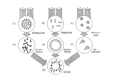
Figure 2: Principle of the procedure of preparing IRIVs
(a) intact influenza virus
(b) mixture of phospholipids
(c) intact, inactivated hepatitis A virions
(d) soluble influenza spike subunit antigens
containing the HA and viral phospholipids
(e) phospholipid membranes without antigens
(f) covalently bound cross-linkers on
the surface of HAV antigen
(g) IRIV containing the reconstituted membrane
with phospholipids extracted from viruses
and other phospholipids carrying the in-
fluenza spike proteins including HA and
the HAV on the surface.
Figure 3: Tolerance of hepatitis A vaccines. Comparison
of IRIV-HAV vaccines versus Al-HAV vaccines.
WO92/19267 PCT/EPg2/01014
2o8683l2
Figure 4: Immunogenicity of hepatitis A vaccines. Com-
parison of IRIV-HAV vaccines versus Al-HAV
vaccines.
Figure 5: Biological fusion activity of different
reconstituted influenza vesicles.
The examples illustrate the invention.
~USPI.~ 1
Prep~r~tion of IRIVs ~ith hep~titis A virus ~ntigen
non-co~lontly bound to their surface
(A) A dispersion of phosphatidylcholine (e.g. lecithin,
SIGMA) (75%), phosphatidylethanolamine (SIGMA) (20%)
and cholesterol (SIGMA) (5%) (all phospholipids 1-2%
(w/v) = 0.013-0.027 M) in 0.l M NaCl contA;ning 0.0l M
Tris/HCl, pH 7.3 was prepared by mixing these compounds
with a VIRTIS homogenizer. Sodium cholate recrystal-
lized as the acid from aceton/water 4:l (v/v) was added
to the milky dispersion in a final concentration of at
least 0.03 M (1.3%) which is required to disintegrate
the multilamellar structures present in unsonicated
phospholipid dispersions.
A pellet of purified influenza virus 90 A/Taiwan
(0.002 M of viral membrane phospholipids) was solubi-
lized in 700 ml of 0.l M Octaethyleneglycol mono(n-
dodecyl)ether (C12E8) (Nikko Chemicals (Tokyo)) in a
buffer containing 7.9 mg NaCl/ml, 4.4 mg/ml trisodium-
citrate dihydrate, 2.l mg/ml 2-morpholinoethane sul-
fonic acid monohydrate (MES) and l.2 mg/ml N-hydroxy-
ethyl-piperazine-N'-2-ethane sulfonic acid in H20 (pH
adjusted with lN NaOH to 7.3). The mixture was centri-
fuged at 170'000 x g for 30 min. and the supernatant
containing the influenza spike proteins (HA) and viral
~! 8 ~8 ~ ~
_ 13
phospholipids was added to the above milky phospholipid mixture.
The whole suspension was stirred for at least one hour at low
temperature (4~C). Subsequently, the suspension was applied to
a Sephadex G-50 (TM) (of medium particle size) column (80 X 15
cm) which was equilibrated and eluted with the same buffer as
used for the preparation of the phospholipid dispersion at 4~C
(flow rate 320 ml/h). The column was embedded in a water bath
connected with an ultrasonification apparatus (Bransonic (TM),
Branson Europe BV, frequency 50 kHz + 10%). 10 seconds of
ultrasonic shocks repeated every minute yielded small unilamellar
IRIVs. The sample volumes and column dimensions were such that
a complete separation of IRIVs eluted at the void volume V0 and
cholate micelles was achieved. The retention of cholate was
tested with 3H-labelled cholate (NEN Chemicals). After the first
Sephadex G-50 chromatography less than 1% of cholate was retained
yielding a phospholipid/cholate molar ratio of >50. A second
chromatography dialysis for 12 hours at 4~C reduced the cholate
amount below the limit of detection, yielding
phospholipid/cholate ratios >500 (i.e. less than 10 cholate
molecules/IRIV). Absence of residual C12E8 was tested by a
conventional hemolysis test: The amount was below the limit of
detection (>100 nM). The IRIVs (Fig. 1) showed a mean diameter
of about 100 nm and were conjugated with the HAV antigen in the
following manner: A purified and inactivated HAV suspension,
strain RG-SB XA112 (CNCM I-1080), containing 1 mg of HAV antigen,
was pelleted by ultracentrifugation (4 h, 100'000 x g). The
IRIVs prepared above were added to the pellet. After
resuspension the suspension was stirred at 20~C over night (16
hours). The HAV antigen spontaneously adsorbed by Vander-Waals
forces onto the surface of IRIVs.
WO92/19~7 PCT/EPg2/01014
208683~l4 --
(B) Purified influenza virus A/Sinqa~ore/6i86 was
stabilized in a buffer cont~i~ing O.l M
octaethyleneglycol mono(n-dodecyl)ether (Nikko
Chemicals, Tokyo, Japan), 7.9 mg/ml NaCl, 4 . 4 mg/ml
trisodium citrate dihydrate, 2.1 mg/ml MES and
1.2 mg/ml N-hydaxylethyl-piperazine-N'-2-ethane sul-
fonic acid, pH 7.3. This mixture was centrifuged at
100,000 x g for 30 minutes and the HA-containing super-
natant was saved.
Phosphatidylcholine (PC; Sigma Chemical Co., St. Louis,
MO) and phosphatidylethanolamine (PE; Sigma) (75%:25%
wt/wt) were Susr~ in O.Ol M Tris - O.l M NaCl,
pH 7.3, and homogenized. Recrystallized sodium cholate
(Sigma) was added to a final concentration of 0.02 M to
disintegrate multilamellar structures. To this solu-
tion was added the HA-cont~; n~ ~g supernatant and the
suspension stirred for l h at 4-C. The suspension was
applied to a Sephadex G-50 column (Pharmacia Fine
Chemicals, Uppsala, Sweden) equilibrated in O.Ol M
Tris - O.l M NaCl, pH 7.3. The sealed column was
placed in a water bath. During elution ultrasonic
shocks (50 KHz; lO s/min) were passed through the water
bath using an ultrasonification device (Bransonic,
Branson Europe BV, The Netherlands). The void volume
fractions, which contained the IRIV, were pooled and
re-chromatographed under identical conditions. The
IRIV possessed an average diameter of approximately
150 nm.
The purified, inactivated HAV suspension with a known
amount of antigen was centrifuged for 4 h at
lO0,000 x g to pellet the virus. An appropriate quan-
tity of the IRIV suspension was added to the pellet and
gently resuspended by shaking. The suspension was
gently stirred at 20~C for 48 h to allow the HAV to ad-
sorb onto the surface of the IRIV. This bulk suspen-
2 ~
_ 15
ion was diluted with sterile phc,sphate bufferedsaline, pH 7.4, to a final concentration of 2 ~g HAV
antigen/ml and bottled.
EXAMPLE 2
Preparation of IRIVs with HAV antigen crosslinked
to the membrane
The preparation of IRIVs with crosslinked HAV antigens is
schematically shown in Figure 2.
The IRIVs were prepared according to Example 1 with the
following modifications:
The HAV antigen molecules were attached to the IRIVs with
a suitable crosslinker molecule. The following procedures
were employed:
(A) Phosphatidylethanolamine (PE) was coupled with N-
succinimidylpyridyl dithiopropionate (SPDP, Pierce) as
follows: 15 mg of PE (20 ~mol) was dried down in a 5
ml glass bottle. The dried PE was redissolved in 2 ml
of dry chloroform (dried over a molecular sieve).
Then 30 ~mol of triethylamine (TEA) !3 mg), followed
by 30 ~mol of SPDP (10 mg) in 1 ml of dried methanol
were added. The mixture was then stirred at room
temperature under nitrogen for 1-2 hours until the
reaction was complete (i.e. no more free PE) . The
reaction product was dried down on a rotary
evaporator. The dried lipids were resuspended in
chloroform and were immediately applied on the top of
a silicic acid chromatography column, which had been
prepared as follows: 2 g of silicic acid were
dissolved in 10 ml of chloroform. The solution was
poured into a 10 ml plastic syringe barrel plugged
with glass fibre. The surplus was allowed to drain
out and the syringe barrel was fitted with a plastic
disposable
W~2/19~7 2 U 8 6 8 31 16 PCT/EP92/01014
three-way tap. After application of the lipids, the
column was washed with 4 ml of chloroform. Finally,
the column was eluted with 4 ml portions of a series of
chloroform-methanol mixtures, first 4:0.25 ~v/v] fol-
lowed by 4:0.5 [v/v], 4:0.75 [v/v] and finally 4:1
[v/v] and 2 ml fractions were collected. The pure
derivative was then located by thin-layer chromato-
graphy (TLC) using silica gel plates developed with
chloroform-methanol-water (65:25:4 by vol.). The deri-
vative runs faster than free PE and the spots are
visualized by phosphomolybdate or iodine.
The fractions containing the desired product were
pooled and concentrated by evaporation at reduced
pressure in a rotary evaporator.
(B) The HAV antigen was thiolated by the following proce-
dure: 5 ml of purified and inactivated HAV was dis-
solved in O.l M phosphate buffer (PBS (pH 7.5)) at a
concentration of 5 mg/ml~1. Then, a SPDP solution at a
concentration of 20 ~mol~~ (6 mg/ml~~) in ethanol was
mixed and 150 ~l thereof was under stirring slowly ad-
ded to 5 ml of the HAV protein solution with a Hamilton
syringe to give a molar ratio of SPDP to protein of
15:l. The ethanol concentration was kept below 5% to
prevent protein denaturation. The mixture was allowed
to react for 30 min. at room temperature (20 C). After
the reaction was stopped, the protein was separated
from the reactants by gel chromatography on Sephadex G-
50, equilibrated with a solution containing 0.05 M
sodium citrate (Na3C6H507 . 2H20, 19.7 g . 1-1), 0.05 M
sodium phosphate (Na2HPO4 . 7H2O, 13.4 g . l-1), and
0.05 M sodium chloride (2.9 g . l-1) pH 7Ø
(C) The pretreated IRIVs and HAV antigens were coupled in
the following manner: The IRIVs were prepared as in
Example l. Instead of PE the PE-SPDP was used.
~ 1 ~,
~ 7 i ~ 1
WO92/1g~7 2 0 8 6 8 31 PCT/EPg2/01014
17
The HAV - SPDP was reduced as follows: The pH of the
HAV - SPDP - solution in citrate-phosphate buffer was
adjusted to pH 5.5 by the addition of l M HCl. lO ~l
of a DTT solution, 2.5 M dithiothreitol (DTT, 380
mg/ml) in 0.2 M acetate buffer, pH 5.5 (165 mg of
sodium acetate in lO ml) was A~e~ for each ml of pro-
tein solution. The solution was allowed to stand for
30 min. Subsequently, the protein was separated from
the DrT by chromatography on a Sephadex G-50 column
equilibrated with a PBS buffer, pH 7Ø In order to
prevent oxidation of thiols all buffers were bubbled
with nitrogen to remove oxygen. The protein fractions
were also collected under nitrogen.
Finally, the IRIVs were mixed with the thiolated pro-
tein by stirring over night at room temperature.
~SAMPL~ 3
Prep~ration of IRIVs with reduced ~AV ~ntigen
The IRIVs were prepared as in Example 2 with the following
modifications: HAV antigen was not coupled with SPDP, but
the disulphide bridges already present at the surface of the
VPl protein were used as precursors for free thiol groups.
This conversion to free thiol groups was carried out as fol-
lows: 5 ml of an HAV solution were prepared in O.l M phos-
phate buffer, pH 7.4 at a final antigen concentration of 5
mg/ml. For each ml of this solution, lO ~l of a DTT solu-
tion (prepared as described in Example 2) were added. The
mixture was allowed to stand for 30 min. Then the protein
was separated from the DTT by chromatography on a Sephadex
G-50 column equilibrated with a PBS buffer at pH 7Ø Pro-
tein fractions were collected under nitrogen and were mixed
with the IRIVs of Example 2.
18
EXAMPLE 4
Preparation of IRIVs containing HAV antigen
The IRIVs were prepared according to Example 1 with the
following modification. 1 mg of purified and inactivated HAV in
suspension was added to a pellet of purified influenza virus
90 A/Taiwan (0.002 M of viral membrane phospholipids) and
incorporated into the IRIVs by the method described in
Example 1.
EXAMPLE 5
Production of an HAV-IRIV vaccine
HAV-IRIVs were prepared according to Examples 1, 2, 3 or 4
and diluted in PBS, pH 7.4 to a final concentration of 500
ng HAV protein ml-1. This bulk solution was sterile
filtrated through a membrane filter of pore size 0.2 µm
(Millipore). A preservative (thiomersal) was added to a final
dilution of 10-4. Aliquots of 0.6 ml of the final bulk
vaccine were filled into vaccine vials under sterile
conditions. Safety and potency tests were performed according to
international regulations.
EXAMPLE 6
Preparation of an anti-idiotype IRIV vaccine
against hepatitis C
The antigen-binding sites of antibody molecules (Ab1), also
known as isiotypes, have been shown to induce the production
of antibodies (anti-idiotypes, or Ab2). Because inoculation
of animals with some Ab2 results in the production of
antibodies (Ab3) that resemble Ab1 in their ability to bind
antigen, it has been assumed that the binding sites of Ab2 act
as a "mirror image" of the antigenic derminants originally
recognized by Ab2 (and subsequently by Ab3) [Jerne, N.K.,
Ann. Immunol. (Paris) 125 C (1974), 373]. The major
advantage of using anti-Id antibodies (Ab2) for eliciting
19
gen-specific antibodies (Ab3) is that the vaccine recipient is
never in contact with infectious agents or materials containing
foreign genes.
The anti-idiotype IRIV vaccine against hepatitis C was prepared
as follows: Sheep were immunized with an Ab1 (dis-solved of a
concentration of a mg/ml in PBS) according to the following
schedule: on day 0 the animals received 4 doses of 2 ml i.m. at
different sites (thighs). On days 7, 14 and 28 they received 2
doses of 2 ml into both hindlegs. On day 42 350 ml of blood was
collected from each sheep. The serum fraction was separated and
further purified by conventional techniques.
The purified anti-Id hepatitis C antibody was cleaved by
digestion with pepsin and the resulting F(ab') 2 fragment was
reduced with DTT (see Example 2) to yield 2 Feb' fragments.
These Fab' fragments contained free sulphhydryl groups which
reacted directly with the IRIVs of Example 2. This preparation
was diluted to a protein concentration of 50 ug/ml with PBS, pH
7.4, and portioned in 0.6 ml aliquots in vaccine vials.
EXAMPLE 7
Safety and Immunogenicity of Inactivated Hepatitis A
Vaccines: Cnm~rision of IRIV-HAV Prepared according
to Example 1 with Alum-absorbed Vaccine
(A) Hepatitis A virus (HAV) was purified after growth on MRC-5
human diploid cells (available from the American Type
Culture Collection under accession number ATCC CCL 171).
The virus was inactivated by treatment with formaldehyde
(0.05~) at 37~C for 10 days. Two vaccine series were
tested. Vaccine series 1 consisted of inactivated virus
linked to IRIVs according to Example
EXAMPLE 8
Biological fusion activity of different reconstituted
influenza virosomes
C
, .. .. ~ .
wo 92,lg~, 2 0 8 6 8 ~1 PCT/EPg2/01014
1 (A) (IRIV-HAV). Vaccine of series 2 was an alum-
adsorbed preparation cont~ini ng 0 . 4% Al (OH) 3 (Al-HAV) .
Both vaccines contained 150 ng of HAV antigen per
0.5 ml dose. Seronegative adult volunteers (two groups
of 15 persons each) received two intram~l~cl~lar in-
jections on day o and a booster injection on day 7 into
the deltoid region. No systemic reaction or al-
terations in the blood chemistry were detected. With
respect to local reactions, IRIV preparations provoked
a significantly lower percentage of reactions than the
alum-absorbed vaccine. The results of these
experiments are summarized in ~igure 3.
It was also found that the IRIV preparations were more
immunogenic than the alum preparation. To test the
anti-HAV immune response, blood samples were taken from
the volunteers on days 21 and 28 after the last injec-
tion. Sera were tested for HAV specific antibodies
using a commercially available RIA (Abbott). The re-
sults are summarized in Figure 4. The numbers on the
columns represent the range of the anti-HAV antibody
titer. Thus, the range of the anti-HAV antibody titers
for the IRIV and alum-adsorbed vaccine formulations on
day 21 was 82-988 and 69-844, respectively. The geo-
metric mean titer (range) for the IRIV and alum-ad-
sorbed vaccine formulations on day 28 was 453 mIU/ml
(92-1210) and 361 mIU/ml (60-929), respectively. Thus,
the IRIV preparations of the present invention are
superior to alum-adsorbed vaccines.
(B) In a phase I clinical study with 120 human volunteers
it could be demonstrated that one single IRIV ad-
juvanted hepatitis A vaccine dose induced protective
antibody titers against hepatitis A which were 7 times
higher than the antibody titer after the alum formula-
tion. Up to now such a high immunopotentiation in man
has never been achieved with any other liposomal, viro-
WO~19~7 2 0 8 6 8 31 PCT/EPg2/01014
21
somal or immunosomal formulation due to the obviouslack of fully biologically active fusion peptides.
A total of 120 HAV seronegative (<10 mIU/ml) healthy
adults were randomized to receive either fluid, alum-
adsorbed, or IRIV vaccine according to Example 1 (B).
The vaccine (0.5 ml) was administered intramuscularly
into the deltoid region. Volunteers were observed for
approximately 30 minutes after vaccination for imme-
diate-type reactions. Each volunteer was asked to re-
cord all adverse reactions on a report sheet for the 4
days following immunization. Serum samples for anti-
HAV antibody determinations were taken at the time of
im--lni7ation and 14 days later.
Each vaccine formulation contained 1 ~g of HAV antigen
per 0.5 ml dose. One dose of the IRIV-HAV formulation
also contAi n~ 10 ~g of influenza HA and 125 ~g total
phospholipids. All three vaccines were found to be
sterile and nontoxic for animals by st~nAArd test
methods. In addition, all 3 formulations elicited a
good anti-HAV antibody response in laboratory animals.
Each formulation was administered intramuscularly to 40
healthy adult volunteers seronegative for HAV antibody.
The y~U~S were well matched in regard to age and sex.
Adverse reactions associated with immunization are
shown in Table I. Pain at the injection site was the
most frequently reported complaint with all the
vaccines. Such discomfort was classified as moderate
by one vaccinee (2.5%) who received the fluid formula-
tion, 9 (23%) who were immunized with the alum-adsorbed
vaccine, and one (2.5%) who received the IRIV prepara-
tion. Severe pain was reported by one subject who re-
ceived the alum-adsorbed vaccine. All other subjects
who reported a "painful" reaction graded it as mild.
Immunization with the alum-adsorbed vaccine was asso-
WO9~19~7 PCT/EPg2/01014
2 0 8 6 ~ 3 122
ciated with a significantly (P < O.Ol) higher incidenceof both pain and swelling/induration as compared to
either the fluid or IRIV formulations. No systemic re-
actions attributable to vaccination were noted.
The anti-HAV antibody Le~G~Ise engendered 14 days after
vaccination is shown in Table II. Immunization with
the fluid vaccine yielded a geometric mean titer (GMT)
of 15.7 mIU/ml with 30% of subjects se~o~G~I~erting (220
mIU/ml). While the alum-adsorbed vaccine induced both
a modestly higher GMT (21.3 mIU/ml) and seroconversion
rate (44%), neither was significantly greater than that
obt~ with the fluid vaccine. In ~ol.L~ast, the IRIV
vaccine formulation elicited a far more vigorous anti-
body response. The GMT of 139.8 was significantly
(P ~ O.OOOl) higher compared to either of the other two
vaccines. All but one vaccinee ros--e~ced >lO0 mIU/ml.
Of greater importance was the fact that all vaccinees
seroconverted by day 14 compared to less than 50% for
the other vaccine formulations (P < 0.005).
7'al~1e I. ~ldverse ReacLions Associated wiL/~ ImmunizaLion '~
Local reactions (%) Systemic reactions (%)
Vaccine
Pain Swelling/ Redness FeverHeadache Malaise
Induration
Fluid 42 01 0 0 0 0 w
Al(0ll)3-adsorbed 88~ 23~ 0 0 0 0
' $~
IRIV 25 5 0 0 0 0 ~~
+ vs * or : P < 0.01
D vs 11 or tt p < O 01
WO 92/lg2G7 2 0 g 6 8 31 PCI/EP92/01014
v
O ~
o v ~P ~P o
o ~ o
., ~ _ _ _
E
. ~ o o o
C o
,~
'J ~l ~ c~ o
O
U~ Z
r
O ~ ~
O O
C I _ ~ O
~J ~ O
.0 ~ ~ -- OU~
r' C ~
C
C ~ ~ ~ ~ ~ O
-- C
O ~ U
~ U
C
~O
~ E o
a
-1 U Ul
4 ~ o o
o o o
o a~
E a
~ o ,~ _
.u ~ O In
~, o o
u o o
._ . .
o o
r
c I C U ~-
O .~ h
~ ~ .~ U~ O
C ~
S i Q~ Ul
~~ 3 -- H .C~~
O ~--~ Y ~ ~C
To study the role of the influenza viral membrane components in
the fusion reaction in detail, it is necessary to be able to
manipulate these components. For this purpose a method is
required for the isolation and reconstitution of the viral spike
proteins, producing reconstituted virosomes with full biological
fusion activity. Most of the methods that have been used to
reconstitute viral envelopes are based on solubilization of the
viral membrane with a detergent.
Several reconstituted influenza virus envelopes using different
methods which are described in the literature have been prepared:
[A] A virosome according to Huang, R.T.C. et al. (Huang,
R.T.C., Wahn, K., Klenk, H.D. and Rott, R., Virology 97
(1979), pp. 212-217) which was prepared using detergents
with a high critical micelle concentration (c.m.c.) [e.g.
octylglucoside].
[B] A virosome according to Kawasaki, K. et al. (Kawasaki, K.,
Sato, S.B. and Ohmiski, S.I., Biochem. Biophys. Acta 733
(1983), pp. 268-290) which was prepared using detergents
with a low c.m.c. (e.g. Triton X-100(TM)).
[C] A virosome according to Hosoka, Y. et al. (Hosaka, Y.,
Yasuda, Y. and Fukai, K., J. Virol. 46 (1983), pp. 1014-
1017) which was prepared using Nonidet P-40 (TM) as
detergent.
[D] An IRIV as it has been described in Example 1.
~.
W~92/19~7 PCT/EPg2/01014
~086831 26
In addition, the following controls have been prepared:
tE] A purified influenza virus suspension as positive con-
trol.
~F] PBS-NaCl, pH 7.4, as negative control.
From each reconstituted influenza virus envelope solution
and the influenza virus control a concentration of 10 ~g/ml
hemagglutinin in bicarbonate-free RPMI 1640 medium,
supplemented with 10 mM NaCl (pH 7.4) was prepared. The
negative control (PBS-NaCl) was diluted in the same medium
1:10. For the vesicle binding and fusion experiment MRC-5
human diploid fibroblasts were grown in 12-well cluster
dishes (NUNC). The cells were C~e~e~ at 34,000 cells/ml per
well and were used 3 days later. At this time, they were
approximately 70-80% confluent.
0.5 ml of the reconstituted envelope solutions were added
per well, for each preparation 20 wells. The vesicles were
allowed to bind to the cells for 30 minutes at room tempera-
ture. During this incubation period the dishes were spun
twice for 3 minutes at 500 g with a 180- rotation between
spins. The centrifugation step ~h~nc~s the binding of
vesicles about 3-fold. After this 30-minute period the
cells were washed four times with PBS-NaCl, pH 7.4, to
remove unbound vesicles. For the fusion experiment, the
fusion activity of the five preparations was induced by
adding a fusion medium (RPMI 1640, supplemented with 10 mM
succinate, 0.2% bovine serum albumin and 35 mM NaCl, pH 5.0)
to each well (0.5 ml/well). After one minute, in two wells
per preparation the fusion reaction was stopped by replacing
the fusion medium with ethanol absolute. This was done
every minute until 10 minutes had passed. The cells were
then stained according to the method of May Grunwald-Giemsa:
-
WO92/19~7 2 0 8 6 8 31 PCT/EP92/01014
27
The cells were then covered with an alcoholic May-
Grunwald solution (Fluka No. 63590). After 5 minutes
the cells were quickly washed with a phosphate buffer,
pH 6.5, and stained with a Giemsa solution (Fluka No.
48900), diluted l:lO with the same phosphate buffer.
After another lO minutes the cells were washed with
running water: Under the microscope the cytoplasm of
the cells appeared in a light blue color, the
membranes in a dark blue color and the nuclei in a
dark red color.
Under the microscope lO sight fields were evaluated
for counting the fused cells (containing at least two
nuclei) and were calculated for each preparation and
time interval. The Figure 5 shows the mean value of
two wells: It was obvious that only the IRIV
preparation shared a fusion activity which was
comparable to the influenza virus control.
Preparation tB] yielded a fusion activity which was
only around 30% compared to the positive control. The
other preparations did not show any fusion activity
(as the negative control did). From these results it
can be concluded that only the described IRIVs show
fully biological fusion activity, whereas the other
methods for influenza envelope reconstitution do not
yield vesicles with fusion activity and lead from a
considerable to a complete loss of fusion activity.
, ,