Note: Descriptions are shown in the official language in which they were submitted.
WO 92/01047 PCT/GB91/01134
203~33~
.'~ETHODS FOR PRODUCING MEMBERS OF
SPECIFT_C BINDING PAIRS
':he present invention relates to methods for producing
members o?: specific binding pairs. The present invention
also relates to the biological binding molecules produced by
these methods.
Owing to their high specificity for a given antigen,
the advent of monoclonal antibodies (Kohler, G. and Milstein
C; 1975 Nature 256: 495) represented a significant technical
break-through with important consequences both
scientifically and commercially.
Monoclonal antibodies are traditionally made by
establishing an inmortal mammalian cell line which is
derived from a single immunoglobulin producing cell
secreting one form of a biologically functional antibody
molecule with a particular specificity. Because the
antibody-secreting mammalian cell line is immortal, the
characteristics of the antibody are reproducible from batch
to batch. The key properties of monoclonal antibodies are
their specificity for a particular antigen and the
reproducibility with which they can be manufactured.
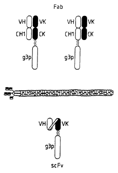
Structurally, the simplest antibody (IgG) comprises
four polypeptide chains, two heavy (H) chains and two light
(L) chains inter-connected by disulphide bonds (see figure
1). The light chains exist in two distinct forms called
kappa (K) and lambda (~~). Each chain has a constant region
(C) and a variable region (V). Each chain is organized into
a series of domains. The light chains have two domains,
corresponding to the C region and the other to the V region.
The heavy chains have four domains, one corresponding to the
V region and three domains (1,2 and 3) in the C region. The
antibody has two arms (each arm being a Fab region), each of
which has a VL and a VH region associated with each other.
~5 It is this pair of V regions (VL and VH) that differ from
one antibody to another (owing to amino acid sequence
variations), and which together are responsible for
recognising the antigen and providing an antigen binding
site ( A8S ) . In even more detail, each V region is made up
from three complementarity determining regions (CDR)
separated by four framework regions (FR). The CDR's are the
most variable part of the variable regions, and they perform
the critical antigen binding function. The CDR regions are
derived arom many potential germ line sequences via a
a5 complex process involving recombination, mutation and
selection.
It has been shown that the function of binding antigens
can be performed by fragments of a whole antibody. Example
binding fragments are (i) the Fab fragment consisting of the
VL, VH. CL and CH1 domains; cii) the Fd fragment consisting
of the VH and CH1 domains; (iii) the Fv fragment consisting
of the VL and VH domains of a single arm of an antibody,
l iv ) the dAb fragment ( Ward , _ . S . et al . , Nature 341, 544-
546 1 1989 ) which consists of a VH domain; ; v ) isolated CDR
WO 92/01047 PCT/GB91/01134
1
2pg6~3~
regions; and (vi) F(ab')2 fragments, a bivalent fragment
comprising two Fab fragments linked by a disulphide bridge
at the hinge region.
Although the two domains of the Fv fragment are coded
Y For by separate genes, it has proved possible to make a
synthetic linker that enables them to be made as a single
protein chain (known as single chain Fv (scFv); Bird, R.E.
et al., Science 242, 423-426 (1988) Huston, ,7.S. et al.,
Proc. Natl. Acad. Sci., USA 85, 5879-5883 (1988)) by .
recombinant methods. These scFv fragments were assembled
from genes from monoclonals that had been previously
isolated. In this application, the applicants describe a
process to assemble scFv fragments from VH and VL domains
that are not part of an antibody that has been previously
isolated.
Whilst monoclonal antibodies, their fragments and
derivatives have been enormously advantageous, there are
nevertheless a number of limitations associated with them.
Firstly, "the 'therapeutic applications of monoclonal
antibodies produced by human immortal cell lines holds great
promise for the treatment of a wide range of diseases
(Clinical Applications of Monoclonal Antibodies. Edited by
S. Lennox. British Medical Bulletin 1984. Publishers
Churchill Livingstone). Unfortunately, immortal antibody
producing human cell lines are very difficuht to establish
and they give low yields of antibody (approximately 1
ug/ml). In contrast, equivalent rodent cell lines yield
high amounts of antibody (approximately 100 pg/ml).
However, the repeated administration of these foreign
rodent proteins to humans can lead to harmful
hypersensitivity reactions. In the main therefore, these
rodent-derived monoclonal antibodies have limited
therapeutic use.
Secondly, a key aspect in the isolation of monoclonal
antibodies is how many different clones of antibody
producing cells with different specificities, can be
practically established and sampled compared to how many
thooratically need to be sampled in order to isolate a cell
producing antibody with the desired specificity
characteristics (Milstein, C., Royal Soc. Croonian Lecture,
Proc. R. Soc. London B. 239; 1-16, (1990)). For example,
the number of different specificities expressed at any one
time by lymphocytes of the murine immune system is thought
to be approximately 107 and this is only a small proportion
of the potential repertoire of specificities. However,
during the isolation of a typical antibody producing cell
with a desired specificity, the investigator is only able to
sample 10~ to 10'~ individual specificities. The problem is ~
worse in the human, where one has approximately 1012
ly~phocyte specificities, with the limitation on sampling of
10~ or 10'~ remaining.
This problem has been alleviated to some extent in
laboratory animals by the use of immunisation regimes.
'~hus, where one wants to produce monoclonal antibodies
~5 having a specificity against a particular epitope, an animal
WO 92/01047 PCT/GB91/01134
~o~s~3s
is immunised with an immunogen expressing that epitope. The
animal will then mount an immune response against the
immunogen and there will be a proliferation of lymphocytes
which have specificity against the epitope. Owing to this
3 proliferation cf lymphocytes with the desired specificity,
it becomes easier to detect them in the sampling procedure.
However, this approach is not successful in all cases, as a
suitable immunogen may not be available. Furthermore, where
one wants to produce human monoclonal antibodies (eg for
therapeutic administration as previously discussed), such an
approach is not practically, or ethically, feasible.
In the last few years, these problems have in part,
been addressed by the application of recombinant DNA methods
to the isolation and production of e.g. antibodies and
fragments of antibodies with antigen binding ability, in
bacteria such as E.coli.
This simple substitution of immortalised cells with
bacterial cells as the 'factory', considerably simplifies
procedures for preparing large amounts of binding molecules.
Furthermore, a recombinant production system allows scope
for producing tailor-made antibodies and fragments thereof.
For example, it is possible to produce chimaeric molecules
with new combinations of binding and effector functions,
humanised antibodies (e. g. murine variable regions combined
with human constant domains or murine-antibody CDRs grafted
onto a human FR) and novel antigen-binding molecules.
Furthermore, the use of polymerise chain reaction (PCR)
amplification (Saiki, R.K., et al., Science 239, 487-491
(1988)) to isolate antibody producing sequences from cells
(e.g. hybridomas and B cells) has great potential for
speeding up the timescale under which specificities can be
isolated. Amplified VH and VL genes are cloned directly
into vectors for expression in bacteria or mammalian cells
(Orlandi, R., et al., 1989, Proc. Natl. Acid. Sci., USA 86,
3833-3837; Ward, E.S., et al., 1989 supra: Larrick, J.W., et
al., 1989, eiochem. Hiophys. Res. Commun. 160, 1250-1255;
Sastry, L. et al., 1989, Proc. Natl. Acid. Sci., USA., 86,
5728-5732). Soluble antibody fragments secreted from
bacteria are then screened for binding activities.
However, like the production system based upon
immortalised cells, the recombinant production system still
suffers from the selection problems previously discussed and
therefore relies on animal immunization to increase the
proportion of cells with desired specificity. Furthermore,
some of these techniques can exacerbate the screening
problems. For example, large separate H and L chain
libraries have been produced from immunized mice and
combined together in a random combinatorial manner prior to
screening (Ruse, W.D. et al., 1989, Science 246, 1275-1281,
W090/14443: W090/14424 and W090/14430). Crucially however,
the information held within each cell, namely the original
pairing of one L chain with one H chain, is lost. This
loses some, of the advantage gained by using immunization
protocols in the animal. Currently, only libraries derived
SS from single VH domains (dAbs; Ward, E.S., et al., 1989,
WO 92/01047 PCT/GB91/01134
'~t~8~i~36
supra.) ao not suffer this drawback. However, because not
all antibody VH domains are capable of binding antigen, more
have to be screened. In addition, the problem of directly
screening many different specificities '_n prokaryotes
remains to be solved.
Thus, there is a need for a screening system which
ameliorates or overcomes one or more of the above or other
problems. The ideal system would allow the sampling of very
large numbers of specificities (eg 106 and higher), rapid
sorting at each cloning round, and rapid transfer of the
genetic material coding for the binding molecule from one
stage of the production process, to the next stage.
The most attractive candidates for this type of
screening, would be prokaryotic organisms (because they grow
quickly, are relatively simple to manipulate and because
large numbers of clones can be created) which express and
display at their surface a functional binding domain eg. an
antibody, receptor, enzyme etc. In the UK patent GB
2137631H methods for the co-expression in a single host cell
of the variable H and L chain genes of immunoglobulins were
disclosed. However, the protein was expressed
intracellularly and was insoluble. Further, the protein
required extensive processing to generate antibody fragments
with binding activity and this generated material with only
a fraction of the binding activity expected for antibody
fragments at this concentration. It has already been shown
that antibody fragments can be secreted through bacterial
membranes with the appropriate signal peptide (Skerra, A.
and Pluckthun, A. 1988 Science 240 1038-1040; Better, M et
al 1988, Science 240 1041-1043) with a consequent increase
in the binding activity of antibody fragments. These
methods require screening of individual clones for binding
activity in the same way as do mouse monoclonal antibodies.
It has not been shown however, how a functional binding
domain eg an antibody, antibody fragment, receptor, enzyme
etc can be held on the bacterial surface in a configuration
which allows sampling of say its antigen binding properties
and selection for clones with desirable properties. In
large part, this is because the bacterial surface is a
complex structure, and in the gram-negative organisms there
is an outer wall which further complicates the position.
Further, it has not been shown that eg an antibody domain
will fold correctly when expressed as a fusion with a
surface protein of bacteria or bacteriophage.
Bacteriophage are attractive prokaryote related
organisms :or this type of screening. In general, their
surface is a relatively simple structure, they can be grown
easily in large numbers, they are amenable to the practical
handling involved in many potential mass screening
programmes, and they carry genetic information for their own
synthesis within a small, simple package. The difficulty
has been to practically solve the problem of how to use
bacteriophages in this manner. A Genex Corporation patent
application number W088/06630 has proposed that the
~5 bacteriophage lambda would be a suitable vehicle for the
WO 92/01047 PCT/G B91/01134
2D~G~j~
expression of antibody molecules, but they do not provide a
teaching which enables the general idea to be carried out.
For example W088/06630 does not demonstrate that any
sequences: (a) have been expressed as a fusion with gene V;
(b) have been expressed on the surface of lambda; and (c)
have been expressed so that the protein retains biological
activity. Furthermore there is no teaching on how to screen
for suitable fusions. Also', since the lambda virions are
assembled within the cell, the fusion protein would be
expressed intracellularly and would be predicted to be
inactive. Bass et al., in December 1990 (after the earliest
priority date for the present application) describe deleting
part of gene III of the filamentous bacteriophage M13 and
inserting the coding sequence for human growth hormone (hGH)
into the N-terminal site of the gene. The growth hormone
displayed by M13 was shown to be functional. (Bass, S., et
al. Proteins, Structure, Function and Genetics (1990) 8:
309-314). A functional copy of gene III was always present
in addition, when this fusion was expressed. A Protein
Engineering Corporation patent application W090/02809
proposes the insertion of the coding sequence for bovine
pancreatic trypsin inhibitor ( HPTI ) into gene VIII of _.M7.3.
However, the proposal was not shown to be operative. For
example, there is no demonstration of the expression of HPTI
sequences as fusions with protein VIII and display on the
surface of M13. Furthermore this document teaches that when
a fusion is made with gene III, it is necessary to use a
second synthetic copy of gene III, so that some unaltered
gene III protein will be present. The embodiments of the
present application do not do this. In embodiments where
phagemid is rescued with M13K07 gene III deletion phage,
there is no unaltered gene III present.
W090/02809 also teaches that phagemids that do not
contain the full genome of M13 and require rescue by
coinfection with helper phage are not suitable for these
purposes because coinfection could lead to recombination.
In all embodiments where the present applicants have
used phagemids , they have used a helper phage and the only
sequences derived from filamentous bacteriophage in the
phagemids are the origin of replication and gene III
sequences.
W090/02809 also teaches that their process needed
information such as nucleotide sequence of the starting
molecule and its three-dimensioned structure. The use of a
pre-existing repertoire of binding molecules to select for a
binding member, such as is disclosed herein, for example
using an immunoglobulin gene repertoire of animals, was not
disclosed. Further, they do not discuss favouring
variegation of their binding molecules in natural blocks of
variation such as CDRs of immunoglobulins, in order to
favour generation of improved molecules and prevent
unfavourable variations. W090/02809 also specifically
excluded the application of their process to the production
of scFv molecules.
In each of the above discussed patents (W088/06630 and
WO 92/01047 PCT/G B91/01134
'~U8oU~6
W090/02809), the protein proposed for display is a single
polypeptide chain. There is no disclosure of a method for
the display of a dimeric molecule by expression of one
monomer as a fusion with a capsid protein and the other
.. protein in a free form.
Another disclosure published in May 1991 (after the
earliest priority date for the present application)
describes the insertion into gene VIII of M13, the coding
sequences for one of the two chains of the Fab portion of an
antibody with co-expression of the other from a plasmid.
The two chains were demonstrated as being expressed as a
functional Fab fragment on the surface of the phage (Kang
A.S. et al., (1991) Proc. Natl. Aced. Sci, USA, _88 p4363-
4366). No disclosure was made of the site of insertion into
gene VIII and the assay for pAb binding activity by ELISA
used a reagent specific for antibody L chain rather than for
phage. A further disclosure published in March 1991 (after
the earliest priority date for the present application)
describes the insertion of a fragment of the AIDS virus
protein gag into the N-terminal portion of gene III of the
bacteriophage fd. The expression of the gag protein
fragment was detected by immunological methods, but it was
not shown whether or not the protein was expressed in a
functional form (Tsunetsugu-Yokota Y et al. (1991) Gene 99
p261-265). ~
The problem of how to use bacteriophages in this way is
in fact a difficult one. The protein must be inserted into
the phage in such a way that the integrity of the phage coat
is not undermined, and the protein itself should be
functional retaining its biological activity with respect to
antigen binding. Thus, where the protein of choice is an
antibody, it should fold efficiently and correctly and be
presented for antigen binding. Solving the problem for
antibody molecules and fragments would also provide a
general method for any biomolecule which is a member of a
specific binding pair e.g. receptor molecules and enzymes.
Surprisingly, the applicants have been able to
construct a bacteriophage that expresses and displays at its
surfac~ a large biologically functional binding molecule (eg
antibody fragments, and enzymes and receptors) and which
remains intact and infectious. The applicants have called
the structure which comprises a virus particle and a binding
molecule displayed at the viral surface a 'package'. Where
the binding molecule is an antibody, an antibody derivative
or fragment, or a domain that is homologous to an
immunoglobulin domain, the applicants call the package a
'phage antibody' (pAb). However, except where the context
demands otherwise, where the term phage antibody is used
generally, it should also be interpreted as referring to any
package comprising a virus particle and a biologically
functional binding molecule displayed at the viral surface.
pAbs have a range of applications in selecting antibody
genes encoding antigen binding activities. For example,
pAbs could be used for the cloning and rescue of hybridomas
vV0 92/01047
PC1'/G B91 /01134
v vrianai , n. , et al ( 1y89 ) PNAS 86 p3833-3837 ) , and in the
screening of large combinatorial libraries (such as found in
Huse, ~l.D. et al., 1989, Science 246, 1275-1281). In
particular, rounds of selection using pAbs may help in
rescuing the i~igher affinity antibodies from the latter
libraries. It may be preferable to screen small libraries
derived from antigen-selected cells (Casali, P., et al.,
(1986) Science 234 p476-479) to rescue the original VH/VL
pairs comprising the Fv region of an antibody. The use of
pAbs may also allow the construction of entirely synthetic
antibodies. Furthermore, antibodies may be made which have
some synthetic sequences e.g. CDRs, and some naturally
derived sequences. For example, V-gene repertoires could be
made in vitro by combining un-rearranged V genes, with D and
J segments. Libraries of pAbs could then be selected by
binding to antigen, hypermutated in vitro in the antigen-
binding loops or v domain framework regions, and subjected
to further rounds of selection and mutagenesis.
As previously discussed, separate H and L chain
libraries lose the original pairing between the chains. It
is difficult to make and screen a large enough library for a
particularly advantageous combination of H and L chains.
For example, in a mouse there are approximately 107
possible H chains and 107 possible L chains. Therefore,
there are 1014 possible combinations of H and L chains, and
to test for anything like this number of combinations one
would have to create and screen a library of about 1014
clones. This has not previously been a practical
possibility.
The present invention provides a number of approaches
which ameliorate this problem.
In a first approach, (a random combinatorial approach,
see examples 20 and 21) as large a library as is practicall
possible is created which expresses as many of the 101
3S potential combinations as possible. However, by virtue of
tha expression of the H and L chains on the surface of the
phage, it is reasonably practicable to. select the desired
combination, from all the generated combinations by affinity
techniques (see later for description of selection formats).
In a second approach (called a dual combinatorial
approach by the present applicants, see example 26), a large
library is cr~ated from two smaller libraries for selection
of the desired combination. This ameliorates the problems
still further. The appr9ach involves the creation of: (i) a
first library of say l07 e.g. H chains which are displayed
on a bacteriophage (as a fusion with the protein encoded by
gene III) which is resistant to e.g. tetracycline; and (ii)
a second library of say 107 e.g. L chains in which the
coding sequences for these light chains are within a plasmid
vector containing an origin of replication for a
bacteriophage (a phagemid) which is resistant to e.g.
ampicillin (i.e. a different antibiotic) and are expressed
in the periplasmic space of a host bacterium. The first
library is then used to infect the bacteria containing the
second library ~o provide 1014 combinations of H and L
WO 92/01047 PCTIG B91/01134
'~(3~ii'~~~ ..
s
chains on the surface of the resulting phage in the
bacterial supernatant.
The advantage of this approach is that two separate
libraries of eg 10~ are created in order to produce 1014
combinations. Creating a 10~ library is a practical
possibility
The 1014 combinations are then subjected to selection
(see later for description of selection formats) as
disclosed by the present application. This selection will
then produce a population of phages displaying a particular
combination of H and L chains having the desired
specificity. The phages selected however, will only contain
DNA encoding one partner of the paired H and L chains
(deriving from either the phage or phagemid). The sample
eluate containing the population is then divided into two
portions. A first portion is grown on e.g. tetracycline
plates to select those bacteriophage containing DNA encoding
H chains which are involved in the desired antigen binding.
A second portion is grown on e.g. ampicillin plates to
select those bacteriophage containing phagemid DNA encoding
L chains which are involved in the desired antigen binding.
A set of colonies from individually isolated clones e.g.
from the tetracycline plates are then used to infect
specific colonies e.g. from the ampicillin .plates. This
results in bacteriophage expressing specific combinations of
H and L chains which can then be assayed for antigen
binding.
In a third approach (called a hierarchical dual
combinational approach by the present applicants), an
individual colony from either the H or L chain clone
selected by growth on the antibiotic plates, is used to
infect a complete library of clones encoding the other chain
(H or L). Selection is as described above. This favours
isolation of the most favourable combination.
In a fourth approach (called a hierarchrical approach
by the present applicants, see examples 22 and 46) both
chains era cloned into the same vector. However, one of the
chains which is already known to have desirable properties
is kept fixed. A library of the complementary chain is
inserted into the same vector. Suitable partners for the
fixed chain are selected following display on the surface of
bacteriophage.
' In a fifth approach (see example 48), to improve the
chances of recovering original pairs, the complexity of the
combinatorial libraries can be reduced by using small B
populations of B-lymphocytes selected for binding to a
desired antigen. The cells provide e.g. mRNA or DNA, for
preparing libraries of antibody genes for display on phage.
' This technique can be used in combination with the above
mentioned four approaches for selection of antibody
specificities.
Phagemids have been mentioned above. The applicants
have realised and demonstrated that in many cases phagemids
will be preferred to phage for cloning antibodies because it
is easier to use them to generate more comprehensive
WO 92/01047 pCT/G B91/01134
._ 208~~3~
libraries of the immune repertoire. This is because the
phagemid DNA is approximately 100 times more efficient than
bacteriophage DNA in transforming bacteria (see example
19 ) . Also, the use of phagemids gives the ability to vary
the number of gene III binding moecule fusion proteins
displayed on the surface of the bacteriophage (see example
17). For example, in a system comprising a bacterial cell
containing a phagemid encoding a gene III fusion protein and
infected with a helper phage, induction of expression of the
gene III fusion protein to different extents, will determine
the number of gene III fusion proteins present in the space
defined between the inner and outer bacterial membranes
following superinfection. This will determine the ratio of
gene III fusion protein to native gene III protein displayed
by the assembled phage.
Expressing a single fusion protein per virion may aid
selection of antibody specificities on the basis of affinity
by avoiding the 'avidity' effect where a phage expressing
two copies of a low affinity antibody would have the same
apparent affinity as a phage expressing one copy of a higher
affinity antibody. in some cases however, it will be
important to display all the gene III molecules derived by
superinfection of cells containing phagemids to have fusions
(e.g. for selecting low affinity binding; molecules or
improving sensitivity on ELISA ) . One way to do this is to
superinfect with a bacteriophage which contains a defective
gene III. The applicants have therefore developed and used
a phage which is deleted in gene III. This is completely
novel.
The demonstration that a functional antigen-binding
domain can be displayed on the surface of phage, has
implications beyond the construction of novel antibodies.
For example, if other protein domains can be displayed at
the surface of a phage, phage vectors could be used to clone
and select genes by the binding properties of the displayed
protein. Furthermore, variants of proteins, including
epitope libraries built into the surface of the. protein,
could be made and readily selected for binding activities.
In effect, other protein architectures might serve as
"nouvelle" antibodies.
The technique provides the possibility of building
antibodies from first principles, taking advantage of the
structural framework on which the antigen binding loops
fold. In general, these loops have a limited number of
conformations which generate a variety of binding sites by
alternative loop combinations and by diverse side chains.
Recent successes in modelling antigen binding sites augurs
well for de novo design. In ar~y case, a high resolution
structure of the antigen is needed. However, the approach
is attractive for making e.g. catalytic antibodies,
particularly for small substrates. Here side chains or
binding sites for prosthetic groups might be introduced, not
only to bind selectively to the transition state of the
substrate, out also to participate directly in bond making
and breaking. '_':~e only auestion is whether the antibody
WO 92/01047 PCT/GB91 /Ol 134
208~~36 ,o
architecture, specialised for binding, is the best starting
point for building catalysts. Genuine enzyme architectures,
such as the triose phosphate isomerase (TIM) barrel, might
be more suitable. Like antibodies, TIM enzymes also have a
framework structure (a barrel of (i-strands and a-helices)
and loops to bind substrate. Many enzymes with a diversity
of catalytic properties are based on this architecture and
the loops might be manipulated independently on the
frameworks for design of new catalytic and binding
properties. The phage selection system as provided by the
present disclosure can be used to select for antigen binding
activities and the CDR loops thus selected, used on either
an antibody framework or a TIM barrel framework. Loops
placed on a e.g. a TIM barrel framework could be further
modified by mutagenesis and subjected to further selection.
Thus, there is no need to select for high affinity binding
activities in a single step. The strategy of the immune
system, in which low affinity evolves to high affinity seems
more realistic and can be mimicked using this invention.
One class of molecules that could be useful in this
type of application are receptors. For example, a specific
receptor could be displayed on the surface of the phage such
that it would bind its ligand. The receptor could then be
modified by, for example, _in vitro mutagenesis and variants
having higher binding affinity for the ligand~selected. The
selection may be carried out according to one or more of the
formats described below~with reference to figure 2 (which
refers particularly to pAbs) in which the pAb antibody is
replaced with a phage receptor and the antigen with a ligand
1.
Alternatively, the phage-receptor could be used as the
basis of a rapid screening system for the binding of
ligands, altered ligands, or potential drug candidates. The
advantages of this system namely of simple cloning,
convenient expression, standard reagents and easy handling
makas the drug screening application particularly
attractive. in the context of this discussion, receptor
moans a molecule that binds a specific, or group of
specific, ligand(s). The natural receptor could be
expressed on the surface of a population of cells, or it
could be the extracellular domain of such a molecule
(whether such a form exists naturally or not), or a soluble
molecule performing a natural binding function in the
plasma, or within a cell or organ.
Another possibility, is the display of an enzyme
molecule or active site of an enzyme molecule on the surface
of a phage (see examples 11,12,30,31,32 and 36). Once the
phage enzyme is expressed, it can be selected by affinity
chromatography, for instance on columns derivatized with
transition state analogues. If an enzyme with ~ different
or modified specificity is desired, it may be possible to
mutate an enzyme displayed as a fusion on bacteriophage and
then select on a column derivatised with an analogue
selected to have a higher affinity for an enzyme with the
desired modified specificity.
WO 92/01047 PLT/GB91/01134
20SG i~
although throughout this application, the applicants
3iscuss the possibility of screening for higher affinity
variants of pAbs, they recognise that in some
applications, .or example low affinity chromatography
lOhlson, S, et al Anal. Hiochem. 169, p204-208 (1988)),
it may be desirable to isolate lower affinity variants.
Examples 21 and 23 show that the present invention
provides a way of producing antibodies with low
affinities (as seen in the primary immune response or in
unimmunised animals). This is made possible by
displaying multiple copies of the antibody on the phage
surface in association with gene III protein. Thus, pAbs
allow genes for these antibodies to be isolated and if
necessary, mutated to provide improved antibodies.
pAbs also allow the selection of antibodies for
improved stability. It has been noted for many
antibodies, that yield and stability are improved when
~he antibodies are expressed at 30°C rather than 37°C.
If pAbs are displayed at 37°C, only those which are
stable will be available for affinity selection. When
antibodies are to be used in vivo for therapeutic or
diagnostic purposes, increased stability would extend the
half-life of antibodies in circulation.
Although stability is important for all antibodies
and antibody domains selected using phage, it is
particularly important for the selection of Fv fragments
which are formed by the non-covalent association of VH
and VL fragments. Fv fragments have a tendency to
dissociate and have a much reduced half-life in
circulation compared to whole antibodies. Fv fragments
are displayed on the surface of phage, by the association
of one chain expressed as a gene III protein fusion with
the complementary chain expressed as a soluble fragment.
If pairs of chains have a high tendency to dissociate,
they will be much leas likely to be selected as pAbs.
Therefore, the population will be enriched for pairs
which do associate stably. Although dissociation is less
of a problem with Fab fragments, selection would also
occur for Fab fragments which associate stably. pAbs
~10 allow selection for stability to protease attack, only
those pAbs that are not cleaved by proteasea will be
capable of binding their ligand and therefore populations
of phage will be enriched for those displaying stable
antibody domains.
X15 The technique of displaying binding molecules on the
phage surface can also be used as a primary cloning
system. For example, a cDNA library can be constructed
and inserted into the bacteriophage and this phage
library screened for the ability to bind a ligand. The
50 ligand/binding molecule combination could include any
pair of molecules with an ability to specifically bind to
WO 92/01047 PC1'/GB91/01134
i
one another e.g. receptor/ligand, enzyme/substrate (or
analogue), nucleic acid binding protein/nucieic acid etc.
If one member of the complementary pair is available,
this may be a preferred way of isolating a clone for the
other member of the pair.
It will often be necessary to increase the diversity
of a population of genes cloned for the display of their
proteins on phage or to mutate an individual nucleotide
sequence. Although in vitro or in vivo mutagenesis
techniques could be used for either purpose, a
particularly suitable method would be to use mutator
strains. A mutator strain is a strain which contains a
genetic defect which causes DNA replicated within it to
be mutated with respect to its parent DNA. Hence if a
population of genes as gene III fusions is introduced
into these strains it will be further diversified and can
then be transferred to a non-mutator strain, if desired,
for display and selection. Example 38 covers the use~of
mutator strains with phage antibodies (an example of in
vitro mutagenesis and selection of phage antibodies is
given in example 45).
Targeted vane transfer
A useful and novel set of applications makes use of
the binding protein on the phage to target the phage
genome to a particular cell or group of cells. For
example, a pAb specific for a cell surface molecule could
be used to bind to the target cell via the surface
molecule. The phage could then be internalised, either
through the action of the receptor itself or as the
result of another event (e. g. an electrical discharge
such as in the technique of electroporation). The phage
genome would then be expressed if the relevant control
signals (for transcription and translation and possibly
replication) were present. This would be particularly
3S useful if the phage genome contained a sequence whose
expression was desired in the target cell (along with the
appropriate expression control sequences). A useful
sequence might confer antibiotic resistance to the
recipient cell or label the cell by the expression of its
product (e. g, if the sequence expressed a detectable gene
product such as a luciferase, see White, M, et al,
Techniques 2(4), p194-201 (1990)), or confer a particular
property on the target cell (e.g. if the target cell was
a tumour cell and the new sequence directed the
expression of a tumour suppressing gene), or express an
antisense construct designed to turn off a gene or set of
genes in the target cell, or a gene or gene product
designed to be toxic to the target cell.
Alternatively, the sequence whose expression is
desired in the target cell can be encoded on a phagemid.
The phagemid DNA may then be incorporated into a phage
displaying an antibody specific for a cell surface
WO 92/01047 pCT/GB91101134
208~~3~
~3
=eceptor. For example, incorporation may be by
superinfection of bacteria containing the phagemid, with
a helper phage whose genome encodes the antibody fragment
specific ior, the target cell. The package is then used
.. to direct the phagemid to the target cell.
This technique of "targeted gene transfer" has a
number of uses in research and also in therapy and
diagnostics. For example, gene therapy often aims to
target the replacement gene to a specific cell type that
is deficient in its activity. Targetting pAbs provide a
means of achieving this.
In diagnostics, phage specific for particular
bacteria or groups of bacteria have been used to target
marker genes, e.g. luciferase, to the bacterial host
'_5 (sec, for example, Ulitzer, S., and Kuhn, J., EPA
85303913.9). If the host range of the phage is
appropriate, only those bacteria that are being tested
for, will be infected by the phage, express the
luciferase gene and be detected by the light they emit.
This system has been used to detect the presence of
Salmonella. One major problem with this approach is the
initial isolation of a bacteriophage with the correct
host range and then the cloning of a luciferase gene
cassette into that ghage, such that it is functional.
The pAb system allows the luciferase cassette to be
cloned into a well characterised system (filamentous
phage) and allows simple selection of an appropriate host
range, by modifying the antibody (or other binding
molecule) specificity that the pAb encodes.
The present applicants have also been able to
develop novel selection systems and assay formats which
depend on the unique properties of these replicable
genetic display packages e.g. pAbs.
TERMINOLOGY
Much of the terminology discussed in this section
has been mentioned in the text where appropriate.
Specific Hindina Pair
This describes a pair of molecules (each being a
member of a specific binding pair) which are naturally
derived or synthetically produced. One of the pair of
molecules, has an area on its surface, or a cavity which
specifically binds to, and is therefore defined as
complementary with a particular spatial and polar
organisation of the other molecule, so that the pair have
the property of binding specifically to each other.
Examples of types of specific binding pairs are antigen-
antibody, biotin-avidin, hormone-hormone receptor,
receptor-ligand, enzyme-substrats~, 1gG-protein A.
Multimeric Member
This describes a first polypeptide which will
associate with at least a second polypeptide, when the
polypeptides are expressed in free form and/or on the
WO 92/01047 PCT/G B91/Ot134
surface of a substrate. The substrate may be provided by
a bacteriophage. Where There are two associated
polypeptides, the associated polypeptide complex is a
dimer, where there are three, a trimer etc. The dimer,
trimer, multimer etc or :,he multimeric member may
comprise a member of a specific binding pair.
Example multimeric members are heave domains based
on an immunoglobulin molecule, fight domains based on an
immunoglobulin molecule, T-cell receptor subunits.
Reolicable Genetic Disolav Package (RQd ) '
This describes a biological particle which has
genetic information providing the particle with the
ability to replicate. The particle can display on its
surface at least part of a polypeptide. The polypeptide
can be encoded by genetic information native to the
particle and/or artificially placdd into the particle or
an ancestor of it. The displayed polypeptide may be any
member of a specific binding pair eg. heavy or light
chain domains based on an immunoglobulin molecule, an
enzyme or a receptor etc.
The particle may be a virus eg. a bacteriophage such
as fd or M13.
Package
This describes a replicable genetic display package
in which the particle is displaying a member of a
specific binding pair at its surface. The package may be
a bacteriophage Which displays an antigen binding domain
at its surface. This type of package has been called a
phage antibody (pAb).
Antibody
This describes an immunoglobulin whether natural or
partly or wholly synthetically produced. The term also
covers any protein having a binding domain which is
homologous to an immunoglobulin binding domain. These
proteins can be derived from natural sources, or partly
or wholly synthetically produced.
Example antibodies are the immunoglobulin isotypes
and the Fab, F(abl)2, scFv, Fv, dAb, Fd fragments.
Immunoalobulin Suoerfamily
This describes a family of polypeptides, the members
of which have at least one domain with a structure
related to that of the variable or constant domain of
immunoglobulin molecules. The domain contains two p-
sheets and usually a conserved disulphide bond (see A.F.
Williams and A.N. Barclay 1988 Ann. Rev Immunol. 6
381-405).
~xample members of an immunoolobulin superfamily are
CD4, platelet derived growth factor receptor (PDGFR),
intercellular adhesion molecule. (ICAM). Except where
the context otherwise 3lctates, refere.~ce to
immunoglobulins and immunoglobulin homologs in this
application includes members of the immunoglobulin
WO 92/01047 PCT/G B91/01134
is
superfamily and homologs thereof.
HomoloQs
This term indicates polypeptides having the same or
conserved residues at a corresponding position in their
primary, secondzry or tertiary structure. The term also
extends to two or more nucleotide sequences encoding the
homologous polypeptides.
Example homologous peptides are the immunoglobulin
isotypes.
Functional
In relation to a sbp member displayed on the surface
of a rgdp, means that the sbp member is presented in a
folded form in which its specific binding domain for its
complementary sbp member is the same or closely analogous
to its native configuration, whereby it exhibits similar
specificity with respect to the complementary sbp member.
In this respect, it differs from the peptides of Smith et
al, supra, which do not have a definite folded
configuration and can assume a variety of configurations
determined by the complementary members with which they
may be contacted.
Genetically diverse po ulation
In connection With sbp members or polypeptide
components thereof, this is referring not only to
diversity that can exist in the natural population of
cells or organisms, but also diversity that can be
created by artificial mutation in vitro or in vivo.
Mutation in vitro may for example, involve random
mutagenesis using oligonucleotides having random
mutations of the sequence desired to be varied. In vivo
mutagenesis may for example, use mutator strains of host
microorganisms to harbour the DNA (see Example 38 below).
Domain
A domain is a part of a protein that is folded
within itself and independently of other parts of the
same protein and independently of a complementary binding
msmbor.
Folded Unit
This is a specific combination of an a-helix and/or
p-strand and/or ~-turn structure. Domains and folded
units contain structures that bring together amino acids
that are not adjacent in the primary structure.
Free Form
This describes the state of a polypeptide which is
not displayed by a replicable genetic display package.
Conditionally Defective
This describes a gene which does not express a
particular polypeptide under one set of conditions, but
expresses it under another set of conditions. An
example, is a gene containing an amber mutation expressed
in non-suppressing or suppressing hosts respectively.
Alternatively, a gene may express a protein which is
WO 92/01047 PCT/G B91/01134
2~~~i:3~6 .-
16
defective under one set of conditions, but not under
another set. An example is a gene with a temperature
sensitive mutation.
Suppressible Translational Stop Codon
S This describes a codon which allows the translation
of nucleotide sequences downstream of the codon under one
set of conditions, but under another set of conditions
translation ends at the codon. Example of suppressible
translational stop codons are the amber, ochre and opal
codons.
Mutator Strain
This is a host cell which has a genetic defect which
causes DNA replicated within it to be mutated with
respect to its parent DNA. Example mutator strains are
NR9046mutD5 and NR9046 mut T1 (see Example 38).
Helper PhaQe
This is a phage which is used to infect cells
containing a defective phage genome and which functions
to complement the defect. The defective phage genome can
be a phagemid or a pha3e with some function encoding gene
sequences removed. Examples of helper phages are M13K07,
M13K07 gene III no. 3; and phage displaying or encoding
a binding molecule fused to a capsid protein.
Vector
This is a DNA molecule, capable of replication in a
host organism, into which a gene is inserted to construct
a recombinant DNA molecule.
Phage Vector
This is a vector derived by modification of a phage
genome, containing an origin of replication for a
bacteriophage, but not one for a~plasmid.
Phaaemid Vector
This is a vector derived by modification of a
plasmid genome, containing an origin of replication for a
bacteriophage as well as the plasmid origin of
replication.
Secreted
Th s describes a rgdp or molecule that associates
with the member of a sbp displayed on the rgdp, in which
tha sbp member and/or the molecule, have been folded and
the package assembled externally to the cellular cytosol.
Repertoire of Rearranged Immunoalobulin Genes
A collection cf naturally occurring nucleotides eg
DNA sequences which encoded expressed immunoglobulin
genes in an animal. The sequences are generated by the
in vivo rearrangement of eg V, D and J segments for H
chains 'and eg the V and J segments for L chains.
Alternatively the sequences may be generated from a cell
line immunised in vitro and in which the rearrangement in
response to immunisation occurs intracellularly.
Library
A collection of nucleotide eg DNA, sequences within
WO 92/01047 PGT/GB91/Oi134
~~~~Jj~
17
clones.
Repertoire of Artificially Rearranged Immunoglobulin
_Genes
A collection of nucleotide eg DNA, sequences derived
wholly or partly from a source other than the rearranged
immunoglobulin sequences from an animal. This may
include for example, DNA sequences encoding VH domains by
combining unrearranged V segments with D and J segments
and DNA sequences encoding VL domains by combining V and
J segments.
Part or all of the DNA sequences may be derived by
oligonucleotide synthesis.
Secretory Leader Peptide
This is a sequence of amino acids joined to the N
terminal end of a polypeptide and which directs movement
of the polypeptide out of the cytosol.
Eluant
This is a solution used to breakdown the linkage
between two molecules. The linkage can be a non-covalent
or covalent bond(s). The two molecules can be members of
a sbp.
Derivative
This is a substance which derived from a polypeptide
which is encoded by the DNA within a selected rgdp. The
derivative polypeptide may differ from -the encoded
polypeptide by the addition, deletion, substitution or
insertion of amino acids, or by the linkage of other
molecules to the encoded polypetide. These changes may
be made at the nucleotide or protein level. For example
the encoded polypeptide may be a Fab fragment which is
then linked to an Fc tail from another source.
Alternatively markers such as enzymes, flouresceins etc
may be linked to eg Fab, scFv fragments.
The present invention provides a method for
producing a replicable genetic display package or
population such rgdps of which method comprises the steps
of:
a) inserting a nucleotide sequence encoding a member of
a specific binding pair eg. a binding molecule
within a viral genome;
b) culturing the virus containing said nucleotide
sequence, so that said binding molecule is expressed
and displayed by the virus at its surface. '
The present invention also provides a method for
selecting a rgdp specific for a particular epitope which
comprises producing a population of such rgdps as
described above and the additional step of selecting for
said binding molecule by contac--ing the population with
said epitope so that individual rgdps with the desired
specificity may bind to said epitope. The method may
comprise one or more of the additional steps of: (i)
separating any bound rgdps from the epitope; (ii)
WO 92/01047 PCT/G B91/O1134
~~Q~~~j~
18
recovering any separated rgdps and (iii) using the
inserted nucleotide sequences from any separated rgdps in
a recombinant system to produce the binding molecule
separate from virus. The selection step may isolate the
nucleotide sequence encoding the binding molecule of
desired specificity, by virtue of said binding molecule
being expressed in association with the surface of the
virus in which said encoding nucleic acid is contained.
The present invention also provides a method of
producing a multimeric member of a specific binding pair
(sbp), which method comprises:
expressing in a recombinant host organism a first
polypeptide chain of said sbp member or a genetically
diverse population of said sbp member fused to a
component of a secreted replicable genetic display
package (rgdp) which thereby displays said polypeptide at
the surface of the package, and expressing in a
recombinant host organism a second polypeptide chain of
said multimer and causing or allowing the polypeptide
chains come together to form said multimer as part of
said rgdp at least one of said polypeptide chains being
expressed from nucleic acid that is capable of being
packaged using said component therefor, whereby the
genetic material of each said rgdp encodes a said
polypeptide chain.
Hoth said chains may be expressed in the same host
organism.
The first and second chains of said multimer may be
expressed as separate chains from a single vector
containing their respective nucleic acid.
At least one of said po~ypeptide chains may be
expressed from a phage vector.
At least one of said polypeptide chains may be
express~d from a phagemid vector, the method including
using a helper phage, or a plasmid expressing
compl~msnting phege genes, to help package said phagemid
genome, and said component of the rgdp is a capsid
protein therefor. The capsid protein may be absent,
defective or conditionally defective in the helper phage.
The method may comprise introducing a vector capable
of expressing said first polypeptide chain, into a host
organism which expresses said second polypeptide chain in
. free form, or introducing a vector capable of expressing
said second polypeptide in free form into a host organism
which expresses said first polypeptide chain.
Each of the polypeptide chain may be expressed from
nucleic acid which is capable of being packaged as a rgdp
using said component fusion product, whereby encoding
nucleic acid for both said polypeptide chains are
packaged in respective rgdps.
The nucleic acid encoding at least one of said first
and second polypeptide chains may be obtained from a
WO 92/01047 PCT/G B91/01134
208i~;
19
library of nucleic acid including nucleic acid encoding
said chain or a population of variants of said chain.
Both the f=rat and second poiypeptide chains may be
obtained from respective said libraries of nucleic acid.
The prese-.t invention also provides a method of
producing a member of a specific binding pair (sbp~, from
a nucleic acid library including nucleic acid encoding
said sbp member or a genetically diverse population of
said type of sbp members, which method comprises:
expressing in recombinant host cells polypeptides
encoded by said library nucleic acid fused to a
component of a secreted replicable genetic display
package (rgdp) or in free form for association with
a polypeptide component of said sbp member which is
'_5 expressed as a fusion to said rgdp component so that
the rgdp displays said sbp member in functional form
at the surface of the package, said library nucleic
acid being contained within the host cells in a form
that is capable of being packaged using said rgdp
component, whereby the genetic material of an rgdp
displaying an sbp member contains nucleic acid
encoding said sbp member or a polypeptide component
thereof.
The nucleotide sequences for the libraries may be
derived from eg animal spleen cells or peripheral blood
lymphocytes. Alternatively the nucleotide sequence may
be derived by the in vitro mutagenesis of an existing
antibody coding sequence.
The present invention also provides a method of
producing a member of a specific binding pair (sbp),
which method comprises:
expressing in recombinant host cells nucleic acid
encoding said sbp member or a genetically diverse
population of said type of sbp member wherein the or
a5 each said sbp member or a polypeptide component
thereof is expressed as a fusion with a component of
a secreted replicable genetic display package (rgdp)
which displays said sbp member at the surface of the
package, nucleic acid encoding said sbp member or a
polypeptide component thereof being contained within
the host cell in a form that is capable of being
packaged using said rgdp component whereby the
genetic material of the rgdp displaying said sbp
member encodes said sbp member or a polypeptide
component thereof, said host organism being a
mutator strain which introduces genetic diversity
into the sbp member to produce said mixed
population.
' The present invention also provides a method of
producing a member of a specific binding pair (sbp),
which method comprises:
expressing in recombinant host cells nucleic acid
WO 92/01047 PCT/GB91/01134
-1
~~~Sa~~~
~o
encoding said sbp member or a genetically diverse
population of said type of sbp member :herein the or
each said sbp member or a polypeptide component
thereof is expressed as a fusion with a component of
-~ a secreted replicable genetic display package (rgdp)
which displays said sbp member in functional Lorm at
the surface of the package, nucleic acid encoding
said sbp member or a polypeptide component thereof
being contained within the host cell in a form that
is capable of being packaged using said rgdp
component whereby the genetic material of the rgdp
displaying an sbp member encodes said sbp member or
a polypeptide component thereof, said fusions being
with bacteriophage capsid protein and the rgdps
being formed with said fusions in the absence of
said capsid expressed in wild-type form.
The present invention also provides a method of
producing a member of a specific binding pair (sbp) which
method comprises:
expressing in recombinant host cells nucleic acid
encoding said sbp member or a genetically diverse
population of said type of sbp member or a
polypeptide component thereof fused to a component
of a secreted replicable genetic display package
(rgdp) which displays said sbp member in functional
form at the surface of the package, nucleic acid
encoding said sbp member or a polypeptide component
thereof being contained within the host cell in a
form that is capable of being packaged using said
rgdp component whereby the genetic material of the
rgdp displaying an sbp member or a polypeptide
component thereof encodes said sbp member or a
polypeptide component thereof, said sbp member or
polypeptide component thereof being expressed from a
phagsmid as a capsid fusion, and a helper phage, or
a plasmid expressing complementing phage genes, is
used along with said capsid fusions to package the
phagemid nucleic acid.
The library or genetically diverse population may be
obtained from:
(i) the repertoire of rearranged immunoglobulin
genes of an animal immunised with complementary
sbp member,
(ii) the repertoire of rearranged immunoglobulin
genes of an animal not immunised with
complementary sbp member,
(iii) a repertoire of artificially rearranged
immunoglobulin gene or genes
(iv) a repertoire of immunoglobulin homolog gene or
genes: or
tv) a mixture of any of (i), (ii), (iii) and (iv).
The capsid protein may be absent, defective or
WO 92/01047 PC1'/GB91/01134
2~SU~J~
~1
conditionally defective in the helper phage.
The host cell may be a mutator strain which
introduces genetic diversity into the sbp member nucleic
acid.
The sbp member may comprise a domain which is, or is
homologous to, an immunoglobulin domain.
The rgdp may be a bacteriophage, the host a
bacterium, and said component of the rgdp a capsid
protein for the bacterophage. The phage may be a
filamentous phage. The phage may be selected from the
class I phages fd, M13, fl, Ifl, lke, ZJ/Z, Ff and the
class II phages Xf, Pfl and Pf3. The phage may be fd or
a derivative of fd. The derivative may be tetracycline
resistant. The said sbp member or polypeptide chain
thereof may be expressed as a fusion with the gene III
capsid protein of phage fd or its counterpart in another
filamentous phage. The sbp member or polypeptide chain
thereof may be inserted in the N-terminal region of the
mature capsid protein downstream of a secretory leader
peptide. The sequence may be inserted after amino acid
+1 of the mature protein. The site for insertion may be
-_ flanked by short sequences corresponding to sequences
which occur at each end of the nucleic acid to be
inserted. For example where 4 the protein domain is an
immunoglobulin domain, the insertion site in the phage
may be flanked by nucleotide sequences which code for the
first five amino acids and the last five amino acids of
the Ig domain. Such flanking nucleotide sequences are
shown in figure 4( 2 ) B and C, wherein the site-flanking
nucleotide sequences encode amino acid sequences QVQLQ
and VTVSS which occur at either end of the VH domain, or
QVQLQ and LEIKR which occur at either end of the Fv
(combined VH + VL) domain. Each of these sequences
flanking the insertion site may include a suitable
cleavage site, as shown in Fig 4.
Alternatively, the flanking nucleotide sequences
shown in figure 4(2)B and C.as described above, may be
used to flank the insertion site for any nucleic acid to
be inserted, whether or not that nucleic acid codes an
immunoglobulin.
The host may be E.coli.
Nucleic acid encoding an sbp member polypeptide may
be linked downstream to a viral capsid protein through a
suppressible translational stop codon.
As previously mentioned, the present invention also
provides novel selection systems and assay formats. In
these systems and formats, the gene sequence encoding the
binding molecule (eg. the antibody) of desired
specificity is separated from a general population of
rgdps having a range of specifies, by the fact of its
binding to a specific target (eg the antigen or epitopel.
Thus the rgdps formed by said expression may be selected
WO 92/01047 PCT/G B91/01134
'~OB~fj~~
or screened to provide an individual sbp member or a
selected mixed population cf said sbp members associated
in their respective rgdps with nucleic acid encoding said
sbp member or a polypeptide chain thereof. The rgdps may
be selected by affinity with a member como_iementarv to
said sbp member.
Any rgdps bound to said second member may be
recovered by washing with an eluant. The washing
conditions may be varied in order to obtain rgdps with
different binding affinities for said epitope~.
Alternatively, to obtain eg high affinity rgdps, the
complementary member (eg an epitope) may be presented to
the population of rgdps (eg pAbs) already bound to a
binding member in which case pAbs with a higher affinity
for the epitope will displace the already bound binding
member. Thus the eluant may contain a molecule which
competes with said rgdp for binding to the complementary
sbp member. The rgdp may be applied to said
complementary sbp member in the presence of a molecule
~0 which competes with said package for binding 'to said
complementary sbp member. Nucleic acid derived from a
selected or screened rgdp may be used to express said sbg
member or a fragment or derivative thereof in a
recombinant host organism. Nucleic acid from one or more
~S rgdps may be taken and used to provide encoding nucleic
acid in a further said method to obtain an individual sbp
member or a mixed population of sbp members, or encoding
nucleic acid therefor. The expression end product may be
modified to produce a derivative thereof.
:0 The expression end product or derivative thereof may
be used to prepare a therapeutic or prophylactic
medicament or a diagnestic product.
The present invention also provides recombinant host
cells harbouring a library of nucleic acid fragments
3S comprising fragments encoding a genetically diverse
population of a type of member of a specific binding pair
(abp), ~ach sbp member or a polypeptide component thereof
being expressed as a fusion with a component of a
secretable replicable genetic display package (rgdp), so
~10 that said sbp members are displayed on the surface of the
rgdps in functional form and the genetic material of the
rgdps encode the associated sbp member or a polypeptide
compoirent thereof. The type of sbp members may be
immunoglobulins or immunoglobulin homologs, a first
.i5 polypeptide chain of which is expressed as a said fusion
with a component of the rgdp and a second polypeptide
chain of which is expressed in free form and associates
with the fused first polypeptide chain in the rgdp.
The present invention also provides a helper ohage
v0 whose genome lacks nucleic acid encoding one of its
capsid proteins, or whose encoding nucleic acid therefor
is conditionally defective, or which encodes said capsid
WO 9Z/01047 PCT/G B91/01134
208~~~6
23
protein in defective or conditionally defective form.
The present invention also provides a bacterial host
cell containing a iilamentous phage genome defective for
a capsid protein thereof and wherein the host cell is
.. capable of exp.-.essing capsid protein complementing said
defect such that infectious phage particles can be
obtained therefrom. The complementing capsid protein may
be expressed in said host from another vector contained
therein. The defective capsid protein may be gene III of
phage fd or its counterpart in another filamentous phage.
The present invention also provides recombinant
E.coli TG1 M13K07 gIII No. 3 (NCTC 12478).
The present invention also provides a phage antibody
having the form of a replicable genetic display package
displaying on its surface in functional form a member of
a specific binding pair or a specific binding domain
thereof.
In the above methods, the binding molecule may be an
antibody, or a domain that is homologous to an
immunoglobulin. The antibody and/or domain may be either
naturally derived or synthetic or a combination of both.
The domain may be a Fab, scFv, Fv dAb or Fd molecule.
Alternatively, the binding molecule may be an enzyme or
receptor or fragment, derivative or anaiogue.of any such
25, enzyme or receptor. Alternatively, the binding molecule
may be a member of an immunoglobulin superfamily and
which has a structural form based on an immunoglobulin
molecule.
The present invention also provides rgdps as defined
above and members of specific binding pairs eg. binding
molecules such as antibodies, enzymes, receptors,
fragments and derivatives thereof, obtainable by use of
any of the above defined methods. The derivatives may
comprise memaers of the specific binding pairs fused to
another molecule such as an enzyme or a Fc tail.
The invention also includes kits for carr~~ring out
thB methods hereof. The kits will include the necessary
vectors. One such vector will typically have an origin
of replication for single stranded bacteriophage and
either contain the sbp member nucleic acid or have a
restriction site for its insertion in the 5' end region
of the mature coding sequence of a phage capsid protein,
and with a secretory leader coding sequence upstream of
said site which directs a fusion of the capsid protein
exogenous polypeptide to the periplasmic space.
The restriction sites in the vectors are preferably
those of enzymes which cut only rarely in protein coding
sequences.
The kit preferably includes a phagemid vector which
may have the above characteristics, or may contain, or
have a site for insertion, of sbp member nucleic acid for
expression of the encoded polypeptide in free form.
WO 92/01047 PCf/G B91/01134
w ~~ w' ~°~' 3
24
The kits will also contain ancillary components
required for carrying out the method, the nature of such
components depending of course on the particular method
employed.
a Useful ancillary components may comprise helper
phage, PCR primers, and buffers and enzymes of various
kinds.
PCR primers and associated reagents for use where
the sbp members are antibodies may have the following
characteristics:
(i) primers having homology to the 5' end of the sense
or anti-sense strand of sequences encoding domains
of antibodies; and
(ii) primers including tag sequences 5' to these
homologous sequences which incorporate restriction
sites to allow insertion into vectors; together with
sequences to allow assembly of amplified VH and VL
regions to enable expression as Fv, scFv or Fab
fragments.
Buffers and enzymes are typically used to enable
preparation of nucleotide sequences encoding Fv, scFv or
Fab fragments derived from rearranged or unrearranged
immunoglobulin genes according to the strategies
described herein.
The applicants have chosen the filamentous F-
specific bacteriophages as an example of the type of
phage which could provide a vehicle for the display of
binding molecules e.g. antibodies and antibody fragments
and derivatives thereof, on their surface and facilitate
subsequent selection and manipulation.
The F-specific phages (e. g. fl, fd and M13) have
evolved a method of propagation which does not kill the
host cell and they are used commonly as vehicles for
recombinant DNA (Kornberg, A., DNA Replication, W.H.
Freeman and Co., San Francisco, 1980). The single
stranded DNA genome (approximately 6.4 Kb) of fd is
extruded through the bacterial membrane where it
sequesters capsid sub-units, to produce mature virions.
These virions are 6 nm in diameter, lEun in length and
each contain approximately 2,800 molecules of the major
coat protein encoded by viral gene VIII and four
molecules of the adsorption molecule gene III protein
(gap) the latter is located at one end of the virion.
The structure has been reviewed by Webster et al. , 1978
in The Single Stranded DNA Phages, 557-569, Cold Spring
Harbor Laboratory Press. The gene III product is
involved in the binding of the phage to the bacterial F-
pilus.
Although these phages do not kill their host during
normal replication, disruption of some of their genes can
lead to cell death (Kornberg, A., 1980 supra.) This
places some restraint on their use. The applicants have
WO 92/01047 PCT/GB91/01134
208~~~u
.'~.. 5
recognized that gene III of phage fd is an attractive
possibility for the insertion of biologically active
foreign sequences. There are however, other candidate
sites including for example gene VIII and gene VI.
.. The protein itself is only a minor component of the
phage coat and disruption of the gene does not lead to
cell death (Smith, G. 1988, Virology 167: 156-165).
Furthermore, it is possible to insert some foreign
sequences (with no biological function) into various
positions within this gene (Smith, G. 1985 Science 228:
1315-1317., Parmley, S.F. and Smith, G.P. Gene: 73 (1988)
p. 305-318., and de la Cruz, V.F., et al., 1988, J. Biol.
Chem., 263: 4318-4322). Smith et al described the
display of peptides on the outer surface of phage but
they did not describe the display of protein domains.
Peptides can adopt a range of structures which can be
different when in free solution, than when bound to, for
example, an antibody, or when forming part of a protein
(Stanfield, R.I. et al., (1990) Science 248, p712-719).
Proteins in general have a well defined tertiary
structure and perform their biological function only when
adopting this structure. For example, the structure of
the antibody D1.3 has been solved in the free form and
when bound to antigen (Shat, T.N. et al., (1990) Nature
347, p483-485). The gross structure of the protein is
identical in each instance with only minor variations
around the binding site for the antigen. Other proteins
have more substantial conformation changes on binding of
ligand, for instance the enzymes hexokinase and pyruvate
dehydrogenase during their catalytic cycle, but they
still retain their overall pattern of folding. This
structural integrity is not confined to whole proteins,
but is exhibited by protein domains . This leads to the
concept of a folded unit which is part of a protein,
often a domain, which has a well defined primary,
secondary and tertiary structure and which retains the
eam~ ov~rall folding pattern whether binding to a binding
partasr or not. The only gene sequence that Smith et
al., described that was of sufficient size to encode a
domain (a minimum of perhaps SO amino acids) was a 335bp
fragment of a p-galctrosidase corresponding to
nucleotides 861-1195 in the a-galactosidase gene sequence
(Parmley, S. ~ Smith, G.P. 1988 supra. This would encode
112 amino acids of a much larger 380 amino acid domain.
Th~refore, prior to the present application, no
substantially complete domain or folded unit had been
displayed on phage. In these cases, although the
infectivity of the virion was disrupted, the inserted
sequences could be detected on the phage surface by use
of e.g. antibodies.
The protein encoded by gene III has several domains
(Pratt, D., et al., 1969 Virology 39:42-53., Grant, R.A.,
WO 92/01047 PCT/G B91/01134
~fl8~~3fl
26
et al., 1981, ~. Biol. Chem. ?56: 539-546 and Armstrong,
et al., FENS Lett. i35: 167-172 1981.) including: (i)
a signal sequence that directs the protein to the cell
membrane and which is then cleaved off; (ii) a domain
3 that anchors the mature protein into the bacterial cell
membrane (and also the phage coat); and (iii) a domain
that specifically binds to the phage receptor, ~he F-
pilus of the host bacterium. Short sequences derived
from protein molecules have been inserted into two places
within the mature molecule (Smith, G., 1985 supra., and
Parmley, S.F. and Smith G.P., 1988 supra.). Namely, into
an inter-domain region and also between amino acids 2 and
3 at the N-terminus. The insertion sites at the N-
terminus were more successful in maintaining the
structural integrity of the gene III protein and
displaying the peptides on the surface of the phage. By
use of antisera specific for the peptides, the peptides
inserted into this position were shown to be on the
surface of the phage. These authors were also able to
purify the phage, using this property. However, the
peptides expressed by the phage, did not possess
measurable biological functions of their own.
Retaining the biological function of a molecule when
it is expressed in a radically different context to its
natural state is difficult. The demands on the structure
of the molecule are heavy. In contrast, retaining the
ability to be bound by specific antisera is a passive
process which imposes far less rigorous demands on the
structure of the molecule. For example, it is the rule
rather than the exception that polyclonal antisera will
recognise totally denatured, and biologically inactive,
proteins oa Western blots (see for example, Harlow, E.
and Lane, D., ,Antibodies, a Laboratory Manual, Cold
Spring Harbor Laboratory Press 1988). Therefore, the
insertion of peptides into a region that allows their
structure to be probed with antisera teaches only that
the region allows the inserted sequences to be exposed
and does not teach that the region is suitable for the
insertion of large sequences with demanding structural
constraints for the display of a molecule with a
biological or binding function. In particular, it does
not teach that domains or folded units of proteins can be
displayed from sequences inserted in this region.
This experience with Western blots is a graphic
practical demonstration which shows that retaining the
ability to be bound by specific antisera imposes far less
rigorous demands on the structure of a polypeptide, than
does folding for the retention of a biological function.
Studies have been carried out, in which E.coli have
been manipulated to express the protein p-adr~nergic
receptor as a fusion with the outer membrane protein
lames. The ~-adrenergic receptor was expressed in a
WO 92/01047 PC1'/G B91 /01134
2~~u~~o
_;
functional form as determined by the presence of binding
activity. However, when an equivalent antibody fusion
was made with lama, the antibody fusion was toxic to the
host cell.
.. The applicants have investigated the possibility of
inserting the gene coding sequence for biologically
active antibody fragments into the gene III region of fd
to express a large fusion protein. As is apparent from
the previous discussion, this approach makes onerous
demands on the functionality of the fusion protein. The
insertion is large, encoding antibody fragments of at
least 100-200 amino acids; the antibody derived domain
must 'fold efficiently and correctly to display antigen-
binding; and most of the functions of gene III must be
retained. The applicants approach to the construction of
the fusion molecule was designed to minimise the risk of
disrupting these functions. In an embodiment of the
invention, the initial vector used was fd-tet (Zacher,
A.N., et al., 1980, Gene 9 127-140) a tetracycline
resistant version of fd bacteriophage that can be
propagated as a plasmid that confers tetracycline
resistance to the infected E.coli host. The applicants
chose to insert after the signal sequence of the fd gene
III protein for several reasons. In particular, the
applicants chose to insert after amino acid 1 of the
mature protein to retain the context for the signal
peptidase cleavage. To retain the structure and function
of gene III itself, the majority of the original amino
acids are synthesized after the inserted immunoglobulin
sequences. The inserted immunoglobulin sequences were
designed to include residues from the switch region that
links VH-VL to CFil-CL (Leak, A., and Chothia, C., Nature
335, 188-190, 1988).
Surprisingly, by manipulating gene .II of
bacteriophage fd, the present applicants have been able
to construct a bacteriophage that displays on its surface
large biologically functional antibody, enzyme, and
racaptor molecules whilst remaining intact and
infectious. Furthermore, the phages bearing antibodies
of desired specificity, can be selected from a background
of phages not showing this specificity.
The sequences coding for a population of antibody
molecules and for insertion into the vector to give
expression of antibody binding functions on the phage
surface can be derived from a variety of sources. For
example, immunised or non-immunised rodents or humans,
and from organs such as spleen and peripheral blood
lymphocytes. The coding sequences are derived from these
sources by techniques familiar to those skilled in the
art (Orlandi, R., et al., 1989 supra; Larrick, ;,.W., et
al., 1989 supra: Chiang, Y.L., et al., 1989 Eio
Techniques 7, p. 360-366; Ward, E.S, et al., 1989 supra;
WO 92/01047 PCT/G B91/01134
2~$u~36 ':
~, a
Sastry, L., et al., 1989 supra.) or by novel linkage
strategies described in examples 14, 33. 40 and 42.
Novel strategies are described in examples 7, ~5, 33, 39
and 40 for displaying dimeric molecules eg Fab and Fv
.. Fragments on the surface of a phage. Each individual pAb
in the resulting library of pAbs will express antibodies
or antibody derived fragments that are monoclonal with
respect to their antigen-binding characteristics.
The disclosure made by the present applicants is
'_0 important and provides a significant breakthrough in the
technology relating to the production of biological
binding molecules, their fragments and derivatives by the
use of recombinant methods.
In standard recombinant techniques for the
:.S production of antibodies, an expression vector containing
sequences coding for the antibody polypeptide chains is
used to transform e.g. E.coli. The antibody polypeptides
are expressed and detected by use of standard screening
systems. When the screen detects an antibody polypeptide
20 of the desired specificity, one has to return to the
particular transformed E.coli expressing the desired
antibody polypeptide. Furthermore, the vector containing
the coding sequence for the desired antibody polypeptide
then has to be isolated for use from E.coli. in further
25 processing steps.
In the present invention however, the desired
antibody polypeptide when expressed, is already packaged
with its gene coding sequence. This means that when the
an antibody polypeptide of desired specificity is
30 selected, there is no need to return to the original
culture for isolation of that sequence. Furthermore, in
previous methods in standard recombinant techniques, each
clone expressing antibody needs to be screened
individually. The present application provides for the
35 selection of clones expressing antibodies with desired
properties and thus only requires screening of clones
from an enriched pool.
8scause a rgdp (eg a pAb) is a novel structure that
displays a member of a specific binding pair (eg. an
.10 antibody of monoclonal antigen-binding specificity) at
the surface of a relatively simple replicable structure
also containing the genetic information encoding the
member, rgdps eg pAbs, that bind to the complementary
member of the specific binding pair ( eg antigen ) can be
S5 recovered very efficiently by either eluting off the
complementary member using for example diethylamine, high
salt etc and infecting suitable bacteria, or by
denaturing the structure, and specifically amplifying the
sequences encoding the member using PCR. That is, there
is no necessity to refer back to the original bacterial
clone that gave rise to the pAb.
For some purposes, for example immunoprecipitation
WO 92/01047 PCT/GB91/01134
2~$C~93~
29
and some diagnostic tests, it is advantageous to use
polyclonal antibodies or antibody fragments. The present
invention allows this to be achieved by either selection
of an enriched pool of pAbs with desired properties or by
mixing individually isolated clones with desired
properties. The antibodies or antibody fragments may
then be expressed in soluble form if desired. Such a
selected polyclonal pAb population can be grown from
stocks of phage, bacteria containing phagemids or
bacteria expressing soluble fragments derived from the
selected polyclonal population. Thus a reagent
equivalent to a polyclonal antiserum is created which can
be replicated and routinely manufactured in culture
without use of animals.
SELECTION FORMATS AND AFFINITY MATURATION
Individual rgdps eg pAbs expressing the desired
specificity eg for an antigen, can be isolated from the
complex library using the conventional screening
techniques (e. g. as described in Harlow, E., and Lane,
D., 1988, supra Gherardi, E et al. 1990. J. Immunol.
meth. 126 p61-68).
The applicants have also devised a aeries of novel
selection techniques that are practicable only because of
the unique properties of rgdps. The general outline of
some screening procedures is illustrated in figure 2
using pAbs as an example type of rgdp.
The population/libraiy of pAbs to be screened could
be generated from immunised or other animals; or be
created in vitro by mutagenising pre-existing phage
antibodies (using techniques well-known in the art such
as oligonucleotide directed mutagenesis (Sambrook, J., et
al., 1989 Molecular Cloning a Laboratory Manual, Cold
Spring Harbor Laboratory Press). This population can be
screened in one or more of the formats described below
with reference to figure 2, to derive those individual
pAbs whose antigen binding properties are different from
sample c.
Binding Elution
Figure 2(i) shows antigen (ag) bound to a solid
surface (s) the solid surface (s) may be provided by a
petri dish, chromatography beads, magnetic beads and the
like. The population/library of pAbs is then passed over
the ag, and those individuals p that bind are retained
after washing, and optionally detected with detection
system d. A detection system based upon anti-fd antisera
is illustrated in more detail below in example 4. If
samples of bound population p are removed under
increasingly stringent conditions, the binding affinity
represented in each sample will increase. Conditions of
increased stringency can be obtained, for example, by
increasing the time of soaking or changing the pH of the
soak solution, etc.
WO 92/OI047 PCTlGB91/Ot 134
~,0~~~~6
~o
Competition
Referring to figure 2(ii) antigen ag can be
bound to a solid support s and bound to saturation by the
original binding molecule c. If a population of mutant
a pAb (or a set of unrelated pAbs) is offered to the
complex, only those that have higher affinity for antigen
ag than c will bind. In most examples, only a minority
of population c will be displaced by individuals from
population p. If c is a traditional antibody molecule,
all bound material can be recovered and bound p recovered
by infecting suitable bacteria and/or by use of standard
techniques such as PCR.
An advantageous application is where ag is used as a
receptor and c the corresponding ligand. The recovered
bound population p is then related structurally to the
receptor binding site/and or ligand. This type of
specificity is known to be very useful in the
pharmaceutical industry.
Another advantageous application is where ag is an
antibody and c its antigen. The recovered bound
population p is then an anti-idiotype antibody which have
numerous uses in research and the diagnostic and
pharmaceutical industries.
At present it is difficult to select directly for
anti-idiotype antibodies. pAbs would give the ability to
do this directly by binding pAb libraries (eg a naive
library) to H cells (which express antibodies on their
surface) and isolating those phage that bound well.
In some instances it may provQ advantageous to pre
select population p. For example, in the anti-idiotype
example above, p can be absorbed against a related
antibody that does not bind the antigen.
However, if c is a pAb, then either or both c and p
can advantageously be marked in some way to both
distinguish and select for bound p over bound c. This
marking can be physical, for example, by pre-labelling p
with biotin; or more advantageously, genetic. .For
example, c can be marked with an EcoH restriction site,
whilst p can be marked with an EcoK restriction site (see
Carter, P. et al., 1985, Nucl. Acids Res. 13, 4431-4443).
Whmn bound p+c are eluted from the antigen and used to
infect suitable bacteria, there is restriction (and thus
no growth) of population c (i.e. EcoB restricting
bacteria in this example). Any phage that grew, would be
greatly enriched for those individuals from p with higher
binding affinities. Alternatively, the genetic marking
can be achieved by marking p with new sequences, which
can be used to specifically amplify p from the mixture
using PCR.
Since the bound pAbs can be amplified us_ng for
example PCR or bacterial infection, it is also possible
to rescue the desired specificity even when insufficient
WO 92/01047 PCT/G B91/01134
2D8u~3~
~1
individuals are bound to allow detection via conventional
techniaues.
The preferred method ~or selection of a phage
displaying a protein molecule with a desired specificity
3 or affinity wil._ often be elution from an affinity matrix
with a ligand (eg example 21). Elution with increasing
concentrations of ligand should elute phage displaying
binding molecules of increasing affinity. However, when
eg a pAb binds to its antigen with high affinity or
avidity (or another protein to its binding partner) it
may not be possible to elute the pAb from an affinity
matrix with molecule related to the antigen.
Alternatively, there may be no suitable specific eluting
molecule that can be prepared in sufficiently high
concentration. In these cases it is necessary to use an
elution method which is not specific to eg the antigen-
antibody complex. Some of the non-specific elution
methods generally used reduce phage viability for
instance, phage viability is reduced with time at pHl2
lRossomando, E.F. and Zinder N.D. J. Mol.Biol. 36 387-399
1968). There may be interactions between eg antibodies
and affinity matrices which cannot be disrupted without
completely removing phage infectivity. In these cases a
method is required to elute phage which does.not rely on
disruption of eg the antibody - antigen interaction. A
method was therefore devised which allows elution of
bound pAbs under mild conditions (reduction of a dithiol
group with dithiothreitol) which do not disrupt phage
structure (example 47).
This elution procedure is just one example of an
elution procedure under mild conditions. A particularly
advantageous method would be to introduce a nucleotide
sequence encoding amino acids constituting a recognition
site for cleavage by a highly specific protease between
the foreign gene inserted, in this instance a gene for an
antibody fragment, and the sequence of the remainder of
gene III. Examples of such highly specific proteases are
Factor X and thrombin. After binding of the phage to an
affinity matrix and elution to remove non-specific
binding phage and weak binding phage, the strongly bound
phage would be removed by washing the column with
protease under conditions suitable for digestion at the
cleavage site. This would cleave the antibody fragment
from the phage particle eluting the phage. These phage
would be expected to be infective, since the only
protease site should be the one specifically introduced.
Strongly binding phage could then be recovered by
infecting eg. E.coli TG1 cells.
An alternative procedure to the above is to take the
affinity matrix which has retained the strongly bound pAb
and extract the DNA, for example by boiling in SDS
solution. Extracted DNA can then be used to directly
WO 92/01047 PCT/G B91/01134
~~'~-~~'~6
32
transform E.coli host cells or alternatively the antibody
encoding sequences can be amplified, :or example using
PCR with suitable primers such as those disclosed herein,
and then inserted into a vector for expression as a
a soluble antibody for further study or a pAb for further
rounds of selection.
Another preferred method for selection according to
affinity would be by binding to an affinity matrix
containing low amounts of ligand.
If one wishes to select from a population of phages
displaying a protein molecule with a high affinity for
its ligand, a preferred strategy is to bind a population
of phage to an affinity matrix which contains a low
amount of ligand. There is competition between phage,
displaying high affinity and low affinity proteins, for
binding to the ligand on the matrix. Phage displaying
high affinity protein is preferentially bound and low
affinity protein is washed away. The high affinity
protein is then recovered by elution with the ligand or
by other procedures which elute the phage from the
affinity matrix (example 35 demonstrates this procedure).
In summary then, for recovery of the packaged DN~4
from the affinity step, the package can be simply eluted,
it can be eluted in the presence of a homologous sbp
member which competes with said package for binding to a
complementary sbp member; it could be removed by boiling,
it could be removed by proteolytic cleavage of the
protein; and other methods will be apparent to those
skilled in the art eg. destroying the link between the
substrate and complementary sbp member to release said
packaged DNA and sbp member. At any rate, the objective
is to obtain the DNA from the package so that it can be
used directly or indirectly, to express the sbp member
encoded thereby.
The efficiency of this selection procedure for pAbs
and the ability to create very large libraries means that
the immunisation techniques developed to increase the
proportion of screened cells producing antibodies of
interest will not be an absolute requirement. The
technique allows the rapid isolation of binding
specificities eg antigen-binding specificities, including
those that would be difficult or even unobtainable by
conventional techniques, for example, catalytic or anti-
idiotypic antibodies. Removal of the animal altogether
is now possible, once a complete library of the immune
repertoire has been constructed.
The novel structure of the pAb molecule can be used in a
number of other applications, some examples of which are:
Signal Amplification
Acting as a novel molecular entity in itself, rgdps
eg pAbs combine the ability to bind a specific molecule
eg antigen with amplification, if the major coat protein
WO 92/01047 PCT/GB91/01134
2Q8u~3~
33
is used to attach another moiety. This moiety can be
attached via immunological, chemical, or any other means
and can be used, for example, to label the complex with
detection reagents or cytotoxic molecules for use in vivo
or in vitro. -
Physical Detection
The size of the rgdps eg pAbs can be used as a
marker particularly with respect to physical methods of
detection such as electron microscopy and/or some
biosensors, e.g. surface plasmon resonance.
Diagnostic Assays
The rgdps eg pAbs also have advantageous uses in
diagnostic assays, particularly where separation can be
effected using their physical properties for example
centrifugation, filtration etc.
In order that the invention is more fully
understood, embodiments will now be described in more
detail by way of example only and not by way of
limitation with reference to the figures described below.
Figure 1 shows the basic structure of the simplest
antibody molecule IgG.
Figure 2 shows schematically selection techniques
which utilise the unique properties of pAbs: 2i) shows a
binding/elution system; and (2ii) shows a. competition
system (p.pAb: ag-antigen to which binding by pAb is
required: c~competitor population e.g. antibody, pAb,
ligand: s~substrate (e. g. plastic beads etc); d-detection
system.
Figure 3 shows the vector fd-tet and a scheme for
the construction of vectors, fdTPs/Hs (for insertion of
VH coding sequences) and fdTPs/Xh for the insertion of
scFv coding saquancea.
Figure 4 shows the nucleotide sequences for the
oligonuclaotidsa and vectors. All sequences are drawn 5'
to 3' and are numbered according to Beck et al., 1978,
Nucl. Acid Rss., 5: 4495-4503. 4.1 shows the sequences
of the oligonuclaotidas used for mutagenasis (oligo's 1
and 2) or sequencing (oligo 3). The sequences shown were
synthesized on an Applied Biosystems, oligonucleotide
synthesizer and are complementary to the single stranded
form of fd-tet (they are in the anti-sense form with
raapect to gene III). 4.2 shows the sequences of the
various constructs around the gene III insertion site.
These sequences are drawn in the sense orientation with
respect to gene III: (A) fd-tat (and fdTBBst) (B)
fdTPs/Hs and (C) fdTPs/Xh. The key restriction enzyme
sites are shown along with the immunoglobulin amino acids
contributed by the vectors, (amino acid single letter
code is used, see Harlow, E., and Lane, D., 1988 supra.).
Figure 5 shows the nucleotide and amino acid
sequences for scFv in the vector scFvDl.3 myc. This
gives the sequence of the anti-lysozyme single chain Fv
WO 92/01047 PGT/G B91/01134
~8a~~~r.
34
and surrounding sequences in scFvDl.3 myc, showing the N-
terminal pel H signal peptide sequence and the C-terminal
myc tag sequence (Ward, E.S., et al., 1989, supra.).
Also shown is the peptide sequence linking the VH and VL
regions. The amino acid sequence is represented above
the nucleotide sequence by the single letter code, see
Harlow, E., and Lane D., 1988 supra.
Figure 6 shows the binding of pAbs to lysozyme and
the effect of varying the amount of supernatant. Each
point is the average of duplicate samples. Lysozyme was
coated at 1 mg/ml in 50 mM NaHC03.
Figure 7 shows the effect of varying the coating
concentration of lysozyme or bovine serum albumin on the
binding of pAbs to lysozyme in graphical form. Each
point is the average of duplicate samples.
Figure 8 shows the sequence around the cloning site
in gene III of fd-CAT2. Restriction enzyme sites are
shown as well as the amino acids encoded by antibody
derived sequences. These are flanked at the 5' end by
the gene III signal peptide and at the 3' end by 3
alanine residues (encoded by the Not 1 restriction site)
and the remainder of the mature gene III protein. The
arrow shows the cleavage site for cutting of the signal
peptide.
Figure 9 shows the binding of pAb (1.3) to
lysozymes. Handing of phage as detected by ELISA to (a)
hen egg-white lysozyme (HEL) (b) turkey egg-white
lysozyme (TEL), (c) human lysozyme (HUL), (d) bovine
serum albumin (BSA). A further control of (e) fdTPs/Hs
to HEL.
Figure 10 shows a map of FabDl.3 in pUCl9.
Figure 11 shows the ELISA results providing a
comparison of lysozyme-binding by phage-Fab and phage-
scFv. Vector~fdCAT2 (example 5); fdscFv(OX)~pAbNQll
(Example 9): fdVHCHl (D1.3)-grown in normal cells (i.e.
no L chain, soe example 7); fdFab(D1.3) i.e. fdVHCHi
(D1.3) grown in cells containing D1.3 L chain; fdscFv
(D1.3)~pAbDl.3.
Figure 12 shows oligonucleotide probing of affinity
purified phage. 1012 phage in the ratio of 1 pAb (D1.3)
in 4 x 10⁴ fdTPS/Hs phages were affinity purified and
probed with an oligonucleotide specific for pAb (D1.3) A
is a filter after one round of affinity purification (900
colonies total) and H is a filter after two rounds (372
colonies total).
Figure 13 shows the sequence of the anti-oxazolone
antibody fragment NQ11 scFv. The sequence contributed by
the linker is shown in the lower case. The sequence for
VH is before the linker sequence and the sequence for VL
is after the linker.
Figure 14 shows the ELISA results for binding pAb
NO11 and pAb D1.3 and vector fdTPs/xh to specified
WO 92/01047 PCT/GB91/01134
2~gG~3u
antigens.
Figure 15 shows the sequence surrounding the phoA
insertion in fd-phoAla166. The restriction sites used
for cloning are shown, as well as the amino acids encoded
5 by phoA around the insertion site. The first five amino
acids of the mature fusion come from gene III.
Figure 16(1) shows the structure of gene III and the
native BamHI site into which a scFv coding sequence was
inserted in example 13 and figure 16(2) shows the natural
10 peptide linker sites A and B for possible insertion of
scFv coding sequences.
Figure 17 shows schematically the protocol for PCR
assembly of mouse VH and VLK repertoires for phage
display described in example 14.
15 Figure 18 shows examples of the final products
obtained with the procedure of example 14. Lanes a and b
show the products of the initial PCR using heavy and
light chain primers respectively; lane c shows the
complete assembled 700bp product before final digestion
20 with Notl and ApaLl ; Ml , M2 markers X174 Hae III digest
and 123 base pair ladder (BRL Limited, P.O. Hox 35,
Washington Road, Paisley, Scotland) r pectively.
Figure 19 shows the binding of l~~I-PDGF-BH to fd h
PDGFB-R phage in immunoprecipitation assay and comparison
25 to fdTPs/Bs and no phage contrgls; binding~is expressed
as a percentage of the total iz5 I-PDGF-BB added to the
incubation.
Figure 20 shows the displacement of 1251-pDGF-BH
bound to fd-h-PDGFH-R phage using unlabelled PDGF-BB
30 measured using an immunoprecipitation assay. Binding is
expressed as a percentage of the total 1251-pDGF-HB added
to the incubation.
Figure 21 shows the displacement of 1251-PDGF-BB
bound to fd-h-PDGFB-R phage using unlabelled PDGF-BS
35 meeaured using an l~c~munoprecipitation assay. Non
specific binding of I-PDGF-H8 to vector phage fdTPs/Bs
in the absence of added unlabelled PDGF was deducted from
each point.
Figure 22 shows the results of an ELISA of lysozyme
binding by pCAT-3 scFv D1.3 phagemid in comparison with
pCAT-3 vector (both rescued by M13K07) and fdCAT2 scFv
D1.3 as described in example 17. The ELISA was performed
as described in example 6 with modifications detailed in
example 18.
Figure 23 shows the digestion pattern seen when
individual clones, selected at random from a library of
single chain Fv antibody genes derived from an immunised
mouse: are digested with BstNi.
Figure 24 shows VH and VK gene sequences derived
from the combinatorial library in example 21 and the
hierarchical library in example 22.
Figure 25 shows a matrix of ELISA signals for clones
WO 92/01047 PC1'/GB91/01134
208~~3~
derived from random combinatorial library. Designation
of the clones is as in figure 24. The number of clones
found with each combination is shown by the numerals.
Figure 26 shows a) the phagemid pHENl a derivative
of pUC119 described in example 24; and b) the cloning
sites in the phagemid pHEN.
Figure 27. The antibody constructs cloned into fd-
CAT2 and pHENl for display on the surface of phage.
Constructs I, II, III and IV were cloned into both fd-
CAT2 (as ApaLI-NotI fragments) and pHENl (as Sfil-Notl
fragments) and pHENl (as SfiI-NOtI fragments). All the
constructs contained the heavy chain (VH) and light chain
(VK) variable regions of the mouse anti-phOx antibody
NQ10.12.5. The constant domains were human CK and CHl (~
1 isotype).
Figure 28. Three ways of displaying antibody
fragments on the surface of phage by fusion to gene III
protein.
Figure 29. Western blot of supernatant taken from
pHENl-Ii(+) or pHENl(-) Cultures in E.COli H82151,
showing secretion of Fab fragment from pHENl-II only.
The anti-human Fab detects both H and L chain. Due to
the attached c-myc tag, the L chain, highlighted by both
anti-c-myc tag and anti-human CK antisera, -is slightly
larger (calculated Mr 24625.) than the H chain (calculated
Mr23145).
Figure 30 is a plot showing the effect of lysozyme
dilution on ratio of ELISA signals obtained using pAbDl.3
or soluble scFv D1.3.
Figure 31 is a plot showing the effect of lysozyme
dilution on ELISA signals obtained using fdTscFvDl.3 and
soluble scFvDl.3.
Figure 32 is a plot showing positive results from an
ELISA screen of phage displaying scFv fragments derived
from tha cell line 013 which express a monoclonal
antibody directed against oestriol.
Figure 33 is a plot showing positive results from an
ELISA screen of phage displaying scFv fragments derived
from the cell line 014 which express a monoclonal
antibody directed against oestriol.
Figure 34 is a Western Hlot showing expression of the
alkaline phosphatase-gene 3 fusion. l6ul of 50 fold
concentrate of each phage sample Was detected on western
blots with either anti-gene 3 antiserum (e-f) or with
anti-alkaline phosphatase antiserum (c-f)
a) fd-phoAla166 grown in TG1 cells
b) fd-phoAla166 grown in KS272 cells
c) fdCCAT2 grown in TGl calls
d) fdCAT2 grown in TGl cells, mixed with i3 ng of
purified alkaline phosphatase
e) fd-phoAlal66 grown in TG1 cells
f) fdCAT2 grown in TG1 cells.
WO 92/01047 PCT/GB91/01134
~~O~~J~
37
Figure 35 is a Western Blot showing ultrafiltration of
phage-enzyme 100u1 of 50 fold concentrate of phage
(representing 5mls of culture supernatant) was
centrifuged through ultrafiltration membranes with
nominal molecular weight retention of 300,000 daltons.
Western blots of flow through and retentate fractions
were detected with anti-alkaline phosphatase antiserum.
The equivalent of 800y~1 of original culture supernatant
was run on the gel.
A. Phage were grown in TG1 cells. a) fd-phoAlal66
before ultrafiltration (short exposure). b) fd-phoAla166
before ultrafiltration. c) fd-phoAla166 material
retained on ultrafiltration membrane.
B. Phage were grown in KS272 cells. a) fd-phoAla166
before ultrafiltration. b) fd-phoAla166 material
retained on ultrafiltration membrane. c) fdCAT2. d)
fdCAT2 mixed with purified alkaline phosphatase before
ultrafiltration. e) Retentate from sample d. f) Flow
through from sample d.
Figure 36 Electrophoresis of samples from stages of
a Fab assembly. Samples from different stages in the PCR
Fab assembly process described in example 33 were
subjected to electrophoresis on a 1% TAE-agarose gel.
Samples from a comparable scFv assembly process (as in
example 14) are shown for comparison. Samples left to
right are:
M = Markers
VHCH1 = sequences encoding VHCH1 domains
VKCK ~Plified by PCR
= sequences encoding VKCK domains
amplified by PCR
-L = Fab assembly reaction performed
in absence of linker
*L - Fab PCR assembly reaction
product VHCHl plus VKCK plus
linker
M ~ Markers
VK = sequences encoding VK domain
amplified by PCR
VL = s8quences encoding VH domains
amplified by PCR
-L - scFv assembly reaction in
absence of linker
+L ~ scFv assembly reaction in
presence of linker
M = Markers
Figure 37. Comparison of ELISA signals With scFv
D1.3 cloned in fd-CAT2 (fd) or pCAT-3. pCAT-3 scFvl.3 has
been rescued with M13K07 (K07). M13K07QgIII No 3 (gIII No
3 ) or M13K07 gII Il~No 2 ( g111No2 ) . Phage antibodies are
compared at 10 times (10x) 1 times (lx) or 0.1 times
WO 92/01047 PCT/GB91/01134
38
(0.1x) concentrations relative to concentration in the
supernatant after overnight growth. The fdCAT2 and pCAT-
3 non-recombinant vector signals were <0.01 at lOx
concentration. M13K07 gIII~No 1 did not rescue at all,
S as judged by no signal above background in this ELISA.
Figure 38. Western blot of PEG precipitated phage
used in ELISA probed with anti-gap. Free gap and the
gap-scFvDl.3 fusion bands are arrowed.
Sample 1 - fd scFvDl.3
Sample 2 - pCAT3 vector
Sample 3 - pCAT3 scFvDl.3 rescued with M13K07, no IPTG
Sample 4 - pCAT3 scFvDl.3 rescued with M13K07, SOUM IPTG
Sample 5 - pCAT3 scFvDl.3 rescued with M13K07, 100uM IPTG
Sample 6 - pCAT3 scFvDl.3 rescued with M13K07 gIIII.~No3
(no IPTG)
Sample 7 - pCAT3 scFvDl.3 rescued with M13K07 gIIIL~ No 2
(no IPTG)
Panel A samples contain the equivalent of 8u1 of
phagemid culture supernatant per track, and 80u1 of the
fd supernatant (10-fold lower phage yield than the
phagemid). Panel B phagemid samples are those used in
panel A at a five-fold higher sample loading (equivalent
to 40u1 of culture supernatant per track) to enable
visualisation of the fusion band in samples rescued with
parental M13K07.
Figure 39 is a graph showing fdCAT2scFvDl.3
enrichment produced from a mixture of fdCAT2scFvDl.3 and
fdCAT2TPH4 by one round of panning.
Figure 40 is a graph showing fdCAT2scFvDl.3
enrichment produced from a mixture of fdCAT2scFvDl.3 and
fdCAT2TPHl by one round of panning.
Figure 41. Western blot of phage proteins of
fdCAT2(1) and fd-tet-SNase(2) with anti-gap antiserum.
Marker molecular weights bands are indicated(kD).
Figure 42. Nuclease assay of soluble SNase ~3 ng)(A
1),fd-tet-SNa:e(4 x 10⁹TU,(B-1),fd-CAT2(2 x 101 TU)(C-1)
artø of a PEG-precipitated fdCAT2 and SNase mixture(2 x
10y0TU and 0.7ug)(D-1) in a 10-fold dilution series (1 to
3 or 4 ) . Marker ( M ) is a Hindl I I digest of . \ -DNA ( New
England Hiolabs).
Figure 43. ELISA signals obtained with fd-tet, fd-
CD4-V1 and fd-CD4-ViV2. In each group of three, the
samples are left to right phage concentrate(SN); phage
concentrate plus soluble CD4(SN + sCD4); phage
concentrate plus gp 120 (SN + gp 120).
Figure 44. shows the DNA sequence of scFv B18 (anti-
NP).
Figure 45 shows a map of the insert of sequences
encoding FvDl.3 present in fd-tet FvDl.3 (example 39).
rbs designates the ribosome binding site. Gene III is
now shown in its full length.
Figure 46. shows an ELISA assay of phages displaying
WO 92/01047 PCT/GB91/01134
39
FvDl.3 or scFvDl.3 by binding to plates coated with
lysogyme. Signals obtained at various dilution factors
are shown. FvDl.3 (~S-Stuffer) which does not express Fv
was used as a control.
Figure 47. shows a schematic representation of steps
involved in the PCR assembly of nucleotide sequences
encoding human Fab fragments. Details are in example 40.
Figure 48. shows A. a map of plasmid pJMl-FabDl.3
which is used for the expression of soluble human Fab
fragments and as a template for the synthesis of linker
DNA for Fab assembly. B. a schematic representation of
sequences encoding a Fab construct. C. The sequence of
DNA template for the synthesis of linker DNA for Fab
assembly.
Figure 49. shows a schmatic representation of steps
involved in the PCR assembly of nucleotide sequences
encoding human scFv fragments. Details are in example
42.
Figure 50. ELISA assay of phage antibodies using
plates coated with turkey egg lysogyme. Two clones B1
and A4 are shown derived by mutagenesis and selection
from pAbDl.3 (example 45). Concentration (x axis) refers
to the concentration of phage for each sample relative to
the concentration in culture supernatant. B1 has raised
binding to turkey egg lysogyme compared to D1.3. A4 has
reduced binding to hen egg lysogyme compared to D1.3.
Figure 51. ELISA of phage antibodies binding to HEL
and TEL. Clone 1 is fdCAT2scFvDl.3. Clones 2 to 10 were
obtained from the library (example 46) after selection.
The background values as defined by binding of these
clones to HSA Ware subtracted.
Figure 52. shows the DNA sequence of the light
chains D1.3 M1F and M21 derived by selection from a
hi~rarchical library in example 46.
Figure 53 shows a Fv lambda expression vector
(~xamplo 48) derived from pUC119. It contains the
r~arrang~d lambdah germ line gene. The heavy and light
Chain cassettes each contain a ribosome binding site
upstream of the pel H leader (Restriction sites shown as:
H~Hind III; Sp~Sphi; B~HamHI, E~EcoRI.
Materials and Methods
The following procedures used by the present
applicants are described in Sambrook, J. et al., 1989
supra.: restriction digestion, ligation, preparation of
competent cells (Hanahan method), transformation,
analysis of restriction enzyme digestion products on
agarose gels, purification of DNA using
phenol/chloroform, 5'-end labelling of oligonucleotides,
filter screening of bacterial colonies, preparation of
2xTY medium and plates, preparation of tetracycline stock
solutions, PAGE of proteins. preparation of phosphate
buffered saline.
WO 92/01047 PCT/G B91/01134
All enzymes were supplied by New England Biolabs (CP
Laboratories, PO Hox 22, Bishop's Stortford, Herts.,
England) and were used according to manufacturer's
inEtructions unless otherwise stated.
5 The vector fd-tet (Zacher, A.N. et al., 1980, supra)
was obtained from the American Type Culture Collection
(ATCC No. 37000) and transformed into competent TG1 cells
(genotype: Kl2b (lac-pro), sup E, thi, hsdDS/F traD36,
pro A+B+, Lac lq, lac 8M15).
10 Viral particles were prepared by growing TG1 cells
containing the desired construct in 10 to 100 mls 2xTY
medium with 15 ug/ml tetracycline for 16-24 hours. The
culture supernatant was collected by centrifugation for
10 mine at 10,000 rpm in an 8 x 50 ml rotor, Sorval RC-5B
15 centrifuge. Phage particles were precipitated by adding
1/5th volume 20% polyethylene glycol (PEG)/2.5M NaCl and
leaving at 4'C for 1 hour. These weFe spun for 15
minutes as described above and the pellets resuspended in
10 mM Tris/HC1 pH 8, 1mM EDTA to 1/100th of the original
20 volume. Residual bacteria and undissolved material were
removed by spinning for 2 minutes in a microcentrifuge.
Single stranded DNA for mutagenesis or sequencing was
prepared from concentrated phage according to Sambrook,
J., et al., 1989 " supra.
25 Index of Examples '
a 1 Design of Insertion Point Link
~rnis example covers the construction of two
derivatives of the phage vector fd-tet: a) fdTPs/Hs for
30 the insertion of VH coding sequences; and b) fdTPs/Xh for
the insertion of scFv coding sequences. The derivative
vectors have a new HstEII site for insertion of
sequences.
Example 2 Insertion of Immunoalobulin Fv Domain into
35 Pheos
This example covers the insertion of scFv coding
sequences derived from an anti-lysozyme antibody D1.3
into fdTPs/Xh to give the construct fdTscFvDl.3.
Example 3 Insertion of Immunoalobulin VH Domain into
40 Phase
This example covers the insertion of VH coding
sequences derived from an anti-lysozyme antibody D1.3
into fdTPa/Hs to give the construct fdTVHDI.3.
Example 4 Analvais of Hindina Specificity of Phase
Antibodies
This example investigates the binding specificities
of the constructs fdTscFvDl.3 and fdTVHDI.3.
Example 5 Construction of fdCAT2
This example covers the construction of the
derivative fdCAT2 of the phage vector fdTPs/Xh. The
derivative has restriction sites for enzymes that cut DNA
infrequently .
WO 92/01047 PCT/G B91/01134
2~8~~~~
41
Example 6 Specific Hinding of Phage Antibody (pAb) to
Antigen
This example shows the binding of pAb fdTscFvDl.3
to lysozyme by ELISA.
Example 7 Expression of FabDl.3
This example concerns the display of an antibody Fab
fragment at the phage surface. The VH-CH1 chain is
expressed by fdCAT2. The VL-CL chain is expressed by
pUCl9 in a bacterial host cell also infected with fdCAT2.
Example 8 Isolation of Specific, Desired Phage from a
Mixture of Vector Phage
This example shows how a phage (e. g. fdTscFvDl.3)
displaying a binding molecule can be isolated from vector
phage by affinity techniques.
Example 9 Construction of pAb Ex ressing Anti-Hapten
Activity
This example concerns the insertion of scFv coding
sequences derived from the anti-oxazolone~ antibody NQ11
into fdTPs/Xh'~to generate the construct pAbNQll. The
example shows the binding of pAbNQll to oxazalone by
ELISA.
Example 10 Enrichment of pAbDl.3 from Mixtures of other
This example shows how a phage (eg. pAbDl.3)
displaying one sort of biding molecule can ~be isolated
from phage (e.g. pAbNQll) displaying another sort of
binding molecule by affinity techniques.
Example 11 Insertion of a Gene Encoding an Enzyme.
(Alkaline Phosphate) into fdCAT2
This example concerns the invention of coding
sequences for an enzyme into the vector fdCAT2 to give
the phage enzyme, fdphoAlail6.
Example 12 Measuring Enzyme Activity Phage - Enzyme
This example shows the functionality of an enzyme
(alkaline phosphatase) when displayed at the phage
surface (fdphoAlal66).
Example 13 Insertion of Binding Molecules into
This example covers the -insertion of scFv coding
d0 sequences derived from a) the anti-lysozyme antibody
D1.3: and b) the. anti-oxazalone antibody NQil into a
BamHl site of fdTPs/Xh to give the constructs fdTBaml
having an NQ11 insert.
Example - ld PCR Assembly of Mouse VH and VLK Reoertc~ires
for PhaQe Display
This example concerns a system for the display on
phage of all VFI and VLK repertoires encoded by a mouse.
The system involves the following steps. l) Preparation
of RNA from spleen. 2) Preparation of cDNA from the RNA
3) Use of primers specific for antibody sequences to PCR
amplify all VH and VLK cDNA coding sequences 4) Use of
PCR to create a linker molecule from linking pairs of VH
WO 92/01047 PCT/GB91/01134
~~~OJJ~
42
and VLK sequences 5) Use of PCR to assemble continuous
DNA molecules each comprising a VH sequence, a linker and
a VLK sequence. The specific VH/VLK combination is
randomly derived 6) Use of PCR to introduce restriction
sites.
Example 15. Insertion of the.Extracellular Domain of a
Human Receptor for Platelet Derived Growth Factor (PDGF)
Isofoxm BB into fdCAT2
This example concerns the insertion of coding sequences
for the extracellular domain of the human receptor for
PDGF into the vector fdCAT2 to give the construct
fdhPDGFHR.
Example 16. Bindin of 125 I-PDGF-BB to the Extracellular
Domain of the Human Rece for for PDGF Isoform BB
Displayed on the Surface of fd PhaQe. Measured using an
Immunoprecipitation Assay.
This example shows that the human receptor PDGF Isoform
HB is displayed on the surface of the phage in a form
which has the ability to bind its ligand.
Example 17. Construction of PhagemidContaininq Gene III
This example concerns the construction of two rphagemids
based on pUC119 which separately contain gene III from
fdCAT2 and the gene III scFv fusion fdCAT2seFvDI.3 to
generate pCAT2 and pCAT3 scFvDI.3 respectively.
Example 18. Rescue of Anti-Lysozvme Antibody Specificity
from pCAT3scFvDl.3'by M13K07
This example describes the rescue of the coding sequence
for the gene IIIscFv fusion from pCAT3scFvDl.3 by M13M07
helper phage growth, phage were.-shown to be displaying
scFv anti-lypozyme activity by EI:ISA.
Example 19. Transformation Efficiency of PCAT-3 and pCAT-
3 scFvDl.~ Phagemids
This example compared the efficiency of the phagemids
pVC119, pCAT-3 and pCAT3scFvDl.3 and the phage fdCAT2 to
transform E.coli.
Exempla 20 PCR Assembly of a Single Chain Fv Library from
an Immunised Mouse
This example concerns a system for the display on phage
of scFv (comprising VH and VL) from an immunised mouse
using the basic technique outlined in example 14 (cDNA
preparation and PCR assembly of the mouse VH and VLK
repertoires) and ligating the PCR assembled sequences
into fdCAT2 to create a phage library of 105 clones.
Testing of 500 clones showed that none showed specificity
against phox.
Example 21. Selection of Antibodies Specific for 2-
phenyl-5-oxazolone from a Repertoire from an Immunised
Mouse.
This example shows that phsge grown from the Library
established in example 20 can be subjected to affinity
selection using phOX to select those phage displaying
WO 92/01047 PCT/GB91/01134
~2~8u~3o
43
scFv with the desired specificity.
Example 22. Generation of Further Antibody Specificities
uy we nssemaly oz Hierarcnial Libraries
This example concerns the construction of hierarchial
libraries in which a given VH sequence is combined with
the complete VLK repertoire and a given VLK sequence is
combined with the complete VH repertoire and selection
from these libraries of novel VH and VL pairings.
.._____, _ .", .._. . . _ _ _
~rms example concerns the separation by affinity
techniques of phages displaying scFv fragments with
differing binding affinities for a given antigen.
Example 24. Construction of Phagemid pHENl for the
Expression of Antibody Fragments Expressed on the surface
unis example concerns the construction of the
phagemid pHENl derived from pUC119. pHENl has the
features shown in Fig. 26.
Example 25. Display of Single Chain Fv and Fab Fragments
Derived from the Anti-Oxazolone Antibody NQ 10.12.5 on
This example describes the display of scFv and Fab
fragment with a specificity against phOx on the surface
of a bacteriophage. For display of scFv the phagemid
pHENl comprises the sequences encoding scFv (VH and VL)
for rescue by either the phages VSM13 or fdCAT2. For
display of Fab the phage fdCAT2 comprises the sequence
for either the H or L chain as a..fusion with gap and the
phagemid pHENl comprises the sequence for the appropriate
H or L chain partner.
a Gene III
0
Tnis example covers the use of phage antibodies
encoding the antibody heavy or light chain to rescue a
phagmmid encoding a gene 3 protein fusion with the
complementary chain and the assay of Fab fragments
display~d on phage in ELISA. The use of this technique
in the preparation of a dual combinatorial library is
discussed.
Example 27 Induction of Soluble scFv and Fab Fragments
This example covers the generation of soluble scFv
and Fab fragments from gene III fusions with sequences
encoding these fragments by expression of clones in pHENl
in an E.coli strain which doss not suppress amber
mutations.
Example 28 Increased Sensitivity in ELISA of Lysozvme
WO 92/01047 PCT/G B91/01134
r~ f~ ,
2~°~~36
44
using fdTscFvDl.3 as Primary Antibody compared to Soluble
scFvDl.3
This example covers the use of fdTscFvDl.3 in ELISA
showing that lower amounts of lysozyme can be detected
S with phage antibody fdTscFvDl.3 than with soluble
acFvDl.3.
Example 29 Direct Rescue and Ex ression of Mouse
Monoclonal Antibodies as Single Chain Fv Fragments on the
Surface of HacterioDhaoe fd
This example covers the display on phage as
functional scFv fragments of two clones directly derived
from cells expressing monoclonal antibodies directed
against oestriol. Both clones were established to be
functional using ELISA.
Example 30 Kinetic Properties of Alkaline Phos hatase
Displayed on the Surface of Bacteriophage fd
This example concerns the demonstration that the
kinetic properties of an enzyme, alkaline phosphatase,
displayed on phage are qualitatively similar to those of
the same enzyme when in solution.
Example 31 Demonstration usin Ultrafiltration that
Cloned Alkaline Phosphatase Behaves as Part of the Virus
Particle
This example concerns the construction of the phage
enzyme fdphoArgl66 and the demonstration that both the
fusion protein made and the catalytic activity observed
derive from the phage particle.
Example 32 Affinity Chromatography of Phage Alkaline
This example concerns the binding of alkaline
phosphatase displayed on phage to an arsenate-Sepharose
affinity column and specific elution of these phage using
the reaction product, phosphate.
Example 33 PCR Assembly of DNA Encoding the Fab Fragment
of an Antibody Directed against Oxazolone
This example covers the construction of a DNA insert
encoding a Fab fragment by separate amplification of
heavy and light chain DNA sequences followed by assembly.
The construct was then inserted into the phage vector
fdCAT2 and the phagemid vector pHENl and the Fab fragment
displayed on the surface was shown to be functional.
Example 34 Construction of a Gene III Deficient Helper
Phage
This example describes the construction of a helper
phage derived from M13K07 by deleting sequences in gene
III. Rescue of pCAT3-scFvDl.3 is described. The
scFvDl.3 is expressed at a high level as a fusion using
the deletion phage, equivalent to expression using
fdCAT2-scFvDl.3.
Example 35 Selection of bacteriophage expressing scFv
fragments directed against lysozvme from mixtures
according to affinity using a oanninQ procedure
WO 92/01047 PCT/GB91/01134
~~bu~a~i
45 ,
This example concerns the selection of bacteriophage
according to the affinity of the scFv fragment directed
against lysozyme which is expressed on their surface.
The phage of different affinities were bound to Petri
dishes coated with lysozyme and, following washing, bound
phage eluted using triethylamine. Conditions were found
where substantial enrichment could be obtained for a
phage with a 5-fold higher affinity than the phage with
which it was mixed.
Example 36 Expression of Catalytically Active
Staphylococcal Nuclease on the Surface of Bacterionhaae
This example concerns the construction of a phage
enzyme which expresses Staphylococcal nuclease and the
demonstration that the phage enzyme retains nuclease
activity.
Example 37 Display of the Two Aminoterminal Domains of
Human CD4 on the Surface of fd Phage
This example covers the cloning of genes for domains
of CD4, a cell surface receptor and member of the
immunoglobulin superfamily, into bacteriophage fd. The
receptor is shown to be functional' on the surface of
phage by binding to the HIV protein gp120.
Example 38 Generation and Selection of Mutants of an
Anti-4-hydroxy-3-nitrophenylacetic acid (NP) Antibody
us
Tnis example covers the introduction of mutations
into a gene for an antibody cloned in phage by growth of
the phage in strains which randomly mutate DNA due to
defects in DNA replication. Several mutations are
introduced into phage which can then be selected from
parent phage.
Example 39 Expression of a Fv Fraoment on the surface of
domains
This example shows that functional Fv fragments can
be expressed on the surface of bacteriophage by non-
covslent association of VH and VL domains. The VH domain
is expressed as a gene III fusion and the VL domain as a
soluble polypeptide. Sequences allowing expression of
these domains from the anti-lysozyme antibody D1.3 in
this form were introduced into phage and the resulting
displayed Fv fragment shown to be functional by ELISA.
Example 40 A PCR eased Techniaue for one step Cloning of
Human V-genes as Fab Constructs
This example gives methods for the assembly of Fab
fragments from genes for antibodies. Examples are given
for genes for antibodies directed against Rhesus-D in a
human hybridoma and a polyclonal l,~mphoblastic cell line.
Example 41 Selection of Phage Displaying a Human Fab
nn
WO 92/01047 PCT/GB91/01134
20~u~3G
46
This example concerns the construction of, and
display of phage antibodies from, a phagemid encoding a
human Fab fragment directed against the Rhesus D antigen.
Phage displaying this antigen were then affinity selected
from a background of phage displaying scFvDl.3 anti-
lysozyme on the basis of binding to Rhesus-D positive red
blood cells.
Example 42 APCR Hased Technique for One Ste Cloning of
Human scFv Constructs
This example describes the generation of libraries
of scFv fragments derived from an unimmunized human.
Examples are given of the preparation for phage display
of libraries in phagemids of scFv fragments derived from
IgG and IgM sequences.
Example 43 Isolation of Binding Activities from a Librar5i
of scFvs from an Unimmunized Human
This example describes the isolation, from the
library of scFv fragments derived from IgM genes of an
unimmunized human, of clones for phage antibodies
directed against HSA, lysozyme and oxazolone. Selection
was by panning or affinity chromatography and analysis of
binding specificity by ELISA. Sequencing of the clones
showed them to be of human origin.
Example 44 Rescue of human IgM library using helper phage
lacking gene 3 ( a3)
This example covers the isolation, from the library
of scFv fragments of unimmunized human IgM genes, of
clones of phage antibodies of clones for phage antibodies
specific for thyroglobulin and oxazolone. In this
example rescue was with M13K07gIII No3 (NCTC12478), a
helper phage defective in gene III. Fewer rounds of
selection appeared necessary for a phagemid library
rescued with this phage compared to one rescued with
M13K07.
Bxam~le 45 Alteration of Fine Specificity of acFvDl.3
electron
This example covers the in vitro mutagenesis of
pCATscP'vDl.3 by replacement, with random amino acids, of
residues known to be of importance in the preferential
recognition of hen egg lysozyme over turkey egg lysozyme
by scFvDl.3. Following selection for phage antibodies
recognising turkey egg lysozyme by affinity
chromatography, clones were analysed for specificity by
d5 FLISA. Two groups of clones were found with mare equal
recognition of hen and turkey lysozymes, one with
increased ELISA signal with the turkey enzyme and one
with reduced signal for the hen enzyme.
Example 46 Modification of the Specificity of an Antibody
by Replacement of the VLK Domain by a VLK Library uerived
s example shows that replacement of the VL domain
WO 92/01047 n ~ ~ ~p~'/GB91/01134
~D~U~~b
47
of scFvDl.3 specific for hen eggwhite lysozyme (HEL) with
a library of VL domains allows selection of scFv
fragments which bind also to turkey eggwhite lysozyme
(TEL). The scFv fragments were displayed on phage and
selection by panning on tubes coated with TEL. Analysis
by ELISA showed clones with enhanced binding to TEL
compared to HEL. Those with highest binding to TEL were
sequenced.
Example 47 Selection of a Pha a Antibody S ecificitv by
binding to an Antigen attached to Magnetic Beads. Use of
a Cleavable Reagent to allow Elution of Bound Phase under
ti
This examples covers the use of a cleavable bond in
the affinity selection method to slow release of bound
phage under mild conditions. pAbNQll was enriched
approximately 600 fold from a mixture with pAbDl.3 by
selection using biotinylated Ox-BSA bound to magnetic
beads. The cleavage of a bond between BSA and the biotin
allows elution of the phage.
Example 48 Use of Cell Selection to provide an Enriched
Pool of Antiaen Specific Antibody Genes, Application to
reducing the Complexity of Repertoires of Antibody
~rnis example covers the use of cell selection to
produce an enriched pool of genes encoding antibodies
directed against 4-hydroxy-3-nitiophenylacetic acid and
describes how this technique could be used to reduce the
complexity of antibody repertoires displayed on the
surface of bacteriophage.
Examflle 1
of insertion Point Linkers and Construction of
The vector fd-let has two BstEII restriction sites
flanking the tetracycline resistance gene (fig 3). Since
the strategy for inserting the VH fragments was to ligate
them into a newly inserted BstEii site within gene III,
it was advantageous to delete the original BstEII sites
lrom !d-tet. This was achieved by digesting fd-tet with
the restriction enzyme BstEII, filling-in the 5'
overhangs and re-ligating to generate the vector fdT6Hst.
Digestion of fd-let with HstEII (0.5 units/y~l) was
carried out in lx KGB buffer (100 mM potassium glutamate,
23 mM Tris-acetate (pH 7.5), 10 mM magnesium acetate, 50
ug/ml bovine serum albumin, 0.5 mM dithiothreitol
(Sambrook, J., et al., 1989, supra.) with DNA at a
concentration of 25 ng/~rl. The 5' overhang was filled
in, using 2x KGB buffer, 250 uM each dNTP's (Pharmacia
Ltd., Pharmacia House, Midsummer Boulevard, Milton
Keynes, Bucks., UK.) and Klenow Fragment (Amershsm
International, Lincoln Place, Green End, Aylesbury,
Hucka., UK) at 0.04 units/pl. After incubating for 1
hour at room temperature, DNA was extracted with
WO 92/01047 ~ 02086936 2001-10-19 p['f/Gg91/01134
48
phenol/chloroform and precipitated with ethanol.
Ligations were carried out at a DNA concentration of
50ng/y~l). Ligations were transformed into competent TG1
cells and plated onto TY plates supplemented with 15
ug/ml tetracycline. This selects for vectors where the
gene for tetracycline resistance protein has reinserted
into the vector during the ligation step. Colonies were
picked into 25 mls of 2xTY medium supplemented with 15
ug/ml tetracycline and grown overnight at 37°C.
Double stranded DNA was purified form the resulting
clones using the gene-clean~""II kit ( Hio101 Inc. , PO Box
2284, La Jolla, California, 92038-2284, USA.) and
according to the small scale rapid plasmid DNA isolation
procedure described therein. The orientation of 5 of the
resulting clones was checked using the restriction enzyme
Clal. A clone was chosen which gave the same pattern of
restriction by Clal as fd-tet, but which had no BstE II
sites.
In vitro mutagenesis of fdTbBst was used to generate
vectors having appropriate restriction sites that
facilitate cloning of antibody fragments downstream of
the gene III signal peptide and in frame with the gene
III coding sequence. The oligonucleotide directed
mutagenesis system version 2 (Amersham International) was
used with oligo 1 (figure 4) to create fdTPs/Bs (to
facilitate cloning of VH fragments). The sequence
offdTPs/Bs (figure 4) was confirmed using the sequenase
version 2.0 kit (USB Corp., PO Hox 22400, Cleveland,
Ohio, 44122, UsA.) with.oligo 3 (figure 4) as a primer.
A second vector fdTPs/Xh (to facilitate cloning of
single chain Fv fragments) was generated by mutagenising
fdTPs/Bs with oligo 2 according to the method of
Venkitaraman, A.R., Nucl. Acid Res. 17, p 3314. The
sequence of fdTPs/Xh (figure 4) was confirmed using the
sequenase version 2.0 kit (USH Corp.) with oligo 3 as a
primer.
Clearly, alternative constructions will be apparent
to those skilled in the art. For example, M13 and/or its
host bacteria could be modified such that its gene III
could be disrupted without the onset of excessive cell
death; the modified fd gene III, or other modified
protein, could be incorporated into a plasmid containing
a single stranded phage replication origin, such as
pUC119, superinfection with modified phage such as K07
would then result in the encapsulation of the phage
antibody genome in a coat partially derived from the
helper phage and partly from the phage antibody gene III
construct.
The detailed construction of a vector such as
fdTPs/Hs is only one way of achieving the end of a phage
antibody. For example, techniques such as sticky feet
cloning/mutagenesis (Clackson, T. and Winter, G. 1989
WO 92/01047 pGT/GB91/01134
20.~~~~i~
49
Nucl. Acids. Res., 17, p 10163-10170) could be used to
avoid use of restriction enzyme digests and/or ligation
steps.
Example 2.
Insertion of Immunoglobulin Fv Domain into PhaQe
The plasmid scFv D1.3 myc (gift from g. Winter and
A. Griffiths) contains VH and VL sequences from the
antibody D1.3 fused via a peptide linker sequence to form
a single chain Fv version of antibody D1.3. The sequence
of the scFv and surrounding sequences in scFvDl.3 myc is
shown in figure 5.
The D1.3 antibody is directed against hen egg
lysozyme (Harper, M. et al., 1987, Molec. Immunol. 24,
97-108) and the scFv form expressed in E.coli has the
same specificity (A. Griffiths and G. Winter personal
Communication).
Digestion of scFv D1.3 myc with Pstl and Xhol (these
restriction sites are shown on Fig. 5), excises a
fragment of 693 by which encodes the bulk of the scFv.
Ligation of this fragment into fdTPs/Xh cleaved with Pstl
and Xhol gave rise to the construct fdTscFvDl.3 encoding
the gene III signal peptide and first amino acid fused to
the complete D1.3 scFv, followed by the mature gene III
protein from amino acid 2.
The vector fdTPs/Xh was prepared for ligation by
digesting with the Pstl and Xhol for 2 hours followed by
digestion with calf intestinal alkaline phosphatase
(Boehringer Mannheim UK Ltd., Hell Lane, Lewes, East
Sussex, HN7 1LG) at one unit/ul for 30 minutes at 37°C.
Fresh calf intestinal alkaline phosphatase was added to a
final total concentration of 2 units/ul and incubated for
a further 30 minutes at 37°C. The reaction was extracted
three times with phenol/chloroform, precipitated with
ethanol and dissolved in water. The insert from scFvDl.3
myc was excised with the appropriate restriction enzymes
(Pstl and Xhol) extracted twice with phenol/chloroform,
precipitated with ethanol and dissolved in water.
Libations were carried out as described in example 1,
except both vector and insert samples were at a final
concentration of 5 ng/ul each. The formation of the
correct construct was confirmed by sequencing as
described in example 1.
To demonstrate that proteins of the expected size
were produced, virions were concentrated by PEG
precipitation as described above. The samples were
prepared for electrophoresis as described in Sambrook J.
et al 1989 supra. The equivalent of 2mls of supernatant
was loaded onto an 18% SDS polyacrylamide gel. After
electrophoresis, the gel was soaked in gel running buffer
( 50 mM tris, 380 mM Glycine, 0.1%SDS ) with 20% methanol
for 15 minutes. Transfer to nitrocellulose filter was
executed in fresh lx running buffer/20% methanol using
WO 92/01047 ~ 02086936 2001-10-19 PCT/GB91/01134
TE70 Semi PhorT""a semi-dry blotting apparatus (Hoeffer,
654 Minnesota Street, Box 77387, San Francisco,
California 94107, USA.).
AFter transfer, the filter was blocked, by incubation
5 for 1 hour in a 2$ solution of milk powder (Marvel ) in
phosphate buffered saline (PBS). Detection of scFv and
VH protein sequences in the phage antibody fusion
proteins was effected by soaking the filter for 1 hour
with a 1/1000 dilution (in 2$ milk powder) of a rabbit
10 polyclonal antiserum raised against affinity purified,
bacterially expressed scFv fragment (gift from G.
Winter ) . After washing with PBS ( 3 x 5 minute washes ) ,
bound primary antibody was detected using an anti-rabbit
antibody conjugated to horseradish peroxidase (Sigma,
15 Fancy Road, Poole, Dorset, HH17 7NH, UK.) for 1 hour.
The filter was washed in PBS/0.1$ triton X-100T""and
developed with 0.5 mg/ml 3,3'-diaminobenzidine
tetrahydrochloride (DAB), 0.02$ cobalt chloride, 0.03$
hydrogen peroxide in PBS.
20 The results showed that with clones fdTVHDI.3 (from
example 3 incorporating sequences coding for VH) and
fdTscFvDl.3 (incorporating sequences coding for scFv) a
protein of between 69,000 and 92,500 daltons is detected
by the anti-Fv serum. This is the expected size for the
25 fusion proteins constructed. This product is not
observed in supernatants derived from fd-tet, fdTbBst or
fdTPs/Xh.
Example 3.
_Insertion of Immunoglobulin VH Domain into Phage Antibody
30 The VH fragment from D1.3 was generated from the
plasmid pSWl-VHD1.3-TAG1 (Ward, E.S. et al., 1989
supra.). Digestion of this plasmid with Pstl and BstEII
generates the fragment shown between positions 113 and
432 in figure 5. Cloning of this fragment into the Pstl
35 and BstEII sites of fdTPs/Bs gave rise to the construct
fdTVHDI.3 which encodes a fusion protein with a complete
VH domain inserted between the first and third amino
acids of the mature gene III protein (amino acid two has
been deleted).
40 The methods used were exactly as in example 2 except
that the vector used was fdTPs/Bs digested with Pstl and
BstEII. '
Example 4.
Analysis of Binding Specificity of Phage Antibodies
45 The binding of the various phage antibodies to the
specific antigen, lysozyme, was analysed using ELISA
techniques. Phage antibodies (e.g. fdTVHDI.3 and
fdTsc/FvDl.3) were grown in E.coli and Phage antibody
particles were precipitated with PEG as described in the
50 materials and methods. Bound phage antibody particles
were detected using polyclonal sheep serum raised against
the closely related phage M13.
CA 02086936 2001-10-19
WO 92/01047 PCT/GB91/01134
51
ELISA plates were prepared by coating 95 well plates
(Falcon Microtest III flexible plate. Falcon: Becton
Dickinson Labware, 1950 Williams Drive, Oxnard,
California, 93030, USA.) with 200 ul of a solution of
lysozyme (lmg/ml unless otherwise stated) in 50 mm NaHC03
for 16-24 hours. Before use, this solution was removed,
the plate rinsed several times in PBS and incubated with
200 ul of 2$ milk powder/PBS for 1 hour. AFter rinsing
several times with PBS, 100 ul of the test samples were
added and incubated for 1 hour. Plates were washed (3
rinses in 0.05$ ~rween 2oTM/PBS followed by 3 rinses in PBS
alone). Hound phage antibodies were detected by adding
200 ul/well of a 1/1000 dilution of sheep anti-M13
polyclonal antiserum (gift from G. Winter, although an
equivalent antibody can be readily made by one skilled in
the art using standard methodologies) in 2$ milk
powder/PHS and incubating for 1 hour. After washing as
above, plates were incubated with biotinylated anti-sheen
antibody (Amersham International) for 30 minutes. Plates
were washed as above, and incubated with streptavidin-
horseradish peroxidase complex (Amersham International).
After a final wash as above, 0.5 mg/ml AHTS substrate in
citrate buffer was added (ABTS - 2'2'-azinobis (3-
ethylbenzthiazoline sulphonic acid); citrate buffer - 50
mM citric acid, 50 mM tri-sodium citrate at a ratio of
54:46. Hydrogen peroxide was added to a final
concentration of 0.003$ and the plates incubated for 1
hour. The optical density at 405 nm was read in a
Titertek multiskan plate reader.
Figure 6 shows the effect of varying the amount of
phage antibody. 100 ul of various dilutions of PEG
precipitated phage were applied and the amount expressed
in terms of the original culture volume from which it was
derived. Signals derived from both the scFv containing
phage antibody (fdTscFvDl.3) and the VH containing phage
antibody (fdTVHDI.3) and the VH containing phage antibody
were higher than that derived from the phage antibody
vector (fdTPS/Xh). The highest signal to noise ratio
occurs using the equivalent of 1.3 mls of culture.
Figure 7 shows the results of coating the plates
with varying concentrations of lysozyme or bovine serum
albumin (BSA). The equivalent of 1 ml of the original
phage antibody culture supernatant Was used. The signals
from supernatants derived from fdTscFvDl.3 were again
higher than those derived from fdTPs/Xh when lysozyme
coated wells were used. There was no significant
difference between these two types of supernatant when
the plates were coated with BSA. Broadly speaking the
level of signal on the plates is proportional to the
amount of lysozyme coated. These results demonstrate
that the binding detected is specific for lysozyme as the
antigen.
WO 92/01047 PCT/G B91/01134
/~ ..
y
Example 5.
Construction of fd CAT 2
It would be useful to design vectors that enable the
use of restriction enzymes that cut DNA infrequently,
thus avoiding unwanted digestion of the antibody gene
inserts within their coding sequence. Enzymes with an
eight base recognition sequence are particularly useful
in this respect, for example Notl and Sfil. Chaudhary et
al (PNAS 87 p1066-1070, 1990) have identified a number of
restriction sites which occur rarely in antibody variable
genes. The applicant has designed and constructed a
vector that utilises two of these sites, as an example of
how this type of enzyme can be used. Essentially sites
for the enzymes ApaLl and Notl were engineered into
fdTPs/Xh to create fdCAT2.
The oligonucleotide:
5'ACT TTC AAC AGT TTC TGC GGC CGC CCG TTT GAT CTC GAG CTC
CTG CAG TTG GAC CTG TGC ACT GTG AGA ATA GAA 3'
was synthesised (supra fig 4 legend) and used to
mutagenise fdTPs/Xh using an in vitro mutagenesis kit
from Ameraham International as described in example 1, to
create fd-CAT2. The sequence of fd-CAT2 was checked
around the site of manipulation by DNA sequencing. The
final sequence around the insertion point within gene III
is shown in figure 8.
N.H. fdCAT2 is also referred to herein by the'alternative
terminologies fd-tet-DOG1 and fdDOGl.
Example 6
Specific Binding of Phage-antibody (pAb) to Antigen
The binding of pAb D1.3 (fdTscFvDl.3 of example 2)
to lysozyme was further analysed by ELISA.
Methods.
1. Phage growth.
Cultures of phage transduced bacteria were prepared
in 10-100 mls 2 x TY medium With 15 ug/ml tetracycline
and Qrown with shaking at 37'C for 16-24 hrs. Phage
supernatant was prepared by centrifugation of the culture
(10 min at 10,000 rpm, 8 x 50 ml rotor, Sorval RC-5H
centrifuge). At this stage, the phage titre was 1 - 5 x
1010/ml transducing units. The phage were precipitated
by adding 1/5 volume 20% PEG 2.5 M NaCl, leaving for 1 hr
at 4'C, and centrifuging (supra). The phage pellets were
resuspended in,l0 mM Tris-HCl, 1mM EDTA pH 8.0 to 1/100th
of the original volume, and residual bacteria and
aggregated phage removed by centrifugation for 2 min in a
bench microcentrifuge.
ELISA
Plates were coated with antigen (1 mg/ml antigen)
and blocked as described in example 4. 2 x 10¹⁰ phage
transducing units were added to the antigen coated plates
in phosphate buffered saline (PHS) containing 2% skimmed
milk powder (MPBS). Plates were washed between each step
WO 92/01047 pCf/G B91/01134
20~~~~~
53
with three rinses of 0.5$ Tween-20 in PBS followed by
three rinses of PBS. Bound phage was developed by
incubating with sheep anti-M13 antisera and detected with
horseradish peroxidase (HRP) conjugated anti-goat serum
(Sigma, Poole, Dorset, UK) which also detects sheep
immunoglobulins and ARTS (2'2'-azinobis (3-
ethylbenzthiazoline sulphonic acid). Readings were taken
at 405 nm after a suitable period. The results (figure
9) show that the antibody bearing-phage had the same
pattern of reactivity as the original D1.3 antibody
(Harper, M., Lema, F., Boulot, G., and Pol~ak, F.J.
(1987) Molec. Immunol. 24, 97-108), and bound to hen egg-
white lysozyme, but not to turkey egg-white lysozyme,
human lysozyme or bovine serum albumin. The specificity
of the phage is particularly illustrated by the lack of
binding to the turkey egg-white lysozyme that differs
from hen egg-white lysozyme by only 7 amino acids.
Example 7.
Expression of Fab D1.3
The aim of this example was to demonstrate that the
scFv format used in example 2 was only one way of
displaying antibody fragments in the pAb system. A more
commonly used antibody fragment is the Fab fragment
(figure 1) and this example describes the construction of
a pAb that expresses a Fab-like fragment on its surface
and shows that it binds specifically to its antigen. The
applicant chose to express the heavy chain of the
antibody fragment consisting of the VH and CH1 domains
from coding sequences within the pAb itself and to co-
express the light chain in the bacterial host cell
infected with the pAb. The VH and CH1 regions of anti-
lysozyme antibody D1.3 ware cloned in fd CAT2, and the
corresponding light chain cloned in plasmid pUCl9. The
work of Skerra and Pluckthun (Science 240, p1038-1040
(1988) and Hatter et al 1988 supra; demonstrated that
multim~ric antigen binding fragments of the antibody
molecule could be secreted into the periplasm of the
bacterial cell in a,functional form using suitable signal
sequences. However, in these publications, special
measures were described as being needed to recover the
binding protein from the cell, for example Skerra and
Pluckham needed to recover the Fv fragment from the
periplasm by affinity chromatography. The present
applicants have shown that it is possible to direct the
binding molecule to the outside of the cell on a phage
particle, a process that requires several events to
occur: correct secretion and folding of the binding
molecule: association of the chains of the binding
molecule: correct assembly of the phage particle; and
export of the intact phage particle from the cell.
Alternatively, it~ is possible however, to express
the light chain from within the pAb genome by, for
WO 92/01047 PCT/G B91/01134
~ ~'~~i~~~6 s4
-w
example, cloning an expression cassette into a suitable
place in the phage genome. Such a suitable place would
be the intergenic region which houses the multicloning
sites engineered into derivative of the related phage M13
(see, for example, Yanisch-Perron, C. et al., Gene 33,
p103-119, (1985)).
The starting point for this example was the clone
Fab D1.3 in pUCl9, a map of which is shown in figure 10.
The regions hybridising with the oligonucleotides KSJ6
and 7 below are shown underlined in fig 10. The sequence
encoding the VH-CH1 region (defined at the 5' and 3'
edges by the oligonucleotides KSJ6 and 7 below ) was PCR
amplified from Fab D1.3 in pUCl9 using oligonucleotides
KSJ 6 and 7, which retain the Pst I site at the 5' end
and introduce a Xho I site at the 3' end, to facilitate
cloning into fd CAT2. The sequences for the
oligonucleotides KSJ6 and 7 are shown below. The
underlined region of KSJ7 shows the portion hybridising
with the sequence for D1.3.
KSJ6:5' AGG TGC AGC TGC AGG AGT CAG G 3'
KSJ7: 5' GGT GAC CTC GAG TGA AGA TTT GGG CTC AAC TTT C 3'
PCR conditions were as described in example II, except
that thirty cycles of PCR amplification were performed
with denaturation at 92'C for 45 seconds, annealing at
55'C for 1 minute and extension at 72'C for. 1 minute.
The template used was DNA from TG1 cells containing Fab
D1.3 in pUCl9 resuspended in water and boiled. The
template DNA was prepared from the colonies by picking
some colony material into 100u1 of distilled H20 and
boiling for 10 mina. lpl of this mixture was used in a
20u1 PCR. This regime resulted in amplification of the
expected fragment of approximately 600bp. This fragment
was cut with Pat I and Xho I, purified from an agarose
gel and ligated into Pst 1/Xho 1-cut fdCAT2. The PCR
mixture was extracted with phenol/chloroform and ethanol
precipitated (Sambrook et al. supra.) before digestion
with P~atl and Xhol (New England Biolabs according to
manufacturers recommendations. The fragment was resolved
on 1$ Tris-Acetate EDTA agarose gel (Sambrook et al.
supra) and purified using Geneclean (BIO 101, Geneclean,
La Jolla, San Diego, California, USA) according to
manufacturers recommendations.
fd-CAT2 vector DNA was digested with Pst 1 and Xho 1
(New England BioLabs) according to manufacturers
recommendations, extracted with phenol/chloroform and
ethanol precipitated (Sambrook et al. supra.).
75ng of Pst 1/Xho 1-digested vector DNA was ligated
to 40ng of PCR-amplified Psti /Xho I-digested hEGF-R
fragment in 12u1 of ligation buffer (66mM TrisHCl
(pH7.6), 5mM MgCl2, 5mM dithiothreitol, (100ug/ml bovine
serum albumin, 0.5mM ATP, O.SmM Spermidine) and 40C units
T4 DNA lipase (New England HioLabs) for 16 hours at 16°C.
WO 92/01047 PCT/GB91/01134
2~gu~~~
Two y~l of the ligation mixture was transformed into
200u1 of competent E.coli MC1061 cells, plated on 2TY
agar containing l5ug/ml tetracycline and incubated at
30°C for 20 hours. A portion of the ligation reaction
5 mixture was transformed into E.coli MC1061 (Available
from, for example Clontech Laboratories Inc, Palo Alto,
California) and colonies identified by hybridisation with
the oligonucleotide D1.3CDR3A as described in example 10.
The presence of the VHCH1 gene fragment was likewise
10 confirmed by PCR, using oligonucleotides KSJ6 and 7. A
representative clone was called fd CAT2VHCH1 D1.3. The
heavy chain was deleted from Fab D1.3 in pUCl9 by Sph I
cleavage of Fab D1.3 plasmid DNA. The pUC 19 2.7Kb
fragment containing the light chain gene was purified
15 from a TAE agarose gel, and long of this DNA self-ligated
and transformed into competent E.coli TG1. Cells were
plated on 2TY agar containing ampicillin (100ug/ml) and
incubated at 30°C overnight. The resulting colonies were
used to make miniprep DNA (Sambrook et al. supra), and
20 the absence of the heavy chain gene confirmed by
digestion with Sph I and Hind .IIi. A representative
clone was called LCD1.3 DHC.
An overnight culture of fd CAT2VHCH1 D1.3 cells was
microcentrifuged at 13, OOOXg for 10 minutes and SOUL of
25 the supernatant containing phage particles added to 50u1
of an overnight culture of LCD1.3 DHC cells: The cells
were incubated at 37 ° C for 10 minutes and plated on 2TY
agar containing ampicillin (100ug/ml) and l5pg/ml
tetracycline. Phage were prepared from some of the
30 resulting colonies and assayed for their ability to bind
lysozyme as described in example.~~6.
The results (Figure 11) showed that when the heavy
and light chain Fab derivatives from the original
antibody D1.3 were present, the pAb bound to lysozyme.
35 pAb expressing the fd VHCH1 fragment did not bind to
lyaozyme unless grown in cells also expressing the light
chain. This shows that a functional Fab fragment was
produced by an association of the free light chain with
VHCHi fragment fused to gene III and expressed on the
40 surface of the pAb.
Examflle 8
The applicant purified pAb (D1.3) (originally called
45 fdTscFvDl.3 in example 2) from mixtures using antigen
affinity columns. pAb (D1.3) was mixed with vector fd
phage (sae table 1) and .approximately 1012 phage passed
over a column of lysozyme-Sepharose (prepared from
cyanogen bromide activated sepharose 4B (Pharmacia,
50 Milton Keynes, Bucks, UK.) according to the manufacturers
instructions. TGl cells were infected with appropriate
dilutions of the elutes and the colonies derived, were
WO 92/01047 PCT/GB91/01134
~Q~S~~~ 56
analysed by probing with an oligonucleotide that detects
only the pAb (D1.3) see Table 1 and Fig. 12. A thousand
fold enrichment of pAb(D1.3) was seen with a single
column pass. By growing the enriched phage and passing
it down the column again, enrichments of up to a million
fold were seen.
Enrichment was also demonstrated using purely
immunological criteria. For example, 1012 phage (at a
ratio of 1 pAb (D1.3) to 4 x 10⁶ fdTPs/Hs) was subjected
to two rounds of affinity selection, and then 26 colonies
picked and grown overnight. The phage was then assayed
for lysozyme binding by ELISA (as example 6). Five
colonies yielded phage with lysozyme binding activities,
see table 1, and these were shown to encode the scFv
(D1.3) by PCR screening (example 13, using 30 cycles of 1
minute at 92°C, 1 minute at 60°C, 1 minute at 72°C using
CDR3PCR1 and oligo 3 (fig. 4) as primers).
Thus very rare pAbs can be fished out of large
populations, by using antigen to select and then screen
the phage.
In this example, affinity chromatography of pAbs and
oligonucleotide probing were carried out as described
below.
Approximately 1012 phage particles in lml MPHS were
loaded onto a 1 ml lysozyme-Sepharose affinity column
which had been prewashed in MPHS. The column was washed
in turn with 10 ml PBS; then 10 ml 50 mM Tris-HC1, 500 mM
Nacl pH 7.5; then lOml 50 mM Tris-HCl 500 mM NaCl pH 8.5:
then 5 mls 50 mM Tris-HC1, 500 mM NaCl pH 9.5 (adjusted
with triethylamine) and then eluted with 5 ml 100 mM
triethylamine. The eluate was neutralised with 0.5 M
sodium phosphate buffer pH 6.8 and the phage plated for
analysis. For a second round of affinity chromatography,
the first column sluate was plated to about 30,000
colonies per petri dish. After overnight growth,
colonies ware then scraped into 5 ml 2 x TY medium, and s
20 y~l aliquot diluted into 10 ml fresh medium and grown
overnight.. The phage was PEG precipitated as described
above, resuspended in 1 ml MPHS and loaded onto the
column, washed and eluted as above.
Oligonucleotides sythesised:
-CDR3PCR1 5'TGA GGA C(A or T) C(A or T) GC CGT CTA CTA CTG
TGC 3'
40 pmole of oligonucleotide VHiFOR (Ward, E. S., at
- al (1989) Nature 341, 544-546), specific to pAb (D1.3)
was phosphorylated with 100 y~Ci a-32P ATP, hybridised
(lpmol~/ml) to nitrocellulose filters at 67°C ~.n 6 x
saline sodium citrate (SSC) Sambrook et al., supra.
buffer for 30 minutes and allowed to cool to room
temperature for 30 mina, washed 3 x 1 min at 60°C in 0.1
x. SSC .
Example 9
CA 02086936 2001-10-19
WO 92/01047 PCT/G B91 /01134
57
Construction of pAb Expressing Anti-hapten Activity
Oxazolone is a hapten that is commonly used for studying
the details of the immune response. The anti-oxazalone
antibody, NQ11 has been described previously (E.
Gherardi, R. Pannell, C. Milstein, J. Immunol. Method 126
61-68). A plasmid containing the VH and VL gene of NQ11
was converted to a scFv form by inserting the BstEII/Sacl
fragment of scFvDl.3 myc (nucleotides 432-499 of Fig. 5)
between the VH and VL genes to generate pscFvNQll, the
sequence of which is, shown in fig. 13. This scFv was
cloned into the Pstl/Xhol site of FdTPs/Xh (as described
earlier) to generate pAb NQ11 has an internal Pstl site
and so it was necessary to do a complete digest of
pscFvNQll with Xhol followed by a partial digest with
Pstl).
The specific binding of pAb NQ11 was confirmed using
ELISA. ELISA plates were coated at 37°C in 50 mM NaHC03
at a protein concentration of 200 ug/ml. Plates were
coated with either hen egg lysozyme (HEL), bovine serum
albumin (BSA), or BSA conjugated to oxazolone (OX-HSA)
(method of conjugation in Makela O." Kartinen M.,
Pelkonen J.L.T., Karjalainen K. (1978) J. Exp. Med.l4 a
1644). Preparation of phage, binding to ELISA plates,
washing and detection was as described in example 6.
Samples were assayed in duplicate and the average
absorbance after 10 minutes presented in figure 14.
This result demonstrates that the pAb NQ11 binds the
correct antigen. Figure 14 also shows that pAb D1.3 and
pAb NQ11 bind only to the antigen against which the
original antibodies were raised.:
Example 10
Enrichment of Ab D1 3 from Mixtures of Other pAb by
Af f inity Pur_i_f ication
3 x 10 phage in 10 mls of PHSM at the ratios of
pAb D1.3 to pAb NQ11 shown in table 2 were passed over a
1 ml lysozyme SepharoseT'"'column. Washing, elution and
other methods were as described in example 8 unless
otherwise stated. Eluates from the columns were used to
infect TG1 cells which were then plated out. Colonies
were probed with a probe which distinguishes pAb D1.3
from pAb NQ11. The sequence of this oligonucleotide
(D1.3CDR3A) is:-
5'GTA GTC AAG CCT ATA ATC TCT CTC 3'
Table 2 presents the data from this experiment. An
enrichment of almost 1000 fold was achieved in one round
and an enrichment of over a million fold in two rounds of
purification. This parallels the result described in
example 8.
Example 11
Insertion of a Gene Encodin an Enzyme (Alkaline
ehos~hatase) into fd-CAT2
As an example of the expression of a functional
WO 92/01047 PCT/GB91/01134
U
58
enzyme on the bacteriophage surface, the applicants have
chosen bacterial alkaline phosphatase, an enzyme that
normally functions as a dimer (McCracken, S. and Meighen,
E., J. Biol. Chem. 255, p2396-2404, (1980)). The
oligonucleotides were designed to generate a PCR product
with an Apa Ll site at the 5' end of phoA gene and a Not
1 site at its 3' end, thus facilitating cloning into fd-
CAT 2 to create a gene III fusion protein. The
oligonucleotides synthesised were:
phoAl:5' TAT TCT CAC AGT GCA CAA ACT GTT GAA CGG ACA CCA
GAA ATG CCT GTT CTG 3' and,
phoA2:5' ACA TGT ACA TGC GGC CGC TTT CAG CCC CAG AGC GGC
TTT C3'
The sequence of the phoA gene is presented in Chang C. N.
et al., Gene 44, p121-125 (1986). The plasmid amplified
(pEK86) contains an alkaline phosphate gene which differs
from the sequence of Chang et al, by a mutation which
converts arginine to alamine at position 16b.
The PCR reaction was carried out in 100u1 of 10 mM
Tris/HCl pH 8.3, containing 50 mM KC1, SmMdNTP 2.5 mM
MgCl2, 0.01% gelatin, 0.25 units/ul of Taq polymerase
(Cetus/Perkin Elmer) and 0.5pg/ml template. The template
was the pEK86 plasmid (described by Chaidaroglou et al.,
Biochemistry 27 p8338-8343, 1988). The PCR was carried
out in a Techne (Techne, Duxford, Cambridge,. UK) PHC-2
dri-block using thirty cycles of 1 min at 92°C, 2 min at
50°C, 3 min at 72'C.
The resultant product was extracted with
phanol:chloroform, precipitated with ethanol, and the
pellet dissolved in 35p1 water. Digestion with 0.3
units/ul of Apa L1 was carried out in 150p,1 volume
according to manufacturers instructions for two hours at
37°C. After heat inactivation of the enzyme at 65°C ,
NaCl was added to a final concentration of 150mM and 0.4
units/ul Notl enzyme added. After incubation for 2 hours
at 37°C, the digest was extracted with phenol: chloroform
and precipitated as above, before being dissolved in 30u1
of water. Tha vector fd-CAT2 was sequentially digested
with Apa Ll and Notl according to the manufacturers
instructions and trented with calf intestinal alkaline
phosphatase as described in example 2. The sample was
extracted three times with phanol:chloroform,
precipitated with ethanol and dissolved in water. The
ligations were performed with a final DNA concentration
of 1-2ng/pl of both the cut fd-CAT2 and the digested PCR
product. The ligations were transformed into competent
TGl cells and plated on 2xTY tet plates. Identification
of clones containing the desired insert was by analytical
PCR performed using the conditions and primers above, on
boiled samples of the resulting colonies. The 'correct
clone containing the phoA gene fused in frame to g:.ne III
was called fd-phoAla 166. The sequence at the function
CA 02086936 2001-10-19
WO 92/01047 PCT/G B91 /0l l 34
59
of the cloning region is given in figure 15.
Example 12
Measuring Enzyme Activity of Phage-enzyme
Overnight cultures of TG1 or KS272 (E.coli cells
lacking phoA. Strauch K. L., and Beckwith J. PNAS 85
1576-1580, 1988) cells containing either fd-phoAla 166 or
fd-CAT2 were grown at 37°C in 2xTY with l5p.g/ml
tetracycline. Concentrated, PEG precipitated phage were
prepared as described earlier. Enzyme assays (Malamy,
M.H. and Horecker B.L., Biochemistry 3, p1893-1897,
(1964)) were carried out at 24°C in a final concentration
of 1M Tris/HC1 pH 8.0, 1mM 4-nitrophenyl phosphate
(Sigma), 1mM MgCl2. 100u1 of a two times concentrate of
this reaction mixture was mixed with 100u1 of the test
I5 sample in a 96 well plate. Absorbance readings were
taken every minute for 30 minutes at a wavelength of
405nm in a Titretek~""Mk 2 plate reader. Initial reaction
rates were calculated from the rate of change of
absorbance using a molar absorbance of 17000 1/mol/cm.
Standard curves (amount of enzyme vs. rate of change
of absorbance) were prepared using dilutions of purified
bacterial alkaline phosphatase (Sigma type III) in lOmM
Tris/HC1 pH 8.0, 1mM EDTA. The number of enzyme
molecules in the phage samples were estimated from the
actual rates of change of absorbance of the phage samples
and comparison to this standard curve.
The results in Table 3 show that alkaline
phosphatase activity was detected in PEG precipitated
material in the sample containing fd-phoAlal66 but not
fd-CAT2. Furthermore, the level of activity was
consistent with the expected number of 1-2 dimer
molecules of enzyme per phage. The level of enzyme
activity detected was not dependent on the host used for
growth. In particular, fd-phoAla166 grown on phoA minus
hosts showed alkaline phospha~ase activity.
Therefore, the phage expressed active alkaline
phosphatase enzyme, from the phoA-gene III fusion, on the
phage surface.
Example 13
Insertion of Binding Molecules into Alternative Sites in
the Phage
The availability of an alternative site in the phage
for the insertion of binding molecules would open up the
possibility of more easily expressing more than one
binding molecule e.g. an antibody fragment in a single
pAb. This may be used to generate single or multiple
binding specificities. The presence of two distinct
binding activities on a single molecule will greatly
increase the utility and specificity of this molecule.
It may be useful in the binding of viruses with a high
mutational rate such as human immunodeficiency virus. In
addition, it may be used to bring antigens into close
WO 92/01047 CA 02086936 2004-11-05 P~/G B91/01134
proximity (e.g. drug targetting or cell fusion) or it
may act as a "molecular clamp" in chemical, immunological
or enzymatic processes.
The vector fd-tet and the derivatives described
S here, have a single Bamlil site in gene 3. This has
previously been used for the expression of peptide
fragments on the surface of filamentous bacteriophage
(Smith GP. (1985) Science 228 p1315-1317 and de la Cruz
et al. (1988) J Hiol. Chem. 263 p4318-4322). This
10 provides a potential alternative site for the insertion
of antibody fragments.
DNA fragments encoding scFv's from D1.3 or NQ11 were
generated by PCR using the primers shown below. These
primers were designed to generate a fragment with BamHl
15 sites near both the terminii, to enable cloning into the
BamHl site of gene3 (see figure 16(1)). The
oligonucleotides used, also ensure that the resulting PCR
product lacks Pstl and Xhol restriction sites normally
used for manipulating the scFv's (see figure 16(1)).
20 This will facilitate subsequent manipulation of a second
antibody fragment in the usual way at the N terminus of
gene 3. The oligonucleotides used were:-
G3Ham1 5'TTT AAT GAG GAT CCA CAG GTG CAG CTG CAA GAG 3'
G3Bam2 5'AAC GAA TGG ATC CCG TTT GAT CTC AAG CTT 3'.
25 Preparation of vector and PCR insert
The PCR reaction was carried out in an 80 ul
reaction as described in example 11 using lng/ul of
template and 0.25U/ul of Taq polymerase and a cycle
regime of 94°C for 1 minute, 60°C for 1 minute and 70°C
30 for 2 minutes over 30 cycles. The template was either
pscFvNQll (example 9) or scFvDl.3 myc (example 2).
Reaction products were extracted with phenol: chloroform,
precipitated, dissolved in water and digested with BamHl
according to manufacturers instructions. The digest was
35 re-extracted with phenol: chloroform, precipitated and
dissolved in water.
The vector fdTPs/Xh was cleaved with BamHl and
treated with calf intestinal phosphatase and purified as
described in example 2. Ligations were set up at a
40 vector concentration of approximately 6ng/ul and a PCR
insert concentration of approximately 3ng/ul. These were
ligated for 2.5 hburs at room temperature before
transforming into competent TG1 cells and plating on TY
tet plates. The resultant colonies were probed as
45 described in example 8. DNA was prepared from a number
of colonies and the correct orientation and insert size
confirmed by restriction digestion with Hind III in
isolation or in combination with BamHl. (One Hind III
site is contributed by one of the primers and the other
50 by the vector).
Two clones containing a D1.3 insert (fdTBaml) and
fdTBam2) and one containing an NQ11 insert (NQllBaml)
WO 92/01047 PC1'/GB91/01134
2~8ri~~o
61
were grown up and phage prepared as described earlier.
ELISAs were carried out as described in example 6. No
specific signal was found for any of these clones
suggesting that the natural HamHl site is not a suitable
site for insertion of a functional antibody (results not
shown).
It may be possible to clone into alternative sites
to retain binding activity. The peptide repeats present
in gene III may provide such a site ( figure 16 blocks A
and B). This can be done by inserting a BamHl site and
using the PCR product described above. To facilitate
this, the natural BamHl site was removed by mutagenesis
with the oligonucleotide G3mut68am shown below (using an
in vitro mutagenesis kit (Amersham International)):-
G3mut6Bam 5' CA AAC GAA TGG GTC CTC CTC ATT A 3'
The underlined residue replaces an A residue, thereby
removing the BamHl site. DNA was prepared from a number
of clones and several mutants lacking BamHl sites
identified by restriction digestion.
The oligonucleotide G3 Bamlink was designed to
introduce a BamHl site at a number of possible sites
within the peptide linker sites A and B, see figufeL
16(2). The sequence of the linker is:
Bamlink 5'CC (G or A) CC ACC CTC GGA TCC (G or A) CC ACC
CTC 3' . '
Its relationship to the peptide repeats in gene III is
shown in figure 16.
Example 14
PCR Assembly of Mouse VH and VL Kavpa (VLK) Repertoires
The principle is illustrated in figure 17. Details
are provided in sections A to F below but the broad
outline is first discussed.
1. cDNA is prepared from spleen RNA from an appropriate
moues and the VH and VLK repertories individually
amplified. Separately, primers reverse and
complementary to VH1FOR-2 (domain 1) and VLK2BACK
(domain 2) are used to amplify an existing scFv
containing DNA by PCR. (The term FOR refers to e.g.
a primer for amplification of sequences on the sense
strand resulting in antisense coding sequences. The
term HACK refers to e.g. a primer for amplification
of sequences on the antisense strand resulting in
sense coding sequences). This generates a 'linker'
molecule encoding the linker with the amino acid
sequence (1 letter code) (GGGGS)3 which overlaps the
two primary (VH and VLK) PCR products.
2. The separate amplified VH, VLK and linker sequences
now have to be assembled into a continuous DNA
molecule by use of an 'assembly' PCR. In the
secondary 'assembly' PCR, the VH, VLK and linker
bands are combined and assembled by virtue of the
WO 92/01047 PC1"/GB91/01134
-..
~~C.~~ vJiL
62
above referred to overlaps. This generates an
assembled DNA fragment that will direct the
expression of VH and one VLK domain. The specific
VH/VLK combination is derived randomly from the
separate VH and VLK repertoires referred to above.
The assembly PCR is carried out in two stages.
Firstly, 7 rounds of cycling With just the three bands
present in the PCR, followed by a further 20 rounds in
the presence of the flanking primers VH1BACK (referring
to domain 1 of VH) and VLKFOR. The nucleotide sequences
for these oligonucleotide primers are provided under the
section entitled 'Primer Sequences' below. This two
stage process, avoids the potential problem of
preferential amplification of the first combinations to
be assembled.
For cloning into the phage system, the assembled
repertoires must be 'tagged' with the appropriate
restriction sites. In the example provided below this is
illustrated by providing an ApaLl restriction site at the
VH end of the continuous DNA molecule and a Not 1 site at
the VLK end of the molecule. This is carried out by a
third stage PCR using tagged primeis. The nucleotide
sequences for these oligonucleotide primers are also
provided under the section entitled 'Primer Sequences'
below. There are however, 4 possible kappa :light chain
sequences (whereas a single consensus heavy chain
sequence can be used). Therefore 4 oligonucleotide
primer sequences are provided for VLK.
For this third stage PCR, sets of primers which
create the new restriction site and have a further 10
nucleotides on the 5' side of the restriction site have
been used. However, long tags may give better cutting,
in which case 15-20 nucleotide overhangs could be used.
Scrupulously clean procedures must be used at all
times to avoid contamination during PCR. Negative
Controls containing no DNA must always be included to
monitor for contamination. Gel boxes must be
dapurinated. A dedicated Genec~lean kit (B10 101,
Genealean, La Jolla, San Diego, California, USA) can be
used according to manufacturers instructions to extract
DNA from an agarose gel. The beads, NaI and the NEW wash
should be aliquoted.
All enzymes were obtained from CP Laboratories, P.O.
Hox 22, Bishop's Stortford, Harts CM20 3DH and the
d5 manufacturers recommendad and supplied buffers were used
unless otherwise stated.
A. RNA Preparation
RNA can be prepared using may procedures well known
to those skilled in the art.- As an example, the
following protocol (Triton X-100 lysis, phenol/SDS RNase
inactivation) gives excellent results with spleen and
hybridoma cells (the addition of VRC (veronal ribosyl
WO 92/01047 PCT/GB91/01134
20~~~3b
63
complex) as an RNase inhibitor is necessary for spleen
cells). Guanidinium isothiocyanate/CsCl procedures
(yielding total cellular RNA) also give good results but
are more time-consuming.
1. Harvest 1 to 5 x 10~ cells by centrifugation in a
beach tope centrifuge at 800xg for 10 minutes at
4°C. Resuspend gently in 50m1 of cold PBS buffer.
Centrifuge the cells again at 800xg for 10 minutes
at 4°C, and discard supernatant.
2. On ice, add 1 ml ice-cold lysis buffer to the pellet
and resuspend it with a lml Gilson pepette by gently
pepetting up and down. Leave on ice for 5 minutes.
3. After lysis, remove cell debris by centrifuging at
1300 rpm for 5 minutes in a microfuge at 4°C, in
precooled tubes.
4. Transfer 0.5 ml of the supernatant to each of two
eppendorfs containing 60u1 10% (w/v) SDS and 250 ul
phenol (previously equilibrated with 100 mM Tris-HC1
pH 8.0). Vortex hard for 2 minutes, then microfuge
(13000 rpm) for five minutes at room temperature.
Transfer the upper, aqueous, phase to a fresh tube.
5. Re-extract the aqueous upper phase five times with
0.5 ml of phenol.
6. Precipitate with 1/10 volume 3M sodium acetate and
2.5 volumes ethanol at 20°C overnight pr dry ice
isopropanol for 30 minutes.
7. Wash the RNA pellet and resuspended in 50 ul to
check concentration by OD260 and check 2 ug on a 1%
agarose gel. 40pg of RNA was obtained from spleen
cells derived from mice.
Lysis buffer is [lOmM Tris-HCl pH 7.4, 1mM MgCl2, 150mM
NaCl, lOmM VRC (New England Hiolabs), 0.5% (w/v) Triton
X-100], prepared fresh.
Lysis buffer is [lOmM Tris-HCl pH 7.4, 1mM MgCl2,
150mM NaCl, lOmM VRC (New England Hiolabs), 0.5% (w/v)
Triton X-100], prepared fresh.
e. cDNA Preparation
cDNA Can be prepared using many procedures well
known to those skilled in the art. As an example, the
following protocol can be used:
1. Set up the following reverse transcription mix:
1
H20 (DEPC-treated) 2
5mM dNTP 10
10 x first strand buffer 10
O.1M DTT 10
FOR primers) (10 pmol/pl) 2 (each) (see below)
RNasin (Promega; 40 U/y~l) 4
NH
i) DEPC is diethylpyrocarbonate, the function of which
is to inactivate any enzymes that could degrade DNA
w'O 92/0104 i CA 02086936 2001-10-19 PCfIGB9 i /O l 131
64
or RNA
ii) dNTP is deoxynucleotide triphosphate
iii) DTT is dithiothreitol the function of which is as an
antioxidant to create the reducing environment
necessary for enzyme function.
iv) RNasin is a ribonuclease inhibitor obtained from
Promega Corporation, 2800 Woods Hollow Road,
Madison, Wisconsin, USA.
2. Dilute 10 ug RNA to 40 ul final volume with DEPC
_ 0 treated water . Heat at 65 ° C f or 3 minutes and hold
on ice for one minute (to remove secondary
structure).
3. Add to the RNA the reverse transcription mix (58 y~l)
and 4 ul of the cloned reverse transcriptase 'Super
_5 RT' (Anglian Biotech Ltd., Whitehall House,
Whitehall Road, Colchester, Essex) and incubate at
42°C for one hour.
4. Hoil the reaction mix for three minutes, cool~~on ice
for one minute end then spin in a microfuge to
~0 pellet debris. Transfer the supernatant to a new
tube.
x first strand buffer is [1.4M KC1, 0.5M Tris-HC1
pH 8.1 at 42°C 80mM MgCl2].
The primers anneal to the 3' end. Examples of kappa
:5 light chain primers are MJK1FONX, MJK2FONX, MJK4FONX and
MJKSFONX (provided under 'Prime. Sequences' below) and
examples of heavy chain primers are MIGG1, 2 (CTG GAC AGG
GAT CCA GAG TTC CA) and MIGG3 (CTG GAC AGG GCT CCA TAG
TTC CA) which anneal to CH1.
0 Alternatively, any primer that binds to the 3' end
of the variable regions VH, VLK, VL, or to the constant
regions CH1, CK or CL can be used.
C. Primarv PCRs
For each PCR and negative control, the following
reactions are set up (e.g. one reaction for each of the
four VLKs and four VH PCRs). In the fol~.owing, the Vent
DNA polymerase sold by (C. P. Laboratories Ltd (New
England Biolabs) address given above) was used. The
buffers are as provided by C.P. Laboratories.
..0
H20 32.5
10 x Vent'~"buf f er 5
x Vent~""HSA 2 . S
5mM dNTPs 1.~
45 FOR primer 10 pmol/pl) 2.S
BACK primer lOpmol/ul 2.S
The FOR and BACK primers are given in the section below
entitled 'Primer Sequences'. For VH, the FOR primer is
0 VH1FOR-2 and the BACK primer is VH1BACK. For VLK the FOR
primers are MJK1FONX,~ MJK2FONX, MJK4FONX and MJKSFONX
(for the four respective kappa light chains) and the BACK
WO 92/0104 i
PCT/GB91 /01134
2osss~s
primer is VK2BACK. Only one kappa iiah~ chaff.~. BACri
primer is necessary-, oecause binding is to a nucleotide
sequence common to the four kappa light chains.
W this mix 5 minutes. Add ?.S ui cDNA preparation
.. (from B above), 2 drops paraffiny oil (Sigma Chemicals.
Poole, Dorset, UK). Place on a cycling heating bloci:,
e.g. PHC-2 manufactured by Techne Ltd. Duxiord UK. o_re
set at 94°C. Add lpl Vent DNA polvmerase under the
paraffin. Amplify using 25 cycles of 94°C 1 min, 72°C 2
min. Post-treat at 60°C for 5 min.
Purify on a 2~ lmp (low melting point aaarose/TAE
( tris=acetate EDTA )gel and extract the DNA to 20 ul i-i~0
per original PCR using a Geneclean kit t see earlier ~ in
accordance with the manufacturers instructions.
D. Preparation of linker
Set up in bulk (e. g. 10 timesi
H20 34.3
10 x Vent buffer 5
20 x Vent HSA
5mM dNTPs 2
LINKFOR primer 10 pmol/N1> 2.5
LINKBACK primer lOpmol/ul 2.5
DNA from fcFv D1.3 (example 2) 1
Vent enzyme 0.2 .
The FOR and BACK primers are given in the section below
entitled 'Primer Sequences'. The FOR primer is LINKFOR
and the HACK primer is LINKBACK. Cover with paraffin and
place on the cycling heating block (see above) at 94°C.
Amplify using 25 cycles of 94°C 1 min, 65°C 1 min,
72°C 2
min. Post-treat at 60°C for 5 min.
Purify on 2$ lmp/TAE gel (using a loading dye
without bromophenol blue as a 93bp fragment is desired)
anc elute with SPIN-n column (Costar Limited. 20~
Broadway, Cambridge, Ma. USA.,) and precipitation. Take
up in 5 )rl HBO per PCR reaction.
E. Assembly PCRs
A quarter o~ each PCR reaction produce (SUl) is
used for earn assembly-. The total volume is 25u1.
For each of the four VLK primers, the following are
set up:
HBO .~ , g 5
l~ x Vent buffer 2.5
20 x Vent HSA _ 25
SmM dNTPs p.g
L'V irradiate this mix for 5 min. Add Sul each ci Vh anc
VK band _rom the primer-: PCRs and ~.:, ul c-_'' linker as
isolates prom the preparative gels and extracted using
the Geneclean ki ~ as described in C and D aaove . Cove_-
WO 92/U104i
PCT/GB91 /01134
~~~8~~3G
00
with paraffin. Place on the cycling heating block preset
at 94°C. Add lul Vent under the paraffin. Amplify using
7 cycles of 94°C .. min, 72°C -~ min. Then return the
temperature to 94°C.
Add ~.~ul eacn or VH1BACK and the appropriate VKFOR
primers MJK1FONX, MJK2FONX. MJK4FONX or MJKSFONX (10
pmol/ul) a~ 94°C. The primers should have been W-
treated as above. Amplify using 20 cycles of 94°C 1.5
min, 72°C 2.5 min. Post-treat at 60°C for 5 min. Purify
on 2% lmp/TAE gel and extract the DNA to 20u1 HBO per
assembly PCR using a Geneclean kit (see earlier) in
accordance with the manufacturers instructions.
F. Addino Restriction Sites
For eacn assembly and control set up:
ul
H20 36.5
10 x Tao buffer 5
5mM dNTPs 2
FOR primer l10 pmol/ul) 2.5
BACK primer l10 pmol/ul) 2.5
Assembly product 1
The FOR and HACK primers are given in the section below
entitled 'Primer Sequences'. The FOR primer is any of
JK1NOT10, JK2NOT10, JK4NOT10 or JKSNOT10 (for the four
respective kappa light chains) for putting a Notl
restriction site at the VLK end. The BACK primer is
HBKAPA10 for putting an ApaLl restriction site at the VH
end.
Cover with paraffin and place on the cycling heating
block preset at 94°C. Add 0.5 ul Cetus Taq DNA
polymerase (Cetus/perkin-Elmer, Beaconsfield, Bucks, UK)
under the paraffin. Amplification is carried out using
11 to 15 rounds of cycling (depends on efficiency) at
94°C :. min, 55'C 1 min, 72'C 2 min. Post-treat at 60°C
for 5 min.
10 r. Taq buffer is (O.1M Tris-HCl pH 8.3 at 25°C,
0.5M KC1. lSmM MgCl2, lmg/ml gelatin]. '
G. Work-uo
Purify once with CHC13/IAA (isoamylalcohol), once
with phenol, once with CHC13/IAA and back-extract
everything to ensure minimal losses. Precipitate and
wash twice i:~ 70% EtOH . Dissolve in 70u1 H-,O .
Digest overnight at 37'C with Notl: - ul
DNA (joined seq) 70
NEB NotI buffer x 10 10
NEB BSA x 10 10
NOtl (10 U/ul) 10
The DNA d omed sequence) above refers to the assembled
DNA seouence comprising in the = to ~ direction
Apa:.l restriction site
VH seauence
WO 92/01047
PCT/GB9i/O1134
208~~3ci
o;
Linker sequence
VLK seauence
Not 1 restriction site.
The VLK sequence may be any one of four possible
kappa chain sequences.
The enzymes Not 1 above, ApaLl below and the buffers
NEB Not 1, NEH HSA above and the NEB buffer 4 (below) are
obtainable from CP Laboratories, New England Biolabs
mentioned above.
Re-precipitate, take up in 80y~1 H20. Add to this
lOpl NEB buffer 4 and 10u1 Apal 1.
Add the enzyme ApaLl in aliquots throughout the day,
as it has a short half-life at 37C.
Purify on 2% lmp/TAE gel and extract the DNA using a
Geneclean kit, in accordance with the manufacturers
instructions. Redigest if desired.
H. Final DNA product
The final DNA product is an approximate 700 by
fragment with Apa Ll and Notl compatible ends consisting
of randomly associated heave and light chain sequences
linked by a linker. A typical molecule of this type is
the scFvDl.3 molecule incorporated into fdscFvDl.3
described in example 3. These molecules can then be
ligated into suitable fd derived vectors, e.g. fdCAT2
(example 5), using standard techniques.
Primer secuences .
Primary PCR oligos (restrictions sites underlined):
VH1FOR-2 TGA GGA GAC GGT GAC CGT GGT CCC TTG GCC CC
VH1HACK AGG TSM ARC TGC AGS AGT CWG G
MJK1FONX CCG TTT GAT TTC CAG CTT GGT GCC
MJK2FONX CCG TTT TAT TTC CAG CTT GGT CCC
MJK4FONX CCG TTT TAT TTC CAA CTT TGT CCC
MJKSFONX CCG TTT CAG CTC CAG CTT GGT CCC
VK2HACK GAC ATT GAG CTC ACC CAG TCT CCA
Ambiguity codes M A or C. R A or G, S = G or C,
W ~ A or T
PCR oligoa to make linker:
LINKFOR TGG AGA CTC GGT GAG CTC AAT GTC
LINKBACK GGG ACC ACG GTC ACC GTC TCC TCA
For adding restriction sites:
HHKAPA10 CAT GAC CAC AGT GCA CAG GTS MAR CTG CAG SAG TCW
GG
JKINOT10 GAG TCA TTC TGC GGC CGC CCG TTT GAT TTC CAG CTT
GGT GCC
JK2NOT10 GAG TCA TTC TGC GGC CGC CCG TTT TAT TTC CAG CTT
GGT CC~
JK4NOT10 GAG TCA TTC TGC GEC CGC CCG TTT TAT. ".'TC CAA
CTm
TGT CCC
JK5NOT10 GAG TCA TT~ TGC GGC C~~ CCG TTT CAG C.TC CAG C':T
Gv~T CC:.
Example 1~
WO 92/U1047 PCf/G B91/01134
2Q8G~36
68
Insertion of the Extracellular Domain of a Human Receptor
for Platelet Derived Growth Factor (PDGF) soform HB into
fd CAT2
A gene fragment encoding the extracellular domain of
the human receptor for platelet derived growth factor
isoform BB (h-PDGFH-R) was isolated by amplification,
using the polymerase chain reaction, of plasmid RP41,
(from the American Type Culture collection, Cat.
No.50735), a cDNA clone encoding amino-acids 43 to 925 of
the PDGF-B receptor (Gronwald, R.G.K. et al PNAS 85
p3435-3439 (1988)). Amino acids 1 to 32 of h-PDGFB-R
constitute the signal peptide. The oligonucleotide
primers were designed to amplify the region of the h-
PDGFB-R gene corresponding to amino acids 43 to 531 of
the encoded protein. The primer RPDGF3 for the N-
terminal region also included bases encoding amino acids
33 to 42 of the h-PDGFB-R protein (corresponding to the
first ten amino acids from the N-terminus of the mature
protein) to enable expression of the complete
extracellular domain. The primers also incorporate a
unique ApaLl site at the N-texininal end of the fragment
and a unique Xhol site at the C terminal end to
facilitate cloning into the vector fdCAT2. The sequence
of the primers is:
RPDGF3 5' CAC AGT GCA CTG GTC GTC ACA CCC CCG GGG CCA GAG
CTT GTC CTC AAT GTC TCC AGC ACC TTC GTT CTG 3'
RPDGF2 5' GAT CTC GAG CTT AAA GGG CAA GGA GTG TGG CAC 3'
PCR amplification was performed using high fidelity
conditions (Eckert, K.A. and Kunkel, T.A. 1990 Nucl Acids
Research 18 3739-3744). The PCR mixture contained: 20mM
TrisHCl (pH7.3 at 70'C, 50mM KCl, 4mM magnesium chloride,
0.01% gelatin, 1mM each of dATP, dCTP, dGTP and dTTP,
500ng/ml RP41 DNA, 1pM each primer and 50 units/ml Taq
polymerase (Cetus/Perkin Elmer, Beaconsfield, Bucks,
U.K.1. Thirty cycles of PCR were performed with
danaturation at 92'C for 1 min, annealing at 60'C for
lmin and extension at 72'C for 1.5 min. This reaction
resulted in amplification of a fragment of ca. 1500bp as
expected.
fdCAT2 vector DNA (see example 5) was digested with
ApaLl and Xhol (New England Biolabs) according to
manufacturers recommendations, extracted With
phenol/chloroform and ethanol precipitated (Sambrook et
al, supra). Cloning of amplified RP41 DNA into this
vector and identification of the desired clones was
performed essentially as in example 7 except that
digestion of the PCR product was with ApaLl and Xho 1.
Colonies containing h-PDGFH-R DNA were identified by
probing with 32p labelled RPDGF2 and the presence of an
insert in hybridising colonies was con~irmed by
analytical PCR using RPDGF3 and RPDGF2 using the
WO 92/01047 PCT/GB91/01134
__ ~~ y~
69
conditions described in example 7.
Example 16
HindinQ of 125I-PDGF-BB to the Extracellular Domain of
the Human Receptor for Platelet Derived Growth Factor
Isoform BB Displayed on the Surface of fd Phaae
Measured using an Immunonreci itation Assay.
Phage particles, expressing the extracellular domain
of the human platelet derived growth factor isoform HB
receptor (fd h-PDGFB-R), were prepared by growing E.coli
MC1061 cells transformed with fd h-PDGFB-R in 50m1 of
2xTY medium with l5ug/ml tetracycline for 16 to 20 hours.
Phage particles were concentrated using polyethylene
glycol as described in example 6 and resuspended in PDGF
binding buffer (25mM HEPES, pH7.4, o.lSmM NaCl, 1mM
magnesium chloride, 0.25% BSA) to 1/33rd of the original
volume. Residual bacteria and undissolved material were
removed by_,spinning for 2 min in a mocrocentrifuge.
Immunoblots using an antiserum raised against gene III
protein (Prof. I. Rashed, Konstanz, Germany) show the
presence in such phage preparations of a geneIII-h-PDGFB
R protein of molecular mass 125000 corresponding to a
fusion between h-PDGFB-R external domain (55000 daltons)
and geneIII (apparent molecular mass 70000 on SDS
polyacrylamide gel).
Duplicate samples of 35p1 concentrated phage were
incubated with 125I_pDGF-BB (78.7fmol, 70nCi, 882Ci/mmol;
Amersham International plc, Amersham, Hucks) for 1 hour
at 37'C. Controls were included in which fdTPs/Bs vector
phage (figure 4) or no phage replaced fd h-HDGFB-R phage.
After this incubation, l0ul of sheep anti-M13 polyclonal
antiserum (a gift from M. Hobart) was added and
incubation continued for 30 min at 20'C. To each sample,
40u1 (20u1 packed volume) of protein G Sepharose Fast
Flow (Pharmacia, Milton Keynes) equilibrated in PDGF
binding buffer was added. Incubation was continued for
30 min at 20'C with mixing by end over end inversion on a
rotating mixer. The affinity matrix was spun down in a
microcentrifuge for 2 min and the supern rant removed by
aspiration. Non-specifically bound 1~5I-PDGF-BB was
removed by resuspension of the pellet in 0.5m1 PDGF
binding buffer, mixing by rotation for 5 min,
centrifugation and aspiration of the supernatant,
followed by two further washes with 0.5m1 0.1% HSA, 0.2%
Triton-X-100. The pellet finally obtained was
resuspended in.100u1 PDGF binding buffer and counted in a
Packard gamma counter. For displacement studies,
unlabelled PDGF-eB (Amersham International) was added to
the stated concentration for the incubation of 12~I-PDGF
BH with phage.
1251-PDGF-HH bound to the fd h-PDGFB-R phage and was
immunoprecipitated in this assay. Specific binding to
WO 92/01047 pCT/GB91/01134
2asa~~G 70
receptor phage was 3.5 to 4 times higher tnan the non-
specific binding with vector phage fdTPs/Bs or no phage
(fig. 19). This binding of 12~I-PDGF-BB could be
displaced by the inclusion of unlabelled PDGF-HH in the
., incubation with phage at 37°C (fig. 20). At 50nM,
unlabelled PDGF-BH the binding of 125I_pDGF-HH was
reduced to the same level as the fdTPs/Hs and no phage
control. Figure 21 shows the same data, but with the
non-specific binding to vector deducted.
These results indicate that a specific saturable
site for 1251-pDGF-BB is expressed on fd phage containing
cloned h-PDGFB-R DNA. Thus, the phage can display the
functional extracellular domain of a cell surface
receptor.
Example 17, Construction of Phacremid Containing GeneIII
fused with the Coding Seauence for a Hindincr Molecule
It would be useful to improve the transfection
efficiency of the phage-binding molecule system and also
to have the possibility of displaying different numbers
and specificities of binding molecules on the surface of
the same bacteriophage. The applicants have devised a
method that achieves both aims.
The approach is derived from the phagemid system
based on pUC119 [Vieira, J and Messing, J. (1987) Methods
Enzymol. 153:3]. In brief, gene III from fd-CAT2
(example 5) and gene III scFv fusion from fd-CAT2 scFv
D1.3 (example 2) were cloned downstream of the lac
promoter in separate samples of pUC119, in order that the
inserted gene III and gene III fusion could be 'rescued'
by M13M07 helper phage [Vieira, J and Messing, J. et
supra.] prepared according to Sambrootz et al. 1989
supra. The majority of rescued phage would be expected
to contain a genome derived from the pUC119 plasmid that
contains the binding molecule-gene III fusion and should
express varying numbers of the binding molecule on the
surface up to the normal maximum of 3-5 molecules of gene
III of the surface of wild type phage. The system has
ba.n exemplified below using an antibody as the binding
molecule.
An fdCAT2 containing the single chain Fv form of the
D1.3 antilysozyme antibody was formed by digesting
fdTscFvDl.3 (example 2) with Pstl and Xhol, purifying the
fragment containing the scFv fragment and ligating this
into Pstl and Xhol digested fdCAT2. The appropriate
clone, called fdCAT2 scFvDl.3 was selected after plating
onto 2xTY tetracycline (l5y~g/ml) and confirmed by
restriction enzyme and sequence analysis.
Gene _II from fd-CAT2 texample 5) and the gene III
scFv fusion from fd-CAT2 scFvDl.3 was PCR-amplified using
the primers :, and H shown below:
Primer ~: TGC GAA GCT TTG GAG CCT T':T T.TT T.TG GAG ATT TTC
AAC G
WO 92/01047 ~CT/G B91/01134
2~~G s3~
Primer °: CAG TGA ATT CCT ATT AAG ACT CCT TAT TAC GCA GTA
TGT TAG C
Primer A anneals to the 5' end of gene III including
the ribosome binding site is located and incorporates a
Hind III site. Pzimer H anneals to the 3' end of gene
III at the C-terminus and incorporates two UAA stop
codons gnd an EcoRl site. 100 ng of fd-CAT2 and fd-CAT2
scFv D1.3 DNA was used as templates for PCR-amplification
in a total reaction volume of 50u1 as described in
example 7, except that 20 cycles of amplification were
performed: 94'C 1 minute, 50'C 1 minute, 72'C 3 minutes.
This resulted in amplification of the expected l.2Kb
fragment from fd-CAT2 and a l.BKb fragment from fd-CAT2
scFv D1.3.
The PCR fragments were digested with EcoRl and Hind
III, gel-purified and ligated into Eco-R1- and Hind III-
cut and dephosphorylated pUC119 DNA and transformed into
E.coli TG1 using standard techniques (Sambrook et al., et
supra). Transformed cells were plated on SOB agar
(Sambrook et al. 1989 supra) containing 100ug/ml
ampicillin and 2% glucose. The resulting clones were
called pCAT-3 (derived from fd-CAT2) and pCAT-3 scFv D1.3
(derived from fd-CAT2 scFv D1.3). _
Example 18, Rsscue of Anti-Lvsozvme Antibody Specificity
from pCAT-3 scFv D1.3 by M13K07
Single pCAT-3 and pCAT-3 scFv D1.3 colonies were
picked into 1.5m1 2TY containing 100ug/ml ampicillin and
2% glucose, and grown 6 hrs at 30'C. 30u1 of these
stationary cells were added to 6mls ZYT containing
100pg/ml ampicillin and 2% glucose in 50m1 polypropylene
tubas (Falcon, Becton Dickinson Labware, 1950 Williams
Drive, Oxnard, CA. USA) and grown for 1.5 hrs at 30'C at
380rpm in a Naw Brunswick Orbital Shaker (New Brunswick
Scisntific Ltd., Edison House 163 Dixons Hill road, North
Minims, Hatfield, UK). Cells were pelleted by
centrifugation at S,OCOg for 25 minutes and the tubes
drainsd on tissue paper. The call pell$ts were then
suspendsd in 6mls 2TY containing 1.25x10 p.f.u. ml-1
M13K07 bactsriophage added. The mixture was left on ice
!or 5 minutes followed by growth at 35'C for 45 minutes
at 450rpm. A cocktail was then added containing 4u1
100y~g/ml ampicillin, 0.5u1 O.1M IPTG and 50p1 lOmg/ml
kanamycin, and the cultures grown overnight at 35'C,
450rpm.
The following day the cultures were centrifuged and
phege particles PEG precipitated as described in example
6. Phage pellets were resuspended in 100u1 TE (tris-EDTA
see example 6) and phage titred on E.coli TG1. Aliquots
of infected cells ware plated on 2TY containing either
100pg/m1 ampicillin to select for pUC119 phage particles,
or 50ug/ml kanamycin to select for the M13 K07 helper
phage. Plates were incubated overnight a~ 37'~ and
WO 92/01047 pCT/GB91/01134
72
antibiotic-resistant colonies counted:
DNA R R
pCAT-3 18x1011 colonies 1a2x109 colonies
pCAT-3scFv D1.3 2.4x10¹¹ colonies 2.0x10⁹ colonies
This shows that the ampR phagemid particles are
infective end present in the rescued phage population at
a 100-fold excess over kanR M13K07 helper phage.
Phage were assayed for anti-lysozyme activity by
ELISA as described in example 6, with the following
modifications:
1) ELISA plates were blocked for 3 hrs with 2%
Marvel/PBS.
2) 50u1 phage, 400u1 lxPBS and 50u1 20% Marvel were
mixed end over end for 20 minutes at room temperature
before adding 150u1 per well.
3) Phage were left to bind for 2 hours at room
temperature.
4) All washes post phage binding were:
2 quick rinses PBS/0.5% Tween 20
3x2 minute washes PHS/0.5% Tween 20
2 quick rinses PBS no detergent.
3x2 minute washes PHS no detergent
The result of this ELISA is shown in figure 22,
which shows that the antibody specificity can indeed be
rescued efficiently.
It is considered a truism of bacterial genetics that
when mutant and wild-type proteins are co-expressed in
the same cell, the wild-type proteins are co-expressed in
same cell, the wild-type protein is used preferentially.
This is analogous to the above situation wherein mutant
(i.e. antibody fusion) and wild-type gene III proteins
(from M13K07) are competing for assembly as part of the
pUC119 phagemid particle. It is therefore envisaged that
the majority of the resulting pUC 119 phage particles
will have fewer gene III-antibody fusion molecules on
their surface than is the case for purely phage system
d~scribod for instance in example 2. Such phagemid
antibodies are therefore likely to bind antigen with a
lower avidity than fd phage antibodies with three or more
copies of the antibody fusion on their surfaces (there is
no wild-type gene III, in the system described, for
instance, in example 2), and provide a route to
production of phage particles with different numbers of
the same binding molecule (and hence different acidities
for the ligand/antigen) or multiple different binding
specificities on their surface, by using helper phage
such as M13K07 to rescue cells expressing two or more
gene III-antibody fusions.
It is also possible to derive helper phage that do
not encode a functional gene III in their genomes (by for
example deleting the gene III sequence or a portion of it
or by incorporating an amber mutation within the gene).
WO 92/01047 PCT/G B91/01134
-. 2080336
73
These defective phages will only grow on appropriate
cells (for example that provide functional gene III in
trans, or contain an amber supressor gene), but when used
to rescue phage antibodies, will only incorporate the
gene III antibody fusion encoded by the phagemid into the
released phage particle.
Example 19. Transformation Efficiency of CAT-3 and
Cp AT-3 scFv D1.3 phaQ_emids
pUC 19, pCAT-3 and pCAT-3 scFv D1.3 plasmid DNAs,
and fdCAT-2 phage DNA was prepared, and used to transform
E.coli TG1, pCAT-3 and pCAT-3 scFv D1.3 transformations
were plated on SOH agar containing 100ug/ml ampicillin
and 2% glucose, and incubated overnight at 30°C. fdCAT-2
transformations were plated on TY agar containing l5ug/ml
tetracycline and incubated overnight at 37°C.
Transformation efficiencies are expressed as colonies per
ug of input DNA.
DNA Transformation efficiency
pUC 19
pCAT-3 1.109
1.108
pCAT-3sCFv D1.3 1.108
fd CAT-2 8,105
As expected, transformation of the phagemid vector
is approximately 100-fold more efficient that the
parental fdCAT-2 vector. Furthermore, the presence of a
scFv antibody fragment does not compromise efficiency.
This improvement in transformation efficiency is
practically useful in the generation of phage antibodies
libraries that have large repertoires of different
binding specificities.
Assembly of a Sincle
To demonstrate the utility of phage for the selection of
antibodies from repertoires, the first requirement is to
be able to prepare a diverse, representative library of
the antibody repertoire of an animal and display this
repertoire on the surface of bacteriophage fd.
Cytoplasmic RNA was isolated according to example 14
from the pooled spleens of five male Halb/c mice boosted
8 weeks after primary immunisation with 2-phenyl-5-
oxazolone kph OX) coupled to chicken serum albumin. cDNA
preparation and PCR assembly of the mouse VH and VL kappa
repertoires for phage display was as described 1n example
14. The molecules thus obtained were ligated into
fdCAT2.
Vector fdCAT2 was extensively digested with Noti and
ApaLl., purified by electroelut~on (Sambrook et a1.a989
supra) and 1 pg ligated to 0.5 pg (5 pg for the
hisrarchial libraries: see example 22) of the assembled
scFv genes in 1 ml with 8000 units T4 DNA ligase (New
WO 9Z/01047 PC1'/GB91/01134
74
England Biolabs). The ligation was carried out overnight
at 16°C. Purified ligation mix was electroporated in six
aliquots into MC1061 cells (W. J. Dower, J. F. Miller &
C. W. Ragsdale Nucleic Acids Res. 16 6127-6145 1988) and
S plated on NZY medium (Sambrook et al. 1989 supra) with
l5ug/ml tetracycline, in 243x243 mm dishes (Nunc): 90-95%
of clones contained scFv genes by PCR screening.
Recombinant colonies were screened by PCR (conditions as
in example 7 using primers VH1BACK and MJK1FONX,
MJK2FONX, MJK4FONX and MJKSFONX (see example 14) followed
by digestion with the frequent cutting enzyme BstNl (New
England Biolabs, used according to the manufacturers
instructions). The library of 2x10⁵ clones appeared
diverse as judged by the variety of digestion patterns
seen in Figure 23, and sequencing revealed the presence
of most VH groups (R. Dildrop, Immunol. Today 5 85-86.
1984) and VK subgroups (Kabat. E.A. et al. 1987 supra)
(data not shown). None of the 568 clones tested bound to
phOx as detected by ELISA as in example 9.
Thus the ability to select antibody provided by the
use of phage antibodies (as in example 21) is essential
to readily isolate antibodies with antigen binding
activity from randomly combined VH and VL domains. Very
extensive screening would be required to isolate antigen-
binding fragments if the random combinatorial approach of
Huse et al. 1989 (supra) were used.
tion of Antibodies Specific for 2-phenyl-5-oxazolone
a Repertoire Derived from an Immunised Mouse
The library prepared in example 20 was used to
demonstrate that ability of the phage system to select
antibodies on the basis of their antibody specificity.
Non~ of the 568 clones tested from the unselected
library bound to phOx as detected by ELISA.
Screening for binding of the phage to hapten was
carried out by ELISA: 96-well plates were coated with 10
~rg/ml phOx-HSA or 10 y~g/ml HSA in phosphate-buffered
saline (PH5) overnight at room temperature. Colonies of
phage-transduced bacteria were inoculated into 200 ul 2 x
TY with 12.5 ~rg/ml tetracycline in 96-well plates ('cell
wells', Nuclon) and grown with shaking (300 rpm) for 24
hours at 37°C. At this stage cult~~es were saturated and
phage titres were reproducible (10 TU/ml). 50 ul phage
supernatant, mixed with 50 ul PBS containing 4% skimmed
milk powder, was then added to the coated plates.
Further details as in example 9.
The library of phages was passed down a phOx
affinity column .(Table 4A), and eluted with hapten.
Colonies from the library preps ed in example 22 were
scraped into 50m1 2 x TY medium3~ and shaken at 37°C for
30 min. Liberated phage were precipitated twice With
polyethylene glycol and resuspended to 1012 TU
WO 92/01047 PCT/GB91/01134
2~~~~3v
(transducing units)/ml in water (titred as in example 8).
For affinity selection, a 1 ml column of phOx-HSA-
Sepharose (0. Makela, M. Kaartinen, J.L.T. Pelonen and K.
Karjalainen J. Exp. Med. 148 1644-1660, 1978) was washed
5 with 300 ml phosphate-buffered saline (PHS), and 20 ml
PBS containing 2% skimmed milk powder (MPBS). 1012 TU
phage were loaded in 10 ml MPBS, washed with 10 ml MPBS
and finally 200 ml PHS. The bound phage were eluted with
5 ml 1 mM 4-E-amino-caproic acid methylene 2-phenyl-
10 oxazol-5-one (phOx-CAP; O. Makela et al. 1978, supra).
About 106 TU eluted phage were amplified by infecting 1
ml log phase E.coli TGl and plating as above. For a
further round of selection, colonies were scraped into 10
ml 2 x TY medium and then processed as above. Of the
15 eluted clones, 13% were found to bind to phOx after the
first round selection, and ranged from poor to strong
binding in ELISA.
To sequence clones, template DNA was prepared from
the supernatants of 10 ml cultures grown for 24 hours,
20 and sequenced using the dideoxy method and a Sequenase
kit (USH), with primer LINKFOR (see example 14) for the
VH genes and primer fdSEQl (5'-GAA TTT TCT GTA TGA GG)
for the Vk genes. Twenty-three of these hapten-binding
clones were sequenced and eight different VH genes (A to
25 H) were found in a variety of pairings. with seven
different Vk genes (a to g) (Fig. 24). Most of the
domains, such as VH-B and Vk-d were 'promiscuous', able
to bind hapten with any of several partners.
The sequences of the V-genes ware related to those
30 seen in the secondary response to phOx, but with
differences (Fig. 24). Thus phOx hybridomas from the
secondary response employ somatically mutated derivatives
of three types of Vk genes - Vkoxl. 'Vkox-like' and
Vk45.1 genes (C. Berek, G. M. Griffiths & C. Milstein
35 Nature 316 412-418 (1985). These can pair with VH genes
from several groups, from Vkoxl more commonly pairs with
the VHoxl gsn~ (VH group 2. R.Dildrop uupra). Vkoxl
genes are always, and Vkox-like genes often, found in
association with heavy chains (including VHoxl) and
40 contain a short five residue CDR3, with the sequence
motif Asp-X-Gly-X-X in which the central glycine is
needed to create a cavity for phOx. Zn the random
combinatorial library however, nearly all of the VH genes
belonged to group l, and must of the Vk genes were ox-
45 like and associated with VH domains with a five residue
CDR3, motif Asp/Asn-X-Gly-X-X (Fig. 24). Vkoxl and VHoxl
were found only once (Vk-f and VH-E), and not in
combination with each other. Indeed Vk-f lacks the Trp91
involved in phOx binding and was paired with a VH (VH-C)
50 with a six residue CDR3.
A matrix combination of VH and VK genes was
identified in phOx-binding clones selected from this
WO 92/01047 PCI'/GB91/01134
~~~~~J~
76
random combinational library. The number of clones found
with each combination are shown in Fig. 25. The binding
to phOx-BSA as judged by the ELISA signal, appeared to
vary (marked by shading in Fig. 25). No binding was seen
to HSA alone.
A second round of selection of the original, random
combinational library from immune mice resulted in 93% of
eluted clones binding phOx (Table 4). Most of these
clones were Vk-d combinations, and bound strongly to phOx
in ELISA (data not shown). Few weak binders were seen.
This suggested that affinity chromatography had not only
enriched for binders, but also for the best.
Florescence quench titrations determined the Kd of
VH-B/Vk-d for phOx-GAHA as 10-8 M (example 23),
indicating that antibodies with affinities representative
of the secondary response can be selected from secondary
response, only two (out of eleven characterised) secrete
antibodies of a higher affinity than VH-B/Vk-d (C. Herek
et al. 1985 supra). The Kd of VH-B/Vk-b for phOx-GAHA
was determined as 10-5 M (example 23). Thus phage
bearing scFv fragments with weak affinities can be
selected with antigen, probably due to the avidity of the
nultiple antibody heads on the phage.
This example shows that antigen specificities can be
isolated from libraries derived from immunised mice. It
will often be desired to express these antibodies in a
soluble form for further study and for use in therapeutic
and diagnostic applications. Example 23 demonstrates
determination of the affinity of soluble scFv fragments
selected using phage antibodies. Example 27 demonstrates
that soluble fragments have similar properties to those
displayed on phage. For many purposes it will be desired
to construct and express an antibody molecule Which
contains the Fc portions of the heavy chain, and perhaps
vary the immunoglobulin isotype. To accomplish this, it
is necessary to subalone the antigen binding sites
idmntified using the phage selection system into a vector
for expression in mammalian cells, using methodology
similar to that described by Orlandi, R. et al. (1989,
supra). For instance, the VH and VL genes could be
amplified separately by PCR with primers containing
appropriate restriction sites and inserted into vectors
such as pSV-gpt HuIgGl (L. Riechmann et al Nature 332
323-327), 1988) which allows expression of the VH domain
as part of a heavy chain IgGl isotype and pSV-hyg HuCK
which allows expression of the VL domain attached to the
K light-chain constant region. Furthermore, fusions of
-VH and VL domains can be made with genes encoding non-
immunoglobulin proteins, for example, enzymes.
Example 22
Generation of Further Antibody Specificities by the
Assembly of Hierarchical Libraries
CA 02086936 2001-10-19
WO 92101047 PCT/GB91101134
77
Further antibody specificities were derived from the
library prepared and screened in examples 20 and 21 using
a hierarchical approach.
The promiscuity of the VH-B and Vk-d domains
prompted the applicants to force further pairings, by
assembling these genes with the entire repertoires if
either Vk or VH genes from the same immunised mice. The
resulting 'hierarchical' libraries, (VH-B x Vk-rep and
VH-rep x Vk-d), each with 4x10⁷ members, were sub~eoted
to a round of selection and hapten-binding clones
isolated (Table 4). As shown by ELISA, mast were strong
binders. Hy sequencing twenty-four clones from each
library, the applicants identified fourteen new partners
for VH-B and thirteen for Vk-d (Fig. 24). Apart from VH-
B and Vk-c, none of the previous partners (or indeed
other clones) from the random combinatorial library was
isolated again. Again the Vk genes were mainly ox-like
and the VH genes mainly group 1 ( as defined in Dildrop,
R. 1984 supra), but the only examples of Vkoxl (Vk-h, -p,
20 -q and -r) have Trp9I, and the VH-CDR3 motif Asp-X-Gly-X
X now predominates. Thus some features of the phOx
- hybridomas seemed to emerge more strongly in the
hierarchial library. The new partners differed from each
other mainly by small alterations in the CDRs, indicating
that much of the subtle diversity had remained untapped
by the random combinatorial approach. More generally it
has been shown that a spectrum of related antibodies can
be made by keeping one of the partners fixed and varying
the other, and this could prove invaluable for fine
tuning of antibody affinity and specificity.
Therefore, again, phage antibodies allow a greater
range of antibody molecules to be analysed for desired
properties.
This example, and example 21, demonstrate the
isolation of individual antibody specificities through
display on the surface of phage. However, for some
purposes it may be more desirable to have a mixture of
antibodies, equivalent to a polyclonal antiserum (for
instance, for immunoprecipitation). To prepare a mixture
of antibodies, one could mix clones and express soluble
antibodies or antibody fragments or alternatively select
clones from a library to give a highly enriched pool of
genes encoding antibodies or antibody fragments directed
against a ligand of interest and express antibodies from
these clones.
Example 23
Selection of Antibodies Displayed on Bacteriophage with
Different Affinities for 2-phenyl-5-oxazolone usincr
The ELISA data shown in example 21 suggested that
affinity chromatography had not only enriched for
binders, but also for the best. To confirm this, the
WO 92/01047 PCT/GB91/01134
78
binding affinities of a strong binding and a weak
binding
phage were determined and then demonstrated that they
could be separated from each other using affinity
chromatography.
Clones VH-B/Vk-b and VH-B/Vk-d were reamplified with
MJK1FONX, MJK2FONX, MJK4FONX and MJKSFONX (see example
14) and VH1BACK-Sfil (5'-TCG CGG CCC AGC CGG CCA TGG
CC(G/C) AGG T(C/G)(A/C) A(A/G)C TGC AG(C/G) AGT C(A/T)G
G), a primer that introduces an SfiI site (underlined)
at
the 5' end of the VH gene. VH-8/Vk-d was cloned into
a
phagemid e.g. pJMl (a gift from A. Griffiths and J.
Marks) as an SfiI-Notl cassette, downstream of the
pelB
leader for periplasmic secretion (M. Better at al.
i supra), with a C-terminal peptide tag for detection
(see
example 24 and figure), and under th control of a
PL
promoter (H. Shimatake & M. Rosenberg Nature 292 128-132
1981). The phagemid should have the following features:
a) unique SfiI and Notl restriction sites downstream
of a
pelH leader; b) a sequence encoding a C-terminal peptide
tag for detection; and c) a ,~ PL promoter controlling
expression. 10 litre cultures of E.coli N4830-1 (M.
E.
Gottesman, S. Adhya & A. Das J.Mol.Biol 140 57-75
1980)
harbouring each phagemid were induced as in K. Nagai
&--H:
C. Thogerson (Methods Enzymol 153 461-481 1987) and
supernatants pracipitated with 50% ammonium sulphate.
The resuspended precipitate was dialysed into PBS
+ 0.2
mM EDTA (PBSE), loaded onto a l.5ml column of
phOx:Sepharose and the column washed sequentially
with
100 ml PBS: 100 ml 0.1 M Tris-HCl, 0.5 M NaCl, pH
8.0:
lOml 50 mM citrate, pH 5.0: 10 ml 50 mM citrate, pH4.0,
and 20 ml 50 mM glycine, pH 3.0, scFv fragments were
eluted with 50 mM glycina, pH 2.0, neutralised with
Tris
base and dialysed against PeSE. VH-B/Vk-b was cloned
into a phagamid vector based on pUCll9 encoding identical
signal and tag sequences to pJMl, and expression induced
at 30'C in a 10 litre culture of E.coli TGl harbouring
th~ phayemid as in D. de Hellis & I. Schwartz (1980
Nucleic Acids Res 18 1311). The low affinity of clone
VH-8/Vk-b made' its purification on phOx-Sepharose
impossible. Therefore after concentration by
ultrafiltration (Filtron, Flowgan),~the supernatant
(100
ml of 600 ml) was loaded onto a 1 ml column of protein
A-
... Sapharosa cpoupled (E. Harlow & D. Lane 1988 supra)
. to
the monoclonal antibody 9E10 (Evan, G. I. et al. Mol.Cell
Hiol.5 3610-3616 1985) that recognises the peptide=tag.
Tha column was washed with 200 ml PBS and 50 m1 PBS
made
.Ø5 M in NaCl:: scFv fragments were eluted with
100 ml
.
_ 0.2M glycine, pH~3.0, with neutralisation and.dialysis
as
. ~fora. .
~ The Kd (1.0 + 0.2 x 10⁻⁸ M) for clone VH-B/Vk-d
was
determined by fluorescence quench titration with 4-E-
amina-butyric acid methylene 2-phenyl-oxazol-5-one
(phOx-
WO 92/01047 PCT/~GB91/01134
20~~~3~
79
GABA Co. Makela et al, 1978 supra). Excitation was at
280 nm, emission was monitored at 340 nm and the Kd
calculated. The Kd of the low affinity clone VH-B/Vk-b
was determined as 1.8+ 0.3 x 10⁻⁵ M (not shown). To
minimise light adsorption by the higher concentrations of
phOx-GABA required, excitation was at 260 nm and emission
was monitored at 304 nm. In addition the fluorescence
values were divided by those from a parallel titration of
the lysozyme binding Fv fragment D1.3. The value was
calculated as in H. N. Eisen Meth.Med.Res. 10 115-121
1964. A mixture of clones VH-B/Vk-b and VH-B/Vk-d,
7x10¹⁰ TU phage in the ratio 20 VH-B/Vk-b . 1 VH-B/Vk-d
were loaded onto a phOx-BSA-Sepharose column in 10 ml
MPBS and eluted as above. Eluted phage were used to
reinfect E.coli TG1, and piage produced and harvested as
before. Approximately 10 TU phage were loaded onto a
second affinity column and the process repeated to give a
total of three column passes. Dilutions~~f eluted phage
at each stage were plated in duplicate and probed
separately with oligonucleotides specific for Vk-b (5'GAG
CGG GTA ACC ACT GTA CT) or Vk-d (5'-GAA TGG TAT AGT ACT
ACC CT). After these two rounds, essentially all the
eluted phage were VH-B/Vk-d (table 4). Therefore phage
antibodies can be selected on the basis of the antigen
affinity of the antibody displayed.
Example 24
Construction of Phagemid HEN1 for the Ex ression of
Antibody Fragments Expressed on the Surface of
The phagemid pHENl (figure 26) is a derivative of
pUC119 (Visits, J. & Messing, J. Methods Enzymol 153 pp
3-11, 1987). The coding region of gap from fdCAT2,
including signal peptide and cloning sites, was amplified
by PCR, using primers G3FUF0 and G3FUBA (given below)
(which contain EcoRI and HindIII sites respectively), and
cloned as a HindIII-EcoRI fragment into pUC119. The
HindIII-Notl fragment encoding the gap signal sequence
was th~ replaced by a pel8 signal peptide (Better, M. ~et
al. Science 240 1041-1043, 1988) with an internal Sfil
site, allowing antibody genes to be cloned as fiI-NotI
fragments. A peptide tag, c-myc, (Munro, S. & Pelham, H.
Call 46 291-300, 1986) was introduced directly after the
Notl site by cloning an oligonucleotide cassette, and
followed by an amber codon introduced by site-directed
mutagenesis using an in vitro mutagenesis kit (Amersham
International) (figure 26b).
G3FUFO,S'-CAG TGA ATT CTT ATT AAG ACT CCT TAT~TAC GCA GTA
TGT TAG C:
G3FUHA,S'-TGC GAA GCT TTG GAG CCT TTT TTT TTG GAG ATT TTC
AAC G:
WO 92/01047 PCT/GB91101134
~OB~~J~
Example 25
Display of Single Chain Fv and Fab Fragments Derived from
the Anti-Oxazolone Antibody NQ10.12.5 on BacterioDhaoe fd
S using pHENl and fdCAT2
A range of constructs (see figure 27) were made from
a clone (essentially construct II in pUCl9) designed for
expression in bacteria of a soluble Fab fragment (Better
et al. 1988 see above) from the mouse anti-phOx (2-
10 phenyl-5-oxazolone) antibody NQ10.12.5 (Griffiths, G. M.
et al. Nature 312, 271-275, 1984). In construct II, the
V-regions are derived from NQ10.12.5 and attached to
human Ck and CH1 (~1 isotype) constant domains. The C-
terminal cysteine residues, which normally form a
15 covalent link between light and heavy antibody chains,
have been deleted from both the constant domains. To
clone heavy and light chain genes together as Fab
fragments (construct II) or as separate chains
(constructs III and IV) for phage display, DNA was
20 amplified from construct II by PCR to introduce a NotI
restriction site at the 3' end, and at the 5' end either
an ApaLI site (for cloning into fd-CAT2) or SfiI sie (for
cloning into pHENl). The primers FABNOTFOK with
VHiBACKAPA (or VH1BACKSFI15) were used for PCR
25 amplification of genes encoding Fab fragments (construct
II), the primers FABNOTFOH with VH1HACKAPA (or
VH1BACKSFI15) for heavy chains (construct III), and the
primers FABNOTFOK and MVKBAAPA (or MVKBASFI) for light
chains (construct IV).
30 The single-chain Fv version of NQ10.12.5 (construct
I) has the heavy (VH) and light chain (Vk) variable
domains joined by a flexible linker (Gly4Ser)3 (Huston,
J. S. et al. Proc. Natl. Acad. Sci. USA 85 5879-5883,
1988) and was constructed from construct II by 'splicing
35 by overlap extension' as in example 14, The assembled
9sn~s were reamplified with primers VK3F2NOT and
VH18ACKAPA (or VHiBACKSFI15) to append restriction sites
for cloning into fd-CAT2 (ApaLI-Notl) or pHENl (Sfil-
NotI).
VH1HACKAPA,5'-CAT GAC CAC AGT GCA CAG GT(C/G) (A/C)A(A/G)
CTG CAG (C/G)AG TC(A/T) GG:
VH18ACKSFI15,5'-CAT GCC ATG ACT CGC GGC CCA GCC GGC CAT
GGC C(C/G)A GGT (C/G)(A/C)A (A/G)CT GCA G(C/G)A GTC
(A/T)GG;
FAHNOTFOH,S'-CCA CGA TTC TGC GGC CGC TGA AGA TTT GGG CTC
AAC TTT CTT GTC GAC:
FAHNOTFOK,S'-CCA CGA TTC TGC GGC CGC TGA CTC TCC GCG GTT
GAA GCT CTT TGT GAC:
MVKHAAPA, 5' -CAC AGT GCA CTC GAC ATT GAG CTC ACC CAG TCT
CCA:
MVKBASFI, 5' -CAT GAC CAC GCG GCC CAG CCG GCC ATG GCC GAC
WO 92/01047 ~ ~ ~ ~ ~ ~ ~ PCT/GB91/01134
81
ATT GAG CTC ACC CAG TCT CCA;
VK3F2NOT, 5' -TTC TGC GGC CGC CCG TTT CAG CTC GAG CTT GGT
CCC.
Restriction sites are underlined.
Rescue of Phage and Phagemid particles
Constructs I-IV (figure 27) were introduced into both fd-
CAT2 and pHENl. Phage fd-CAT2 (and fd-CAT2-I,II,III or
IV) was taken from the supernatant of infected E.coli TG1
after shaking at 37°C overnight in 2xTY medium with
12.5ug/ml tetracycline, and used directly in ELISA.
Phagemid pHENl (and pHENl-I and II) in E.coli TG1 (supE)
were grown overnight in 2 ml 2xTY medium, 100 ug/ml
ampicillin, and 1% glucose (without glucose, expression
of gap prevents later superinfection by helper phage).
10u1 of the overnight culture was used to innoculate 2 ml
of 2xTY medium, 100pg/ml ampicillin, 1% glucose, and
shaken at 37°C for 1 hour. The cells were washed and
resuspended in 2xTY, 100 ug/ml ampicillin, and aphagemid
particles rescued by adding 2 ul (108pfu) VCSM13 helper
phage (Stratagene). - After growth for one hour, 4ul
kanamycin (25 mg/ml) was added, and the culture grown
overnight. The phagemid particles were concentrated 10-
fold for ELISA by precipitation with polyethylene glycol.
ELISA
Detection of phage binding to 2-phenyl-5-oxazolone (phOx)
was perfoxined as i~n example 9. 96-well plates were
coated with 10 ug/ml phOx-BSA or 10 ug/ml BSA in PHS
overnight at room temperature, and blocked with PBSS
containing 2% skimmed milk powder. Phage (mid)
supernatant (50 ul) mixed with 50 ul PHS containing 4%
skimmed milk powder was added to the wells and assayed.
To detect binding of soluble scFv or Fab fragments
secreted from pHENl, the c-myc peptide tag described by
Munro and Pelham 1986 supra, was detected using the anti-
myc monoclonal 9E10 ( Evan, G. I . et al. Mol Cell Hiol 5
3610-3616, 1985) followed by detection with peroxidase-
conjuQated Qoat anti-mouse immonoglobulin. Other details
are as in example 9.
. _.. The constructs in fdCAT2 and pHENl display antibody
fragments of the surface of filamentous phage. The phage
vector, fd-CAT2 (figure 8) is based on the vector fd-tet
(Zacher, A. N. et al. Gene 9 127-140, 1980) and has
restriction sites (ApBLI and NotI) for cloning antibody
genes (or other protein) genes for expression as fusions
to the N-terminus of the phage coat protein gap.
Transcription of_ the antibody-gap fusions in fd-CAT2 is
driven from the gene III promoter and the fusion protein
taryetted to the periplasm by means of the gap leader.
Fab abd scFv fragments of NQ10.12.5 cloned into fd-CAT2
5Q for display were shown to bind to phOx-BSA (but not BSA)
by ELISA (table 5). Phage were considered to be binding
if A405 of the sample was at least 10-fold greater that
WO 92/01047 PCT/GB91/01134
~asa~~a
-w
82
the background in ELISA.
The phagemid vector, pHENl (fig. 26), is based upon
pUC119 and contains restriction sites (SfiI and NotI) for
cloning the fusion proteins. Here the transcription of
antibody-gap fusions is driven from the inducible lacZ
promoter and the fusion protein targetted to the
periplasm by means of the pelB leader. Phagemid was
rescued with VCSM13 helper phage in 2xTY medium
containing no glucose or IPTG_: under these conditions
there is sufficient expression of.antibody-gap. Fab and
scFv fragments of NQ10.12.5 cloned into pHENl for display
were shown to bind to phOx-HSA (but not BSA) by ELISA
(Table 5) using the same criterion as above.
An alternative methodology for preparing libraries
of Fab fragments expressed on the surface of phage would
be to:
1. Prepare a library of phage expressing heavy chain
(VHCH) genes from inserts in the phage genome.
2. Prepare a library of light chain genes in a plamid
expression vector in E.coli, preferably a phagemid, and
isolate the soluble protein light chins expresed from
this library.
3. Bind the soluble protein light chains fromt he
library to the heavy chain library displayed on phage.
4. Select phage with the desired properties of affinity
and specificity.
These will encode the heavy chain (VHCH) genes.
5. Isolate the light chain genes encoding fight chains
which form suitable antigen binding sites in combination
with the selected heavy chains, preferably by using
superinfectin of bacteria, containing phagemid expressing
the light chain, with phage expressing the selected heavy
chain (as described in example 20) and then assaying for
antig~n binding.
Examol~ 26
or
a Gene III
~ompiemenzary r~nmvoay c:nain uisptayea on Pnage anct tae
Use of this Technique to Make Dual Combinatorial
40- Libraries
With random combinatorial libraries there is a
limitation on the potential diversity of displayed Fab
fragments due to the transformation efficiency of
bacterial. cells. Described here is a strategy (dual
d5 combinatorial libraries) to overcome this problem,
potentially increasing the number of phage surveyed by a
factor of 107.
For assembly of heavy and light chains expresses
from different vectors, phagemid (pHENl-III or IV) was
50 grown in E.coli HB2151 (a non-supressor strain) .o allow
production of soluble chains, and rescued as above
(example 27) except that helper phage were used
WO 92/0104' ~ ~ ~ ~ ~ J ~ PCT/GB91/01134
83
expressing partner chains as fusions to gap (109 TU fd-
CAT2-IV or III respectively) and 2 ul tetracycline (12.5
mg/ml) in place of kanamycin.
Separate Vectors to Encode Fab Heavy and Light
Chains
The heavy and light chains of Fab fragments can be
encoded together in the same vector (example 25) or in
different vectors. To demonstrate this the heavy chain
(construct III) was cloned into pHENl (to provide soluble
fragments) and the light chain (construct IV) into fd-
CAT2 (to make the fusion with g3p). The phagemid pHENl-
III, grown in E.coli HB2151 (non-supressor) was rescued
with fd-CAT2-IV phage, and phage(mid) shown to bind to
phOx:BSA, but not to BSA (Table 5). This demonstrates
that soluble light chain is correctly associating with
the heavy chain anchored to the gap, since neither heavy
chain nor light chain alone bind antigen (Table 5).
Similar results were obtained in the reverse
experiment (with phagemid pHEN-1-IV and fd-CAT2-III
phage) in which the heavy chain was produced as a soluble
molecule sad the light chain anchored to gap ( Table 5 ) .
Hence a Fab fragment is assembled on the surface of phage
by fusion of either heavy or light chain to gap, provided
the other chain is secreted using the same or another
vector (figure 28). .
The resulting phage population is a mixture of phage
abd rescued phagemid. The ratio of the two types of
particle was assessed by infecting log phase E.coli TG1
and plating on TYE plates with either 15 ug/ml
tetracycline (to select for fd-CAT2) or 100 y~g/ml
ampicillin (to s~~ect for pHENl). The titre of fd-CA~
phaQe was 5 x 10 TU/ml and the titre of pHENl 2 x 10
TU/ml, indicating a packaging ratio of 25 phage per
phagemid.
Demonstrated here is an alternative strategy
involving display of the heterodimeric antibody Fab
fragments on the surface of phage. One of the chains is
fused to gap and the other is secreted in soluble form
into the periplasmic space of the E.coli where it
associates non-covalently with the gap fusion, and binds
specifically to antigen. Either the light or heavy chain
can be fused to the g3p~ they are displayed on th~ phage
as Fab fragments and bind antigen (Figure 28). Described
are both phage and phagemid vectors for surface display.
Phagemids are probably superior to phage~ vectors for
creation of large phage display libraries. Particularly
in View of their higher transfection efficiencies (Two to
three orders of magnitude higher), allowing larger
libraries to be constructed. The phagemid vector, pHENl
also allows the expression of soluble Fab fragments in
non-suppressor E.coli.
Also demonstrated here is that heavy and light
WO 92/01047 PGT/G B91/01134
208GJ36 :-
chains encoded on the same vector ( construct II ) , or on
different vectors (constructs III and IV) can be
displayed as Fab fragments. This offers two distinct
ways of making random combinatorial libraries for
display. Libraries of heavy and light chain genes,
amplified by PCR, could be randomly linked by a 'PCR
assembly' process (example 14) based on 'splicing by
overlap extension', cloned into phage(mid) display
vectors and expressed from the same promoter as part of
the same transcript (construct II) as above, or indeed
from different promoters as separate transcripts. Here
the phage(mid) vector encodes and displays both chains.
For.a combinatorial library of 107 heavy chains and 107
light chains, the potential diversity of displayed Fab
fragments (1014) is limited by the transfection
efficiency of bacterial cells by the vector (about 109
clones per ug cut and ligated plasmid at best) (W. J.
Dower et al Nucl. Acids. Res. 16 6127-6145, 1988).
Libraries thus prepared are analogous to the random
combinatorial library method described by Huse, W.D. et
al Science 246 1275-1281 (1989), but have the important
additional feature that display on the surface of phage
gives a powerful method of selecting antibody
specificities from the large number of clones generated.
Alternatively, libraries of heavy and light chains
could be cloned into different vectors for expression in
the same cell, with a phage vector encoding the gap
fusion and a phagemid encoding the soluble chain. The
phage acts as a helper, and the infected bacteria
produced both packaged phage and phagemid. Each phage or
phagemid displays both chains but encodes only one chain
and thus only the genetic information for half of the
antigen-binding site. However, the genes for both
antibody chains can be recovered separately by plating on
the selective medium, suggesting a means by which
mutually complementary pairs of antigen binding heavy and
light chain combinations could be selected from random
combinatorial libraries. For example, a light chain
r~p~rtoire on fd phage could be used to infect cells
harbouring a library of soluble heavy chains on the
phagemid. The affinity purified phagemid library could
then be used to infect E.coli, rescued with the affinity
pusified phage library, and the new combinatorial library
subjected to a further round of selection. Thus,
antibody heavy and light chain genes are reshuffled after
each round of purification. Finally, after several
rounds, infected bacteria could be plated~and screened
individually for antigen-binding phage: Such 'dual'
combinatorial libraries are potentially more diverse than
those encoded or,~ a single vector. Hy combining separate
libraries of 10 light chain phage(mid)s, he diversity
of displayed Fab fragments (potentially 1010 is limited
CA 02086936 2001-10-19
WO 92/01047 PCT/GB91/01134
only by the number of bacteria (1012 per litre). More
simply, the use of two vectors should also facilitate the
construction of 'hierarchical' libraries, in which a
fixed heavy or light chain is paired with a library or
5 partners (example 22), offering a means of 'fine-tuning'
antibody affinity and specificity.
Example 27
Induction of Soluble scFv and Fab Fragments using
Phaqemid pHENl
10 Further study of antibodies which have been
expressed on the surface of phage would be greatly
facilitated if it is simple to switch to expression in
solution.
E.coli HB2151 was infected with pHEN phagemid
15 (pHENl-I or II), and plated on YTE, 100ug/ml ampicillin
plates. Colonies were shaken at 37'C in 2xTY medium, 100
ug/ml ampicillin, 1$ glucose to OD550=0~S to 1Ø Cells
were pelleted, washed once in 2xTY medium, resuspended in
medium with 100 ug/ml ampicillin, 1 mM isopropyl ø-D
20 thiogalactoside (IPTG), and grown for a further 16 hours.
Cells were pelleted and the supernatant, containing the
secreted chains, used directly in ELISA.
The phagemid pHENl has the advantage over phage fd-=
CAT2, in that antibody can be produced either for phage
25 display (by growth in supE strains of E.coli) or as a
tagged soluble fragment (by growth in non-suppressor
strains), as a peptide tag (example 24) and amber codon
were introduced between the antibody and gap. Secretion
of soluble Fab fragments from pHENl-II or scFv fragments
30 from pHENl-I was demonstrated after growth in E.coli
HH2151 and induction with IPTG using Western blots
(Figure 29). For detection of secreted proteins, lOpl
supernatant of induced cultures were subjected to SDS-
PAGE and proteins transferred by electroblotting to
35 Immobilon-PT"'(Millipore). Soluble heavy and light chain
Were detected with goat polyclonal anti-human Fab
antiserum (Sigma) and peroxidase conjugated rabbit anti-
goat immunoglobulin (Sigma), each at a dilution of
1:1000. The tagged VK domain was detected with 9E10
40 antibody (1:1000) and peroxidase conjugated goat anti-
mouse immunoglobul:in (Fc specific) (1:1000) (Sigma) or
with a peroxidase labelled anti-human CK antiserum
(Dako). 3,3'-diaminobenzidine (DAB;Sigma) was used as
peroxidase substrate (Harlow E., et al. 1988 Supr). With
45 the scFv, the fragments were detected using the 9E10
anti-myc tag antibody (data not shown). With the Fab,
only the light chain was detected by 9E10 (or anti-human
CK) antibody, as expected, while the anti-human Fab
antiserum detected both heavy anu light chains. Binding
50 of the soluble scFv and Fab fragments to phOx-HSA (but
not to BSA) was also demonstrated by ELISA (Table SH).
Thus scFv and Fab fragments can be displayed on phage or
WO 92/01047 PCT/G B91/01134
,..-,
k a
86
secreted as soluble fragments from the same phagemid
vector.
Example 28
Increased Sensitivity in ELISA assay of Lysozyme using
FDTscFvDl.3 as Primary Antibody Compared to Soluble
scFvDl.3
In principle the use of phage antibodies should
allow more sensitive immunoassays to be performed than
with soluble antibodies. Phage antibodies combine the
ability to bind a specific antigen with the potential for
amplification through the presence of multiple (ca.2800)
copies of the major coat protein (g8p) on each virion.
This would allow the attachment of several antibody
molecules directed against M13 to each virion followed by
the attachment of several molecules of peroxidase-
conjugated anti-species antibody (anti-sheep) IgG in the
case below) . Thus for every phage antibody bound to
antigen there is the potential for attaching several
peroxidase molecules whereas when a soluble antibody is
used as the primary antibody this amplification will not
occur.
ELISA plates were coated overnight at room
temperature using 200p1 of 10 fold dilutions of hen egg
lysozyme ( 1000, 100, 10, 1, 0.1 and 0.01 y~g/ml ) in 50mM
NaHC03, pH9.6. ELISA was performed as described in
example 4 except that (i) incubation with anti-lysozyme
antibody was with either FDTscFvDl.3 (pAb;lOll phage per
well; l.6mol) or soluble affinity purified scFvDl.3 (l8pg
par well; 0.7nmol) (ii) incubation with second antibody
was with 1/100 dilution of sheep anti-M13 serum for
FDTscFvDl.3 samples or with or '1/100 dilution of rabbit
anti-scFvDl.3 serum (from S. Ward) for soluble scFvDl.3
samples (iii) peroxidase-conjugated rabbit anti-goat
immunoglobulin (Sigma: 1/5000) was used for FDTscFvDl.3
samples and peroxidase-conjugated goat anti-rabbit
immunoglobulln (Sigma; 1/5000) was used for soluble
scFvDl.3 samples. Absorbance at 405nm was measured after
15h. The results are shown in Figures 30 and 31. In
these figures lysozyme concentrations for coating are
shown on a log scale of dilutions relative to lug/ml.
(i.e. log ~ -3 -lmg/ml ; log ~ 2 - O.Oiug/ml)
Higher signals were obtained with FDTscFvDl.3 at all
concentrations of lysozyme (Fig.31) but the difference
was very marked at the greatest dilutions, where antigen
quantities are most limiting (Figs. 30 and 31). This
suggests that phage antibodies may be particularly
valuable for sandwich type assays where the.capture of
small amounts of antigen by the primary antibody will
generate an amplified signal when phage antibodies
directed against a different epitope are used as the
second antigen binding antibody.
Example 29
WO 92/01047 PCT/G B91/01134
~0~~~30
s7
Direct Rescue and Expression of Mouse Monoclonal
Antibodies as Single Chain Fv Fragments on the Surface of
Bacteriophage fd.
The principle is very similar to that described in
example 14. It consists of the PCR assembly of single
chain antibodies from cDNA prepared from mouse
monoclonals. As an example, the rescue and expression of
two such antibodies from monoclonals expressing
antibodies against the steroid hormone oestriol is
described.
A. RNA Preparation
RNA can be prepared using many procedures well known
to those skilled in the art. In this example, the use of
Triton X-100 lysis, phenol/SDS RNase inactivation gave
excellent results.
1. The mouse monoclonal cells that were used here had
been harvested by centrifugation and resuspended in serum
free medium. They were then centrifuged and resuspended
in saline and after a final centrifugation step,
resuspended in sterile water at 1 x 10⁷ cells per ml.
(Normally cells would be washed in PBS buffer and finally
resuspended in PBS buffer, but these particular cells
were supplied to us as described frozen in water.).
2. To 750u1 of cells was added 250u1 of ice cold 4X
lysis buffer (40mM Tris HCl pH 7.4/4mM MgCl2/600mM
NaCl/40mM VRC (Veronyl ribosyl complex)/2% Triton X-100).
The suspension was mixed well and left on ice for 5
minutes.
3. Centrifugation was carried out at 4°C in a microfuge
at 13000 rpm for 5 min.
The supernatant is then phenol extracted three times,
phenol chloroform extracted three times and finally,
ethanol precipitated as described in the materials and
methods. The precipitate was resuspended in 50u1 water.
4. The optical density of the RNA at 260nm with a 2.5u1
sample in iml water was measured. The RNA was checked by
electrophoresis of a tug sample on a 1% agaroae gel. RNA
in the range of 32ug to 42ug was obtained by this method.
H. cDNA Preparation
The method used is the same as that described 1n
example 14. Two cDNA preparations were made. These were
from RNA extracted from the monoclonals known as cell
lines 013 and 014 which both express antibodies against
eh steroid hormone, oestriol.
C. Primary PCRs
The method used is essentially the same as that
described in example 14. The VH region was amplified
. with the primers VH1BACK and VHiFOR-2. For the Vkappa
region, four separate reactions were carried out using
the primer VK2BACK and wither MJK1FONX, MJK2FONX,
MJK4FONX or MJKSFONX. Samples (5u1) were checked on a
1.5% agarose gel. From this it was observed. that for
WO 92/01047 CA 02086936 2001-10-19 PCT/G B91 /01134
88
cDNA prepared from the two oestriol monoclonals the
primers VK2BACK and MJK1FONX gave the best amplification
of the Vkappa region. The VH bands and the Vkappa bands
amplified with VK2HACK/MJK1FONX were purified on 2$ low
melting point agarose gels for each monoclonals. The DNA
bands were excised from the gel and purified using a
dedicated Geneclean kit as described in example 14.
D. Preparation of linker
The method used is essentially the same as that
described in example 14. In this case, the amplified
linker DNA was purified on a 2$ agarose gel and recovered
from the gel with a dedicated "Mermaid"T""kit (HIO 101,
Geneclean, La Jolla, San Diego, California, USA) using
the manufacturers instructions.
E. Assembly PCRs
The method used is essentially the same as that
described in example 14. In this case, the assembled PCR
product was purified on a 2$ agarose gel and recovered
from the gel with a dedicated "Mermaid" kit.
F. Addinq restriction sites and work-up
The assembled product was "tagged" with Apa LI and
Not I restriction sites. The DNA was then digested with
Apa LI and Not I to give the appropriate sticky ends for
cloning and then purified on a 2$ low melting point
agarose gel and extracted using a Geneclean kit. The
method used is the same as that described in example 14.
G. Cloning into Vector fd-CAT2
A total of l5ug of CsCl purified fd-CAT2 DNA was
digested with 100 units of the restriction enzyme Not I
(New England Biolabs) in a total volume of 200u1 1X NEH
Not I buffer with 1X NEH acetylarted BSA for a total of 3
hours at 37°C. The vector DNA was the treated twice with
15u1 Strataclean (a commercially available resin for the
removal of protein), following the manufacturers
instructions (Stratagene, 11099 North Torrey Pines Road,
La Jolla, California, USA). The DNA was then ethanol
precipitated and redissolved in TE buffer (Sambrook et
al., 1989 supra). The DNA was then digested with 100
units of the restriction enzyme Apa LI (New England
Biolabs) in a total volume of 200u1 1X NEB Buffer 4
overnight at 37°C. The vector was then purified with a
Chroma Spin 1000 column following the manufacturers
instructions (Clontech Laboratories Inc, 4030 Fabian way,
Palo Alto, California, USA). This step removes the Apa
LI/Not I fragment to give cut vector DNA for maximum
ligation efficiency.
Ligation reactions were carried out with 2.5-long of
the DNA insert and long of vector in a total volume of
l0ul of 1X NEH ligase buffer with lul of NEB ligase (New
England Biolabs) at 16°C overnight (approx 16 hours).
H. Transformation and growth
E.coli strain TG1 was made competent and transfox~ned
WO 92/01047 ~ ~ ~ ~ ~ ~ ~ PCT/GB91/01134
89
with the fdCAT2 recombinant DNA as described by Sambrook
et al, 1989 Supra. The cells were plated out on LBtet
plates (lOg tryptone, 5g yeast extract, lOg NaCl, 15g
bacto-agar per litre with l5ug/ul of tetracycline added
just before pouring the plates) and grown overnight.
Single well isolated colonies were then inoculated
into 10 ml of LBtet broth (LB medium with l5ug/ul of
tetracycline) in 50 ml tubes. After overnight.growth at
35'C/350rpm in a bench top centrifuge. The supernatants
were transferred to 15 ml centrifuge tubes and 2m1 20%
PEG 8000/2.5M NaCl added to each. After incubating at
room temperature for 20-30 minutes, the recombinant phage
was pelleted by centrifugation at 9000rpm in a Sorval
SM24 rotor for 30 minutes. The PEG supernatant was
discarded. Any remaining PEG was removed With a pasteur
pepette after a brief (2 minutes) centrifugation step.
This last step was repeated to make sure that no PEG
remained. The phage pellet was then resuspended in 500u1
PBS buffer. This was transferred to a microcentrifuge
tube and spun at 13000 rpm to remove any remaining cells.
The phage supernatant Was transferred to a fresh tube.
I. Assay for antibody expression
Bacteriophage fd recombinants were screened for the
expression of antibody against oestriol by ELISA. This
method is described in example 6. In:this case the
following alterations are relevant.
1. Microtitre plates were coated overnight with 40ug/ml
oestriol-6 carboxymethyloxime-BSA (Steraloids, 31
Radcliffe Road, Croydon, CRO 5QJ, England).
2. 1st antibody was the putative phage anti oestriol
antibody. 50u1 of phage in a final volume of 200u1 of
sterile PHS combining 0.25% gelatin was added to each
well.
3. 2nd antibody was sheep anti M13 at 1:1000 dilution.
4. 3rd antibody was peroxidase conjugated rabbit anti
goat immunoQlobulin.
Racombinnnts expressing functional antibody were
detected by incubation with the c~romogenic substrate
2'2' axinobis (3-ethyl benzthiazoline sulphonic acid).
The results are shown in figures 32 and 33.
ies of
This example demonstrates that kinetic.properties of
an enzyme expressed on phage are qualitatively similar to
those in solution. Hacteriophage fd displaying alkaline
phosphatase fusions of gene 3 with either the native
arginine (see example 31) or the mutant residue alanine
at position 166 (see example l.) were prepared by PEG
precipitation as described in the materials and methods.
The kinetic parameters of alkaline phosphatase
expressed on the surface of fd phage were investigated in
WO 92/01047 PCT/G B91/01134
.-- i
2t3~~936
1M Tris/HC1, pH8.0 at 20°C with lml 4-nitrophenyl
phosphate as substrate. The reactions were initiated by
the addition of 100p1 of a phage-alkaline phosphatase
fusion preparation, 50 fold concentrated with respect to
S the original culture supernatant. The rate of change of
absorbance was monitored at 410nm using a Philips 8730
spectrophotometer and the initial reaction rate
calculated using a molar absorbance of 16200 1/mol/cm.
For the fdphoAla 166 enzyme but not fdphoArgl66 a lag
10 phage was seen following this addition, the reaction rate
accelerating until a steady state was obtained after
approximately 60 to 90 sees. This steady state rate was
used for determination of kinetic parameters. No
deviation form Michaelis Menten kinetics was apparent for
15 either phage enzyme. Values of ICm and kcat were derived
from plots of s/v against s and are shown in Table 6.
Because of the difficulty in establishing the
relationship between the number of phage particles an the
number of active enzyme dimers formed on the phage kcat
20 values are expressed not as absolute values, but as
relative values between the two enzyme forms. Western
blots (carried out as in example 31 using antig3p
antiserum) of the phage enzyme preparations used in this
experiment showed approximately equal intensities for the
25 full length fusion band with the Arg~166 and A1a166
enzymes when detected using antibody directed against
gene3. In these preparations the intact fusion
represents approximately 30% of the detected material.
The two preparations were therefore assumed to be
30 expressing approximately the same concentrations of
intact fusions.
Table 6 summarises the kinetic data from this
experiment and compares it with data from Chaidaroglou,
A. et al (Hiochemistxy 27, 8338-8343 (1988)) obtained
35 with soluble preparations of the wild type and mutant
enzyme forms. The same substrate and assay conditions
were ussd in both experiments. Soluble alkaline
phosphatase was also tested in parallel in our
experiments ( ICm~8 5pt~l; kcat-3480 mol 'substrate converted
40 mol enzyme-1 min-1).
The effect of mutating arginine at position 166 to
alanine is qualitatively similar for the phage enzyme as
for the soluble enzyme. is increased about 15 fold
and the relative kcat is ~ecreased to 36$ of that for
45 wild type. This increased Km would reflect a reduction
in subatrate~affinity in the phage enzyme on mutation of
Argl66; as was proposed for the soluble enzyme
(Chaidaroglou et al, 1988 supra), assuming the same
kinetic mechanism applies. There are, however, some
50 quantitative differences in the behaviour of lCi. of the
phage enzyme. The 1Cm of 73uM observed for fdphoArgl66
compares with a ICm of 12.7uM for the free enzyme; the ICm
WO 92/01047 PCT/G B91/01134
--
91
for fdphoAla166 is 1070uM whereas the free mutant enzyme
has a Km of 1620uM. One can speculate that the higher Km
for fdphoArg 166 and the lower Km for fdphoAlal66,
compared to the soluble enzymes result from the
'anchored' alkaline phosphatase fusion molecules
interacting to form dimers in a different manner to the
enzyme in free solution.
The relative values of kcat for the Arg166 and
Alal66 forms are however very similar for both the phage
enzymes and the soluble enzymes, a reduction occurring on
mutation to 35 to 40% of the value for the native enzyme.
The rate limiting step, determining kcat, for soluble
phoArg166 is thought to be dissociation of non-covalently
bound phosphate from the enzyme (Hull W.E. et al.
Biochemistry 15, 1547-1561 1976). Chaidaroglou et al
(1988) supra suggest that, for the soluble enzyme,
mutation of Arg166 to alanine alters additional steps,
one of which may be hydrolysis of the phosphoenzyme
intermediate. The similarity in the reduction in kcat on
mutation of Arg166 to alanine for the phage enzymes
suggests that the same steps may be altered in a
quantitatively similar manner in the mutant phage enzyme
as in the mutant soluble enzyme.
Thus, enzymes displayed on phage show qualitatively
similar characteristics to soluble enzymes.
trafiltration that Cloned Alkaline
Part of the Virus Particle
The construct fdphoAla166 (derived in example 11)
was converted back to the wild type residue (arginine) at
position 166 by in vitro mutagenesis (Amersham
International) using the printer
APARG166:5' TAGCATTTGCGCGAGGTCACA 3'.
This construct with the wild type insert was called
fdphoArg166.
E . aoli TGl or KS272 cells ( cells with a deletion in the
endogenous phoA gene, Strauch and Heckwith, 1988 Supra)
containing either fd-phoAla166, fdphoArg166 or fd-CAT2
were grown for 16 hours at 37'C in 2xTY with l5pg/ml
tetracycline. Concentrated phage were prepared as
follows. Phage-enzyme cultures are clarified by
centrifugation (15 min at 10,000 rpm, 8 x 50 ml rotor,
aorval RC-5H centrifuge). Phage are precipitated by
adding 1/5 volume 20% polyethylene glycol, 2.5 M Nacl,
leaving for 1 hr at 4'C, and centrifuging (as above).
. Phage pellets are resuspended in 10 mM Tris-HC1, pH 8.0
to 1/100th of the original volume, and residual bacteria
and aggregated phage removed by centrifugation for 10 to
15 minutes in a bench microcentrifuge at 13000 rpm at
4'C.
SDS/Polyacrylamide gel electrophoresis and western
blotting were basically as described previously (example
WO 92/01047 PGT/GB91/01134
2.~~~~3~
.::-.,
92
2). Denatured samples consisting of 16u1 of a 50
fold
concentrate of phage were separated using a 10~
SDS/polyacrylamide gel and detected with polyclonal
antiserum raised against either E.coli alkaline
phosphatase (Northumbria Biologicals, South Nelson
Industrial Estate, Cramlington, Northumberland,
NE23 9HL)
or against the minor coat protein encoded by gene
3 (from
Prof. I. Rasched, Universitat Konstanz, see Stengele
et
al, 1990) at 1 in 1000 dilution. This was followed
by
incubation with peroxidase-conjugated goat-anti-rabbit
immunoglobulin ( Sigma 1 in 5000 ) and detection
with the
ECL Western blotting system (Amersham International).
The presence of fusion proteins was confirmed by
western blotting of proteins from phage particles
derived
from fd-phoAla166 (phage-enzyme) or fd-CAT2 (vector
phage). Detection with antiserum raised against
the gene
3 protein reveals a product of apparent relative
molecular mass (Mr) of 63,000 in vector phage (figure
34e). Although this is different from the predicted
molecular weight based on the amino acid sequence
(42,000), the natural product of gene 3 has previously
been reported to exhibit reduced mobility during
electrophoresis (Stengele et al, 1990).
In the fd-phoAla166 sample the largest band has
an
apparent Mr of 115,000, (fig. 34). Taking into account
the aberrant mobility of the gene 3 portion of the
fusion, this is approximately the size expected
from
fusing with an alkaline phosphatase domain of 47
kD.
This analysis also reveals that a proportion of
the Gene3
reactive material in this phage-enzyme preparation
is
present at the size of the'native gene3 product,
suggesting that degradation is occurring. In the
preparation shown in figure 34, approximately 5-10%
of
the gene 3 fusions are intact. In more recent
preparations and in all the preparations used in
this
example and example 32, approximately 30-60% of
fusions
are full length.
The protein of Mr 115,000 is the major protein
oboerved in Western blots of phage-enzyme derived
from
TG1 cells when probed with antiserum raised against
E.coli alkaline phosphatase -(anti-BAP), confirming
the
assignment of this band to intact fusion. Further,
when
phage enzyme is prepared using ICS272 cells, which
have a
... deletion in the endogenous phoA gene (Strauch &
Beckwith,
~45 1988, supra:) it is also the major band. There are
'~-'~ additional bands at Mr 95000 and 60000 reactive
with
anti-HAP antiserum'which may indicate degradation
of the
fusion product.
The anti-HAP antiserum also reacts wit material
running With the dye front and with a molecule of
Mr
45,000 but evidence suggests that this material
is not
alkaline phosphatase. This pattern is detected in
PEG
WO 92/01047 PCT/GB91/01134
~~8~i~3~
93
precipitated vector phage samples (figure 34c) and is not
therefore contributed by protein expressed from the
cloned phoA gene. These bands are detected in culture
supernatants of cells carrying fd-CAT2 but is not
detected in the supernatant of uninfected cells (not
shown) and so either represents cross-reactivity with
phage encoded material or with a PEG precipitable
cellular component leaked from infected cells (Boeke et
al, Mol. Gen. Genet. 186, 185-192 1982). Although the
fragment of Mr, 45,000 is close to the size of free
alkaline phosphatase (47,000), it is present in phage
preparations from KS272 cells which have a deletion in
the phoA locus. Furthermore its mobility is different
from purified alkaline phosphatase and they can be
distinguished by electrophoresis (figure 34d).
Ultrafiltration was used to confirm that the fusion
protein behaved as though it we~~e part of a larger
structure, as would be expected for an enzyme bound to a
phage particle. Phage samples (100u1 of a 50 fold
concentrate) Were passed through ultrafiltration filters
with a nominal molecular weight limit of 300000 daltons
(Ultrafree-MC filters, Millipore) by centrifugation for 5
to 15 minutes at 13,000 r.p.m. in an MSE microcentaur
microfuge. Retained material was recovered by
resuspending in 100u1 of lOmM Tris, pH 8.0;.
Phage-enzyme or free alkaline phosphatase (83ng)
mixed with vector phage ware passed through filters with
a nominal molecular weight limit of 300,000 daltons
(Ultrafree-MC filters, Millipore). Figure 35 A again
shows that the band of Mr, 115,000 is the major product
reactive with anti-HAP antiserum. This and the other
minor products reactive with anti-HAP are present in
material retained by the ultrafiltration membrane.
Analysis of retained and flow through fractions of phage
preparations derived from KS272 demonstrates that
different molecular species are being separated by the
ultrefiltration membranes. Figure 35b shows the protein
of Mr 115,000 is retained by the filter whereas the
putative degradation products of Mr 95,000 and 60,000
found in phage preparations derived from KS272 cells, are
not retained.
In mixture of alkaline phosphatase and vector phage
Figure 35c-f, free alkaline phosphatase (dimer size of
94,000 daltons) is detected in the flow through as a
monomer band with Mr 47,000 on denaturing polyacrulamide
gels (figure 35H), while the cross reactive molecule
found in vector phage preparations (Mr 45,000) is in
retained on the filter (figure 35B). This suggests that
the cross reactive molecule is pert of the phage particle
and underlines the fact that the ultrafiltration
membranes are effecting a separation. Thus the expected
fusion band in this phage-enzyme is present in material
WO 92/01047 P(.'T/GB91/01134
208~~36
94
retained on ultrafiltration membranes demonstrating that
it is part of a larger structure as would be expected for
viral bound enzyme.
Catalytic activity has been demonstrated on phage
particles expressing alkaline phosphatase. Table 7 shows
that the wild type alkaline phosphatase gene expressed on
phage (fd-phoArg166) has a specific activity (moles of
substrate converted per mole of viral particles) of
3,700/min. This is close to the turnover value of
4540/min found for purified alkaline phosphatase by
Malamy and Horecker, Biochemistry 3, 1893-1897 1964).
Chaidaroglou et al, 1988 supra have shown that
substituting alanine for arginine at the active site
(residue 166) leads to a reduction in the rate of
catalysis. Preparations of phage displaying alkaline
phosphatase with this mutation derived from TG1 and KS272
show reduced specific activities of 380 and 1400 mol
substrate converted/mol phage/min respectively. Enzyme
activity was measured in the retained and flow-through
fractions prepared by ultrafiltration, shown in figure
35. The bulk of activity from phage-enzyme was retained
on the filters whereas the majority of activity from free
_ enzyme passes through. Therefore, the enzyme activity in
these fusions behaved as would be expected for virally
associated enzyme (not shown). Little or no catalxtic
activity is measured in preparations of vector phage from
either TG1 or KS272 cells (Table 7), indication that the
catalytic activities above are due to phage enzyme and
not contamination with bacterial phosphatase. Addition
of phage particles to soluble enzyme does not have a
significant effect on activity (Table 7).
Therefore, both the catalytic and immunochemical
activity of alkaline phosphatase have been demonstrated
to be due to enzyme which is part of the phage particle.
Example 32
kaline ohoschatase
.. ..~...i ...»....... ~..y-..i...l , ,~ ..b -.... _r.._---.. ___.__
properties of enzymes has proved to be a very powerful
method for their purification. The purification of
phage-enzymes by this approach would. enable the genetic
material encoding the enzyme to be isolated with the
enzyme itself. Thus, mutagenesis of cloned enzymes
expressed on the surface of filamentous bacteriophage
will lead to a whole population of enzyme variants, from
which variants with desired binding properties could be
isolated.
Soluble alkaline phosphatase (from calf intestine)
has been purified by binding to immobilised arsenate (a
competitive inhibitor), and eluting with inorganic
phosphate, which is a product (and competitive inhibitor)
of the enzyme reaction (Hrenna,0. et al, Biochem. J. 151
291-296 1975). The applicants have determined that
WO 92/01047 PCT/GB91/01134
soluble alkaline phosphatase from E.coli is also retained
by this matrix (not shown). In this example it is
demonstrated that phage displaying E.coli alkaline
phosphatase binds to arsenate-Sepharose and can be
5 specifically eluted.
Arsenate-Sepharose was prepared by coupling 4-(p-
aminophenylazo) phenyl arsonic acid to tyraminyl-
Sepharose according to the method of Breena et al, (1975;
supra). Affinity chromatography of phage enzyme
10 fdphoArg166 (example 31) was carried out in a disposable
chromatography column with a 0.5 ml column volume.
Columns were prewashed with 100 volumes of column buffer
( 100mM Tris pH 8.4, 1mM MgCl2, 0.1 mM ZnCl2, 0.1% Tween
20, Hrenna et al, 1975, supra.) lml of a 40 fold
15 concentrate of phage-enzyme (in column buffer; prepared
as in example 31) was loaded and washed through with 100
volumes of column buffer. Bound phage-enzyme was eluted
with 5mls of column buffer containing 20mM NaHP04. The
eluate and wash fractions were quantitated by dot
20 blotting onto nitrocellulose and comparing with known
amounts of phage-enzyme. The blots were detected using
sheep anti-M13 antiserum (gift from M. Hobart), anti-
sheep peroxidase (Sigma) and enhanced chemiluminescent
substrate (Amersham). A range of exposures were taken.
25 Table 8 shows the results of affinity chromatography
of phage displaying alkaline phosphatase on arsenate-
Sepharose. In separate experiments phage particles
expressing either mutant (fdphoAla 166: example 11) and
or wild type (fdphoArg 166) forms are retained on
30 arsenate-Sepharose and eluted ~rith inorganic phosphate.
Approximately 0.5 to 3% of added phage enzyme particles
loaded ('input phage') were specifically eluted with
phosphate ('output phage') compared to only 0.05% of
vector particles. Arsenate is a competitive inhibitor
35 with Ki of 20phf with respect to 4- nitrophenyl phosphate.
Phags particles antibodies have previously been isolated
on the basis o~ interactions with similar affinities
(example 23). This association is in within the range of
a large number of enzyme-ligand interactions suggesting
40 wide applicability for this approach.
Table 8 also shows that the infectivity of phage
particles expressing enzyme is reduced with compared with
vector phage particles. This makes titration of
infectious particles an inappropriate means of
45 quantitating the number of phage enzyme particles. For
this reason the number of phage were measured by dot
blotting and phage were detected with anti-M13 antiserum
as above.
Whereas, overall recovery of catalytic activity may
50 be an important consideration in enzyme purification,
this is not critical with phage-enzymes. Even if only
low levels of phage-enzyme bind to and are specifically
WO 92/01047 ~? ~ ~ ~~ F~.'f/GB91/01134
~~~gu~~~
96
eluted from affinity columns, this will generate clones
which can subsequently be grown up in bulk as phage-
enzymes or can be transferred to expression vectors
yielding soluble products.
Example 33
PCR Assembly of DNA encoding Fab Fragments of an Antibody
directed against Oxazolone
Example 25 showed that genes encoding Fab fragments
could be subcloned into vectors fdCAT2 and pHENl and the
protein domains displayed on the surface of phage with
retention of binding function. This example shows that
the VHCH and VKCK domains can be amplified separately and
then joined by a linker allowing the expression of the
light chain as a geneIII protein fusion and the VHCH
fragment as a soluble molecule. A functional Fab
fragment is then displayed on phage by association of
these domains. The assembly process, described in this
example, is required for display of a__.library of Fab
fragments derived from the immune repertoire if both
heavy and light chain domains are to be encoded within a
single vector.
The VHCH1 and VKCK domains of a construct (example
25; construct II in pUCl9) derived from antibody NQ10
12.5 directed against 2-phenyl-5-oxazolone were amplified
using PCR. For cloning into the vector fdCAT2 the
oligonucleotides VH1BACKAPA (example 25) and HuIgGl-4
CH1FOR (example 40) were used to amplify the VHCH1
domains. For cloning into pHENi VH1BACKSFFI5 (example 25)
replaced VH1BACKAPA for this amplification. For cloning
into both vectors the VKCK domains were amplified using
VK2BACK (example 25) and CKNOTFOR (example 40). A linker
oligonucleotide fragment containing the bacteriophage fd
gene 8 terminator and the fd gene 3 promoter was prepared
by amplifying the region containing them from the vector
fdCAT2 by PCR using the oligonucleotides.
VK-TERM-FOR
5' TGG AGA CTG GGT GAG CTC AAT GTC GGA GTG AGA ATA GAA
AGG 3' (overlapping with VK2BACK [example 14]) .
and
CHl-TERM-HACK -
5'AAG CCC AGC AAC ACC AAG GTG GAC AAG AAA GTT GAG CCC AAA
TCT AGC TGA TAA ACC GAT ACA ATT AAA GGC 3' ( overlapping
with HuIgGl-4 CHl-FOR)
Assembly of the Fab fragment from the amplified VHCHl and
VKCK domains and the linker prepared as above was as
described in example 14E except that the primers
VH18ACKAPA-(when' cloning into fdCAT2) or VH1BACKSFHS
(when cloning into pHENl) and CKNOTFOR were used for the
final reamplification, thereby introducing restriction
sites for cloning into fdCAT2 (Apall-Notl) or pHENl
(Sfil-NOtI) the assembled Fab fragment is shown in figure
34. No assembled product was seen in the absence of
WO 92/01047 ~ ~ pCT'/GB91 /01134
20~~J36
97
linker. An assembled scFv prepared according to example
14 is shown for comparison.
Phage antibodies were prepared as in example 25 and
ELISA was performed with oxazolone as antigen according
to example 6. Results were as expected for Fab fragments
cloned in both fdCAT2 and pHENl samples, phage particles
bound to oxazolone as detected by a positive ELISA
signal.
Example 34
Construction of a Gene III Deficient Helper Phage
To fully realise the potential of the phagemid
cloning system, a helper phage lacking gene III
is
desirable. Rescue of gene III fusions with such
a helper
phage would result in all the progeny phagemids
having a
gene III fusion on their capsid, since there would
be no
competition with the wild type molecule.
Control over the number of fusion molecules
contained on each phage will provide particularly
useful.
For example, _a gene III deficient helper phage
can be
used to rescue low affinity antibodies from a naive
repertoire, in which high avidity will be necessary
to
isolate those phage bearing the correct antibody
specificity. The unmutated helper phage can then
be used
when higher affinity versions are constructed, thereby
reducing the avidity component, and permuting selection
purely on the basis of affinity. This will prove
a
surprisingly successful strategy for isolation and
affinity maturation of antibodies from naive libraries.
The strategy chosen to construct the helper phage
was to partially delete gene III of M13K07 using
exonuclease Hal 31. However,~'phage lacking gene
III
protein are non-infective so an E.coli strain expressing
gene III was constructed. Wild type M13 gene III
was
PCR-amplified with primers gIIIFUFO and gIIIFUHA,
exactly
as described in example 24. The PCR product was
digested
with Eco RI and Hind III and inserted into Eco RI
and
Hind III-cut pUCl9 (not a phagemid as it lacks the
filam~atoua phage origin of SS DNA replication)
under
control of the lac promoter. The plasmid was transformed
into E.coli TG1, and the resulting strain called
TGl/pUCI9gIII. This strain provides gIII protein
in
trans to the helper phage.
There is a single unique Ham HI site in M13K07,
which is approximatlely in the centre of gIII. Double-
stranded M13K07 DNA was prepared by alkaline.lysis
and
~'t~~ caesium chloride centrifugation (Sambrook et al,
et
supra. 1989): twenty pg of DNA was cut with Ham
Hl,
phenol extracted and ethanol precipitated then
i , resuspended in 50p1 of Bal 31 buffer (600mM NaCl,
20mM
Tris-HCl pH 8.0, 12 mM CaCl2, l2mM MgCl2 and 1mM
EDTA)
and digested for 4 minutes with 1 unit of Bal 31
(New
England HioLabs). This treatment removed approximatley
WO 92/01047 ~ ~ ~ U ~ ~ ~ PCT/G B91/Oi134
98
1Kb of DNA. EGTA was added to 20mM and the reaction
phenol extracted and ethanol precipitated prior to
purification of the truncated genome on an agarose
gel.
The DNA was repaired with klenow enzyme and self-ligated
with T4 DNA ligase (New England BioLabs).
Aliquots of the ligation reaction were transformed
into competent TGl/pUCI9gIII and plated on SOH medium
containing ampicillin at 100ug/ml and kanamycin at
50ug/ml. Colonies were screened for the presence
of a
deletion by PCR with primers gIIIFUBA and KSJ12
(CGGAATACCCAAAAGAACTGG).
KSJ 12 anneals to gene VI which is immediately
downstream of gIII in the phage genome, so distinguishing
gIII on the helper phage from that resident on the
plasmid. Three clones gave tructated PCR products
corresponding to deletions of ca. 200, 400 and 800bp.
These clones were called M13K07 .gIII Q Nos 1,2 and
3
respectively. No clones were isolated from the earlier
Bal 31 time points, suggesting that these are in
some way
lethal to the host cell. Several clones were isolated
from later time points, but none of these gave a
PCR
product, indicating that the deletion reaction had
gone
too far.
M13K07 .gIII /S No.s 1,2 and 3 Were cultured and
the
resulting helper phage tested for their ability to
rescue
an antibody gIII fusion (scFv D1.3) by ELISA, exactly
as
described in example 18. As shown in figure 37, only
one
clone, M13K07,gIII ~ No3 was found to rescue the
antibody
well; in fact the signal using this helper was greater
than that observed with the parent M13 K07. M13K07
gIIIG
No3 rescued phagemids should have a much higher density
of antibody fusions on their surfaces. That this
was
indeed the case was demonstrated when the phage used
in
this ELISA were analysed by Western blotting with
anti
gIII protein antiserum (fig. 38). This analysis enables
estimation of the amount of gIII fusion protein versus
tree giiI protein present on the phage(mid) particles.
Only a minute fraction of the gIII protein on the
M13K07-rescued material~is present as an intact fusion
' 40 (fiQ 38). The fusion protein band is induced by IPTG,
so
is indisputably that synthesised 'by the phagemid.
As
expected, even when the lac promoter driving gIII
fusion
protein synthesis is fully induced (100uM IPTG),
wild
type gIII protein, at a lower copy number and driven
from
a far weaker promoter, predominates. This is in contrast
to the pattern generated by .. the same . clone rescued
with
M13K07 .gIII/~No3, and the pattern generated by fd
CAT2-
scFv D1.3.. In both of these latter cases, there
is no
competition with wild-type gIII and the fusion protein
band is correspondingly stronger.
It is worthy of note that construction of M13K07
gILI:.'lNo3 was immensely inefficient: one clone
from 20ug
WO 92/01047 2 ~' ~ a ~ 3 ~ PCf/GB91/01134
99
of starting DNA. Moreover, the yield of gIII helper
phage from overnight cultures is extremely low ca.106
cfu/ml compared with ca. 1011 cfu/ml for the parental
phage. Despite this, M13K07 gIII No3 rescues the
phagemid as well as the parental phage, as judged by the
number of phagemid particles produced after overnight
growth. This indicates that trans replication and
packaging functions of the helper are intact and suggest
that its own replication is defective. Hence it may be
that inactivation of gIII is normally toxic to the host
cell, and that M13K07 gIII D No3 was isolated because of a
compensating mutation affecting, for example,
replication. Phage fd-tet is unusual in that it
tolerates mutations in structural genes that are normally
lethal to the host cell, since it has a replication
defect that slows down accumulation of toxic phage
products; M13K07 gIIIb No3 may also have such a defect.
M13K07g III No 3 has been deposited at the National
Collection of Type Cultures, 61 Colindale Avenue, London,
NW9 6HT, UK (Accession No. NCTC 12478). On 28 June 1991,
in accordance with the regulations of the Budapest
Treaty. It contains a deletion of the M13 genome from
bases 1979 to 2768 inclusive (see Van Wezenbeek, P.G.M:F~.
et al., Gene II p129-148, 1980 for the DNA sequence of
the M13 genome).
Example 35
Selectionof bacteriophage expressing scFv fragments
to
arssnitY using a panning procedure
For isolation of an antibody with a desired high
affinity, it is necessary to be able to select an
antibody with only a few fold higher affinity than the
remainder of the population. This will be particularly
important when as antibody with insufficient affinity
has been isolated, for example, from a repertoire derived
from an immuniasd animal, and random mutagenesis is used
to prepare derivatives with potentially increased
affinity. In this example, mixtures of phage expressing
antibodies of different affinities directed against hen
egg lysozyms were subjected to a panning procedure. It
is demonstrated that phage antibodies give the ability to
select for an antibody with a Kd of 2nM against one with
a Kd of l3nM.
The oligonucleotidea used in this example are shown
in the list below:
OLIGONUCLEOTIDES
~~ VHHHD13APA : 5'- CAC AGT GCA CAG GTC CAA CTG CAG GAG AGC
GGT
VHFHD13 . 5'- CGG TGA CGA GGC TGC CTT GAC CCC
HD13HLIN . 5'- GGG GTC AGG GCA GCC TCG TCA CCG
HD13FLIN3 . 5'- TGG GCT CTG GGT CAT CTG GAT GTC CGA T
VKHHD13 . 5'- GAC ATC CAG ATG ACC CAG AGC CCA
WO 92/01047 PCT/G 591/01134
t. '~'~ .
~~~u~.36 ~ .
loo
VKFHD13NOT : 5'- GAG TCA TTC TGC GGC CGC ACG TTT GAT TTC
CAC CTT GGT CCC
MURD13SEQ . 5'- GAG GAG ATT TTC CCT GT
HUMD13SEQ . 5'- TTG GAG CCT TAC CTG GC
FDPCRFOR . 5'- TAG CCC CCT TAT TAG CGT TTG CCA
FDPCRBAK . 5'- GCG ATG GGT GTT GTC ATT GTC GGC
Phage displaying scFv fragments directed against lysozyme
were
derived
from
cloned
Fab
fragments
in
plasmids.
Heavy
and
light
chain
variable
regions
were
amplified
by
the
polymerase
chain
reaction
(PCR)
from
plasmids
containing
humanized
VH-CH1
or
VK-CK
inserts
suitable
for
production
of
Fab
fragments
(gift
of
J.
Foote).
The
dissociation
constant,
Kd
for
different
combinations
of
the
two
plasmids
combined
as
Fabs,
are
shown
below:
Heavy
Chain
Plasmid
Light
Chain
Plasmid
Kd
HuH-1
HuK-3
52
nM
HuH-1
HuK-4
180
nM
HuH-2
HuK-3
13
nM
HuH-2
HuK-4
(not
determined)
Primar y PCR
T he primary PCR of the variable regions was
performed
by
combining
the
following:
36.5
Nl
Water
'
5 ul
PCR
buffer
(10x)
2 pl
dNTP
(5mM)
2.5
ul
Back
oligo
(10
pmoles/ul)
(VHBHD13APA
or
VKBHD13)
2.5
pl
Forward
oligo
(10
pmoles/pl)
(VHFHD13
or
VKFHD13NOT)
The
reaction
is
decontaminated
by
W irradiation
to
destroy
foreign
DNA
for
5 minutes,
and
1 ul
of
plasmid
DNA
add~d
(0.1
ug/pl).
The
pcr
mixture
was
covered
with
2 drops
of
paraffin
oil,
and
placed
on
the
pcr
block
at
94'C
for
5 minutes
before
the
addition
of
0.5
ul
of
Taq
DNA
polymerase
under
the
paraffin.
The
cycling
conditions
used
were
94'C
1 min,
40'C
1 min,
72'C
1.5
min
17 cycles.
The
linker
(Gly4-Ser)3,
was
amplified
from
the
anti-
phOx
(2-phenyloxazol-5-one)
clone
fd-CAT2-scFv
NQ11,
using
the
oligos
HD13HLIN
and
HD13FLIN3,
with
O.ly~g
of
plasmid
DNA.
The
PCR
cycling
used
was
94'C
1 min,
25'C
1.5
min,
for
17
cycles.
Amplified
DNA
was
purified
by
running
the
samples
on
.-,.;,j 45 a 2%
low
melting
point
agarose
gel
at
90
mA,
excising
the
appropriate
bands
and
extracting
the
DNA
using
the
Geneclean
II
Kit
(BIO
101
Inc.)
for
the
VH
and
VK,
or
by
using
Spin-X
filter
units
(Costar)
for
the
linker.
A
final
volume
of
10
ul
was
used
to
resuspend
the
extracted
DNA.
PCR
Assembly
Assembly
of
the
four
single
chain
Fv
Humanized
D1.3
WO 92/01047 PCT/GB91 /01134
20869~~
101
(scFv HuDl.3) constructs was by the process of 'assembly
by overlap extension' example 14.
The following were combined:
34.5 ul Water
5 ul PCR Buffer (10x)
2 pl dNTP (5 mM)
2.5 pl Back oligo (10 pmoles/ul) (VHBHD13APA)
2.5 N1 Forward oligo (10 pmoles/ul) (VKFHD13NOT)
Once again, the reaction is decontaminate by W
treatment for 5 minutes before the addition of 1 ul of
the primary PCR products VH-1 or VH-2, VK-3 or VK-4,
plus the linker DNA. The reaction was covered with 2
drops of paraffin, and heated at 94°C for 5 minutes
before the addition of 0.5 ul of Taq Polymerise. The PCR
cycling conditions used were 94°C 1 min, 60°C 1.5 min,
72°C 2.5 min for 20 cycles. ~ _
The aqueous layer under the paraffin was extracted
once with phenol, once with phenol: chloroform, once with
ether, ethanol precipitated, and resuspended in 36 ul of
water. To this was added, 5 ul of lOx Buffer for NotI, 5
y~l 1 mg/ml BSA, and 4 ul (40 U) of NotI (New England
Biolabs). The restriction was' incubated at 37°C
overnight.
The DNA was ethanol precipitated and resuspended in
36 ul of water, and 5 ul lOx NEB Buffer 4, 5 ul 1 mg/ml
BSA, and 2 ul (40 U) of ApaLI (New England Biolabs).
This was incubated at 37°C for 5 hours; a further 2 ul of
ApsLI was added and the reaction incubated at 37°C
overnight.
The cut DNA was extracted by gel purification on a
1.3% low melting point agarose gel followed by treatment
with Geneclean, to yield the insert DNA for cloning.
Vector fd CAT2 (prepared and digested with ApaLI and
NotI as in example 20 ) and the scFv DNA were ligated as
in example 20.
Analysis Of Clones
Colonies from the ligations were first screened for
inserts by PCR screening. The PCR mixture was prepared
in bulk by combining 14.8 uL lx PCR Buffer, 1 ul dNTP (5
mM), 1 ul Hack oligo (FDPCRHAK), 1 ul Forward oligo
(FDPCRFOR), and 0.2 ul Taq polymerise per colony
screened. 20 ul of this PCR mixture was aliquoted into a
96 well Techne plate. The top of a colony, was touched
with a toothpick and twirled quickly into the PCR mixture
and the colony rescued by placing the toothpick in a
Cellwell plate (Nunc) containing. 250 ul of 2x.TY medium.
The PCR mixture is covered with 1 drop of paraffin and
the plate placed on the block at 94°C for 10 minutes
before cycling at 94°C 1 minute, 60°C 1 minute, 72°C 2.5
minutes.
The clones thus derived were named as below. The
affinity of scFv fragments derived the Fab fragments was
wo 92/a 1047 ~, O ~ a ~ ~ G
PCT/G B91 /01134
;;
102
not determined but previous results suggests that these
are closely related although not necessarily identical
(R. E. Bird & B.W. Walker TIBTECH 9 132-137, 1991).
Construct Affinity
Name Composition of Fab (Kd)
TPB1 VH-HuH2-(Gly4-Ser)3-VK-HuK3 13 nM
TPB2 VH-HuHl-(Gly4-Ser)3-VK-HuK4 180 nM
TPB3 VH-HuH2-(Gly4-Ser)3-VK-HuK4 (Unknown)
TP84 VH-HuHl-(Gly4-Ser)3-VK-HuK3 52 nM
Preparation of phage and ELISA was as described in
example 6. The clones generated in fd CAT2 were shown to
bind lysozyme as expected.
Affinity selection
Selection of Highest Affiaity Binding Phage
Mixing experiments were performed in which fd-CAT2
scFvDl.3 phage (example 19) were mixed with either fd
CAT2 TPBl, fd-CAT2 TPB2, or fd-CAT2 TKPB4, and used in
one round of panning.
The general method used for affinity selection by
panning is that detailed below. Any deviation from this
protocol is described at the relevant point. Panning
plates were placed on a rocking -platform between
manipulations.
Falcon 35 mm Tissue Culture dishes were coated
overnight with 1 ml of Lysozyme (various concentrations)
dissolved in 50 mM Sodium Hydrogen Carbonate, pH 9.6, and
blocked with 2 ml 2% MPHS at room temperature for 2
hours. Phage were prepared in 1 ml 2% MPBS and rocked at
room temperature for 2 hours. Plates were washed for 5
minutes with 2 ml of the following solutions; 5 times
with PHS, PHS-Tween, 50 mM Tris-HC1, pH 7.5; 500 mM
Sodium Chloride, 50 mM Tris-HCl, pH 8.5: 500 mM Sodium
Chlorid~, 50 mM Tria-HCl,_ pH 9.5: 500 mM Sodium Chloride,
50 mM Sodium Hydrogen Carbonated, pH 9.6; 500 mM Sodium
Chloride. Phage were than eluted by adding 1 ml 100 mM
Triethylamine and rocking for 5 minutes before removing
the eluate which was neutralised with 100 y~l 1.0 M Tris-
HCl, pH 7.4.
- Plates were coated overnight with Lysozyme at the
concentration listed below.
w Colonies frpm the single round of panning were
probed with either MURDSEQ (for fdCAT2 scFvDl.3) or
HUMD13SEQ (for fdCAT2 TPH constructs).
Circles of nitrocellulose (Schleicher & Schuell, BA
85, 0.45 pm) were labelled in pencil and lowered gently
onto the colonies derived from the panning experiments
and left for one minute. The filters were then pulled
off quickly from one edge and placed colony side up on a
piece of 3MM paper (Whatman) soaked in Denaturing
CA 02086936 2001-10-19
fVO 92/01047 PCT/G B91 /01134
103
solution (500 mM Sodium Hydroxide; 1.5 M Sodium Chloride)
for 5 minutes. They were then transferred to 3MM soaked
in Neutralizing Solution (3.0 M Sodium Chloride; 500 mM
Tris-HC1, pH 7.5) for 1 minute, and then to 3MM soaked in
5x SSC; 250 mM Ammonium Acetate for 1 minute. The
filters were then air dried before baking in an 80°C
vacuum oven for 30 minutes.
The oligonucleotide probe was prepared by combining
the following:
2 ul oligonucleotide (1 pmoles/p.l)
2 ul ~~ -32P ATP (3000 Ci/mmole) (Amersham International
plc)
2 ul 10 x Kinase buffer ( 0 . 5 M Tris-HC1, pH 7 . 5 ; 100 mM
Magnesium Chloride; 10 mM DTT)
12 ul Water
2 pl Polynucleotide Kinase (20 Units)
This was incubated at 37°C for 1 hour.
Hybridization was performed in the Techne HB-1
Hybridises. The baked filters were pre-hybridized at
37°C in 40 ml of Hybridization Buffer (10 ml 100 mM
Sodium pyrophosphate; 180 ml 5.0 M Sodium chloride; 20 ml
50x Denharts Solution; 90 ml 1.0 M Tris-HC1, pH 7.5; -24
ml 250 mM EDTA; 50 ml 10$ NP40; made to 1 litre with
water; 60.3 mg rATP; 200 mg yeast RNA (Sigma)), for 15
minutes before the addition of the 20 ul of the kinased
oligo. The filters were incubated at 37°C for at least
one hour, and then washed 3 times with 50 ml of 6x SSC at
37°C for 10 minutes (low stringency wash). Filters were
air dried, covered with Saran wrap and exposed overnight
with Kodak X-ART"'film.
Selection of fd-CAT2 scFv D1.3 from fd-CAT2 TPB4
Figure 39, summarizes the results from panning
experiments using a mixture of the high affinity fd-CAT2
scFv D1.3 phage (Kd-2 nM) and the fd-CAT2 TPB4 construct
(Kd-52 nM).
At a coating concentration of 3000 pg/ml Lysozyme,
little or no enrichment could be obtained. It was
however, possible to get enrichment for the scFv D1.3
phage when a lower concentration of Lysozyme was used for
coating the plates. , The best enrichment value obtained
was from 1.5$ fd-CAT2 scFv D1.3 in the starting mixture,
to 33$ fd-CAT2 scFv D1.3 in the eluted faction, on a
plate coated overnight with 30 pg/ml Lysozyme.
Selection of fd-CAT2 scFv D1.3 from fd-CAT2 TPB1
Enrichment for the high affinity scFv D1.3 phage
over the fd-CAT2 TPH1 phage (Kd-13) nM, could only be
shown from experiments where the plates had been coated
overnight with low concentrations of Lysozyme, as shown
in Figure 40.
Zn summary, single chain Fv versions of a series of
humanized D1.3 antibodies have been constructed in phage
fd-CAT2. By affinity selection of fd-CAT2 phage
WO 92/01047 PCT/GB91/01134
~~Jb~i~~~ ...-
l04
mixtures, by panning in small petri dishes, it was shown
that the high affinity scFv D1.3 phage, could be
preferentially selected for against a background of lower
affinity scFv HuDl.3 phage.
Example 36
Expression of Catalytically Active Staphylococcal
Nuclease on the Surface of Bacteriophaqe fd
Examples 11 and 12 showed that alkaline phosphatase
from E.coli can be expressed as a catalytically active
enzyme on the surface of bacteriophage fd. Here we show
that Staphylococcal nuclease can also be expressed in a
catalytically active form suggesting that this
methodology may be general.
The gene for the enzyme Staphylococcal nuclease
(SNase) was amplified from M13 mpl8 - SNase (Neuberger,
M.S. et al Nature 312 604-608, 1984) by PCR using primers
with internal ApaLI (5'
GGAATTCGTGCACAGAGTGCAACTTCAACTAAAAAATTAC-3') and NotI
(5'-
GGGATCCGCGGCCGCTTGACCTGAATCAGCGTTGTCTTCG-3') restriction
sites, cloned into phage vector fd-CAT2 after digestion
with ApaLI-NOtI restriction enzymes and the nucleotide
sequence of the SNase gene and junctions with gene III
checked by DNA sequencing. The fd-tet-SNase phage was
prepared from the supernatant of infected E.coli TG1
cultures by three rounds of PEG precipitation, and the
fusion protein demonstrated by SDS-gel electrophoresis
and Western blotting using rabbit anti-gap antiserum
(Prof. I. Rasched, Konstanz) and peroxidase-labelled goat
anti-rabbit antibodies (Sigma) (Fig.41) as described in
example 27. As well as the fusion protein band
(calculated Mr 59749, but runs at a higher position due
to the aberrant gap behaviour), a smaller (proteolytic ?)
product is seen.
The fusion protein was shown to be catalytically
active by incubation of the fd-tet-SNase phage (4 x 10
tetracyclin resistant colonies [TU]) with single stranded
DNA (1 pg) for 1 hr at 37'C in the presence of Ca2+, and
analysis of the digest by agarose gel electrophoresis
(Figure 42). Nuclease ~~tivity was not detected with the
parent fd-CAT2 ( 2 x 10 TU ) phage alone or after three
ro~~ds of PEG precipitation of mixtures of fd-CAT2 (2 x
10 TU) with SNase (0.7 y~g). Thus the nuclease activity
results from the display of the enzyme on the surface of
the phage and not from co-precipitated or soluble SNase
set free by degradation of the fusion protein. The
nuclease activity of fd-tet-SNase (Figure 42) lies in the
same order of magnitude, ( 2 x 10⁸ TU and assuming three
copies of SNas~ per TU ) as an equimolar amount of SNase
(0.03 ng or 10 particles), and like the authentic SNas
was dependent on Ca2+, since incubation with 40 mM MgCl
and 25 mM EGTA blocked activity (not shown).
WO 92/01047 PCT/GB91/01134
2~~~~3,G
105
Example 37: Display of the Two Aminoterminal Domains of
Human CD4 on the Surface of fd PhaQe
The protein CD4, a member of the immunoglobulin
superfamily, is a cell surface receptor involved in MHC
class II restricted immune recognition. It is also
recognised by the protein gp120 derived from the human
immunodeficiency virus (AIDS virus). The first two
domains (named V1 and V2, residues 1-178) of the surface
antigen CD4 were amplified from pUCl3-T4 (gift from T.
Simon) containing the human cDNA of CD4, by PCR using
primers with internal ApaLI ( 5' -GGA ATT CGT GCA CAG AAG
AAA GTG GTG CTG GGC AAA AAA GGG G-3') and NotI (5'-GGG
ATC CGC GGC CGC AGC TAG CAC CAC GAT GTC TAT TTT GAA CTC-
3') restriction sites. After digestion with these two
enzymes, the PCR-product was cloned into fdCAT2, and the
complete nucleotide sequence of the CD4-V1V2 DNA and
,junctions with gene III checked by dideoxy sequencing
using oligonucleotides fd-seql (5'-GAA TTT TCT GTA TGA
GG), CD4-seql (5'-GAA GTT TCC TTG GTC CC-3') and CD4-seq2
(5'-ACT ACC AGG GGG GCT CT-3'). In the same way, a fd-
CD4-V1 version was made, linking residues 1-107 to the N-
terminus of gene III, using previously mentioned primers
and oligonucleotide 5'-GGG ATC CGC GGC CGC GGT GTC AGA
GTT GGC AGT CAA TCC GAA CAC-3' for amplification, PCR
conditions and cloning ware essentially as described in
example 15 except that digestion was with ApaLI and NotI
(used according to the manufacturers instructions).
Both fd-CD4-V1 and fd-CD4-V1V2 phages ware prepared
from the supernatant of infected E.coli TG1 cultures by
three rounds of PEG precipitation, thereby concentrating
the sample 100-fold for ELISA analysis. The fusion
protein was detected in a Western blot (results not
shown) with a rabbit anti-gene III antiserum, and
revealed bands of the expected size.
Binding of the CD4 moiety to soluble gp120
(rmcombinant HIV-IIIB gp120 from CFIO cells, ADP604,
obtained from the Aids Directed Programme, National
Institute far Biological Standards and Controls, South
Minima, Potters Bar, UK) was analysed in an ELISA, using 5
y~g/ml gp120 for coating (overnight, in PBS). Anti-M13
antiserum was used to detect bound phage: all other
conditions were as in Example 9. Figure 43 shows the
ELISA signals of wild-type phage (fd-tet) and both CD4-
phages. Both CD4-phages can bind gp120, but fd-CD4-ViV2
binds much stronger to gp120 than fd-CD4-Vl. The binding
competitors,-soluble CD4 (recombinant soluble CD4 from
Haculovirus, ADP 508: from the AIDS Directed Programme)
(25 pg/ml) or soluble gp120 (20 ug/ml), added together
with the 50 ul phage stock sample during the ELISA,
decreased the signal to background level. These results
indicate that phage binding to gp120 is mediated by the
CD4 molecule displayed at its surface " and that binding
WO 92/01047 PCT/GB91/01134
~,O~G~3~
106
is stronger when the two aminoterminal domains of CD4 are
presented.
Thus, CD4 is a cell surface receptor molecule which
is active when displayed on bacteriophage fd. Like the
PDGF-HH receptor, the functional display of which is
described in examples 15 and 16, CD4 is a member of the
immunoglobulin superfamily and this result suggests that
this class of molecule may be generally suitable for
display on the surface of phage.
Example 38 Generation and Selection of Mutants of an
Anti-4-hydroxy-3-nitrophenylacetic acid (HP) Antibody
expressed on Phage using Mutator strains
It will sometimes be desirable to increase the
diversity of a pool of genes cloned in phage, for example
a pool of antibody genes, or to produce a large number of
variants of a single cloned gene. There are many
suitable in vitro mutagenesis methods. However, an
attractive method, particularly for making a more diverse
population of a library of antibody genes, is to use
mutator strains. This has the advantage of generating
very large numbers of mutants, essentially limited only
by the number of phage that can be handled. The phage
display system allows full advantage to be taken of this
number to isolate improved or altered clones.
Nucleotide sequences encoding an. antibody scFv
fragment directed against 4-hydroxy-3-nitrophenylacetic
acid (NP), scFvHlB, derived as in example 14 from a
monoclonal antibody against NP were cloned into fdCAT2
using ApaLI and NotI restriction sites as in example 11
to create fdCAT2scFvBlB or into fdDOGKan (fdCAT2 with its
tetracycline resistance gene removed and replaced by a
kanamycin resistance gene) using PstI and Noti
restriction sites to create fdDOGKanscFvHlB or into the
phagemid vector pHENl using the restriction sites Sfil
and Notl as a fusion protein with gene III to create
pHENlscFv818.
The following mutator strains (R. M. Schaaper & R.L.
Dunn J. Mol. 8iol.. 262 1627-16270, 1987; R. M. Schaaper
Proc. Natl. Aced. Sci. U.S.A. 85 8126-8130' 1988) were
used:
NR9232: nra, thi, mutDS-zafl3::Tn10, prolac, F'prolac
NR9670: era, thi, azi, mutTi, leu::TnlO, prolac
NR9292: era, thi, mutH101, prolac, F'prolac
NR9084: era, thi, mutTl, azi, prolac, F'prolacl-Z-/1M15
M15
NR9046: era, thi, supE, rif, nalA, metB,w argE(am),
prolac, F'prolac
were kind gifts of: Dr. R. M. Schaaper (Department of
Health & Human Services, N1H, PO Hox 12233, Research
Triangle Park, N.C. 27709)
NR9046mutD5: NR9045 mutDS::TnlO
NR9046mutTl: NR9046 mutTl::TnlO
CA 02086936 2001-10-19
~WO 92/01047 PCT/GB91/01134
107
were constructed by P1 transduction according to standard
procedures. Mutator strains were transfected with
fdCAT2scFvHl8 of fdDOGKanscFv818 and transfectants
selected for antibiotic resistance. -Transfectants were
grown for 24h at 37°C before mutant phage was harvested
by PEG precipitation. The mutant phage were selected on
a lml NIP (4-hydroxy-3-iodo-5nitrophenylacetic acid)-
BSA-Sepharose affinity column (prepared according to the
manufacturers instructions) prewashed with 200m1 of PBS
and blocked by 20m1 MPBS. Phage were loaded on the
column in lOml MPBS. and unbound material reapplied to
ensure complete binding. The column was subsequently
washed with lOml of MPBS and 500m1 of PBS. Phage bound
to the affinity matrix was eluted with 5 column volumes
of 0.33 mM NIP-Cap (example 48).
Phage eluate was incubated for 30min to lh with log
phase (2x10⁸ cells/ml) E.coli mutator strains without
antibiotic selection. The infected cells were then
diluted 1:100 in 2xTY and grown for 24h with antibiotic
selection (l5ug/ml tetracyclin or 30pg/ml kanamycin for
fdCAT2scFvBlB or fdDOGKanscFvHl8 respectively). Phage
from this culture was used for another round of affinity
selection and mutation.
Binding of phage antibodies was assayed by ELISA as
in example 9 except that ELISA plates were coated with
NIP-HSA (4-hydroxy-3-iodo-5-nitrophenylacetyl-BSA; 0.4
mg/ml). Culture supernatants were prepared following
growth in Cellwells~'"as described in example 21 and 20u1
of culture supernatant was added to each well diluted to
200u1 with MPHS.
Phage samples giving signals in ELISA of more than
twice the background were tested ELISA as above for non-
specific binding against lysozyme, BSA or Ox-BSA (example
9). Specificity for NIP was further confirmed by an
ELISA in which serial dilutions of NIP-CAP were added
together with phage antibodies. Addition of increasing
concentrations of NIP-CAP reduced the ELISA signal to the
background level.
Phage giving positive signals in ELISA were
sequenced and 2 different mutants were subcloned into
pHENl phagemid and~transformed into HH2151 for soluble
expression and TG1 for phage display (example 27).
For expression of soluble scFv fragments,
transformants in E.coli HB2151 were grown at 37'C in 1
litre 2xTY, 0.2$ glucoe, O.lmg/ml ampicillin to an OD600
of 1 and expression of soluble scFv fragments induced by
adding IPTG to lmM. Cultures were shaken at 30°C for
16h.
Soluble scFvBl8 was concentrated from crude
~0 bacterial supernatant in a FLOWGEN ultrafiltration unit
to a volume of 200m1.
The concentrate was passed two times over a 2m1
WO 92/01047 PCT/GB91/01134
108 ' .
column of NIP-BSA-Sepharose prewashed with 200m1
of PBS.
The column was washed with 500m1 of PHS and 200m1
of O.1M
Tris pH7.5, 0.5M NaCl and phage antibodies eluted
with
50mM Citrate buffer pH2.3. The eluate was immediately
neutralised with lMTris pH8. The eluate was dialysed
against two changes of 1 litre PBS, 0.2mM EDTA,
Precipitated protein was removed by centrifugation
at
10000g and protein yield was determined by measuring
the
absorbance at 280nm of the supernatant.
After 4 rounds of mutation and selection, isolated
clones were screened and in one or two rare examples
strongly positive ELISA signals were obtained from
phage
antibodies derived from the mutation of each of
fdCAT2scFvBlB and fdDOGKanscFvBlB in the ELISA.
The
ELISA conditions were such that the parent phage
fdCAT2scFvHlB only generated weak signals. These
phage
antibodies giving strongly positive ELISA signals
were
enriched in further rounds by a factor of roughly
2.5 per
round. Forty phage antibodies giving strongly positive
signals ware sequenced snd they each displayed single
mutations in six different positions in the scFvBl8
nucleotide sequences, five of which reside in the
light
chain. More than 70% of the mutations occurred at
positions 724 and 725 changing the first glycine
in the J
segment of the light chain (framework 4) to serine
(in 21
cases) or aspartate (in 3 cases). The mutations
found
are shown in Table 9. The sequence of scFvBl8 is
shown
in Figure 44.
The nucleotide sequences encoding the scFv fragments
of a framework mutant with the above glycine to
serine
mutation, as well as a mutant where Tyr in the CDR3
of
the light chain had been mutated to aspartate, were
amplified by PCR from the phage antibody clones
and
subcloned into pHENl phagemid (essentially as in
example
25). This avoids possible problems with geneIII
mutations caused by the mutator strains. The same
pattern of ELISA signals was seen wh~n the mutants
were
displayed on phage following rescue of the phagemid
with
:lelper phage (as described in example 25) as when
the
mutants were assayed when expressed from the phage
genome
as above.
The scFv fragments from scFvBlB and the scFv
fragments containing the glycine to serine and tyrosine
to aspartate mutations respectively were expressed
in
solution (following transformation into E.coli HH2151
as
in example 27) at 30'C. They showed no differences
in
the ELISA signals between wild-type B18 snd the
framework
mutant. The signal obtained from the phage antibody
with
the Tyr mutated to aspartate in CDR3 of scFvBlB
was about
lOx stronger. Expression yields were found to be
camparable as fudged by Western blotting using an
antiserum raised against gap (as described above).
WO 92/01047 PCT/GB91/01134
208u~~~
.09
Affinity measurements were performed using Fluorescence
auenching as described in example 23. affinity
measurement o= affinity purified scFv fragments however
showed scFvHl8, and the scFvBl8 (Gly->Ser) and
scFv818(Tyr->Asp) mutants all to have e. como_arable
affinity of 20nM for NIP-CAP.
A Western blot using an anti-geneIII antibody showed
the framework mutant had suffered significantly less
proteolytic cleavage than scFvHl8.
Hence, the use of mutator strains generates a
diverse range of mutants in phage antibodies when they
are used as hosts for clones for gene III fusions. In
this case some of the clones exhibit higher ELISA signals
probably due to increased stability to proteolyic attack.
The mutator strains can therefore be used to introduce
diversity into a clone or population of clones. This
diversity should generate clones with desirable
characteristics such as a higher affinity or specificity.
Such clones may then be selected following display of the
proteins on phage.
Example 39 Ex~_ ression of a Fv Fragment on the Surface of
Hacterio~haqe by Non-Covalent Association of VH and VL
This example shows that functional Fv fragments
can be expressed on~the surface of bacteriophage by non-
covalent association of VH and VL domains. One chain is
expressed as a gene III fusion and the other as a soluble
polypeptide. Thus Fv fragments can be used for all the
strategies discussed for Fab fragments including dual
combinatorial libraries (example.26).
A useful genetic selection system for stably
associated Fv fragments could be established if the
expression of Fv fragments as fusion proteins on the
phage surface would be possible such that one V domain is
~5 fused to the gene III protein and the other V domain is
expressed separately in secreted form, allowing it to
associate with the V domain on the fusion protein
provided the interaction strength is sufficiently high.
This idea was tested in a model experiment using the V
,10 domains from the anti-hen egg lysozyme antibody D1.3 by
fusing the D1.3 VK gene to gene III and separately
expressing the D1.3 VH domain.
Experimentally this was achieved as follows: The
vector fd-DOG1 was digested with the restriction enzymes
.i5 Pstl and Xhol. From the Fv expression plasmid pSWl
VHD1.3-VKDl.3myc version 3/pUC119 (Ward et al., 1989
supra) a Pst 1/Xho I-digested restriction fragment was
isolated that carries the VH domain coding sequence
(terminated by 2 stop codons), a spacer region between VH
5J and VK genes _ncluding a ribosome-binding site for
expression of the VK gene, a pel8 leader sequence, and,
following is _rame, the VK gene. This fragment was
WO 92/01047 PCf/G B91/01134
2~8~336
.lo '
cloned into she digested fd-DOG vector ~o generate the
construct fd-tet Fv D1.~. as shown on the map in Fig.45,
the dicistronic VH/VK-gene III operon is transcribed from
the gene III promoter; secretion of the VH domain is
., achieved by the gene III protein leader, secretion of the
VK-geneIII ~usion protein by the pelB leader sequence.
For control purposes a second construct with the name fd-
tet Fv D1. 3 (L1S-Stuffer ) was made by a similar route as
described above: the VH used in this construct carries an
insertion of a 200 by fragment in the Sty I restriction
site at the junction of VH CDR 3/FR4, thus interrupting
the VH with several in frame stop codons. It is known
from previous work that this insertion sufficiently
disrupts the VH structure to abolish binding to the
antigen lysozyme when expressed either as a soluble Fv or
single-chain Fv fragment or as a single-chain Fv fragment
on phage surface. This construct was used as a control.
TG1 bacteria carrying either the fd-tet Fv D1.3, fd-tet
Fv D1.3 (~;S-Stuffer) or as single-chain wild-type control
fd-tet scFv D1.3 plasmids were grown in liquid culture
(medium 2xTY containing 15 ug/ml tetracycline) for 24h to
produce phage particles in the supernatant. After
removal of bacterial cells by centrifugation the phage
titer in the supernatants was determined by re-infecting
exponentially growing TG1 cells with di:lutions of the
supernatants and scoring tetracycline-resistants colonies
after plating on tetracycline-plates. The infectious
phage titers achieved were 1x10¹¹ tetR transducing
units/ml for the single-chain wild-type control fd-tet
scFv D1.3 and 2x10¹⁰ tetR transducing units/ml for Fv
phsge constructs id-tet Fv D1. 3 and fd-tet Fv D1. 3 (:~ S-
Stuffer).
ELISA of hen egg lysozyme was perfozmad as in example 2.
The results are shown in Fig.46. Phsge derived from
:,5 bacteria carrying and expressing the Fv construct fd-tet
Fv D1.3 bind to the immobilised hen egg lysozyme, and
when taking the phage titer into account, indeed
apparently better than the single-chain Fv bearing phages
produced by fd-tet scFv D1.3 carrying bacteria. The
specificity of the reaction and the requirement for a
functional VH domain is demonstrated by the fd-tet Fv
D1.3 (,S-Stuffer) control in which disruption of the VH
domain and consequently of the Fv fragment association
eliminates binding to lysozyme.
As a final control of the expected structure of the
VK/geneIII fusion protein a Western Blot was carried out.
20 ul of phage suspensions concentrszed 100 fold by two
sequential precipitations with PEG were applied to a 10%
SDS-PAGE gel, electrophoretically separated and then'
transferred to a PVDF membrane (Immobilon, ~lillip~re) in
a semi-dry Western transfer apparatus tHoefer).
Remaining binding sites on the filter were blockea by lh
WO 92/01047 PCT/GB91/01134
~~~6936
111
incubation with 3g HSA in PHS, and detection of the gene
III protein accomplished by incubation with a 1:1000
diluted rabbit anti-geneIII antiserum for 2h, several
washes in PBS/0.1~ Tween 20, incubation with oeroxidase-
.. conjugated goat anti-rat immunoglobulin antibodies,
washes and development with the chromogenic substrate
diaminobenzidine/CoCl2/0.03$ H202. The Fv phage fd-tet
Fv D1.3 yields a band for the gene III fusion protein
(data not shown), that is intermediate in size between
the bands obtained for a wild-type gene III protein from
fd-DOG1 and the scFv-gene III fusion protein from fd-tet
scFv D1.3, thus proving the presence of a single
immunoglobulin domain covalently fused to the gene III
product int he Fv phage.
In summary, Fv-gene III fusions in which one V
domain is fused to the gene III protein and the other V
domain associates non-covalently can be presented in
functionally active form on the surface of filamentous
phage. This opens the possibility to genetically select
for stably associated Fv fragments with defined binding
specificities from V gene libraries expressed in phages.
Example 40 A PCR Based Technique for one step Clonin4 of
Human V-genes as Fab Constructs
This example describes a PCR based technique to
"assemble" human Fabs by splicing together the heavy and
light chain DNA with a separate piece of 'linker' DNA. A
mixture of universal primers is used which should make
the technique applicable to all human V-genes.
The general technique for PCR assembly of human V
genes to create a Fab construct is described. The
efficiency of this technique was assessed by
"assembling", cloning and expressing a human anti rhesus
D (Rh-D) Fab from a IgG-K monoclonal hybridoma. We also
demonstrate the potential to rescue human monoclonal
antibodies from polyclonal cell populations by
assembling, cloning, expressing and isolating an IgG-
lambda monoclonal anti-Rh-D Fab from a polyclonal
lymphoblnstic.cell line (LCL).
The overall strategy for the PCR assembly is shown
in fig.47 and is described in more detail below. For Fab
assembly, the VH-CH1 and VK-CK or V lambda-C lambda light
chains are amplified from first strand cDNA and gel
purified. Heavy and light chain DNA are than combined
together with linker DNA and flanking oligonucleotides in
a new PCR reaction. This results in a full length Fab
construct since the 5' end of the linker DNA is
complementary to the 3' end of the CHl domain and the 3'
end of the linker is complementary to the 5' end of the
light chain domain. The linker DNA contains terminal
residues of the human CH1 domain, the bacterial leader
sequence lpelH) ~or the light chain and the initial
residues of the VK or V lambda light chain lfig.2).
WO 92/01047 PLT/GB91/01134
n!'~1~ ,"1
20bu~~~
.i2
:finally, after gei purification, the Fab construct is
reamplified with Tanking oligonucleotides containing
restriction sites for cloning.
Oligonucleotide rimers: In order to develop the PCR
cloning o= human V genes it was necessary to design a new
range of human specific oligonucleotide primers.
The PCR primers at the 5' end of the VH and VK and
Vlambda gene exon (BACK primers) are based on sequence
data extracted from the Kabat database, (Kabat, E.A. et
al, Sequences of Proteins of Immunological Interest. 4th
Edition. US Department of Health and Human Services.
1987) the EMBL database, the literature (Chuchana, P., et
al, Eur J. Immunol. 1990. 20:1317) and unpublished data.
The sequence of the VH, VK and Vlambda primers are given
in table 1. In addition, extended VH primers with SfiI
sites at the 5' end were also designed (Table 10) for
adding a restriction site after assembly.
Table 10 also shows the 3' primers (FORWARD primers)
designed for the PCR based cloning of human V genes.
There are two sets of these depending on whether a Fab or
scFv is to be produced. For Fab assembly, the forward
primer was based at the 3' end of the CH1 domain, CK
domain and Clambda domain. In addition, the CK and C2
FORWARD primers were also synthesized as extended
versions with Notl sites at their 5' ends.:
Primers complementary to the CHl forward primers and
the VkK and V lambda back primers were synthesized to
permit generation of linker DNA by PCR amplification of a
plasmid template containing the Fab linker (Table 10).
To ensure adequate amplification, the primers were
extended into the actual linker sequence.
A RNA preparation
This is essentially the same as described in Example
14, but using material of humnn origin. In the results
"., given in this example human hybridoma and human
polyclonal lymphoblastic cell lines were used.
B cDNA preparation
Approximately 4pg of total RNA in 20u1 water was
heated at 65'C for 3 minutes, quenched on ice and added
to n 30 ul reaction mixture resulting in a 50u1 reaction
mixture containing 140mM KC1, SOmM Tris, HCl (pH8.1 @
42'C), 8mM MgCl2, lOmM DTT, 500uM deoxythymidine
triphosphate 500 uM deoxycytosine triphosphate, 500 uM
deoxyadenosine triphosphate and 500 uM deoxyguanosine
triphosphate, 80 units of human placental RNAse inhibitor
and lOpmol of. the appropriate Forward primer (HulgG1-
4CH1FOR, HuIgMFOR, HuCKFOR, HuCLFOR). Two ul (50 units)
of avian myeloblastosis virus (AMV) reverse transcriptase
was added, the reaction incubated at 42°C for 1 hour,
~0 heated to 100'C ~or 3 minutes, quenched on ice and
centrifuged for 5 minutes.
C Primary PCRs
WO 92/01047 PCT/GB91/01134
~~~~~J~
113
For the primary PCR amplifications, an equimolar
mixture of the appropriate family based BACK and FORWARD
primers was used. (See specific examples 40a and 40b
given later in this example). A 50u1 reaction mixture
was prepared containing 5u1 of the supernatant from the
cDNA synthesis, 20 pmol total concentration of the
FORWARD primers, 250 uM dNTPs, 50mM KC1, 100mM Tris. HC1
(pH 8.3), 1.5 mM MgCl2, 175ug/ml BSA and lul (5 units)
Thermus aquaticus (Taq) DNA polymerise (fetus,
Emeryville, CA). The reaction mixture was overlaid with
paraffin oil and subjected to 30 cycles of amplification
using a Techne thermal cycler. The cycle was 94°C for 1
minute (denaturation), 57°C for 1 minute (annealing) and
72°C for 1 minute (extension). The product was analyzed
by running 5ul on a 2% agarose gel. The remainder was
extracted twice with ether, twice with phenol/chloroform,
ethanol precipitated and resuspended in 50u1 of H20.
D Preparation of linker
To make the Fab linker DNA, 13 separate PCR
reactions ware performed using HulgG1-4CH1FOR and each of
the reverse VK or V lambda oligonucleotides. The
template was approximately lng of pJM-lFab D1.3 (fig.48)
The PCR reaction reagents were as described above and the
cycle was 94':1 min, 45°:lmin and 72°:1 min. The linkers
were analyzed on a 4% agarose gel, purified on a 2%
agarose gel, eluted from the gel on a Spin-X column and
ethanol precipitated.
E Assembly PCRs
For PCR assembly of a human Fab approximately lug of
a primary heavy chain amplification and lug of a primary
light chain amplification were mixed with approximately
250ng of the appropriate linker DNA in a PCR reaction
mixture without primers and cycled 7 times (94°: 2 min,
72':2.5 min> to join the fragments. The reaction mixture
was than amplified for 25 cycles (94':1 mi, 68°-72°:1
min, 72':2.5 min) after the addition of 20 pmol of the
appropriate flanking HACK and FORWARD primers.
F Adding Restriction Sites
The assembled products were gel purified and
reamplified for 25 cycles (94':1 min, 55°:1 min, 72':
25min) with the flanking oligonuceotides containing the
appended restriction sites. PCR buffers and NTPs were as
described previously.
Specific examples of PCR assembly of human immunoclobulin
genes .
a° PCR assembly of a Fab from a human hybridoma: the
human monoclonal anti Rh-D cell lines Fog-1 (IgG-k) was
derived from EHV transformation of the PBLs of a Rh-D
negative blood donor immunized with Rh-D positive blood
and has been previously described (Melamed, M.D., et al.,
J. immunological Methods. 1987. 104:245) (Hughes-Jones
N.C., et al., Hiochem. J. 1990. 268:135) (Gorick, B.D. et
WO 92/Oi047 ~ ~ ~ ~ PCT/G B91/01134
_14
al., Vox. Sang. 1988. X5:165) '_'otal RNA was prepared
'rom approximately 107 zybridoma cells. First strand
cDNA synthesis was performed as described above using the
primers HulgG1-4CH1FOR and HuCKFOR. Primary PCRs were
performed for the VH-CH1 using a mixture of the 6
HuVHBACK primers and HuIgGl-~~CG1FOR and for the VK-CK
using a mixture of the 6 HuVKBACK primers and HuCKFOR. A
Fab construct was assembled as described above,
restricted with Sfil and Notl, gel purified and ligated
into pJM-lFab D1.3 restricted with SfiI and NotI. The
ligation mixture was used to transform competent E.coli
E.M.G. cells. Ninety-six clones were toothpicked into
media in microtitre plate wells, grown to mid-log phase
at 30°C and then expression of the Fab was induced by
heat shocking at 42°C for 30 min followed by growing for
4 hours at 37°C. The ninety-six clones were then
screened for anti-Rh-D activity as described below.
b. assemblv_ of human Fabs from a polyclbnal (LCL): A
polyclonal LCLw "OG" was derived from EBV transformation
of approximately 107 peripheral blood lymphocytes (PBLs)
from a Rh-D negative donor immunized with Rh-D positive
red blood cells. The cells were plated at a
concentration of approximately 105 cells per well.
Positive wells were identified by screening the cells
harvested and then subcloned once. Typihg of the well
indicated that an IgG-lambda antibody was being produced.
At this stage, total RNA was prepared from approximately
10° cells. First strand cDNA synthesis was.performed as
described above using the primers HulgG1-4CG1FOR and
HuCLFOR. Primary PCRs were performed for the VH-CH1
using a mixture of the 6 HuVHBACK2 primers and HulgGl-4
CG1FOR and for the V lambda-C lambda using a mixture of
the 7 HuV HACK primers and HuC FOR. Restriction,
cloning and screening proceeded as described. To
determine the diversity of the clones, the VH and V
lambda genes of 15 clones were PCR amplified, restricted
with the frequent cutting restriction enzyme HstNl and
analyzed on a 4% agarose gel (see example 20).'
Assay for anti-Rh-D activity and demonstration of
specificity: A 5% (vol/vol) suspension of either Rh-D
positive (OR2R2) or Rh-D negative (Orr) erythrocytes in
phosphate buffered saline (PHS, pH 7.3) were incubated
with a papain solution for 10 min at 37°C. The
erythrocytes were washed three times in PHS and a 1%
(vol/vol) suspension of erythrocytes was made up in PBS
supplemented' with 1% (vol/vol) of bovine serum albumin
(HSA1. ~ Fifty ul of a papain treated erythrocyte
suspension and 50u1 of phage supernatant were placed in
the wells of round bottom microtitre plates and the
~0 plates were placed on a TItertek plate shaker for 2 min.
After 15 min incubation at 37°C 100 ul of PHS/HSA was
added to each well. :'he plates were centrifuged at 200 g
WO 92/01047 c~[~ ,~ (, ,~ ~? PCT/GB91/01134
~~C~uu~~
115
for 1 min and the supernatant was discarded. The
erythrocytes were resuspended in the remaining PBS/BSA
and the Fab fragments were crosslinked by addition of
the 9E10 monoclonal antibody (50u1 a lug/ml solLLion in
PBS/HSA) directed against the myc peptide tag (Ward,
E.S., et al., Nature 1989. supra). The plates were
placed at room temperature (RT) until sedimentation had
occurred. Agglutination of erthrocytes caused a diffuse
button of erythrocytes and the results were evaluated
macroscopically. Specificity was confirmed with a
standard prepapainized (as above) panel of 9 erythrocyte
suspensions in PBS (all suspensions blood group O, 4 D
positive and 5 D negative) known to have homozygous
expression of all the clinically relevant erythrocyte
blood group alloantigens. The number of copies of the D
antigen on the D positive cells varied between 10,000 and
20,000 per erythrocyte depending on the Rh genotype.
Briefly, 50 ul phage supernatant in PBS supplemented with
2% (vol/vol) skimmed milk was mixed with 50 ul of a 2%
erythrocyte suspension in PBS in glass tubes and
incubated for 15 min at 3?°C. After one wash with
PBS/HSA the erythrocytes were pelleted and resuspended in
50 ul donkey anti-human lambda light chain (Sigma L9527,
diluted 1:40 in PHS/HSA). The tubes were centrifuged for
1 min at 200g and agglutination was read macroscopically
using "tip and roll" method.
Results
a PCR assembly of a'Fab from a human hybridoma: A single
band of the correct size was obtained after
amplification. Thirty-eight of 96 clones (40%) screened
specifically agglutinated Rh-D positive but not Rh-D
negative red blood cells. The results demonstrate a high
frequency of successful splicing in the assembly process
and the potential of this technique for one step cloning
of human hybridomas.
b Assembly of human Fabs from a polyclonal lymphoblastic
cell line (LCL): Analysis of the diversity of the clones
indicated that 3 different heavy chain families and 2
different light chains families were present. Five anti-
Rh-D specific clones were identified out of 96 screened.
The VH and V l~ chains had identical nucleotide sequences
in each clone and were-typical of anti-Rh-D V-genes
(unpublished results): The results demonstrate the
potential of this technique to assemble, clone and
isolate human antibody fragments from polyclonal cell
populations (sae also section on isolation of specific
binding activities from an 'unimmunized' human library
(examples 42 and 43).
Example 41
Selection of PhaQe Displaying a Human Fab Fragment
WO 92/01047 PCT/G B91/01134
208G~3G ;
~i6
directed against the Rhesus-D Antigen by binding to Cells
displav_ina the Rhesus D Antigen on their Surface
A large number of important antigens are integral
components of cell surface membranes, i.e. they are cell
:, surface antigens. These include tumor specific antigens
and red and white blood cell surface antigens. In many
instances,. it would be important to isolate antibodies
against these antigens. For example, antibodies directed
against the rhesus-D (Rh-D) antigen on red blood cells
are used both diagnostically and therapeutically. Many
of these antigens are difficult to purify and some, like
Rh-D, are not biologically active when isolated from the
membrane. Thus, it would be useful to be able to
affinity purify antibody fragments displayed on the
surface of .bacteriophage directly on cell surface
antigens. To test the feasibility of affinity
purification on cell surface antigens, the anti-Rh-D
human monoclonal antibody Fog-H was displayed as a Fab
fragment on the surface of bacteriophage fd. The
displayed Fog-B Fab fragment bound antigen as determined
by agglutination assay and could be affinity purified on_
the basis of its binding on the surface of Rh-D positive
red blood cells but not Rh-D negative red blood cells.
Materials and Methods
Construction of a clone encoding an anti-Rh-D Fab
fragment in phagemid pHENI and display of the Fab
fragment on the surface of bacteriophage fd.
The human hybridoma Fog-B has been previously
described (N.C. Hughes-Jones et al Biochem, ,z, 268 135
(1990). It produces an IgG-1/lambda antibody which binds
the Rh-D antigen. RNA was prepared from 10~ hybridoma
cells using a modified method of Cathala (as described
in example 14) and 1st strand cDNA synthesized using
specific immunoglobulin heavy and light chain primers
(HuVHIFOR [example 40] and HuC~~ FOR (S'-GGA ATT CTT ATG
AAG ATT CTG TAG GGG CCA C-3')) as described in example
14. The VH gene was subsequently amplified from an
aliquot of: the ~lst strand cDNA using HuVH4aBACK and
HuVHIFOR. The V.\ gene was amplified using a Vi\primer
specific for Fog-B (V~\Fog-B, 5'-AAC CAG CCA TGG CC AGT
CTG TGT TGA CGC AGC C-3'). The PCR conditions were as
described in example 40. The PCR products were analyzed
by running 5u1 on a 2% agarose gel. The remainder was
extracted twice with ether, twice with phenol/chloroform,
ethanol precipitated.and resuspended in 50u1 of H20. The
amplified VH DNA was~digested with Pstl and BstEII, and
the amplified V ~-C.' DNA .with Ncol and EcoRi. The
fragments were purified on~a 2% agarose gel, extracted
using Geneclean, and sequentially ligated into the
~0 soluble expression vector pJM-1 Fab D1.3 (Fig 48).
Clones containing the correct insert were initially
identified by restriction analysis and verified by assay
WO 92/01047 PCT/G B91/01134
~~Su~3(i
;1,,
of expressed soluble Fab (see example 23 for induction
conditions). The Fog-B Fab cassette was amplified from
pJM-1 by PCR using HuVH4BACK-Sfi and Hu C~\-Not, digested
with the appropriate restriction enzymes and ligated into
pHENl. Clones containing the correct insert were
identified initially by restriction analysis and
subsequently by assay (see example 25 for induction
conditions).
Assay for soluble Fog-B Fab fragment and phage
displayed Fog-B Fab fragment for anti-Rh-D activity and
documentation of specificity.
Assay of the soluble expressed Fab was performed on
unconcentrated E.coli supernatant. Assay of Fog-B
displayed on the phage surface was performed on phage
that had been concentrated 10 fold by PEG precipitation
and then resuspended in PBS. the assays for activity and
specificity are as described in example.
Cell surface affinity purification of phage
displaying Fog-B anti-Rh-D Fab fragment
Purified Fog-B phage was mixed with purified phage
Fd-Tet CAT-1 displaying the anti-lysozyme scFv D1.3
(pAbDl.3) in a ratio of approximately 1 Fog-B:50
scFvDl.3. Prepapainized erythrocytes (OR2R2 [RhesBs
positive] or Orr [Rhesus negative]) were suspended in PHS
supplemented with 2% skimmed milk~powder in a
concentration of 4x10⁷/ml. One ml of this suspension was
mixed with 1011 phage suspended in 2 ml of PBS
supplemented with 2% skimmed milk and incubated for 30
min at room temperature under continuous rotation. The
erythrocytes were washed three times with an excess of
ice-cold PHS (10 ml per wash) and subsequently pelleted.
The phage were eluted from the cells by resuspending in
200 ul of 76 mM citric acid pH 2.8 in PHS-for 1 min. The
cells were then pelleted by centrifugation for 1 min at
3000 rpm and the supernatant containing the eluted phage
was neutralized by adding 200 pl of 240 mM Tris-base,
22mM Disodium hydrogen phosphate in 1% w/vol albumin.
Serial dilutions of the eluate was used to infect TGl
cells. Fog-H Fab phage were selected on ampicillin
plates and scFvDl.3 phage on tetracycline plates and the
titre of each determined prior to selection, after
selection on rhesus-D negative cells and after selection
on rhesus-D positive cells.
Results
Fog-H Fab fragment displayed on the surface of the
phage derived from the phagemid pHEN clone specifically
agglutinated rhesus-D positive but not rhesus D-negative
red blood cells. Affinity purification of the Fog-1 Fab
phagemid on Rh-D positive red bland cells resulted in an
enrichment from 1:50 to 1500:1 (Fog-H Fab:scFvDl.3),
whereas purification on Rh-D negative red blood cells
demonstrated essentially no enrichment (10 fold>.
WO 92/01047 PCT/GB91/01134
~..'_l
208G~3~ 118
TITRE RATIO
Fog-B Fab scFvDl.3 Fog-B FAb/scFvDl.3
Prior to selection 1.0 x 10⁸ 5.0 x 10⁹ 1:50
Selection on Rh-D 2.0 x 10'~ 1.0 x 10⁵ 1:5
negative cells
Selection on Rh-D 6.0 x 10⁶ 4.0 x 10³ 1500:1
positive cells
42 A PCR Based Technique for One Step Cloning of
c.. n.....~.i-~......+.~.
Assembly of human scFv is similar to the assembly of
mouse scFvs described in example 14. To develop the PCR
cloning of human V genes it was necessary to design a new
range of human specific oligonucleotide primers (table
10). The use of these primers for the generation of
human Fabs is described in example 40. The assembly of
human scFvs is essentially the same but requires a set of
FORWARD primers complementary to the J segments of the
VH, VK and V lambda genes. (For ,Fabs FORWARD primers
complementary to the constant region are used.) The J
segment specific primers were designed based on the
published JH, JK and J lambda sequences (Kabat, E.A. et
al, Sequences of Proteins of Immunological Interest. 4th
Edition. US Department of Health and Human Services.
1987).
In addition, a different linker is needed for scFvs
than for Fabs so for human scFvs a new set of primers was
needed to prepare the linker. Primers complementary to
the JH forward primers and the VK and V lambda back
primers were synthesized to permit generation of linker
DNA by PCR amplification of a plasmid template containing
th~ scFv linker (Table 10, Fig. 49). To ensure adequate
amplification, the primers were extended into the actual
linker seQuence. Using these primers to make the scFv
linker DNA, 52 separate PCR reactions were performed
using each of the 4 reverse JH primers in combination
with each of the 13 reverse VK and V lambda
oligonucleotides. The template was-approximately lng of
pSW2scDl.3 (Ward, E.S. 1989 supra) containing the short
peptide (Gly4Ser)3 (Huston, J.S. et al., Gene 1989.
77:61)
A specific example of PCR assembly of a human scFv
library
This example describes the generation of a human
library of scFvs made from an unimmunized human:
500m1 of blood, containing approximately 108 B
cells, was obtained from a healthy volunteer blood donor.
The white cel~.s were separated on Ficoll and RNA was
prepared as described in example 14.
Twenty percent of the 'RNA, containing the genetic
WO 92/01047 PCT/GH91/01134
~~vu~3G
119
material from approximately 2 x 10' B-cells, was used for
cDNA preparation as described in example 40. Heavy
chains originating from IgG and IgM antibodies were kept
separate by priming cDNA synthesis with either an IgG
specific primer (HuIgGl-4CH1FOR) or an~ IgM specific
primer (HuIgMFOR). Aliquots of the cDNA was used to
generate four separate scFv libraries (IgG-K, IgG-lambda,
IgM-K and IgM-lambda) as described in example 40. The
resulting libraries were purified on 1.5% agarose,
electroeluted and ethanol precipitated. For subsequent
cloning, the K and lambda libraries were combined giving
separate IgG and IgM libraries.
Cloning of the library: The purified scFv fragments (1
4ug) were digested with the restriction enzymes NotI and
either Sfil or Ncol. After digestion, the fragments were
extracted with phenol/chloroform, ethanol precipitated.
The digested fragments were ligated into either SfiI-Notl
or NcoI-NotI digested, agarose gel electrophoresis
purified pHENl DNA (hug) (see example 24), in a 100 ul
iigation mix with 2,000 U T4 DNA ligase (New England
Hiolabs) overnight at room temperature. The ligation mix
was purified by phenol extraction and ethanol
precipitated. The ligated DNA was resuspended in 10 ul
of water, and 2.5 dal samples were electroporated into
E.coli TG1 (50 ul). Cells were grown in'1 ml SOC for 1
hr and then plated on 2 x TY medium with 100 ug/ml
ampicillin and 1% glucose (AMP-GLU), in 243 x 243 mm
dishes (Nunc). After overnight growth colonies were
scraped off the plates into 10 ml 2 x TY containing AMP-
GLU and 15% glycerol for storage at -70°C as a library
stock.
Cloning into SfiI-Notl and NcoI-Notl digested pHENl
yielded libraries of 10~ and 2 x 10~ clones respectively
for the IgM libraries and approximately 5 x 10~ clones
for each of the two IgG libraries.
Example 43 Isolation of binding activities from a library
of scFvs trom an unimmunized human
The ability to select binding activities from human
antibody libraries displayed on the surface of phage
should prove even more important than isolation of
binding activities from murine libraries. This is
because the standard way of generating antibodies via
hybridoma technology has not had the success with human
antibodies that has been achieved with mouse. While in
some instances it will be possible to. make libraries from
immunized humans, in many cases, it will not prove
possible to immunize due to toxicity or lack of
availability of an appropriate immunogen or ethical
SO considerations. Alternatively, binding activities could
be isolated from libraries made from individuals with
diseases in which therapeutic antibodies are generated by
WO 92/01047 PCT/GB91/01134
~-1
~~.o; ,~~ t ,
N~~~~~ _~o
the immune response. ~iowever, in many cases, the
antibody producing cells will be located in the spleen
and not available in the circulating pool of peripheral
blood lymphocytes (the most easily accessible material
.. nor generating the library . In addition, in diseases
associated with immunosuppression, therapeutic antibodies
may not be produced.
An alternative approach would be to isolate binding
activities from a library made from an unimmunized
individual. This approach is based on estimates that a
primary repertoire of 10~ different antibodies is likely
to recognize over 99% of epitopes with an affinity
constant of 10~ M-1 or better. (Pewrelson, A.S. Immunol.
Rev, (1989) 110:5). While this may not produce high
affinity antibodies, affinity could be boosted by
mutation of the V-genes and/or by using the isolated VH
domain in a hierarchical approach with a library of light
chains (or vice versa). In this section " we demonstrate
the feasibility of this approach by isolating specific
antigen binding activities against three different
antigens from a library of scFvs from an unimmunized
human.
Materials and Methods
The generation of the human scFv library used for
the isolation of binding activities described in this
example is detailed in example 42.
Estimation of diversity of original and selected
libraries: Recombinant clones were screened before and
after selection by PCR (example 20) with primers LMB3
(which sits 5' of the pel8 leader sequence and is
identical to the reverse sequencing primer (-40 n) of
pUCl9) and fd-SE01 (see example 37) followed by digestion
with the frequent-cutting enzyme BstNl. Analysis of 48
clones from each unselected library indicated that 90% of
the clones had inset, and the libraries appeared to be
extrem~ly diverse as judged by the BstNI restriction
pattern.
Rescue of Phagemid libraries for enrichment experiments:
To rescue phagemid particles from the library, 100 ml 2 x
TY containing AMP-GLU (see example 42) was inoculated
with 10 bacteria taken from the library (prepared in
example 42) (approx. 10 ul) and grown for 1.5 hr, shaking
at 37°C. Cells were spun down (IEC- centrifuge, 4 K, 15
min ) and resuspended in 100 ml prewarm d ( 37 ° C ) 2 x TY-
AMP (see example 41) medium, 2 x 101 pfu of VCS-M13
(Stratsgene) particles added and incubated 30 min at 37°
without shaking. Cells were then transferred to 900 ml 2
x TY containing ampicillin (100 ug/ml) and kanamycin (25
pg/ml) (AMP-KAN), and grown overnight, while shaking at
37°C. Phage particles were purified and concentrated by
three PEG-precipitations isee materials and methods) and
resuspended in PHS to 1013 TU/ml (ampicillin resistant
WO 92/01047 PCT/GB91/01134
2~8u3~~
'21
clones).
Enrichment =or ohOx:BSA binders by selection on tubes:
For enrichment, a 75 x i2 mm Nunc-immunotuDe ( Maxisorp;
Cat. No. .~-44202) was coated with 4 ml phOx:BSA (1 mg/ml;
~ 14 phOx per BSA in 50 mM NaHC03 pH 9.6 buffer) overnight
at room temperature. After washing three tines with PHS,
the tube was incubated for 2 hr at 37°C with PHS
containing 2% Marvel (2% MPBS) for blocking. Following
three PBS washes, phagemid particles (1013 TU) in 4 ml of
2% MPBS were added, incubated 30 min at room temperature
on a rotating turntable and left for a further 1.5 hours.
Tubes were then washed with 20 washes of PBS, 0.1% Tween
and 20 washes PBS (each washing step was performed by
pouring buffer in and out immediately). Bound phage
15 particles were eluted from the tube by adding 1 ml 100 mM
triethylamine pH 11.5 and rotating for 15 min. The
eluted material was immediately neutralised by adding 0.5
ml 1.0 M Tris-HC1, pH 7.4 and vortexed. Phage was stored
at 4°C.
~0 Eluted phage (in 1.5 ml) was used to infect 8 ml
logarithmic growing E.coli TG1 cells in 15-ml 3 x TY
medium, and plated on AMP-GLU plates as above yielding on
average 10~ phage infected colonies.
For selection of phOx:HSA binders, the rescue-tube
enrichment -plating cycle Was repeated '4 times, after
which phagemid clones were analysed for binding by ELISA.
Enrichment for lvsozyme binders by anning and on
columns: A petri dish (35 x 10 mm Falcon 3001 Tissue
culture dish) was used for enrichment by panning. During
all steps. the plates were rocked on an A600 rocking
plate (Raven Scientific). Plates were coated overnight
with 1 ml turkey egg white lysozyme (3 mg/ml) in 50 mM
sodium hydrogen carbonate (pH 9.6), washed three times
with w ml PBS, and blocked with 2 ml 2% ~1PBS at room
~5 temperature for 2 hours. After three PHS washes
approximately 1012 TU phage particles in 1 ml 2% MPBS
ware added ger plate, and left rocking for 2 hr at room
temperature. Plates were washed for 5 min with 2 ml of
the following solutions:. .. times PBS, PBS-Tween (0.02%
Tween-20 ) , 30 mM Tris-HCl ( pH 7 . 5 ) + 500 mM NaCl , 50 mM
Tris-HCl (pH 8.5) + 500 mM NaCi, 500 mM Tris-HCl (pH 9.5)
+ 500 mM NaCl and finally 50 mM sodium hydrogen carbonate
pH 9.6 Bound phage particles were then eluted by adding
1 ml 100 mM triethylamine pH 11.5 and rocking for S min
before neutralising with = M Tris-HC1 (pH 7.4) (as
above). Alternatively, 1 ml turkey egg white lysozyme-
Sepharose columns were used for affinity purification
(McCafferty, ~.. et al., Nature 1990..348: 552) Columns
were washed extensively with PHS, blocked with 15 ml 2%
~IPBS, and phage (1012 TU) in 1 ml 2% MPHS loaded. After
washing with 50 ml ?BS, .0 ml PHS- Tween (PHS + 0.02%
Tween-20), ., ml of 50 mM Tris-HC1 (pH 7.5) r 500 mM NaCl,
WO 92/01047 PCT/GB91/01134
122
mM Tris-HC1 9pH 8.5) + 500 mM NaCl, 5m1 of 50 mM Tris
HC1 (pH 9.5) + 500 mM NaCl and finally 5 ml of 50 mM
sodium hydrogen carbonate pH 9.6. Bound phage was eluted
using 1.5 ml 100 mM tri2thylamine and neutralised with 1
5 M Tris-HC1 (pH 7.4).
For selection of turkey egg white lysozyme binders,
the rescue-tube enrichment-plating cycle or rescue-
column-plating cycle was repeated 4 times, after which
phagemid clones were analysed for binding by ELISA.
Rescue of individual phac7emid clones for ELISA: Clones
resulting from reinfected and plated phage particles
eluted after 4 rounds of enrichment, were inoculated into
150 ul of ~ 2 x TY-AMP-GLU in 96-well plates ( cell wells,
Nunclon), grown with shaking (250rpm) overnight at 37"C.
A 96-well plate replicator ('plunger') was used to
inoculate approximately 4 ul of the overnight cultures on
the master plate into 200 ul fresh 2 x TY-AMP-GLU. After
1 hr, 50 ul 2 x TY-AMP-GLU containing 108 pfu of VCS-M13
was added to each well , and the plate incubated at 37 ° C
for 45 min, followed by shaking the plate at 37'C for 1
hr. Glucose was then removed by spinning down the cells
,_ (4K, 15 min), and aspirating the supernatant with a drawn
out glass pasteur pipet. Cells were resuspended in 200
ul 2 x TY-AMP-KAN ( Kanamycin 50 ug/ml ) and grown 20 hr,
shaking 37'C. Unconcentrated supernatant containing
phage was taken for analysis by ELISA.
ELISA
Analysis for binding to phOx:HSA, HSA or lysozyme was
performed by ELISA (see example 9), with 100 pg/ml
phOx:HSA or BSA, or 3 mg/ml turkey egg white lysozyme
used for coating. Determination of cross reactivity to
unrelated antigens with the isolated clones was also
determined by ELISA on plates coated with 100 ug/ml of an
irrelevant antigen (keyhole limpet haemocyanin (KLH),
ovalbumin, chymotrypsinogen, cytochrome C, thyroglobulin,
GAP-DH (fllyceraldehyde-3-phosphate dehydrogenase), or
trypsin inhibitor).
Characterization of ELISA ositive clones: ' All antigen
specific clones isolated were checked for cross
reactivity against a panel of irrelevant antigens as
described above. The diversity of the clones was
determined by PCR screening as described above and at
least two clones from each restriction pattern were
sequenced by the dideoxy chain texlnination method.
Results
Isolation and characterization of hOx:HSA binders:
After 4 rounds of selection, ELISA-positive clones were
isolated for phOx:BSA. All clones originated from the
IgM library. Of 96 clones analysed, 43 clonE~ were
binding to both phOx:BSA and BSA, with ODs ranging from
0.4 to 1.3 (background 0.125). These clones are
WO 92/01047 PCT/GB91/01134
20~~~3~
designated as BSA binders. The binding to SSA seemed to
be specific, since none of the 11 clones analysed gave a
signal above background when used .:~ an ELISA with KLH,
ovalbumin, c~ymotrypsinogen, cytochrome .., _vsozyme,
.. thyroglobulin, GAP-DH, or trypsin inhibitor. all HSA
binding clones had the same HstNI restriction pattern,
and 14 clones were completely sequenced. '~hirteen of the
fourteen clones had the same sequence, the VH was derived
from a human VH3 family gene and the VL from a human V
lambda 3 family gene (Table 1). The other BSA binder was
derived from a human VH4 family gene and a human Vkl
family gene (data not shown).
One clone was isolated which bound to phOx:BSA only
(OD 0.3), and bound phage could be completed off
completely by adding 0.02 mM 4-E-amino-caproic acid
methylene 2-phenyl-oxazol-5-one (phOx-CAP) as a
competitor. Also no binding above background could be
detected to the panel of irrelevant proteins described
above. The sequence revealed a VH derived from a human
30 VHl family gene and a VL derived from a human V lambda 1
family gene (Table 11).
Isolation and characterisation of lvsozvme binders:
After .~ rounds of selection, 50 ELISA-positive clones
were isolated for turkey lysozyme. The majority of the
clones, greater than 95$, were from the IgM library. The
binding to lysozyme seemed to be specific, since none of
the clones analysed gave a signal above background when
used in an ELISA with KLH, ovalbumin, chymotrypsinogen,
cytochrome C, thyroglobulin, GAP-DH, or trypsin
inhibitor. The lysozyme binding clones gave 3 different
estNl restriction patterns, and at least '' clones from
each restriction pattern were completely sequenced. The
sequences indicated the presence of 4 unique human VH-VL
combinations, (Table 11).
3S Conclusion
The results indicate that antigen binding activities
can be isolated from repertoires of scFvs prepared from
IgM cDNA from human volunteers that have not been
specifically immunized.
Examflle 44
library using hel
This example describes the rescue of gene 3 fusions
from a human library using a helper phage with a gene 3
.15 deletion.
100 y~l of bacterial stock of the IgM phagemid
libr~ry prepared as described (example 42), containing
5x10 bacteria, was used to inoculate 100m1s of 2xTY
medium containing 100y~g/ml ampicillin, 2$ glucose
(TY/Amp/Glu). This was grown at 37'C for ~.5 hours. 10
mls of this culture was added to 90 mls of prewarmed
TY/Amp/Glu and infection carried out by adding lOmls of a
WO 92/01047 PCT/GB91/01134
208G~3G ,
i24
200 fold concentrate of K07 helper phage lacking gene 3
(M13K07gIIId,No.3) (example 34) and incubating for 1 hour
at 37°C ~rithout shaking. Preparation of M13K07gIII No.3
was as described in example 34. After centrifugation at
., 4,000 r.p.m. for 10 minutes the bacteria were resuspended
in 100 mls of 2 x TY medium containiny 100 ug/ml
ampicillin (with no glucose). Titration of the culture
at this point revealed that there were 1.9x10a infected
bacteria as judged by their ability to grow on plates
containing both ampicillin (100ug/ml) and kanamycin
(50ug/ml). Incubation was continued for 1 hour with
shaking before transferring to 2.5 litres of 2xTY medium
containing 100ug/ml ampicillin, 50ug/ml kanamycin,
contained in five 2.5 litre flasks. This culture was
i5 incubated for 16 hours and the supernatant prepared by
centrifugation. (10-15 minutes at 10,000 r.p.m. in a
Sorvall RCSB centrifuge at 4°C). Phage particles were
harvested by adding 1/5th volume of 20% polyethylene
glycol, 2.5 M-°NaCl-, standing-at 4°C for 30 minutes and
centrifuging as above. The resulting pellet was
resuspended in 40m1s of lOmM Tris, O.lmM EDTA pH 7.4 and
bacterial debris removed by centrifugation as above. The
packaged phagemid preparation was then re-precipitated,
collected as above and resuspended in lOmls of lOmM Tris,
O.lmM EDTA pH 7.4. The litre of this preparation was
4.1x10¹³ transducing units/ml (ampicillin resistance).
Tubes coated with OX-BSA were prepared as described
in example 45 for panning the phagemid library from
example 42. The rescued library was also panned against
tubes coated with bovine thyroglobulin (Sigma). These
were coated at a concentration of lmg/ml thyroglobulin in
50mM NaHC03 pH9.6 at 37'C, overnight. Tubes were blocked
with PHS containing 2% milk powder (PBS/M) and incubated
with lml of the rescued phagemid library (the equivalent
of 250m1s of culture supernatant) mixed with 3mls of
P8S/M for 3 hours. Washing, elution, neutralisation and
infection were as described in example 45.
Results: Panninc against oxazalone - BSA
The first round of panning against OX-BSA yielded
.i0 2.8x10⁶ phage. A large bacterial plate with 1.4x10°
colonies derived from this eluate was scraped into lOmls
of 2xxTY, 20% glycerol, shaken for 10 minutes, aliquoted
and stored. This was also used to inoculate a fresh
culture for rescue with M13K07gIIi No.3. (Bacteria and
rescued phage derived from first round panning against
OX-BSA are namedw OXPAN1. Bacteria or rescued phage
derived from second and third round pannings are named
OXPAN2 and OXPAN3 respectively) Rescue of phagemid with
M13K07gIII No.3 after each round of panning was
30 essentially as described above but using Sml volumes for
the initial cultures in TY/Amp/Glu. using lml of helper
phage and Transferring to 100-500m1s of 2xTY medium
WO 92/01047 PCf/G B91/01134
__ 2~~~~~~
'_25
containing i00ugjml ampicillin, 50ug/ml kanamycin.
Second and third round panning steps were as described
above for the first round, but using 0.8-l.Omls of 100
fold concentrated phage (the equivalent of 80-100 mls of
~ culture supernatant). The eluate from the second round
panning contained 8x10a infectious particles and the
eluate from the third round panning contained ...3x10
infectious particles.
Panning ag_ainst thyroQlobulin
The first round panning against thyroglobulin
yielded 2.52x10 infectious particles. Half of the
eluate was used to generate 1.26x10 bacterial colonies
on a large plate. These colonies were scraped into lOmls
of 2xTY, 20$ glycerol, shaken for 10 minutes, aliquoted
and stored. These bacteria and rescued phage derived
from them are termed THYPAN1, and used to inoculate a
fresh culture for rescue with M13K07gIII No.3 to give a
polyclonal rescued phage preparation. Material similarly
derived from second and third round pannings are termed
THYPAN2 and THYPAN3 respectively. Second and their round
pannings with thyroglobulin were as described for second
and third round OX-HSA panning. The eluate from the
second round panning contained 8x10⁷ transducing units
and the eluate from the third round panning contained
6x10⁷ infectious particles. '
ELISA screening of clones derived by panning
40 colonies derived form the third round of panning
against thyroglobulin (THYPAN3) were picked into a 96
well plate and grown overnight at 37°C in 200u1 of
TY/Amp/Glu. Similarly 48 colonies from two rounds and 48
colonies from three rounds of panning against OX-BSA were
grown (OX-PAN2 and OX-PAN3). Polyclonal phage were
prepared at the same time. Next day Sul from each
culture was transferred to 100u1 of ~resh prewarmed
TY/Amp/Glu grown for 1.5 hours and M13K07gIII Vo.3 added
(2 x 10⁵ infectious phage per well in i00u1 of
TY/Amp/Glu). these were incubated for 1 hour at 37°C
without shaking, centrifuged at 4,000 r.p.m. for 10
minutes, resuspended in 1501r1 of 2xTY medium containing
.10 100ug/ml ampicillin and incubated for a further hour with
shaking before adding to 2mls of medium containing
100ug/ml ampicillin, 50ug/ml kanamycin. After overnight
growth the cultures were centrifuged at 4,000 r.p.m. for
10 minutes and the supernatants collected. ELISA plates
used to screen THYPAN3 clones were coated at 37°C
overnight with 200ytg/ml thyroglobulin in 50mM
NaHC03pH9.6. Plates used for OXPAN2 and OXPAN3 were
coated at 100ug/ml OX-BSA in PHS at 37°C overnight.
120u1 of culture supernatant was mixed with 30u1 of
:,0 5x PHS, 10% milk powder and incubated at room temperature
for 2 hours at room temperature. ~LISAs were carried out
as described in example 18.
WO 92/01047 PCT/GB91/01134
~08u9~~
126
For thyrogiobulin, 18 out of 40 clones were positive
(0.3-2.0 O.D. after 30 minutes). (A phage control
(vector pCAT3) gave a reading of 0.07 O.D.). In
addition, positives were also seen on the polyclonal
phage preparations THYPAN1 '(0.314 O.D.) and THYPAN2
(0.189 O.D.) compared with phage derived from the
original non-panned phagemid library (0.069 O.D.). All
polyclonal phage were PEG precipitated and used at a 10
fold concentration.
PCR reactions and BstNl digests were carried out on
the positive clones as described above and six different
patterns of DNA fragments were obtained showing that at
least six different clones had been isolated.
For OX-HSA after two rounds of panning, 30 of 48
clones were positive by ELISA and after three rounds , 42
of 48 were positive. In a separate experiment, positive
signal was obtained from the polyclonal phage
preparations OXPANl (0.988 OD) and OXPAN2 (1.717 OD)
compared with phage derived from the original non-panned
phagemid library (0.186 O.D.) after 30 minutes.
Specificity of clones for thyroglobulin or OX-HSA
Selected clones (11 anti-thyroglobulin, 5 anti-OX-
HSA) representing each of the different BstNI restriction
digest patterns were assayed for binding to a panel of
irrelevant antigens. ELISA plates were coated with
antigen (100 ul/ml in 50 mM NaHC03, pH 9.6)~by overnight
incubation at 37'C. The panel of antigens consisted of
keyhole limpet haemocyanin, hen egg lysozyme, bovine
serum albumin,ovalbumin, cytochrome c, chymotrysinogen,
trypsin inhibitor, GAP-D11 (glyceraldehyde-3-phosphate
dehydrogenase), bovine thyroglobulin and oxazolone-BSA.
Duplicate samples of phage supernatant (80 Nl + 20 pl 5 x
PHS, 10% milk powder) were added to each antigen and
Incubated for 1 hour at room temperature. the ELISA was
carried out as described in example 18.
8ach of the thyroglobulin specific clones (11 from
11) were positive for thyroglobulin (OD 0.12 - 0.76) but
after 60 minutes showed no binding (OD<0.03) to any of
the 9 irrelevant antigens. Similarly of the 5 OX-HSA
specific clones 3 had an OD 0.07 - 0.52 compared to ODs <
0.02 for the irrelevant antigens. None of the 5 clones
had any binding to BSA alone.
Thus positive clones can be isolated after only two
rounds of panning by rescuing with M13K07gIII No.3. In
addition there is a greater likelihood with this helper
of generating phage particles with more than one intact
antibody molecule. This will potentially increase the
avidity of phage-antibodies and may enable isolation of
clones of weaker affinity.
Example 45: Alteration of fine specificity of scFV D1.3
on
WO 92/01047 PCT/G B91/01134
a~~~, 7
-~~~v:~~.6
.27
'~he D1 . 3 antibody binds hen egg, iysozvme ( HEL )
with an affinity constant of ='.5 :c 10'M-~ whereas it
binds turkey egg :ysozvme (TEL) with an affinity of
;1x10~M-1. (Harper et al (1987) Molecular T_mmunology _24
_ X97-i08, Amit et ai (1986) Science 233 p747-753).
It has been suggested that this is because the
glutamine residue present at position 121 of HEL ;g1n121)
is representated by histidine residue at the same
position in TEL. Thus mutagenising the D1.~ antibody
residues which interact with g1n121 of HEL may facilitate
binding to TEL.
According to Amit et al, supra, tyrosine at amino
acid position 32, phenylalanine at position 91 and
tryptophan at position 92 of the light chain interact
with g1n121 of HEL. In addition tyrosine at position 101
of the heavy chain also interacts. None of these
residues are predicted to be involved in determining the
main chain conformation of the antibody variable regions
(Chothia and Lesk (1987) Journal of Molecular Biology
196, p901-917).
Mutagenesis of oCAT3SCFvDI.3
The oligonucleotides mutL91,92, was prepared toe
randomise phenylalanine at position 91 (L91) and
tryptophan at position 92 (L92) of the light chain. The
oligonucleotides mutL32, was prepared to randomise
tyrosine at light chain position 32 (L32) and the
oligonucleotides mutH101 was prepared to randomise
tyrosine at position 101 of the heavy chain (H101).
mutL91,92:
5' CGT CCG AGG AGT ACT NNN NNN ATG TTG ACA GTA aTA 3'
mutL32:
5' CTG ATA CCA TGC TAA NNN ATT GTG ATT ATT CCC 3'
mutH101:
°' CCA GTA GTC AAG CCT NNN ATC TCT CTC TCT GGC
.N represents a random insertion of equal amounts of
A.C,G or T) in vitro mutagenesis of the phagemid vector,
pCAT3scFvDl.3 (example 17) with the oligonucleotide
mutL91,92 was carried out using an in vitro muzagenesis
kit (Amersham). The resultant DNA was transformed by
.l0 electroporation into TG1 cells using a 9io-Rad
electroportor. 78,000 clones were obtained and these
were scraped into l5mls of 2xTY/20% glycerol. This pool
was called D1.3L91L92. Single stranded DNA was prepared
by rescue with M13K07 as described in Sambrook et al,
a5 1989 supra, and sequenced with the primer FDTSEQ1, using
a Sequenase sequencing kit (United States Biochemical
Corporation .
This revealed hat the DNA had been successfully
mutagenised as judged by the presence of bands in all
your DNA sequencing cracks at the nucleotide o_ositions
encoding L91 and L92. This mutagenised single stranded
DNA was subjected =o a further round of mutaaenesis as
WO 92/01047 CA 02086936 2001-10-19 pCT/GB91/01134
.28
above using either mutL32 or mutHlOI oligonucieotides.
Mutagenesis with mutL32 gave rise to 71,000 clones (pool
called D1.3L32) while mutH101 gave 102,000 clones (pool
called D1.3H101). These clones were scraped into l5mls
:, of 2xTY/20% glycerol. Single stranded DNA derived _rom
each pool was sequenced with the oligonucleotides D1.3L40
and LINKSEQ1 respectively, as described above, and shown
to be correctly randomised.
D1.3L40:
~' CAG GAG CTG AGG AGA TTT TCC 3'
LINKSEQ1:
TCC GCC TGA ACC GCC TCC ACC 3'
Preparation of rescued ohaqe for affinity Dur~~ication
10-20u1 of bacteria derived from each mutagenised
pool (plate scrapes) was used to inoculate 5mls of
TY/Glu/Amp. All bacterial growth was at 37°C. After 2-3
hours growth, lml was diluted in 5mls of prewarmed
TY/Glu/Amp and infected by addition of 0.5 mis of a 200
fold concentrate of the M13K07gIII J No.3 preparation
described in example 34. After _ hour of infection the
cultures were centrifuged at 4,000 r.p.m. for 10 minutes,
resuspended in 2xTY, 100ug~m1 ampicillin, incubated for a
further hour, transferred to 500 mls of 2xTY medium
containing 100 ug/ml ampicillin, 50 ug/ml kanamycin and
grown for 16 hours. The remaining steps of phage
preparation were as described in example 44. Phage were
finally dissolved in lOmM Tris, 1mM EDTA pH7.4 at 1/100th
the original culture volume.
Affinity purification
lOmls of turkey egg lysozyme at a concentration of
l0mg/ml in O.1M NaHC03, 0.5MNaC1 pH8.3 was mixed with an
equal volume of swollen Cyanogen Bromide Activated
Sepharose 4B (Pharmacia), covalently linked and washed
according to manufacturers instructions. Before use this
matrix (TEL-Sepharose) was washed with 100 volumes of P5S
followed by i0 volumes of PBSM. The TEL-Sepharose was
resuspended in an eaual volume of PBSM and lml was added
to lml of a SO fold concentrate of phage in PHSM and
incubated on a rotating platform for 30 minutes at room
temperature. The actual phage used for this step was
prepared by mixing equal volumes of the independent
preparations of the three randomised pools (D1.3L9192,
D1.3H101 and D1.3L32). After this binding step, the
suspensions were loaded onto a disposable polypropylene
column (Poly-PrepT''"columns, Bio-Rad) and washed with 200
volumes of ?BS containing 0.1% Tween 20. Bound phage
were eluted with lml of 100mM triethylamine and
neutralised with O.~ml ~M Tris (pH7.4), a dilution
series was prepared from the eluate and used to infect
~0 TG1 cells and plated out on TY plates containing 100t:g/mi
amoicillin, 2% glucose. Plates carrying approximately
10° colonies were scraped into 3mls of 2xTY, 20% glycerol
WO 92/01047 PCf/G B91I01134
2080030
X29
and stored at -i0°C. i0u1 of this was used to initiate a
second round culture which Was rescued with M13K07gIII/,
No.3 as described above (using a final culture volume of
i00m1s1. Second and third round affinity column
.. purification steps were carried out as described above
for the first round.
Analysis by ELISA
40 colour es derived from the third round of column
purification on TEL-Sepharose were picked into a 96 well
plate and grown overnight at 37°C in 200u1 of TY/Amp/Glu.
Phagemid particles were rescued and prepared for ELISA as
described in example 18. ELISA plates were coated
overnight at 37°C with hen egg lysozyme (HEL) or turkey
egg lysozyme (TEL) at a concentration of 200ug/ml in 50mM
NaHC03 pH9.6 ELISAs were carried out as described in
example 18.
After 15 minutes incubation in substrate, 13 clones
were found to be negative ( OD<0. 05 on HEL and TEL ) . In
all positives, a signal of 0.1-0.78 was scored on HEL
with the exception of one where signal on HEL was 0.078
but signal on TEL (OD 0.169) brought it in to the
positive group. The control phagemid preparation had a
percentage ratio of signal TEL:HEL of 22%. Clones were
deemed to have an unaltered binding if the ratio of
TEL:HEL was less than 40%. 9 clones fell .into this
category. 18 samples were. scored as having altered
binding with a ratio of signal on TEL:HEL of between 40-
200%.
A dilution series was made on 10 clones which ware
analysed by ELISA in 6 of these clones the profile of
binding to HEL was the same' as the original clone
(pCAT3SCFvDl.3) while the signal with TEL was increased
(see figure 50 clone B1). In th~ remaining 4 clones, the
increased signal with TEL was accompanied by a decrease
in signal on HEL Isee figure 50 clone A4).
Competition with soluble anticxen
All of the isolated clones retained binding to HEL
to varying extents. In order to determine whether a
soluble antigen could compete with the immobilised
antigen, a parallel experiment was carried out, as above,
but with the addition of hen egg lysozyme (lmg/ml) to
TEL-Sepharose before incubating with the phage
preparation. This experiment was carried through 3
rounds of Column purification and 40 colonies were
picked. None of these clones bound HEL or GEL
demonstrating that the soluble antigen had been
successful in competing out binding to the immobilised
antigen.
Modification of the Soecificitv_ of an ~ntibodv b
Replacement of the 'JLK Domain by a ~7LK :,~brarv derive
prom 8n Unimmunised Mouse
WO 92/01047 PCT/GB91/01134
208936
~3
130
When an antibody .specificity is isolated it will
often be desirable to alter some of its properties
particularly its affinity or specificity. This example
demonstrates that the specificity of an antibody can be
., altered by use of a different VL domain derived form a
repertoire of such domains. This method using display on
phage would be applicable to improvement of existing
monoclonal antibodies as well as antibody specificities
derived using phage antibodies. This example shows that
replacement of the VL domain of scFvDl.3 specific for Hen
eggwhite lysozyme (HEL) with a library of VL domains
allows selection of scFv fragments with bind also to
Turkey eggwhite lysozyme (TEL). More generally this
experimental approach shows that specificities of
antibodies can be modified by replacement of a variable
domain and gives a further example of the hierarchical
approach to isolating antibody specificities.
The D1.3 heavy chain was amplified from an existing
construct (pSWl-VHD1.3, Ward et al., 1989 supra) by PCR
using the primers VH1BACK and VH1FOR, the light chain
library was amplified from a cDNA library derived from
the spleen of an unimmunised mouse, which was synthesized
by using the MJKFONX primers 1,2,4,5 for the first strand
as in example 14. The subsequent amplification was
performed with the same forward primers and the VK2BACK
primer. The PCR assembly of the D1.3 heavy chain with
the light chain library was mediated by the signal chain
Fv linker as described in example 14.
Cloning the assembled PCR products (scFv sequences)
was done after an additional PCR step (pull-through)
using a BACK primer providing an ApaLI site and forward
primers which contained a Not 1 site as described in
example 14. ApaLl/Not 1 digested PCR fragments were
cloned into the similarly digested vector fdCAT2 as in
example 11. 5x10 transformations were obtained after
electroporation of the ligation reaction into MC1061
c~lls.
Screening of the phage library for TEL binders was
pex'formed by panning. Polystyrene Falcon 2058 tubes were
coated (16 hrs) with 2 ml of TEL-PHS (3 mg/ml) and
blocked for 2 hrs with 4 ml MPHS (PHS containing 2%
skimmed milk powder). Phage derived from the library
(5x10¹⁰ transducing unites) in 2 ml of MPHS (2%) were
incubated in these tubes for 2 hrs at room temperature.
The tubes ware washed 3x with PHS, lx with 50 mM Tris-
HCl, pH 7.5, 0.5 M NsCl; lx with 50mM Tris-HCl, pH8.5,
0.5 M NaCl. 50 mM Tris-HCl, pH 9.5 M NaCl. Finally phage
were eluted with 100 mM triethylamine. Eluted phages
were taken to infect TGl cells, the cells were plated on
2xTY plates containing 15 ug/ml tetracycline ar.3 grown
for 16h.~ The colonies were scraped into 25m1 of 2xTy
medium and the phages were recovered by PEG
WO 92/01047 PCT/~GB91/01134
_ ~ 2~~~~36
131
precipitation. After a second round of selection for TEL
binders ELISAs were performed as described (example 2).
Analysis of 100 clones from the library before
affinity selection by ELISA on plates coated w_th TEL
showed no binders. In contrast, after two rounds of
selection for TEL binding phages about 10$ of the phage
clones showed positive ELISA signals. ELISA signals were
scored positive with values at least two fold higher than
the fdCAT2 vector without insert. A more detailed
analysis of binding properties of TEL binding phages is
shown in figure 51.
As shown in figure 51, several clones were found
which bind equally to TEL and HEL in contrast to the
original D1.3 scFv, which binds almost exclusively to
HEL. None of the clones bound to BSA. These findings
indicate that the specificity of these scFvs was broader-
in comparison to D1.3, since both lysozymes (HEL and TEL)
are recognized, but specificity for lysozyme was retained
since other BSA was not recognized. The deduced amino
acid sequences (derived by DNA sequencing) of two light
chains from clones MF1 and M21, which correspond to
clones 3 and 9 in figure 51 are shown in figure 52.
In the case of isolated antibodies the experimental
approach as described in this study may be particularly
useful if recognition of a wider range of different but
closely related antigens is desired. For example,
monoclonal antibodies against viral antigens viral
antigens like V3 loop of HIV-1 gp120 are in most cases
quite specific for one particular virus isolate because
of the variability in this part of the HIV-1 env gene.
The modification of such antibodies in the way described
in this example may lead to antibodies which cross react
with a wider range of HIV-1 isolates, and would
th~refore be of potentially higher therapeutic or
diagnostic value.
A similar approach could be taken in which a light
chain variable domain of desired properties is kept fixed
and combined with a library of heavy chain variable
domains. Some heavy chains, for example VHD1.3 retain
binding activity as single domains. This may allow a
strategy where VH domains are screened for binding
activity when expressed on phage and then binding domains
combined with a library of VL domains for selection of
suitable light chain partners.
Example 47
Selection of a Phage Antibody Specificity by Binding to
an Antigen attached to Magnetic Beads. Use of a
CleaVable Rea4ent to 'allow elution of Bound Phase under
When a phage antibody binds to its antigen with high
affinity or avidity it may not be possible to elute the
phage antibody from an affinity matrix with a molecule
WO 92/01047 PCT/G B91/01134
20~G~3~
. . ,;
_32
related to the antigen. Alternatively, there may be no
suitable specific eluting molecule that can be prepared
in sufficiently high concentration. In these cases it is
necessary to use an elution method which is not specific
:. to the antigen-antibody complex. Unfortunately, some of
the non-specific elution methods disrupt phage structure,
for instance phage viability is reduced with time at pHl2
(Rossomando, E.F. and Zinder, N.D. ,1. Mol. Biol. 36 387-
399 1968). A method was therefore devised Which allows
elution of bound phage antibodies under mild conditions
(reduction of a dithiol group with dithiothreitol) which
do not disrupt phage structure.
Target antigen was biotinylated using a cleavable
biotinylation reagent. BSA conjugated with 2-phenyl-5
oxazolone ( O. Makela et al . supra ) was modified using a
biotinylation reagent with a cleavable dithiol group
(sulphosuccinimidyl 2-(biotinamido) ethyl-1,3-
dithiopropionate from Pierce) according to the
manufacturers instructions. This biotinylated antigen
was bound to streptavidin coated magnetic beads and the
complex used to bind phage. Streptavidin coated magnetic
beads (Dynal) were precoated with antigen by mixing 650pg
- of biotinylated OX-BSA in 1 ml PBS, with 200u1 of beads
for at least 1 hour at room temperature. Free antigen
was removed by washing in PHS. One fortieth of the
complex (equivalent to 5u1 of beads and an input of 17.5
ug of OX-BSA) was added to 0.5m1 of phage in PBSM (PBS
containing 2% skimmed milk powder) containing 1.9x10 01
phage particles mixed at the ratios of pAbDl.3 directed
against lysozyme (example 2) to pAbNQll directed against
2-phenyl-5-oxazolone (example 11) shown in Table 12.
After 1 hour of incubation with mixing at room
temperature, magnetic beads were recovered using a Dynal
MPC-E magnetic desperation device. They were then washed
in PHS containing 0.5% Tween 20, (3x10 minutes, 2x1 hour,
2x 10 minutes) and phage eluted by 5 minutes incubation
in 50~r1 PHS containing lOmM dithiothreitol. The eluate
was used to infect TG1 cells and the resulting colonies
probed with the oligo NQ11CDR3
(5' AAACCAGGCCCCGTAATCATAGCC 3')
derived from CDR3 of the NQ11 antibody (This hybridises
to pAbN011 but not pAb D1.3).
A 670 fold enrichment of pAbNQll (table 12) was
achieved form a background of pAbDl.3 in a single round
of purification using the equivalent of 17.5ug of
biotinylated OX-BSA.
This elution procedure is just one example of an
elution procedure under mild conditions. A particularly
advantageous method would be to introduce a nucleotide
sequence encoding amino acids constituting a recognition
site for cleavage by a highly specific protease between
the foreign gene inserted, in this instance a gene for an
WO 92/01047 PCT/G B91/01134
O ~v.~,J
.33
antibody fragment, and the seauence of the remainder of
gene III. Examples of such highly specific proteases are
Factor X and thrombin. After binding of the phage to an
affinity matrix and elution to remove non-specific
.. binding phage and weak binding phage, the strongly bound
phage would be removed by washing the column with
protease under conditions suitable for digestion az the
cleavage site. This would cleave the antibody fragment
from the phage particle eluting the phage. These phage
would be expected to be infective since the only protease
site should be the one specifically introduced. Strongly
binding phage could then be recovered by infecting e.g.
E.coli TG1 cells.
Example 48
Use o= Cell Selection to provide an Enriched Pool of
Antigen Specific Antibody Genes Application to reducing
the Complexity of Repertoires of Antibody Fragment
displayed on the Surface of BacterioDhaQe
There are approximately 10 different combinations
of heavy and light chains derived from the spleen of an
immunised mouse. If the random combinatorial approach is
.. used to clone heavy and light chain fragments into a
single vector to display scFv, Fv or Fab fragments on
pha a it is not a practical proposition to display all
101 combinations. One approach, described in this
example, to reducing the complexity is to clone genes
only from antigen selected cells. (An alternative
approach, which copes with the complexity is the dual
combinatorial library described in example 26).
The immune system uses the binding of antigen by
surface immunoglobulin to select the population of cells
that respond to produce specific antibody. This approach
of selecting antigen binding cells has been investigated
to reduce the number of combinatorial possibilities and
so increase the chance of recovering the original
combination of heavy and light chains.
The immunological response to the hapten 4-hydroxy-
3-nitrophenylacetic acid (NP) has been extensively
studied. Since the primary immune response to NP uses
only a single light chain the applicants were able to
examine the use of the combinatorial method using a fixed
light chain and a library of heavy chains to examine the
frequencies genes that code for antibodies binding to NIP
(4-hydroxy-3-iodo-5-nitrophenylacetic acid). The
applicants have thus used this system to investigate the
merits of selecting cell populations prior to making
combinatorial libraries for display on phage.
Methods
2.1 Haoten con~uoates
SO Chick gamma globulin (CGG, Sigma, Poole, UK) and
Hovine serum albumen (HSA, Boehringer, Mannheim, Germany)
ware conjugated with NP-O-succinimide or NIP-caproate-O-
WO 92/01047 PCT/GB91/01134
~~~~~J~
134
succinimide (Cambridge Research Biochemicals, Northwich,
UK) based on the method described by Brownstone
(Brownstone A., Mitchison, N.A. and Pitt-Rivers, R.,
Immunology 1966. 10: 465-492). The activated compounds
were dissolved ~n dimethylformamide and added to proteins
in 0.2 M sodium hydrogen carbonate. They were mixed with
constant agitation for 16 hours at 4°C and then dialysed
against several changes of 0.2 M sodium hydrogen
carbonate. They were finally dialysed into phosphate
buffered saline (PBS). The conjugates made were NP12CGG,
NIP10BSA. The NIP10BSA derivative was subsequently
biotinylated using a biotinylation kit purchased from
Amersham (Amersham International, Amersham, UK).
2.2 Animals and immunisation
Mice of the strain C57BL/6 were immunised by
intraperitoneal injection of 100ug NP-CGG in Complete
Freunds Adjuvant at 10 weeks of age.
2.3 Spleen preparation
Seven days after immunization cells from the spleen
were prepared as described by Galfre and Milstein
(Galfre, G, and Milstein, C. Methods Enzymol. 1981. 73:3
46). Rad cells were lysed with ammonium chloride (Boyle;
W. Transplantation 1968.6:71) and when cell selection was
performed dead cells were removed by the method described
by von Boehmer and Shortman (von Boehm~r, H. and
Shortman, K, J. Immunol, Methods 1973:1:273). The cells
were suspended in phosphage buffered saline (PBS), 1%
Bovine serum albumen, 0.01% sodium azide; throughout all
cel l selection procedures the cells were kept at 4 ° C in
this medium.
2.4 Cell Solution
Biotinylated NIP-HSA was coupled to streptavidin
coupled magnetic beads (Dynabeads M280 Streptavidin,
Dynal, Oslo, Norway) by incubating 108 beads with 100~g
of biotinylated protein for 1 hour, with occasional
agitntion, and then washing five times to remove unbound
antigen. The coupled beads ware stored at 4°C in medium
until required. For selection of antigen binding cells
the calls (2-4x10⁷/ml) were first incubated for 30
minutes with uncoupled beads, at a bead: cell ratio of
l:l, to examine the degree of non-specific binding. The
beads were then separated by placing the tube in a
magnetic device (MPC-E Dynal) for 3-5 minutes. The
unbound cells were removed and than incubated with NIP-
HSA coupled magnetic beads, at a bead:cell ratio of
0.1:1, for 60 minutes, with occasional agitation. The
beads and rosetted cells were separated as described
above. The beads were than resuspended in 1 ml of medium
and the separation repeated: this process was repeated 5-
7 times until no unbound cells could be detected when
counted on a haemocytometer.
For the depletion of surface immunoglobulin positive
~
CA 02086936 2001-10-19
VVO 92/01047
135
PCT/G B91 /01134
cells the cells were incubated with 20ug biotinylated
goat anti-mouse polyvalent immunoglobulin (Sigma, Poole,
UK). The cells were then washed twice with meaium and
added to streptavidivin coupled magnetic beads at a bead
to cell ratio of 30:1. After 30 minutes incubation the
beads and rosetted cells were separated by applying the
magnetic device three times - taking the supernatant each
time.
2.4 DNA/cDNA preparation, PCR amplification and cloning
DNA was prepared by a simple proteinase-K digest
method that was particularly convenient for small numbers
of cells (PCR Protocols: A Guide to Methods and
Applications. Ed Innis M.A., Gelfand D. H., Sninsky J.J.
and White T. J. Academic Press). RNA preparation and
subsequent cDNA synthesis was performed as described by
Gherardi et al (Gherardi E., Pannell R. and Milstein C.
J. Immunol. Methods, 1990. 126:61-68). PCR and cloning
of the heavy chain libraries was performed using the
primers and conditions described by Ward et al (Ward,
E.S., Gussow, D., Griffiths, A.D., Jones, P.T. and
Winter, G., Nature, 1989. 341: 544-546); 40 cycles of
PCR amplification were performed. The VH and Fv
expression vectors used were adapted from those
previously described by Ward et al. They were both
subcloned into pUC119 (Veira and Messing see later) and
the Fv expression vector was modified to include a
germline lambda-1 light chain (obtained as a gift from T.
Simon (originally cloned by Siegfried Weiss, Basel
Institute of Immunology)). THe vector is shown in Figure
53.
2.5 Expression and ELISA
For screening single colonies were picked into
individual wells of microtitre plates (8ibby) in 200u1 2
x TY/Ampicillin 100ug/ml/0.1~ glucose and then incubated
at 37°C for 5-6 hours with agitation, Isopropyl-~i-D-
thiogalactopyranoside (IPTG, Sigma, Poole, UK) was then
added to a final concentration of 1 mM and the incubation
continued for a further 16 hours at 30°C before
harvesting the supernatants. The wells of Falcon ELISA
plates (Becton Dickenson, N.J., USA) were coated
overnight at room temperature with NIP10-BSA (40ug/ml in
PHS) and then blocked with 2$ skimmed milk powder in PHS
for 2 hours at room temperature. The bacterial
supernatants were added and incubated at room temperature
for 1 hour and then the plates were washed three times
with PHS. Peroxidase conjugated-Goat anti-mouse lambda-
cha=n (Southern Biotechnology, Hixzningham, USA) was added
and again incubated for 1 hour at room temperature be=ore
washing six times with PBS and then developing with 2.2'-
~0 Azino-bis (3-ethvlbenzthiazoline-6-sulfonic acid) (Sigma,
Poole, UK) as the peroxidase substrate. The optical
density at 405nm was measured using a ThermomaxT""
WO 92/01047 PCf/G B91/01134
2~0~~~~ -.
i36
microplate reader (Molecular Devices, Menlo Park, USA)
after 30 minutes. Western blotting using the C-terminal
myc tag as described in example 27.
3.1 Comparison of RNA/DNA and antiqen selected cells
The results of antigen selection are shown in Table
13. Less than 1% of cells bind to NIP-BSA coated beads
and the non-specific binding is very low. Assessment of
the proportion of expressed genes from each VH library
using western blotting showed that full length VH domains
were expressed in 95% (19/20) of all clones when RNA was
used as the starting material but only 60% (12/20) of
clones when DNA (either selected cells or from total
spleen) was used as the starting material. This
difference probably results from the fact that many re-
arranged pseudogenes could be amplified with our primers
and it appears that there must be some degree of
selection, at the level of transcription, for functional
genes.
A variable number of clones from each type of
library were screened for the production of Fv fragments
that bound to NIP. Initial screening ELISAs were
performed and positives taken to include those with an
optical density of at least twice the background. The
initial positives were retransformed and the binding
checked in duplicate: it was confirmed that the binding
was specific to NIP and-not to BSA. The frequency of
confirmed positive NIP binding clones for each starting
material are shown in Table 14. U~ing DNA as the
starting material for the PCR amplification is
approximately equivalent to sampling the cells present as
there is only one functional re-arranged heavy chain gene
and at most one re-arranged pseudogene per B-cell.
Amplifying from the RNA of an animal of course biases the
repertoire to the reacting H-cells and in a recently
immunised animal this would be expected to give some bias
towards the immunogen. The data in Table 14 clearly
shows how powerful this selection is with the number of
antigen specific genes being enriched at least 96 fold
when RNA made one week after primary inununisation is used
as the starting material. The data also show that
selection for antigen binding calls also provides an
alternative powerful method of selection for the required
genetic starting material.
3.2 Comparison of Total Soleen/surface immunoglobulin
depleted Spleen
To examine the cellular basis of the selection
achieved by using RNA as the starting material we
depleted the spleen of surface immunoglobulin. positive
calls using biotinylated anti-polyvalent immunoglobulin
and streptavidin conjugated magnetic beads. Prior FRCS
analysis had demonstrated that this method removed over
96% of surface immunoglobulin positive cells. RNA was
WO 92/01047 PCT/GB91/01134
n r c~ n
137
prepared from both surface immunoglobulin depleted and
non-depleted factions of a spleen and VH libraries made
from each. The ELISA results (Table 14) show that the
number of positives is certainly not decreased by this
a depletion suggesting that the major portion of the
selective effect of using RNA may come from surface
immunoglobulin negative G-cells (probably plasma cells).
Conclusions
The applicants have demonstrated the importance of
the amplification of specific RNA produced by
immunisation to enable binding activity to be obtained
with any reasonable frequency from a combinatorial
library. The applicants have also demonstrated an
alternative strategy which mimics that of the immune
system itself. Using a simple method of selecting for
antigen binding cells gave comparable enrichment and has
the added advantage of using a broader range of genes.
At first sight the random combinatorial approach would
appear unlikely to produce the original combination of
heavy and light chain because of the vast diversity of
the immunoglobulin genes. The applicants show here,
however, that following immunisation, with a good
antigen, 10% of the VH genes from total splenic RNA
isolated come from antigen specific cells so the
effective size of the repertoire is greatly reduced.
This together with the fact that promiscuity of the heavy
and light chains occurs (examples 21 and 22) accounts for
the fact that combinatorial system does produce antigen
binding clones with reasonable frequency. The data also
suggests that the bulk of the antigen specific RNA comes
from surface immunoglobulin negative cells which are most
likely plasma cells.
The data also show that this simple method of
antigen selection may be useful in reducing the
complexity of the combinatorial library. In this case an
enrichment of antigen specific genes of at least 56 fold
hen been achieved which in tha normal case where heavy
and light chains are unknown would' result in a reduction
of the complexity of the combinatorial library by a
factor of over 3000. A further advantage of using
antigen selected cells (and amplifying from DNA to reduce
any bias due to the state of the cell) is that this
results in a broader range of antibody genes amplified.
It may be that a simple call selection such as that the
applicants have described here in combination with phage
selection would be ideal. From this example it can be
seen that by combining cell and phage selection methods
one could reasonably expec= to screen all the
combinations of heavy and light chain (approximately
4x10 ) and would thus be able to screen all binding
combinations although this would not, at present, b
possible from whole spleen (approximately 4x10 1
combinations, assuming 50% B-cells).
WO 92/01047
PCT/G B91 /01134
93 s
Table 1. Enrichment of pAb (D1.3) from vector population
INPUT RATIO' OUTPUT RATIO ENRICHMENTd
oligob ELISA'
pAb:fd-CAT1 pAb:total phagepAb:total
phage
Single Round
1:4x10' 43/124 1.3x10³
1:4x10' 2/82 1.0x10³
Two Rounds
I 1:4x10' 197/372 2.1x10'
1:4x10s 90/356 3~11E 1.0x10⁵
1:4x10⁶ 27/183 5 a.6 5.9x10⁵
1:4x10' 13/278 1.8x10⁶
Footnotes: aApproximately 1012 phage with the stated
ratio of pAb (D1.3) . FDTPs/Bs were applied to 1 ml
lysozyme-sepharose columns, washed and eluted. bTGl
cells were infected with the eluted specific binding
phage and plated onto TY-tet plates. After overnight
incubation at 30-37'C, the plates ware analysed by
hybridisation to the 32p, labelled oligonucleotide VH1FOR
(Ward et al op cit) which is specific to pAb D1.3. c
Single colonies from overnight plates were grown, phage
purified, and tested for lysozyme binding. dEnrichment
was calculated from the oligonucleotide probing data.
WO 92/01047 PGT/G B91/01134
208u036
Table 2 Enrichment of pAb (D1.3) from mixed pAb population
Input Ratiol Output R o~ Enrichment
(pAbDl.3:pAbNQl1) (pAb D1.3:Tota1 phage)
Single Round
1 2.5 x 10' 18/460 0.98 x 10'
1 : 2.5 x 10' 3/770 0.97 x 10³
1 : 2.5 x 10⁶ 0/112 -
pAb NQ11 only 0/460 -
Second Round
1 : 2.5 x 10' 119/170 1.75 x 10'
i, 1 : 2.5 x 10' 101/130 1.95 x 10⁵
1 : 2.5 x 10⁶ 102/204 1.26 x 10⁶
1 : 2.5 x 10' 0/274 -
1 : 2.5 x 10 0/209 -
pAb NQ11 only 0/170 -
Notes
1. 1010 phage applied to a lysozyme column as in table
1.
2. Plating of cells and probing with oligonucleotide as
in table l, except the oligonucleotide was
D1.3CDR3A.
WO 92/01047 PCf/G B91/01134
208u~3~ .i~ ~.
Table 3: Enzyme activity of phage-enzyme
Input ng of enzyme Rate No. of
or No. of phage(OD/hr) molecules
of Enzyme
equivalent
( x10'1'
)
Pure Enzyme 335 34 24.5
Pure Enzyme 177.5 17.4 12.25
Pure Enzyme 88.7 8.7 6.125
Pure Enzyme 44.4 4.12 .3.06
Pure Enzyme 22.2 1.8 1.5
Pure Enzyme 11.1 0.86 0.76
No Enzyme 0 0.005 0
fd-phol~ln166/TGl 1.83x101' 5.82 4.2
fd-CAT2/TGl l.OxlO'~ 0.155 0.112
fd-phoAla166/KS2727.1x101 10.32 7.35
fd-CAT2/KS272 8.2x101 0.038 - 0.027
WO 92/01047 PCi'/GB91/01134
~~J-3G~36
L~
i I I I
i I ~ : i _
I
I I I x
I
n
i , , i r
I I
I
I I I
-
_ I I I =L
i I
L I i o I
-
I I ,,
DO 1 , 1
' _
~ J
0C 00 ' ' j
V ~ Pi
~ .
: -.
I I /
y ~ I I I
i j t I r
i i I I I '
L I i I I
y I
LQ I g I
a ~ e: N E~' I c
I ' r I ~
V
1
r x ~",O I Y:
I
~
I ~ ~' N i Y' '
1 ~ y
i
~ ~ vo T
~ ~ ' i tt h
N
~r ~ ~ I ~
Z I H ~ v _o :u v
n
_ i a : ~
= ~ s
1 ? :J
~ ~
I I ~ T 3
i
v
N X
w ~ ~ J
Y
.
=
I .~ L ' ~' I ~ I ' , j C
Z ~ ~
J
a
? ' ~ = > > y
'
~r I~ = L 1C I W 7l ~ 3
C = '~'
'
C '
~ ? G
_ v =
C L ~ y i ~ V
I r r ~ > > n G ~ J ?
~ i ~
L I 1 1 J
, 1
I
1 ~ v , ~- r
WO 92/01047 PCT/GB91/01134
11.~ 2
--..
Table 5
,
-- j = ~
- ~
_ ~ = _~
-- .
_ _
v a ...
~
.
j
?v . r
.
> > ~ > >
J
i ~ ~ ~.
J: ~ v
~ .
>: CO
.
. ~ J
, v7
i = I y :.
~
~
I ,
J
> ~ > ~ 1 T" ~ ?v ~ 7 ~ ~
.
-
=
.. _ ~ _ _
~_
><
~ ' ~ ~ .:
~
~ Z . ~
s
~7 S
= t ~ ,~ ~ ._ y
~
_ y
:~ , ~ amm
i
b
~ ' _ ~ ~ ~ 3: ~ ~ S0 .d. /v
_ ~ m i 1 G ~
i
~ . _
, S = i ! S ~ ~ ~ a1
'i S
'
. ; "'.C~ J
' -'
as I ~ _ _ -'
1
a ! , . ;;~ , ;. ;
_
_
~
~
.~
_
~'
i iii ~ y i
! ~ ~ y~ ~ ~~ '~
~~~ j - ~
j > . >,. p L :~,
-.>~
.
r
' ~ ~ ~
-... ~ ~~ ~
! l1 ~~~__v~.
i v1
!
~ N N fV J
1 ~V ~, .~
N
aC ! ~~y '~-' N J _
t i ~~ ... v..
! ... _' '~. '
1 1 1 1 ~ ~ ~ ~ :1
> > gy
V N N ~ ~ . _ ,-,y '
~ ~ y N ., p
i-~ 1- _ c.. . . a
i-~ ~
~. .~
;
ZZ~- a cZZ'~" j o o
:d :.7 J C? . " 'I r
'~ ~ ~ 'vL'~
~ 4 _ ~... ....
W0 92/01047 PCT/G B91 /01134
~20~~~~~3~
Soluble enzyme PhaQe enzyme
(Data from Chaidaroglu Data from this
study)
et al 1988
phoArg166 phoAla166 phoArQ166 phoAla166
Km (~M) 12.7 1620 73 1070
Relative Km 1 127 1 14.6
Relative 1 0.397 1 0.360
kcat
Relative 1 0.0032 1 0.024
kcat~Km
Table 6. Kinetic parameters
of soluble and phage-bound
alkaline phosphatase. Relative
values of k~at and Km for
the
soluble enzyme and for the phage enzyme were derived by
_ for wild type enzyme (phoArg
comparing with the values 166) and
the phage-wild type enzyme
(fdphoArg166).
WO 92/01047 ~ ~ V U ~ J ~ PCT/GB91/01134
Enzyme Activity of Phage Samples
SAMPLE INPUT PHAGE RATE SPECIFIC
(Construct: PARTICLE (pmol substrateACTIVITY
host)
(pmol: converted/min)(mol substrate
converted/mol
hase/min )
fdphoArg166 2.3 8695 3700
:TG 1
fdphoAla166 5.6 2111 380
:TG 1
fdphoAla166 1.8 2505 1400
'
KS272
fdCAT2: 3 . 3 <1 <0.3
TG 1
fdCAT2: 5 .6 7 0 1 2
KS272
Table ~'
WO 92/01047 , 4 ~ ~ ~ ~ ~ ~ j ~ pCT/GB91 /01134
Table 8. Affinity chromato~raphv of phase-enzymes
SAMPLE INFECTIVITY INPUT PHAGE OUTPUT PHAGE
(Percentage PARTICLE PARTICLE
of
phage particles(x 109) (x 109)
which are
infectious
)
fd hoAr 166 0.37% 5 160 3 0
fd hoAlal66 0.26% 3040 90
fdCAT2 4.75% 4000 2
WO 92/01047 ~~ , . Pt'T/GB91101134
~ ~8i~~3ci ~ui:
Nucleotide mutation Amino acid mutation Number
(base position)
308 Ala->VaI (VH FR3) 3
X03 Tyr->Asp (VL CDR3) 1
706 Ser-> Gly (VL CDR3) 1
724 Gly-> Ser (VL FR4) 21 -
Gly-> Asp ( VL FR4) 3
734 Thr-> Ile (VL FR4) 1
Table 9 Mutations in scFvBl8 selected by display on phase
t'ollowinp ~rowtb in mutator strains
WO 92/01047 ~~ ~ ~ ~ ~ ~ ~ j~('/GB91/01134
_ _
.,.
=~ w ... =~ _ C C C ~ C r
~. ~ C C C C C "
'v'... ~ C G < < <
.:, < < < < < <
~. <
~''..
G
C
~ '-'
'"
.~ w w~w ~ ~_
~ N w ~~r'1'. ~ ~ ~ 1~ W L ~ ~ a
'' O (11 '~ f.~7 L L -
O- " C= ~
-~ - ~.,~aAp~~ c~bc~~"..--
'.-: ~ ~ w
o a apaa >aaaa>
o
n " n~n~n~ ~~~~~~: w
a
~ T
J y ~
1 1 1 ~ ~ r:
~
1
Cn Ut 'p Cn Cn Cl1 CIt Cn CIi .~., (~
Cn C>1 Cn Cn U1 Cn CIi
Cn - _ -
J I I I ~' i I I i I 'J
I
n Ci
y ~ G
= c~ n ~ ~ c~ c~ c~ c~
~ n c
_
r ~ ~ ~_ ~ n 1
~
~
~ ~
7 "'7 ~'7 J (;
~7 ~': .. ~i
4J W W - - - - - =_
W -
ul ~ ~ ~ W
0.
Q
n
~_ V
~
a I
v W
~ ' C
rr
n .~ /' ~
~ .-.
.II~'~VWn
I I i I I
I
w W w w v.i
w
WO PCT/GB91/01134
92/01047
. _ _ _ _ _ _ ~ .~_ _ _ ~a
_
_ _ .=... _ C C C C C C ~ o N
~C7SX 7S.; . ~ i
~..
X
~ . W_N ~. '- ~. a
7. :11.C.W N ~ P cXn.xa,w rya~ C ac
C7~ '-1 '-7-~ X c~ ~ n
'-t
_
O O O O C ""' ~ L1CJ:JCJ_:>>, ~ ~ O
r
n n n n n ~ ~ N N N w ~~ n
T T 7:T 7C n n ~ n n n ~ ~'
Z Z Z . Z < ~ ~ ~ ~ x ~ x' --
Z ~
C ~ o c ~ r. O
N
. ~. .
/. y
I I I I 1 I I 1 1
G7G7G7C~nn C~~ln C~~, ~ ~ ~ ~ -;
G: G~ G7G7G7 ~
G7
C7 H
,.5 ~ ~ ~ ~ r'_'-~~ a a n a ,.~~a
r~~ '-y ~'7~-J~"~ ~7 G~C7~J~"~C_. L J
'-'~ 1 ,
.-~a a ...~~.: r~,~ .~ ~ G7G~G;~ ~ C'
'-
-5 ~ ~ ~ ' ' ~ ..-m--. ~J.' r
- '
r , .~.-~ ,~H ~ c ~-c5c c ,.,
5 ~
-: '_'~ C'~ ~ ~ "'' r~
~ ~ ~ ~ ~ ~
. .~L ~ ~ t ~~ ~ ~ ~ ~ In
J J
~ ~ ~ n ~
~ ~ ~ '
c~ n c~c c ~ c~~ ~: ?
c~ J
ri
C7 ~ ~ I I I I I
I
2y~ ~ w w w w w ~~'w w w w w I I
_ _ _ ~ _ w w
y
rr~...~
r.i1-.
"7
~y n
t
_ ~ . ~
I
:~ ~ ~ n
~ ~ .
~
I I I , I
~,.,~w w w w
WO p~/G
92/01047 891/01134
14 L' ~
~
~
~
~
~
~
~_
.r Y Y Y YY Y Y
Y Y '1 Ul .GWW N '-- s
N J N ~ Y ?, 7C 7C
w " Q ~ C:~ p
~
c:~~ c_'r,c~.,C ~ > > > ~~ > > %; r' C P
~ J'
0 0 ~ c o ~ ~ ~ n n ~~ n n
N
-- a ~ z a ~ J 7C 7C 7Cxx 7~7~
x Z Z O
_r
'' Z Z O ~ .-
.- /1 O ....j r. N .-.
~
J
~
X11(J1(J1 CnLJ1LI1~ ~I1(J1CJlU1CJl~JlV7 (J1 L1 (J1
~J~'~ ~ r~~,n '~ H I I
n
~
W G~~ W:C~~: H GS n hn L7G7
,..5 a '~ ~ ~ n
S
n c
n ~ ~ ~ ~
c~ ~ c~ c5
~ ~ ~ ~ ~ ~ ' '
c c ' c cc~ c~
-~~, ~ ~ ~ V ,.
/
;
,
i
t ,.l-~~- c~ c~ c~c~ c~cI
G~ G~'
n ~ ~ ~ ~ ~ ~n ~ n
I I I w w w ww w w I'
J ~' w W ~,.; .. _ __ _ _ ~ a W
~ ~' _
1,J
! _ ii
_
J Vi
.l Hi
n ~
r
'
s -::
1 r.I .. ~I
' l
' ,-_.
.. r,~. ,. ..
.-tii~-' - c;
,
l J
I w w
w .a
_.:
I
WO ~~~ PCT/GB91/01134
92/Oi047 n
'
' v
1 C : w'1
:,
H N j,.
r
_
'' : _N..~ ~ ~ ..N,r' .N..~ ..N.. ~ .~
-., -, n -
~ ~ ~ ~' n n n
C C < < < < < _ ~ ~ . ~ - _
7; 7C7C7S7;X < T ~ ... .,.. Y Y Y
~ ... ,_, ~
.r
Ov V7~LaW N .-. X ' '1 .:. .-.w ~ l~ v
:J CJJ 'S1~ f~ ~ n ; W N _ ~
'~ CIl ~
~ N N N
... Q Q A Q CJQ ,y ~ .a "'
.~ r~'~'y y 'rte
n
n n n n n n
~ T. T 7C~ T.~; - - ~. N .
_ -.~-.7~.7~.~-- '
C ~ ~ ~ - ~ :.
_ _
7c" i <
(T~ CnCnCWn Cn ,.~.~ ..~,cn Cn Cn x cn .w
cJ~ Cn =; Cn L~
I I _ p
~ I
~
J 7
'
C~~. H J
n ~:c n c ~ i~ ~
~ c~ r;
~ ~ c~', ~ ~ ~i ~'iC~~ ~
~ r
c~c,c~c~c a ...3
GS~ n ~ : ~ ~ ~
b
r
~ p~~
Y~ ~ ~ i ~ - '
- n ~ : ~S c c~ . .-= ~,
S cS ;
__ ~ r5~ ~ n ~: ~ ~ ~ ~ ~ ~
~
r h
~ ~ '~~ ~ c p
c 5 s
c
/ 'I V1
7 ~% ~ w
~ c~ c,
'
~..z~~~ ,,~~~. I I I ,
~ ,.. I ,.,
w w
m
iI %5
w ..,~w w w w
_ _ _ _ _ _
r
~I V ,
J ~:
~7 V i
~f S~!
I
w ~ n
n
.. /
i1
a
I
WO, I , P~/G
92/01047 ~ ~ B91
I :-. /01134
.~.0
~
~
~
J
~
, .~
~ a
v
it
..
~ ~ ~ ~ ~ ~ ~
_ _ _
C ~ C C C _ ~ _ ~ ~ _~~ C r~o = _C
~ rD = C C C C ~
_
C < C C C C C C C C C C < C C CC C C <
<
~ G;Cr W ~7G1~ ?' Y ?'~ Y a Utea N ~
F
.. ~ Q ~ ~. , (O
'r ~ > ~ ~ ~ D ~ > > 'D ~ '~~ "~ :J J. W ~J
~ :J c
'~ :J
n n n n n n n ~ n n n n ~ n > j~ > j ~ p
n
~ ~ w w N '' ~~ ~ x ~ x ~ x N n ~n n n n
~
_ -.. ~ Cl1viaW W N C
I - ~ ~ ' '"'~"'~'~'~n--. ':1 7C 7CT 7C7C7C
- ~ :
J
J :JiJ . -fa CJ ! < C "-7"-7C~ C ~ _-....,.._....
rr r,_,rr-~ Coa"~ . < :~:~ ~ : :~~ c
y i
CT'Q' < < ~ ~J r . c
J c
~n cn~n cntncncn ~' ~n cncn ~n_~n_~n_ rxo ~n ~~cn cn~nv~
tn
a ~ ~ ~_ ~ ~ I
I I I
~i ~i
r r G~ ~ ~
'~'~ ~ t.
7 ~7
~
,'S r5 %'Sr5~5 ~SC5~5 j ~''; r r r;
C'S ~. : ;
~ ri~ ~ ~ ~~ ' ~ ~ ~
~
i . .~ ~s n
.~ ~.
a y '-~~~ ~ n ~ C~'~H ~ ~ ~('~'. ~ rl~ ~ ~ ~
b
~' ~ ~ ~ ~ ~ n ~ ~ ~ ~~ ~ . ~
c G G , ~GS ~ ~ G~
S o .~
r'S
'y ~ ~ ~3 ~ I C:
N
'J, ~ ~' ~'~ ~ b '~ ~ (~ n n C7 ~ 'rJ J ~ .~'J
- '' C ~ '''~'~
S !')
r-'
.~ : ~ . _ ._.~._
~ a 4 a V . .
n I n I r v
a
l
a
si ~ w a.jv.D~.~a .-Jr.r,,: ~ r.r,~.r -
..r Ti ~1 ~.v.
~ "7~ ~ "7 r~ ~ r~ ~ ~
ry
'r~ r V ~ ~ ~ !"~~ ~ n..~b.:'~!.-~I~.I!~ts..~
% ' ~ ' ' ~:-t , ._ .._
~ . ,
~J~J ~-!~ .. G CSG G~ I yI I I I
i S S G'S
r: ~'1~~
vJ..v1... lwi
~; -~~ c _-~~,
I I ~ I I I ~ '~ I I n
...~w w w w w w ,.,~w w w w I
w
w
WO 92/01047 ~ ~ ~ ~ ~ ~ PCT/G B91/01134
~SZ
,~ e~~ ,~~ W <r ~~ O -1
a a a o ~ T a a o ~ W G1 a D N yn y
..;..:....~ ~....:.~ H N~ ~~'
aC QJ a '7 a
~yn~~n~~~ p40 ~ ~' ~(rqj~ ~~
C" CN" P G' N r r C U7 ~ r fZJI ~ tA N ... Q ~~,.
~D < ?
2~' '- ~ ' v .
i' N < fL C
r
N N _fi_
<
3
0
._ . OD~fO
f4 m N N cS
r
00
..N~.. <
K m K K . K ~-. n
K K K a
n
t~ a
w<H< p O ,~' C.
C
.,
Y
n . n
K K
WO 92/01047 ~ 5 3 ~ p ~ ~ ~ ~ ~ GT/GB91/01134
Table 12
Enrichment of pAbNQll from pAbDl.3 background by
affinity selection using Ox-BSA biotinylated with a
cleavable reagent and binding to streptavidin magnetic
heads
Input Ratio( Output Ratio= Enrichment
tpAbDl.3:pAbNQ111 tpAh NQII: Total phase!
-__ ''235:1 __ 61/197 690
'2350:1 SI202 544
1. 1.9x10~~ phage in O.SmI mixed for /hour with Sul streptavidin-magnetic
heads precoated with antigen (OX-BSA).
3. Colonies prohed with the ttligonucleotide NQ11CDR3
15 4
WO 92/01047 FCT/GB91/01134
20aG~~6
i3
Table : Results of antigenic cell selection
dumber ~'o of total
of Cells cells
Total spleen cells 4x 10~
Cells bound to 0.8x 10⁴ 0.02
uncoated beads
Cells bound to MP-BSA ?2x 10⁴ 0.55
coated beads
,15 5
W(> 92/01047 PCTlGB91/01134
20~u036
.,
Table : Results of Fv NIP binding ELISAs from selected cell populations:
Positives 'Degree of
Enrichment
Cell Population
DNA from total spleen 01940
RNA from total Spleen 29/182 > 96
DNA from antigen 17/182 >56
binding cells
Surface tR Selection
RNA fromSurface Ig g~g4 _
negative traction
RNA from total Spleen .ilg4
De~roe of enrichment compared to total DNA.