Note: Descriptions are shown in the official language in which they were submitted.
2087132
8TENT CAPABLE OF ATTACHMENT TO ARTERIAL ~ALL
BACKGROUND OF THE INVENTION
Field Of The Invention
The present invention generally relates to
oxpandable endoprosthesis devices, in particular
expandable intraluminal vascular grafts, generally called
stents, which are adapted to be implanted into a
patient's lumen, such as a blood vessel, to maintain the
patency of the vessel. These devices are frequently used
in the treatment of atherosclerotic stenosis in blood
vessels, especially after percutaneous transluminal
coronary angioplasty (PTCA) procedures, to prevent
restenosis of a blood vessel. The present invention also
relates to an expandable intraluminal vascular graft that
can be used in any body lumen, and can be used for drug
delivery.
Descri~tion Of Related Art
In expandable stents that are delivered with
expandable catheters, such as balloon catheters, the
stents are positioned over the balloon portion of the
catheter and expanded from a reduced diameter to an
enlarged diameter greater than or equal to the artery
wall, by inflating the balloon. Stents of this type can
be expanded and held in an enlarged diameter by
deformation of the stent (e.g., U.S. Patent No. 4,733,665
to Palmaz), by engagement of the stent walls with respect
to one another (e.g., U.S. Patent No. 4,740,207 to
Kreamer; U.S. Patent No. 4,877,030 to Beck et al.; and
U.S. Patent No. 5,007,926 to Derbyshire), and by one-way
engagement of the stent walls together with endothelial
growth into the stent (e.g., U.S. Patent No. 5,059,211 to
Stack et al.).
Dockct No. ACS 33007 ~ 6180)
2087132
-2-
SUMMARY OF THE INVENTION
The present invention is directed to providing
a stent, adapted to be attached within a body vessel,
designed to expand and remain in an enlarged diameter
form by engaging, through protuberances on the stent,
both the vessel walls that the stent is expanded against
and the stent walls themselves. The stent has a
plurality of projections on the stent walls that engage
and interact with a plurality of apertures, thereby
providing a locking mechanism as the stent is expanded.
Further, some of the projections engage the vessel walls
of a patient to secure the stent at a desired location in
the vessel.
A further object of the present invention is to
provide a stent having a plurality of different geometric
projections on its walls, in any combination of sizes and
shapes, for securing the stent in a vessel.
Another object of the present invention is to
design a stent that is capable of localized therapeutic
drug delivery.
Yet another object of the present invention is
to design a stent that is bioabsorbable.
These and other advantages of the invention
will become more apparent from the following detailed
description thereof when taken in conjunction with the
accompanying drawings.
BRIEF DESCRIPTION OF THE DRAWINGS
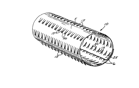
FIG. 1 shows a perspective view of one embodi-
ment of the present invention, showing a stent in a non-
expanded form.
FIG. 2 shows an axial view of another embodi-
ment of the present invention, employing shallower
dimensioned protrusions.
FIG. 3 shows the dimensions of the protrusions
in FIG. 2.
Docket No . ACS 33007 ( 6180 )
2~87132
-3-
FIG. 4 shows an axial view of a third
embodiment of the present invention, employing a random
dispersion of protrusions.
DETAILED DESCRIPTION OF THE INVENTION
5As shown by the embodiment of Fig. 1, a stent
10 is formed, in its natural state prior to expansion, in
a cylinder having protrusions or teeth 15 on one side of
the stent, forming a roughened outer wall or exterior
surface 5 that is adapted to lie contiguous with an
10arterial wall. Teeth 15 of roughened outer wall or
exterior surface 5 of stent body 10 are designed to
engage the arterial walls and endothelium layer of a
blood vessel, and, in general, would engage the walls of
a body lumen to help retain the stent in place.
15The teeth 15 are cut away from a sheet of
material which is then curled into a cylinder. The
cylinder has a wall thic~ness 25 at its end. In the
embodiment of Fig. 2 the wall thickness of the stent
tapers towards the ends, to form a beveled end surface,
20to give greater flexibility and vessel compliance to the
stent, as explained below.
Apertures 20 correspond to the recesses of body
10 left behind when teeth 15 were cut away from the body.
Apertures 20 thus have the same outline as teeth 15.
25As can be seen in the Fig. 1 embodiment, teeth
15 are arranged in rows extending axially
(longitudinally) along the stent body, and have a sharp,
narrow "V" shape. Although in this embodiment a sharp
"V" shape is employed, in general, practically any shaped
30protrusion may be employed. The teeth create a roughened
surface texture on the stent body.
Furthermore, while in all the embodiments shown
the protrusions are found on one side of the stent body,
namely the outer wall of the stent body or the side lying
35against the artery, it is possible to have protrusions or
projections on either or both sides of the body.
Docket No. ACS 33007 (6180)
2087132
-4-
In the embodiment of Fig. 1, teeth 15 project
out from the exterior surface or roughened outer wall 5
of stent body 10, the side adjacent to the artery wall.
The teeth can thus engage the stent wall in a positive
manner. The interior surface or inner wall 6 of stent
body 10 is the side facing the bloodstream, and is
substantially free of any projections.
In the preferred embodiment shown in Fig. 1,
the teeth are formed from stent body 10 in a one-piece or
unitary manner, being cut from the same sheet of material
that constitutes the stent body. The portion of the
stent body thus removed becomes a tooth or protrusion,
leaving an aperture 20 in the body having substantially
the same shape as the tooth. Apertures 20 are thus
defined by the portion of the stent body removed to form
the teeth or protrusions. The teeth may be for~ed by
injection-molding, casting, lasing, etching, plasma and
corona techniques, or machining.
Also in the preferred embodiment of Fig. 1,
stent body 10 is formed from a sheet of material curled
into a cylinder, with the sheet having overlapping edge~,
as can be seen in Fig. 1. Also, and as is readily
apparent from Fig. 1, the overlapping edges allow the
exterior surface or outer wall 5 to contact the interior
surface or inner wall 6 of the cylinder forming the
stent. The protrusions 15 on the outer wall, which make
the outer wall rougher than the inner wall, permit the
outer wall to engage the apertures in the inner wall,
and, together with the engagement of the protrusions with
the blood vessel, hold the stent in place in an enlarged
diameter form in the patient's vasculature.
While in the preferred embodiments disclosed
herein protrusions 15 were formed from the body in the
form of triangular teeth, in general, the protrusions may
be formed in any shape and in any manner, including by
adding the protrusions to a smooth stent body made of the
same or different material from the protrusions, or
treating the stent body to create a roughened surface
Docket No. ACS 33007 (6180)
2087132
texture, which texture can be with or without apertures
in the stent body.
Furthermore, while any material may be employed
to form the stent of the present invention, preferably a
Food and Drug Administration (FDA) approved material for
use in this environment is employed, that is, a bio-
compatible material. Metals such as stainless steel, Ni-
Ti, platinum (Pt), NitinolTM, tantalum (Ta) or gold (Au)
may be used.
A plastic that is biocompatible may be used.
The biocompatible plastic may also be bioabsorbable, that
is, biodegradable. For instance, a polymer from the
linear aliphatic polyester family, such as poly(lactic
acid), poly(glycolic acid) or polycaprolactone, and their
associated copolymers, may be employed. Degradable
polymers such as polyorthoester, polyanhydride, poly-
dioxanone and polyhydroxybutyrate may also be employed.
When the stent is expanded by an expanding
device, such as a balloon catheter, teeth 15 engage
apertures 20 to lock the stent open in an expanded
diameter form. A plurality of teeth 15 hold the stent in
an expanded diameter form by engaging apertures 20 and
the arterial walls. By engaging the arterial walls,
teeth 15 on roughened outer wall 5 also prevent the stent
from being axially displaced along the artery.
Expansion of stent 10 from a reduced diameter
form into an expanded diameter form is preferably
performed by a balloon catheter. Any other means for
expanding the stent, however, may be used. Briefly, and
in general terms, when the stent is to be deployed in a
coronary artery the stent is placed over a balloon
catheter that has been prepared for PTCA angioplasty.
The catheter is percutaneously introduced into a vessel,
following a previously positioned guidewire in an over-
the-wire angioplasty catheter system, and tracked by a
fluoroscope, until the balloon portion and associated
stent are positioned at the point where the stent is to
be placed. Thereafter, the balloon is inflated and the
Docket No. ACS 3âO07 (6180)
2087132
stent is expanded by the balloon portion from a reduced
diameter form to an expanded diameter form. After the
stent has been expanded to its final expanded diameter,
the balloon is deflated and the catheter is withdrawn,
leaving the stent in place.
It should be understood that the present stent
is not limited to use in coronary arteries and over-the-
wire angioplasty catheter systems, but the stent may be
deployed in any body lumen by any suitable mechanical
means, which includes hydraulic expansion.
To facilitate the placement of the stent of the
present invention, the stent may be impregnated with a
radiopaque material, making it opaque, and, therefore,
visible, to X-rays. Suitable radiopaque materials
include iodine-based materials and solutions thereof, and
barium salts, including materials containing iodipamide
(sold commercially under the trade name Cholografin),
iopanoic ~cid (sold under the trade name Telepaque),
barium sulfate, bismuth trioxide, bismuth oxychloride, or
powdered metals, such as tantalum, gold, platinum and
palladium.
It is further envisioned that the stent may be
impregnated with a therapeutic agent to provide localized
drug delivery.
When the stent has been expanded to its final
form, the stent is affixed in place by a combination of
the teeth in the body engaging apertures in the body of
the stent, as well as the teeth engaging the walls of the
artery, including the endothelium layer. It is believed
that the endothelium layer of the artery will grow into
the stent over a short period of time (in 7 to 21 days),
to further help retain the stent in place. The stent may
be made of a bioabsorbable material, such as any of the
materials disclosed above, so that eventually the stent
will dissolve. Endothelium layer growth into the stent
and the modified surface texture of the stent ensures
that pieces of the stent will not discharge into the
bloodstream and cause an embolism as the stent is
Docket No. ACS 33007 (6180)
2087132
dissolved.
It can be seen that one of the features of the
present invention includes maintaining an expandable
stent in an enlarged diameter or operative form in a body
lumen, by providing a roughened surface texture on the
outside surface of the stent that engages both the lumen
wall and the stent itself.
Turning now to Fig. 2, a protrusion pattern for
a second embodiment of the present invention is
disclosed. The stent of the second embodiment
substantially corresponds in structure to the embodiment
disclosed in Fig. 1, the principle difference being the
geometric shape and spacing of the teeth, and the
beveling of the ends, to be described below. In the Fig.
1 embodiment teeth 15 form a sharp, narrow "V" shape,
whereas in the second embodiment teeth 15 are less
pointed, and form a wider "V" shape. As shown in Fig. 3,
height 35 of the apex of the "V" is 0.010 in, width 40 at
the mouth of the "V" is 0.020 in., and the length of side
45 is 0.014 in. Sides 50 of each tooth are spaced at 90
to one another at the apex. The teeth are spaced along
the circumference of the stent in rows, each row spaced
0.0189 in. from one another, as indicated by reference
No. 57.
As can be seen in Fig. 2, another modification
of the stent of the second embodiment is the presence of
beveled ends 55. As can be seen from the drawings,
beveled ends 55 are defined as those portions of the
cylindrical stent that lie along the longitudinal (axial)
axis of the cylinder, spaced from one another by approx-
imately the longitudinal length of the stent. Beveled
ends 55 allow the stent to be more compliant and flexible
at its ends, so that the ends can more easily match the
flexibility of the vessel walls that the stent is
embedded in, and allow the stent to be more vessel
compliant. Beveled ends 55 taper from a larger wall
thickness away from the ends to a smaller wall thickness
at the very end of the stent, as can be seen from the
Docket No. ACS 33007 (6180)
2087132
--8--
drawing. For instance, on the right hand side the stent
is beveled along a portion of the stent starting from
point 65, defining the right hand end of the stent, to
75. In a symmetrical fashion the left hand side of the
stent end is beveled, with both beveled ends having an
angulation ~. As is readily apparent the wall thickness
of the stent along the beveled portion is less than the
wall thickness of the stent outside the beveled portion.
The height of the bevel from points 60 and 65
is 0.055 in., while the length of the straight portion of
the stent from points 70 and 75 is 0.455 in, with the
overall length from end to end being 0.600 in.
Another embodiment of the present invention is
shown in Fig. 4. In this embodiment, the tooth pattern
is substantially random, with protrusions 71 scattered
throughout the body of the stent, which is formed from a
sheet curled into a cylinder, as in the other embodi-
ments. The operation of this embodiment of stent is the
same as the other embodiments, with the teeth engaging
apertures in the body, as well as engaging the walls of
the vessel the stent resides in, when the stent is
expanded into an enlarged diameter form.
Again it should be understood that while in the
above embodiments the protrusions on the body were formed
from the stent body, in general, the protrusions may be
formed in any manner, including adding the protrusions to
a smooth stent body made of the same or different
material from the protrusions, or treating the stent body
to create a roughened surface texture, with or without
apertures in the stent body. As before, the surface
texture forming the protrusions may be formed via plasma
techniques, corona techniques, molding, casting, lasing,
etching, machining, or any other technique that changes
the surface texture of the body.
Furthermore, it should be understood that the
dimensions set forth for the above embodiments are not
intended to limit the invention to only those dimensions.
For example, while certain dimensions might be
Docket No . ACS 33007 ~ 6180 )
2087132
- 9 -
appropriate for a stent used in a coronary artery, these
same dimensions might not be suitable for a stent used in
other parts of a patient's vasculature or body lumen.
Other modifications can be made to the present
invention by those skilled in the art without departing
from the scope thereof.
Docket No. ACS 33007 (6180)
