Note: Descriptions are shown in the official language in which they were submitted.
W O 92/02276 PCT/US91/05292
~,3~9~
FIBE~ OPTIC LASER C~THETER AND ~ETHOD OF USE
Backqround of the Invention
This invention relates generally to fiber optic
laser catheters and, more particularly, to catheters of
this kind that are adapted to selectively position the
optical fiber's distal tip to facilitate the sculpting out
and debulking of an obstruction or occlusion in a vessel
o- lumen of a living body.
10Catheters of this type are particularly useful
- in removing obstructions or occlusions such as those
associated with arteriosclerosis lesions. In such
procedures, the catheter is inserted into the diseased
blood vessel and moved through the vessel to a position
where its distal end lies immediately adjacent to the
occlusion to be removed. A laser beam, commonly generated
by a pulsed excimer laser, is then directed through the
fiber and emitted from its distal tip, to ablate or
otherwise remove the lesion. In this particular
application, t~r laser catheter is being used to perform
angiosurgery, a procedure that is commonly referred to as
laser angioplasty.
In order to produce a successful angiosurgery
outcome, a sufficiently larger laser-generated lumen must
be created, without inflicting any damage on the remaining
tissues. ~lood vessels normally range in diameter from 1-
millimeters. Therefore, any successful laser
angiosurgery must be capable of producing a relatively
large lumen that is typically 70-80~ of the diameter of
the native healthy vessel. In orde_ to produce a
sufficiently large lumen, the fiber op'ic lase- ligh_
guide either mus~ have a very large light-emitting area,
. .
wo92/02776 PCT/US9l/05292
~ J ~ 2-
nearly the size of the vessel, or must use a small
diameter fiber and manipulate its distal tip to various
positions within the vessel so as to sculpt out a large
area. The large area fiber approach makes the catheter
less flexible and its use therefore may be limited to non-
tortuous vessels. The large area fiber approach also has
the disadvantage of requiring a large catheter introducer
sheath. The small fiber approach allows the catheter to
have superior flexibility, but requires a method of fiber
tip position control to create a lumen larger than the
fiber diameter.
In the past, catheters of the large area type
have been positioned using a guide wire that extends along
the vessel and through the lumen of a stenosis or partial
occlusion. This has not always proven to be a
satisfactory means of positioning, however, because the
hole in the occlusion is not always centrally located
within the vessel. Moreover, in the case of a total
occlusion, the hole is entirely absent and the use of a
guide wire is not possible.
Catheters of the small diameter type include
various kinds of structures for controlling either the X-
Y or the radial and circumferential positions of the
optical fiber's distal tip. This requires both a precise
positioning of the catheter relative to the vessel and a
precise positioning of the fiber tip within the catheter.
Even when the catheter can be precisely
positioned within the vessel, it has generally proven to
be difficult to sculpt away a sufficient amount of the
occlusion without at the same time mechanically or
- thermally damaging the vessel. Such damage can occur by
the mere physical rubbing of the catheter on the vessel
lining, injuring the endothelial cells, or by excessive
W092/02276 PCT/~S91/05292
-3~ 4 9 ~
heating of various parts of the vessel wall by the laser
beam. Such physical or thermal damage can lead to a
significant rate of restinosis, in which hyperplasia, the
excessive growth of smooth muscle cells, within the vessel
is triggered, leading in some cases to even more severe
blockages than were provided by the original occlusion
being removed.
It should therefore be appreciated that there is
a need for a fiber optic laser catheter that can be used
to more reliably and more thoroughly remove an obstruction
or occlusion in a vessel of a living body, in such a
fashion so as not to inflict any further injury to the
vessel. The present invention fulfills this need.
~ - ' .
Summary of the Invention
The present invention is embodied in a small
diameter fiber optic laser-catheter apparatus, and related
method for using it, that is adapted to cooperate with a
laser in substantially removing obstructions or occlusions
from a vessel of a living body, with reduced risk of
mechanically or thermally injuring the vessel. The
apparatus includes a tubular catheter sheath sized to be
received within the vessel, with a distal end of the
catheter sheath being located proximal to the occlusion,
and positioning means for engaging the vessel wall and
positioning the catheter sheath at a selected radial
position, e.g., centrally, within the vessel. An optical
fiber extends through the sheath and includes a proximal
end attachable to the laser and a distal end located
immediately adjacent to the occlusion to be removed. In
accordance with the invention, the apparatus further
includes fiber guide means located within the catheter
sheath and adapted to support the small diameter optical
fiber's distal end. The small diameter fiber may be a
single fiber or may be a bundle of smaller fibers. The
W092/02276 PCT/~S91/05292
'3 ~
fiber guide means is rotatable within the sheath, about
the sheath's longitudinal axis, and it is configured to
allow the fiber's distal tip to be positioned at any
selected radial and circumferential location relative to
the sheath. Since the only relative mechanical motion
during use of the catheter apparatus is within the fiber
guide means or between the fiber guide means and the
sheath, the vessel is isolated from that motion, whereby
the risk of damage to the vessel is substantially reduced.
In addition, since the catheter apparatus is itself
precisely positioned relative to the vessel, the risk of
vessel perforation during operation of the laser,
likewise, is substantially reduced.
During use of the catheter apparatus, the .iber
guide means is typically continuously rotated at a low
rate (e.g., less than 1000 rpm) while the laser is
energized and the laser beam removes successive bits of
tissue along a circumferential path. The use of a pulsed
excimer laser, or other pulsed laser with a wavelength
that is verv highly absorbed in tissue, is quire
advantageous in this application. With this type of
laser, tissue is discretely removed only at or very near
the fiber's distal tip and results in the generation of a
very smooth surface lumen. The fiber guide means'
rotation rate and the laser's pulse repetition rate are
approximately adjusted so that successive laser pulses do
not spatially overlap on the beam's circumferential path.
This reduces any thermal build-up and minimizes damage in
the surrounding tissues.
The fiber guide means can have any of several
alternative preferred configurations. In one preferred
embodiment, the fiber guide means includes a body and
means defining a channel within the body sized to receive
the optical fiber's distal end, with the channel having an
axis skewed relative to the outer sheath's axis. The
w092/02276 PCT/US91/05292
~5~ ~ 19~
optical fiber is selectively movable axially within the
channel such that the fiber's distal tip moves radially
relative to the fiber guide means and the sheath.
Selecting the distal tip's radial position facilitates the
sculpting away of large diameter obstructions. Extending
the optical fiber axially toward the lesion can even
provide a positioning of the fiber's distal tip radially
outside the sheath.
In an alternative embodiment, the fiber guide
means includes a body and means defining a slot within the
body sized to receive the optical fiber's distal end and
to permit that distal end to be moved laterally within the
slot, along an axis aligned substantially radially
relative to the sheath. In this embodiment, the fiber
guide means further includes biasing means for moving the
fiber's distal end to a selected radial location within
the slot. This biasing means can include an inflatable
bladder and means for inflating the bladder such that it
enlarges in size and forcibly urges the fiber's distal end
radially within the slot.
In another embodiment, the fiber guide means
includes spring bias means for yieldably urging the
optical fiber's distal end radially relative to the sheath
by an amount that varies as the optical fiber is moved
axially relative to the sheath. In this embodiment, the
fiber guide means further includes a body having a central
passageway that terminates in a flared opening, and an
elongated carrier for carrying the optical fiber's distal
end. The carrier is sized to be received in the body
passageway and to be axially and rotatable movable within
the passageway. The spring bias means is located on the
exterior of the elongated carrier and is adapted to engage
the flared opening and thereby move the optical fiber's
distal end radially relative to the sheath by an amount
that varies in accordance with the fiber's axial position.
'':''
:, . .
, .
WO 92/022~6 PCr/US91/05292
6--
~ J~b'~9~
In yet another embodiment, the fiber guide means
includes a body having a plurality of channels formed
within it, each channel sized to receive the optical fiber
and to hold its distal tip at a unique radial position.
In use, the fiber is placed sequentially from one such
channel to the next, with the fiber guide means being
rotated relative to the sheath during each such placement,
so as to sculpt out a series of concentric rings from the
lesion.
In yet another embodiment, the fiber guide means
includes a set of optica'l fiber holders, each having a
separate channel formed in it for receiving the optical
fiber's distal end. Each holder positions the fiber's
distal tip at a unique radial position. In use, the
separate holders are used sequentially, each sculpting out
of the lesion a ring-shaped segment of unique radius.
Other features and advantages of the present
invention should become apparent from the following
description of the preferred embodiments, taken in
conjunction with the accompanying drawings, which
illustrate, by way of example, the principles of the
invention.
Brief Description of the Drawin~q
FIG. 1 is an illustration of a fiber optic
catheter assembly constructed in accordance with the
invention, for use in removing obstructions or occlusions
from a vessel of a living body, the assembly being shown
connected to a laser and a fiber guide rotation device.
FIG. 2 is a side cross-sectional view of the
distal end of a first embodiment of a catheter assembly in
.. . .
.'' ,' `, ' . .
.,
W092/02276 PCT/US91/05292
-7~
accordance with the invention, depicted within a vessel in
a position to sculpt away part of an occlusion.
FIG. 3 is an end view of the catheter assembly
of FIG. 2.
FIG. 4 is a side, cross-sectional view of the
distal end portion of the catheter assembly of FIG. 2, but
showing the optical fiber axially extended from a fiber
guide contained in the assembly.
FIG. 5 is a side, cross-sectional view similar
to FIG. 4, but with the optical fiber extended even
further, to a position where its distal tip lies radially
beyond the catheter outer wall, allowing a hole to be
created that is larger than the catheter diameter.
FIG. 6a and FIG. 6b are a side, cross-sectional
view and an end view, respectively, of the distal end of
a second embodiment of a catheter assembly in accordance
with the invention, shown with a positioning bladder
deflated and the optical fiber positioned at one extreme
of its range of lateral motion.
FIG. 7a and FIG. 7b are views similar to FIG. 6a
and FIG. 6b, respectively, but with the bladder
pressurized and the optical fiber urged to the opposite
extreme of its range of lateral motion.
FIG. 8 is a side, cross-sectional view of a
third embodiment of a catheter assembly in accordance with
the invention, shown with the optical fiber retracted such
that its distal tip is located centrally within a fiber
holder of the assembly.
FIG. 9 is a side, cross-sectional view of the
distal end of the catheter assembly of FIG. 8, but with
. . .
.,' , .
.. '' ' ' :
,
w092/02276 PCT/US91/05292
~ g~ -8-
the optical fiber axially extended such that its distaltip is urged laterally by a leaf spring biased against a
flared openlng in the fiber holder.
FIG. 10 is an end view of a fourth embodiment of
a catheter assembly in accordance with the invention,
showing three channels sized to receive the optical fiber
and position its distal tip at a three selected radii.
FIG. lla and FIG. llb are a side, cross-
sectional view and an end view, respectively, of the
distal end of a fifth embodiment of a catheter assembly in
accordance with the invention, showing one of several
optical fiber guides used sequentially, the depicted guide
positioning the fiber's distal tip centrally within the
assembly.
FIG. 12a and FIG. 12b are views similar to FIG.
lla and FIG. llb, respectively, but showing a second
optical fiber holder, this holder positioning the fiber's
distal tip at an intermediate radial position.
FIG. 13a and FIG. 13b are views similar to FIG.
2C lla and FIG. llb, respectively, but showing a third
optical fiber holder, this holder positioning the fiber's
distal tip at a maximum radial position.
,.. ~ :
~ .
w092~02276 PCT/US91/OS292
_g_ J ~
Description of the Preferred Embo~iments
With reference now to the drawings, and
particularly to FIG. 1, there is shown a fiber optic laser
catheter assembly 21 adapted for use with a laser 23 in
sculpting away a partial obstruction or total occlusion in
a vessel of a patient 24. The catheter assembly is
particularly useful with an excimer laser in removing
arterial occlusions resulting from arteriosclerosis. The
assembly includes an elongated, tubular sheath 25 sized
to be insertable freely into the vessel, with an optical
fiber 27 extending fully through the sheath. The fiber's
proximal end is connected to the laser, and the fiber's
distal end is located at or near the sheath's distal end.
As is conventional, the assembly's proximal end includes
an input device 29 having various fittings adapted for
connection through fluid connection parts 31a and 31b to
supplies for various solutions, etc.
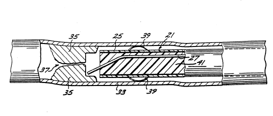
As shown in FIG. 2, a first embodiment of a
fiber optic catheter assembly 21 is positioned within a
vessel 33, with its distal end located immediately
adjacent to an occlusion 35. In this case, the occlusion
is only partial, with a narrow opening 37 located near the
vessel's centerline. In addition to the sheath 25 and
optical fiber 27, the assembly further includes a
mechanical device such as an inflatable balloon or bladder
39 for use in centering the assembly within a healthy part
of the vessel. The assembly is moved axially through the
vessel to its desired location, after which the bladder is
inflated by pressurizing it with saline solution, or radio
opaque contrast media, provided through one of the tubes
3la or 3lb connected to the assembly via the input device
29. The entire procedure may be easily monitored by
conventional x-ray fluoroscopy.
An elongated fiber guide or holder 41 is
WO 92/02276 PCI/US91/05292
10-
positioned within the tubular sheath 25, for supporting
the optical fiber 27. In particular, a cylindrical
channel is formed along the guide's entire length, the
channel being of a si~e to slidably receive the optical
fiber. The fiber guide has an outside diameter
approximately the same as the inside diameter of the
sheath, at least in the region of the assembly's distal
end. The abutting surfaces of the fiber guide and the
sheath are of high lubricity, to facilitate a convenient
rotation with minimum torque of the fiber guide about its
longitudinal axis. As shown in FIG. 1, a conventional
stepper motor 43 controllably rotates the fiber guide
through a collar 45 secured to the holder at its proximal
end.
With particular reference now to FIGS. 2-5, it
will be observed that the channel in the fiber guide 41
that slidably receives the optical fiber 27 is angularly
skewed at its distal end relative to the guide's
centerline. The channel emerges from the guide's distal
end at a point substantially coincident with the guide's
centerline. Thus, when the optical fiber's distal tip is
coterminous with the fiber guide's distal end (FIG. 2),
the laser beam being transmitted along the fiber will be
emitted at a point at or very near the vessel's
centerline.
As previously mentioned, the optical fiber 27
is selectively slidable along the channel formed in the
fiber guide 41. When the fiber has been slid axially
forwardly to the location depicted in FIG. 4, the catheter
assembly 21 will emit the laser beam at a point radially
spaced from the vessel's centerline. Consequently, in
this configuration, the catheter assembly can be used to
remove a section of the occlusion 35 located radially
outwardly from the vessel's centerline. Rotating the
fiber guide 41 relative to the sheath 25 using the motor
.
?
- ~ .
. .
.
WO 92/02276 PCI/US91/0~292
--11-- , ~
43 and collar 45 will cause the exposed distal end of the
optical fiber to sweep in a conical motion, whereby a
ring-shaped section of the occlusion, concentric with the
vessel's centerline, can be removed.
S The rotation rate of the fiber guide 41 is
preferably less than about 1000 rpm, and the pulse
repetition rate of the laser 23 (FIG. 1) is preferably
synchronized with the rotation rate, so that successive
laser pulses do not spatially overlap on the beam's
circumferential path. This reduces any thermal build-up
and minimizes damage in the surrounding tissues.
Alternatively, the fiber guide 41 may be rotated manually.
Such manual rotation is particularly effective when
removing an obstruction from a vessel in an intraoperative
procedure, in which the catheter assembly is very short
and inserted into the vessel through an incision located
near the obstruction.
It will be appreciated that by incrementally
advancing the optical fiber 27 axially through the channel
in the fiber guide 41, a series of rings of progressively
larger radius can be removed from the occlusion 35. At
its extreme, the optical fiber can even be moved axially
forwardly through the fiber holder to a point where its
distal tip projects radially beyond the sheath 25. This
enables the removal of a ring-shaped section of the
occlusion larger even than the sheath. Such a position
for the optical fiber is depicted in FIG. 5.
Rotating the fiber holder 41 about its
longitudinal axis causes relative motion between the
optical fiber 27 and the holder and between the holder and
the sheath 25. The optical fiber and the sheath both
remain rotationally stationary, although, as described
above, the skewing of the fiber holder's channel causes
the fiber's angular orientation to vary with the rotation.
- .
WO 92/022-6 PCr/~;S91/05292
12-
Significantly, the lack of relative motion
between the catheter assembly's sheath 25 and the wall of
the vessel 33 avoids the possibility of the vessel wall
tissue being mechanically damaged. In the absence of the
sheath, rotation of the fiber holder could otherwise rub
the vessel wall so as to injure endothelial cells and
thereby trigger a hyperplastic response, which could lead
to restinosis in the vessel. The sheath effectively
isolates the rotating fiber holder from the vessel wall
and thus obviates this problem.
After the catheter assembly 21 has been used to
remove a central segment and a series of contiguous ring-
shaped segments, as described above, the assembly can be
advanced incrementally forwardly within the vessel 33 and
the process repeated to remove an additional layer of
contiguous ring-shaped segments from the occlusion. This
sequential process can be repeated until the occlusion has
been completely removed, or at least removed sufficiently
to enable adequate blood flow through the vessel. The
assembly is preferably used in treating vessels having
occlusions that occur in relatively non-tortuous regions
and that have lengths of a few centimeters or less.
Otherwise, difficultly might be encountered in maintaining
the assembly's distal end properly centered wlthin the
vessel. In order to treat very tortuous vessels, it would
most likely be necessary to augment the catheter apparatus
with an additional form of guidance and visualization,
such as that provided by angioscopy or intravascular
ultra-sound. Ideally, the inflatable bladder 39 for use
in centering the assembly's distal end always engages
portions of the vessel wall that are proximal to the
occlusion and proximal to those portions of the occlusions
removed during the initial stages of its removal.
With reference now to FIGS. 6(a) and (b) and
W092/022,6 PCT/~S91/05292
-13- ~u~
FIGS. 7(a) and (b), there is shown the distal end of a
second preferred embodiment of a catheter assembly 51 in
accordance with the invention. This second embodiment is
similar to the first embodiment of FIGS. 2-5, except that
the distal end of its fiber guide 53 supports the optical
fiber 27 in a slot 55 having a non-circular cross-section.
The slot is sized in the circumferential direction to be
substantially the same as the optical fiber's diameter,
but is sized in the radial direction to be much wider.
This permits the fiber's distal end to be moved radially,
thereby facilitating the use of the catheter assembly to
sculpt out large portions of occlusions formed in the
vessel.
To controllably move the optical fiber's distal
tip radially within the slot 55, this second embodiment
51 further includes an inflatable bladder 57 positioned
within the fiber guide 53 so as to apply a lateral
pressure to the fiber 27. The bladder is controllably
inflated using a pressurized saline solution delivered to
it via one of the tubes associated with the assembly's
input device 29 (FIG. 1). FIGS. 6(a) and 6(b) show the
bladder in its fully deflated condition, such that the
fiber's distal end remains centrally located within the
fiber holder 53. FIGS. 7(a) and 7(b), show the bladder
fully inflated such that the fiber's distal end is urged
radially outwardly to its furthest outward position. The
bladder may be inflated in either a continuous fashion or
a step-wise fashion.
This second catheter assembly embodiment 51 is
used in much the same fashion as the embodiment 21 of
FIGS. 2-5, the only significant difference being in the
manner in which the optical fiber's distal end is moved
incrementally in the radial direction. Rather that moving
the fiber 27 axially forwardly to increase the radial
position of its distal tip, the bladder is incrementally
:.
, . .
, .
:' ,
,, ,
'
W O 92/02276 PCT/~591/05292
-14-
inflated. In each incremental position, either a central
segment or a ring-shaped segment is removed from the
occlusion 35. After an entire layer of contiguous ring-
shaped sections have been removed, the catheter assembly
is moved incrementally forwardly within the vessel 33, to
facilitate the removal of a succeeding layer. It will be
noted that the axial position of the fiber's distal tip
remains substantially the same for all of its successive
radial positions. This ensures that each successive layer
of contiguous, ring-shaped sections that is removed from
the occlusions is substantially planar.
FIGS. 8 and 9 depict the distal end of a third
catheter assembly embodiment 61 in accordance with the
invention. In this embodiment, an elongated fiber guide
63 terminates within a body 65 at the distal end of the
assembly's sheath 66. The body and sheath include a
central passageway 67 of circular cross-section, for
receiving the fiber guide and allowing the fiber guide to
rotate relative to it, about its longitudinal axis. The
distal end of the passageway flares outwardly to define a
generally conical surface 69 in the body.
Attached to the exterior surface of the fiber
guide, near its distal end, is a leaf spring 71. When the
fiber guide is axially positioned with the leaf spring
located within the passageway 67 of the body 65 and sheath
66, the leaf spring is forced to a straight position,
parallel with the fiber guide's longitudinal axis. As the
fiber guide 63 and optical fiber 27 are moved axially
forwardly to bring a portion of the spring into the region
of the conical surface 69, however, the spring and fiber
are yieldably urged apart from each other, so as to
position the fiber's distal tip at a radial location
spaced from the centerline of the body and vessel.
Eventually, continued forward axial movement of the fiber
will cause the fiber's distal tip to be moved to the
w092/02276 PCT/US9l/05292
-15- ~9 ~
maximum deflected angle. The optical fiber can be moved
axially within the spring-deflected guide, to control the
radial position of the fiber's distal tip relative to the
assembly's centerline.
As with the first two embodiments, this third
catheter assembly embodiment 61 is used to sequentially
remove first a center section and then a series of
contiguous ring-shaped sections from the occlusion. After
a single layer of such sections has been removed, the
catheter assembly is advanced forwardly within the vessel
to facilitate the removal of a second and subsequent
layers. As with the first two embodiments, this third
catheter assembly embodiment also can be used to remove
ring-shaped sections from the occlusion having a radius
greater than that of the assembly's sheath.
- FIG. 10 is an end view of the distal end of a
fourth catheter assembly embodiment 81 in accordance with
the invention. This emhodiment includes a fiber holder 83
having a set of three separate channels 85a, 85b and 85c
for slidably receiving the optical fiber 27. Each such
channel is located at a different radial position within
the fiber holder. The catheter assembly is used by
placing the optical fiber sequentially in each of the
separate channels, each time the assembly being used to
sculpt out of the occlusion either a central section or a
concentric ring-shaped section. The separate channels are
sized and positioned such that the successive sections are
all contiguous with each other. Some overlap is required
to compensate for the presence of cladding on the optical
fiber's outer surface.
FIGS. 11-13 depict a fifth catheter assembly
embodiment 91 in accordance with the invention. In this
embodiment, three separate fiber guides 93a, 93b and 93c
are utilized in sequence. Each guide supports the optical
., -
WO 92/02276 PCI/I_IS91/05292
1 J ~ ~ ~ .3 ~, -16-
fiber 27 at a different radial position. The guide 93a
(FIGS. ll(a) and ll(b)) supports the fiber in a central
position, the guide 93b (FIGS. 12(a) and 12(b)) supports
the fiber at an intermediate radial position, and the
guide 93c (FIGS. 13(a) and 13(b)) supports the fiber at an
extreme radial position.
Initially, the fiber guide 93a is used such that
the catheter assembly 91 removes a central section,
approximately the diameter of the fiber 25, from the
occlusion in the vessel. With this fiber guide 93a in
position, the fiber, itself, can be moved axially
forwardly through the guide so as to remove a succession
of centrally-located sections from the occlusion. Next,
the fiber guide 93b is substituted for the guide 93a and
a ring-shaped section, concentric with the initially-
removed section, is removed. Again, the optical fiber can
be moved incrementally forwardly through the guide 93b to
remove a succession of ring-shaped sections from the
occlusion. Finally, the fiber guide 93c is substituted
for the guide 93b and the process repeated for a
succession of further ring-shaped sections.
It should be appreciated from the foregoing
description that the present invention provides an
improved fiber optic laser catheter assembly for use in
removing, or largely debulking, obstructions or occlusions
from a lumen in a living body. In all of the disclosed
embodiments, the obstructions are removed by controllably
positioning an optical fiber's distal tip in a succession
of selected radial and circumferential positions so as to
sculpt away the obstruction without substantial risk of
mechanically or thermally damaging the vessel. Several of
the embodiments are configured such that the portion of
the occlusion that can be reliably sculpted away has a
radius greater even than the radius of the catheter
assembly itself.
; ,
.: ' '- ' ` ' '
wog2/02~ PCT/US91/05292
Although the invention has been described in
detail with reference to the presently preferred
embodiments, those of ordinary skill in the art will
appreciate that various modifications can be made without
departing from the invention. Accordingly, the invention
is defined only by the following claims.
..... : . ~ ,.
.`', ,.,, ,1~ ~
.. . .
' . , :
.