Note: Descriptions are shown in the official language in which they were submitted.
Immunological assay method for the determination of
antibodies in biological fluids and kit for
carrying out the method
The invention relates to an immunological assay method
for the determination of antibodies in biological
fluids, in particular of autoantibodies, the detection
of which permits the diagnosis of an autoimmune
disease.
Various immunological assay methods play a very
important role in medical diagnostics. In addition to
those assay methods aimed at the qualitative and/or
quantitative determination of antigens or haptens, for
example hormones, there are also many assay methods of
this type for the determination of antibodies in
biological fluids, in particular human sera.
Antibodies are proteins which are designated as
immunoglobulins (Ig) and which are formed by the body
as a reaction to an antigen. Since antigens normally
208~8'~~
have many antigenic determinants, antibodies are
polyclonal and therefore represent a population of
proteins having different binding properties relative
to the antigen against which they are directed. They
are normally formed to counteract exogenous antigens in
order to protect the body from substances which have
corresponding antigenic determinants. If the immune
system of the body incorrectly recognises certain
endogenous cells or cell structures as being exogenous,
however, antibodies can also be formed against
antigenic determinants of endogenous elements. Such
endogenous elements are then designated as
autoantigens, and the antibodies formed to counteract
them are designated as autoantibodies.
Known assay methods for antibodies realise, in one form
or another, various basic principles, two of which are
shown schematically, for example, at the top of column
3 of U.S. Patent B1 3 654 090. According to a variant
which corresponds to the classical radioimmunoassay
(RIA), a deficiency of an immobilised antigen is used,
and a labelled form of the antibody to be determined is
added in a known amount to the sample to be
investigated. Information about the presence or
concentration of the required antibody can be obtained
from the degree of binding of the labelled antibody to
the immobilised antigen. The antigen is required in
highly pure form for this test based on the competition
principle.
According to a second principle, a known amount of the
antibody to be determined or of a suitable derivative
thereof is immobilised on a solid substrate, and the
antibody present in the sample to be investigated and
the immobilised antibody are then allowed to compete
for a labelled antigen added to the reaction system.
The presence or amount of the antibody to be determined
~~ ~ ~ iJ Pg
3
is obtained from the reduction in the binding of the
labelled antigen to the immobilised antibody and
therefore to the solid phase.
In the last-mentioned method of determination, a
labelled form of the associated highly pure antigen is
required, and the antibody to be determined and the
immobilised antibody must be present in amounts such
that effective competition can occur between the
immobilised antibody and the antibody in the sample to
be investigated, the fact that affinities to the
labelled antigen may not be completely identical being
taken into account.
According to a further principle, in a procedure
analogous to the sandwich test well known for antigen
determination an excess of an antigen, usually in
immobilised form, is first taken, by means of which the
total amount of the antibody to be determined is bound,
and, by a subsequent second immunological reaction with
a second labelled "antigen", against the antibody bound
in the first step, the latter is labelled with
formation of a sandwich-like immune complex. The
second "antigen" is frequently an anti-antibody (double
antibody method), or it is used for labelling the
labelled so-called protein A, a protein which is
obtained from bacteria and binds unspecifically to many
IgG antibodies. In this method, the amounts of antigen
required are such that the binding capacity is
sufficient for bindina all antib~ciiP~ nrP~Ant ;n ;-ho
sample. If larger amounts of the antibodies to be
determined are expected, the samples must therefore
generally be highly diluted before they can be used in
the test. This applies in particular to the coated
tube technique, which is often preferred for practical
reasons, and the microtitre plate technique, in which
an antigen-coated test tube has a binding capacity of
~~~~5~~
4
only about 1-2 pg of human antibodies.
In the determination of antibodies against exogenous
antigens, the requirements for the functioning of the
various tests can frequently be met without great
difficulties. The antibody concentrations against
exogenous antigens are normally relatively low, and the
associated antigens or haptens can frequently be
synthesised in sufficient amounts by a chemical or
biotechnological method or can be isolated from natural
material and concentrated.
However, if it is intended to determine autoantibodies
by one of the principles described, a number of
difficulties, some of them considerable, are
encountered, both as a result of the autoantibody
concentrations which occur and as a result of the
nature of the autoantigens.
The determination of autoantibodies is very important
for detecting the presence of an autoimmune disease, in
particular for correctly interpreting the observed
symptoms and avoiding harmful incorrect treatments.
Known autoimmune diseases, some of which are extremely
severe, are for example rheumatoid arthritis, diabetes
mellitus type 1, myasthenia gravis and some autoimmune
diseases associated with the thyroid, such as Basedow's
hyperthyreosis (also referred to as Graves' disease),
anaemic myxoedema and Hashimoto's thyroiditis. In the
case of the thyroid autoimmune diseases,
thyreoglobulin, THS receptor and/or thyroid peroxidase
(TPO) act as autoantigens, depending on the type of
disease, and recent discoveries have shown that the
latter is identical to the so-called microsomal
antigen. The present invention is described below in
particular with respect to the determination of thyroid
autoantibodies, in particular of antibodies against
5
hTPO, but the novel principle on which the invention is
based is not restricted to these specific
determinations but can also usefully be applied to the
determination of other autoantibodies. In the
determination of other antibodies, it may in specific
cases also have advantages over the assay methods based
on the known principles.
Reviews of the current state of knowledge in the area
of the thyroid autoimmune diseases are to be found in
the scientific literature, for example in the article
by Marian Ludgate and Gilbert Vassart in: Autoimmunity,
1990, Volume 7, pages 201-211; in the Review by Jadwiga
Furmaniak and Bernard Rees Smith in: Autoimmunity,
1990, Volume 7, pages 63-80; and in the article by P.-
M. Schumm-Drager, H.J.C. Wenisch in: Akt. Endokr.
Stoffw. 10 (1989), pages 9-102 (special edition), where
an overview of the methods for the. detection of thyroid
autoantibodies is given.
The immunodiagnostic determination of thyroid
autoantibodies or autoantibodies generally with
corresponding use of one of the determination types
mentioned at the outset encounters the fundamental
difficulty that the autoantibodies are very frequently
directed against autoantigens which are anchored in the
cell membrane and are difficult to obtain in the high
purity and amount required for the usual procedure. In
the case of the human thyroid peroxidase (hTPO), an
enzyme which, as an autoantigen, is responsible for
Hashimoto's thyroiditis, it is, for example, a
glycosylated haemoprotein which is bound to the thyroid
membranes. Its antigenic properties, including the
types of epitopes present on its surface, are described
in the article by P. Carayon et al. in: Endocrinology,
Vol. 125, No. 3, pages 1211 to 1218. In order to have
this thyroid peroxidase available in sufficient purity
6
and amount as an antigen for the immunodiagnostic
determination method based on the known principles, the
thyroid peroxidase must be removed from the membrane by
a proteolytic method or with the aid of detergents and
purified via immune adsorbents or by means of
conventional chromatography methods over various
separation stages, for example by means of gel
filtration, ion exchange chromatography, chromatography
via hydrophobic interactians, chromatography via
aromatic interactions, adsorption chromatography and
chromatography using concanavalin A. These methods are
complicated and entail the risk of unintentional
changes in the enzyme to be isolated and high material
loss. Highly purified natural thyroid peroxidase (TPO)
is therefore available only in small amounts and at
high prices. As an alternative to isolating the
thyroid peroxidase from thyroid glands, attempts were
therefore also be made and methods developed to permit
the production of TPO by genetic engineering. However,
the TPO obtained in 'this manner is also available only
in limited amounts and at high prices, and the identity
of the material obtained by genetic engineering with
the natural thyroid peroxidase, particularly with
regard to the antigenic properties, is not guaranteed
in every case.
A further difficulty also arises from the fact that the
antigenic properties of the TPO can be very greatly
impaired by chemical effects, particularly if, as a
result, the three-dimensional structure is changed
and/or the disulphide bridges are broken (cf. the
stated article by P. Carayon). However, in order to be
able to use TPO as a labelled antigen in the classical
method for antibody assay, a label must be chemically
bonded to the TPO. In addition to the difficulty of
obtaining pure TPO, there is at this stage the risk
that, as a result of the reactions associated with the
2fl~~~'~~
labelling, the antigenic properties of the TPO will be
influenced so that it no longer corresponds to the
natural TPO and is therefore suitable only to a limited
extent as an antigen for the detection of
autoantibodies. For example, the changes caused by the
isolation and/or labelling of the TPO may result in
only some of the polyclonal TPO autoantibodes reacting
with a TPO labelled in this way.
To avoid at least some of the problems associated with
the isolation and labelling of TPO, a test in which a
TPO which is not highly purified but used in crude form
is employed as an immobilised antigen was developed as
a modification of the sandwich test described at the
outset for the assay of antibodies. In this test,
however, there is the danger that the immobilised crude
TPO may also contain other substances having antigenic
properties which lead to immobilisation of antibodies
other than the required antibodies, and that these will
then be labelled in the subsequent, relatively
unspecific labelling by a double antibody method or by
labelled protein A and will give false positive
results.
From practical points of view, in. particular with
regard to the required work and the achievable accuracy
of determination, a further problem in the
determination of autoantibodies is that, when an
autoimmune disease is present, they occur in extremely
large amounts in the biological fluids, particularly
the patients' sera. For example, up to 20 ug of human
autoantibodies are to be expected in a sample volume of
20 u1 of serum. In order to be able to determine them,
particularly by the coated tube technique or microtitre
plate technique, it is therefore normally necessary to
dilute the patients' sera several-fold, which is
labour-intensive and time-consuming and constitutes an
w
CA 02089870 2002-11-25
E3
additional source ~f er.ro.r=.
In view of the difficulties c'escr.ibed in the
determinatic>n of au~;.oanti.bod.ies by the known
immunodiagnost.ic assay methods, thex-e is therefore an
urgent need for a novel method for the immunodiagnostic
determination of eiutoamtibodies in biological fluids
which permits a safe qualitative determination of
antibodies in biological fluids, which, as a result of
suitable calibration and opt~imisatiorl of the parameters
of the method, is also suitable for the reliable
quantitative determination of aIlt:~_bodies, in which
undiluted sera can be used and in which it is not
necessary to use significant amounts of highly purified
antigens for the antibodies to be determined.
It is the object of the present invention to provide
such a method.
This object is achieved by an immunological assay
method as described herE~in.
In the assay method according to the invention, a
procedure is adopted in which, for the detection of
antibodies (Ak) , in particular of autoantibodies, the
disturbance of the formation of a sandwich complex of
a first immobilised antibody (Ak~~), an added antigen
(Ag), in particular a crude antigen, and a further
antibody (Ak ) which carries a detectable label is
determined, said disturbance being due to the presence
of the antibodies (Ak) to be determined in the
biological fluid. The inhibition of the formation of
a sandwich complex. of the stated type is evident in a
reduction in the binding of the labelled antibody (Ak )
~~~~~"l~
9
to the solid phase. The disturbance of the formation
of the sandwich by the antibody or autoantibody (Ak) to
be determined and present in the sample can occur in
principle at each individual binding site for the
synthesis of the sandwich or at both simultaneously.
Substances which act as autoantigens and can be formed
against the autoantibodies are, as a rule, complexes of
large molecules, generally of a protein nature, which
have more than two regions or epitopes involved in the
antibody binding, and the antibodies formed are
polyclonal. Depending on the choice of the antibodies
selected for the test as immobilised or labelled
antibodies, it is therefore possible to construct
different types of sandwich complexes which - as far as
the bonds involved and the disturbance thereof are
concerned - may behave very differently with regard to
the presence of the autoantibodies (Ak). The
construction of such sandwiches with specification of
suitable properties for the detection of certain
autoantibodies (Ak) can be tailored in particular when
both the immobilised antibody (Ak~~) and the labelled
antibody (Ak ) are suitable monoclonal antibodies. In
the case of the determination of autoantibodies with
respect to TPO, the epitopes present on this antigen
are relatively well characterised, and various
monoclonal antibodies are available which are directed
against specific epitopes of this antigen. In this
connection, reference may be made to the above
mentioned article by P. Carayon et al. in
Endocrinology, Volume 125, No. 3, pages 1211-1218.
If such an antibody, in particular a monoclonal
antibody, which binds TPO in a region in which
autoantibodies against TPO are also bound is chosen as
the immobilised antibody against TPO, and if the
labelled antibody Ak is chosen so that its binding is
1~
not influenced by autoantibodies, the disturbance of
the formation of the sandwich is due to competition
between the immobilised antibody Ak~~ and the antibody
Ak to be determined for the complex antigen-labelled
antibody (Ag-Ak ), which may be regarded as an
indirectly labelled antigen. If the opposite approach
is adopted and the immobilised antibodies Ak~~ is
chosen so that its binding to the antigen is not
disturbed by the antibodies (Ak) to be determined,
while the labelled antibody Ak binds to regions or
epitopes of the antigen where the antibodies or
autoantibodies to be determined also bind, the
disturbance of the formation of a sandwich which
contains the labelled antibody Ak takes place owing to
*
the competition between Ak and Ak for the indirectly
immobilised antigen. If both antibodies Ak~~ and Ak*
bind to those regions of the antigen where the
autoantibodies (Ak) also bind, the presence of such
autoantibodies (Ak) results in double impairment of the
sandwich structure and its immobilisation and hence may
give rise to an increase in sensitivity.
A very significant advantage of the method according to
the invention is that the antigen Ag need not be
present in highly purified form as in the assay methods
known to date but can be used as crude antigen, for
example in the form of an organ extract. The double
antibody specificity which is required for the
formation of a sandwich which contains a label means
that unspecific disturbances of the test by foreign
components which are introduced into the assay together
with the crude antigen can be eliminated without
difficulties, so that sandwich formation is limited to
the required immunological binding partners. While in
the case described above, where the crude antigen is
immobilised together with accompanying substances,
11
there is the danger that not only antibodes against the
immobilised antigen but also immunological binding
partners of other antigenic substances present in the
crude antigen are bound to the solid phase and
labelled, such disturbances are very improbable in the
method according to the invention. The possibility of
using a crude antigen in the form of a suitable organ
extract has the further advantage that it is possible
to use relatively gentle recovery methods for the
antigen and to dispense with purification steps by
means of which the natural structure of the antigen is
attacked. The antigen can therefore very much more
exactly represent the antigen present in the organism
and its complete immunological binding spectrum. The
danger that only a fraction of the autoantibodies
formed in the organism will be detected by the test is
thus smaller.
0n the other hand, by a suitable choice of the
monoclonal antibodies used for the test, it is also
possible to ensure that the test responds very
specifically only to certain autoantibodies and can
thus be made more selective. If, for example, certain
symptoms associated with the autoimmune disease can be
attributed to very specific clones of the polyclonal
autoantibodies formed by the body, the method for the
determination of such autoantibody clones can be made
specific by using monoclonal or, optionally, selected
polyclonal antibodies.
In the method according to the invention, the expected
high autoantibody concentrations present no problem
since the crude antigen, for example the crude hTPO,
can be used in sufficient amounts comparable with the
amounts of autoantibodies without making the test
excessively expensive. It has been found that the
possibility of using the crude antigen is so
12
advantageous in terms of cost that the two added
antibodies (Ak~~, Ak ) required in the method according
to the invention do not give rise to any cost
disadvantage compared with methods operating with
highly purified antigens but only one antibody.
The assay method according to the invention is also
very suitable for optimisation with regard to the
required sensitivity by adapting the intended amounts
of antigen Ag or labelled antibody Ak* or immobilised
antibody Ak~~ in wide limits to the test conditions.
This is possible without major problems, owing to the
good availability and the relatively low price of the
crude, natural antigen used. The amount of antigen can
be matched directly with the expected or possible
amounts of autoantibody, which also means that the
biological fluids, i.e. in particular sera, can be used
in undiluted form.
Regarding the amounts of immunological reactants to be
used, it is self-evident to one skilled in the art that
the amount of added antigen should not be so large that
both all antibodies Ak to be determined and all
labelled antibodies Ak or all immobilised antibodies
Ak~~ are saturated and there is no longer any
significant competition between them for the antigen.
In such a case, significant formation of an
immobilised, labelled sandwich can be suppressed in
certain circumstances, and no quantitative conclusions
about the actual amounts of autoantibodies present in
the sample are possible.
If a deficiency of, for example, labelled antibody Ak
is used relative to the amount of the added antigen, so
that only a part of the amount of antigen is labelled
with formation of an immune complex Ag-Ak , the amount
13
*
of the labelled Ak must not of course be so small that
the amount of complex finally bound is too small owing
to its competition with unlabelled antigen for the
immobilised antibody Ak~~. An analogous situation
applies, in another described variant of the method,
for the amount of the immobilised antibody Ak~~.
The concentrations of antigen Ag, immobilised antibody
Ak~~ and labelled antibody Ak which are required or
most advantageous for a certain test can be determined,
taking into account the expected amounts of the
antibodies Ak to be determined, without significant
difficulties in routine optimisations by varying the
sensitivity of the test (calibration curve) via, for
example, the added amounts of antigen.
Antibodies which can be used in the method according to
the invention may essentially be all antibodies
suitable for immunological assay methods. Their
affinity constants are usually in the range from 1012 to
10$ 1/mol.
With regard to the solid substrates which can be used
for immobilised antibodies Ak~~ and the conditions of
the immunological reaction (pH between 4 and 9,
presence of buffers; temperature in the range from 0 to
about 55°C; reaction times), the procedure does not
differ fundamentally from other conventional
immunological assay methods. The immunological
reaction can be carried out under conditions such that
the equilibrium between all reactants is reached;
however, it is in principle also possible to stop the
reaction at an earlier predetermined time and to
determine the ratios at this earlier time.
The method according to the invention is described in
w
14
detail below with reference to an embodiment which
relates to the determination of autoantibodies against
human thyroid peroxidase (hTPO) with the use of two
added monoclonal antibodies against hTPO and of crude
hTPO in the form of an extract from human thyroid
glands as added antigen, arid .its efficiency.is compared
with that of the known methods for the determination of
the same autoantibodies.
In the Figures,
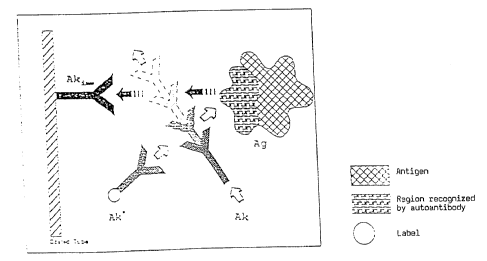
Figure 1 shows a schematic diagram of the method
according to the invention in a first variant in which
the immobilised antibody Ak~~ binds to a region of the
antigen Ag which is also recognised by the antibody Ak
which is to be determined and which is preferably an
autoantibody, so that the antibody Ak competes with the
immobilised antibody Ak~~ for the antigen Ag and thus
interferes with its fixation on the solid phase; the
disturbance caused by the antibody Ak is represented
here and in the following Figures 2 and 3 symbolically
as an intervening movement of the antibody Ak,
although, as is quite clear to one skilled in the art,
the observed disturbance is due to competition for the
antigen.
Figure 2 shows a further variant of the method
according to the invention, in which the antibodies Ak
to be determined and the labelled antibodies Ak
compete for the region of the antigen Ag which is
indirectly immobilised via Ak~~ and is recognised by
the autoantibody Ak.
Figure 3 shows a third variant of the method according
to the invention in which both the immobilised antibody
Ak~~ and the labelled antibody Ak bind to a region of
the antigen Ag which is also recognised by the
15
autobody Ak, so that the presence of the antibody Ak in
the sample causes a double disturbance.
Figure 4 shows a graphic representation of the standard
curve in an assay for the detection of human
autoantibodies against human thyroid peroxidase (hTPO)
as a function of the amount of antigen used (crude
hTPO).
Example
The following Example describes the procedure and the
practical advantages of an assay in which the method
according to the invention is realised, with reference
to a preferred embodiment which relates to the
detection of human autoantibodies against human thyroid
peroxidase (hTPO).
1. Immobilisation of a monoclonal anti-hTPO antibody
(Ak~~) on a solid phase
A purified monoclonal antibody which binds to a region
of hTPO which is recognised by human autoantibodies
against hTPO was chosen as the antibody which is
coupled to the solid phase. The purified monoclonal
antibody used was a monoclonal mouse anti-hTPO antibody
which had been prepared by the process described in the
publication by P. Carayon in: Endocrinology, Vol. 125,
No. 3, page 1212, bottom of left column, top of right
column, and which was chosen according to the selection
criteria described in the same publication so that it
recognises hTPO in a region which is also recognised
by human anti-hTPO autoantibodies.
The coupling of the stated monoclonal mouse anti-hTPO
antibody to the solid phase in the form of the walls
of a test tube was carried out by known methods, as
~~s~ ~~
~~,tj=,~ ~'~
16
follows:
Test tubes (STAR Tubes 12 x 75 mm from NUNC, catalogue
No. 470/319) were each filled with 1 ug of anti-mouse-
IgG (SIGMA, catalogue No. MA 8642) in 300 p1 of an
aqueous buffer solution at pH 7.8, which had a
concentration of 10 mmol of TRIS/HC1 and 10 mmol of
NaCl. After incubation for 20 hours at room
temperature, the tubes were washed twice. The tubes
were saturated with a solution of 0.5% BSA (bovine
serum albumin; SIGMA, catalogue No. A 3294), i.e. the
tubes were filled with the saturation solution and
incubated for 2 hours at room temperature, after which
the content was decanted. In a subsequent third step,
the added monoclonal mouse anti-hTPO antibody was bound
to the solid phase by immunoextraction from a solution
of the stated monoclonal antibody which contained the
latter in an amount of 0.2 ug in,300 u1 of the above-
mentioned buffer solution, incubation being carried out
for 20 hours at room temperature for this purpose.
Thereafter, the tubes were washed and a final
saturation was carried out using the same saturation
solution as above. The tubes containing the
immobilised monoclonal antibody were then freeze-dried.
2. Preparation of an hTPO extract of human thyroid
Frozen human thyroids (60 g) were comminuted, buffer
(200 ml of phosphate-buffered saline solution, PBS) was
added and homogenisation was then carried out by means
of a homogeniser (Ultraturrax from IKA Werke).
Centrifuging was carried out fox one hour at 100,000 g,
after which the supernatant was removed and the pellet
obtained was rehomogenised in the same way as the
comminuted thyroids. This was followed by further
centrifuging at 100,000 g for 1 hour. The pellet now
obtained was again rehomogenised in PBS (200 ml) which
17
additionally contained, as a detergent, 0.5~ Triton X
100 from PIERCE (catalogue No. 28314), and stirring was
carried out for 1 hour at 4°C. Finally, the homogenate
obtained was centrifuged at 100,000 g for 2 hours. The
resulting supernatant is the hTPO extract which is used
as crude natural antigen Ag in the method according to
the invention for the determination of autoantibodies
against hTPO.
R
3. Preparation of an anti-hTPO antibody (Ak ) labelled
with a chemiluminescent label
A monoclonal mouse anti-hTPO antibody which was
obtained in principle by the same method as the
corresponding monoclonal antibody described above under
1. but which was chosen so that it binds hTPO outside
the region which is also recognised by human
autoantibodies against hTPO is labelled with acridinium
ester by known methods. For this .purpose, the pure
monoclonal antibody (100 ug in 100 u1 of PBS) is
reacted with acridinium ester (2 ug in 2 u1 of
acetonitrile) for 10 min at room temperature. The
antibody labelled with the acridinium ester is then
separated from unreacted free acridinium ester by HPLC.
4. Determination of human autoantibodies against hTPO
a) For the determination of human autoantibodies
against hTPO, in the present case the labelled antibody
Ak was first reacted, in a molar ratio of 1 . 1, with
the hTPO extract used as antigen Ag. The reaction was
carried out in the course of 20 hours at 4°C in a
buffer which contained the following components in the
concentrations stated below: 50 mmol of sodium
phosphate, 0.1~ by weight of Triton X 100, 0.2o by
weight of EDTA, 0.3% of BSA (bovine serum albumin),
100 pg/ml of mouse IgG (SIGMA, No. I 5381) and 10 pg/ml
18
of bovine IgG (SIGMA, No. 5506). The pH of the buffer
was 7.8.
b) For the detection of human autoantibodies against
hTPO or for the calibration, the following procedure
was used in each case:
1. 20 u1 of the sample to be investigated or of the
standard or serum were pipetted into test_ tubes which
had been coated with mouse anti-hTPO antibodies by the
process step described under 1.
2. 300 u1 of the cocktail prepared according to 4a) and
containing the antigen hTPO in the form of the extract
from human thyroids and the labelled mouse anti-hTPO
antibody reacted therewith were then pipetted.
3. After the end of the addition, the reaction mixture
obtained was incubated for 3 hours at room temperature
while shaking. The test tubes were then washed, and
the amount of the acridinium ester tracer bound to the
wall of the test tube was measured in a Berthold
Autoclinilumat LB 952116 in a manner known per se by
means of the light reaction.
Figure 4 shows the measured curves obtained for various
amounts of autoantibodies in the sample to be
investigated or in the standard used, for assays with
different amounts of added antigen (hTPO) in the form
of a human thyroid extract, the "TPO dilution" data
relating to the thyroid membrane extract described
above under 2. Figure 4 clearly shows that the shape
of the curve and hence the measurement sensitivity can
be varied by varying the amount of antigen used ( amount
of hTPO) .
S. Clinical data
In a clinical study, the results obtained with the
novel method described were compared with those
19
obtained using existing immunological assay methods for
the determination of autoantibodies against hTPO for 29
positively reacting patients.
The results are summarised in Table 1.
J ~ C9 ~~
Table 1
Autoantibodies units/ml
a)* b)* c)* d) Novel
method
Standard all neg. all neg. all neg. all neg.
group
Patient pool
(N = 25)
Patient No.
1 156 neg. 168 138
2 neg. neg. neg. neg.
3 129 83 240 207
4 3869 1163 8706 3742
5 2225 1414 2163 2235
6 182 91 249 147
7 198 598 2596 1728
8 3232 100b 5283 4834
9 1015 110 362 784
10 neg. neg. 110 neg.
11 433 198 886 600
12 1049 359 1171 1473
13 140 89 168 135
14 neg. neg. neg. neg.
15 134 86 255 94
16 2768 7496 5106 5159
17 2154 1412 4090 2791
18 341 105 neg. 109
19 898 530 1173 1552
20 1478 476 1125 1471
21 3753 2215 2593 5069
22 274 108 141 237
23 784 516 =E61 414
24 921 472 521 451
405 341 798 709
26 1415 424 997 1032
27 1914 1011 2608 2170
28 183 704 2631 1747
29 161 neg. 130 378
. Results of comparative methods
21
In this Table 1, columns a), b) and c) relate to the
results of assay methods of the prior art which are
explained in more detail below, while d) shows the
results obtained by the novel method carried out as
described above.
The methods of the prior art which were used as
comparative methods and compared with the method
according to the invention were specifically:
a) A method in which an anti-hTPO antibody on the
solid phase is used. Radiolabelled and
purified hTPO is displaced from the solid phase
by human autoantibodies. The test used is a
commercial test which is commercially available
as the Applicant's DYNOtest Anti-TPO.
B) In this test, protein A is immobilised on the
solid phase. Antibodies in a sample are
detected by their binding to the solid phase
and their subsequent detection with the aid of
labelled purified hTPO. Tn a specific case,
radioiodine-labelled hTPO is used in the
Applicant's IMMUtest Anti-TPO.
c) In this test, crude TPO (as so-called
microsomal antigen) is used in immobilised form
on the solid phase. The binding and the
detection of the bound autoantibodies are
carried out with the aid of a labelled protein
A. The comparative test is the Applicant's
PROMAK assay.
Table 1 with the comparative results clearly shows that
comparative method a) (purified labelled hTPO as
tracer) and comparative method b) (protein A on a solid
phase, purified labelled hTPO as tracer) in some cases
give lower values than the assay method according to c)
and method d) according to the invention. Thus, assay
method a) gives substantially lower values than the
~~~~'~~
22
other methods for individual patient samples (patients
7, 16, 28), while assay method b) gives negative
results for certain patients which are slightly
positive in the other methods (patients 1, 29) or gives
lower values for certain patients than in the other
assay methods (patients 4, 8, 11, 12, 17, 19, 20, 27).
It should be pointed out that, in the case of patients
1 and 29, the samples were even determined incorrectly
as being autoantibody-free by assay method b). In the
case of patients 7 and 28, assay method a) finds the
patients only extremely slightly positive, whereas they
are strongly positive in the other methods.
Method c) has the disadvantage that, in addition to
antibodies against hTPO, a large number of other
autoantibodies against accompanying proteins are also
measured, which leads, for example, to autoantibodies
against hTPO being detected for patient 10, who is free
from autoantibodies against hTPO according to all other
methods.
The principle on which the method according to the
invention is based is variable in many respects, as
explained in detail at the outset. Since an antibody
is labelled, any currently known label can be used for
labelling. Instead of the preceding reaction of
antigen (crude hTPO) with the labelled antibody as
described in the specific case, it is also possible to
carry out the method as a synchronous reaction, i.e.
instead of the crude antigen (hTPO) being indirectly
prelabelled with the labelled antibody in a separate
step prior to the addition of the sample to be
investigated, the incubation is carried out as follows
The sample to be investigated for autoantibodies is
first pipetted into the test tube coated with the
immobilised antibody, the labelled antibody Ak is then
E~~~~~"~~
23
added and finally the antigen solution (hTPO solution)
i_s added, with the result that the reaction is started.
Such a procedure is advantageous when the binding
conditions are such that human autoantibodies also
influence and weaken the interaction between the
*
labelled antibody Ak and the antigen Ag (hTPO),
similarly to the scheme according to Figure 3.
I0 Other pipetting schemes are also possible. For
example, it is also possible not to immobilise the
first antibody Ak~~ until during the assay procedure by
placing a solid phase with a binder for Ak~~ in the
test vessel and then pipetting in, for example, a
mixture of Ak~~ and Ak and finally starting the
reaction by adding antigen Ag.
Although the lack of the necessity to predilute the
samples is an important advantage of the method
according to the invention, it is of course also
possible, where required, to vary the test so that the
samples are used in prediluted form.
As already mentioned at the outset, the novel principle
of the method is not restricted to the determination
of autoantibodies against hTPO. Similar advantages are
also expected for the determination of other
autoantibodies which are formed against membrane-
associated and other autoantigens. In this context, it
is possible to mention the determination of
autoantibodies against acetylcholine receptors of the
nicotine type, the occurrence of which is generally
believed to be characteristic of the severe autoimmune
disease myasthenia gravis.
However, the method according to the invention can of
I
~~C~~~~~',~~
24
course also be used for the determination of antibodies
which are not autoantibodies and may occur in the
biological fluids in very much lower concentrations
than autoantibodies. Here too, the method according to
the invention can in individual cases have the
advantage that antigens obtainable in pure. form only
with difficulty can be used in crude form and/or that
direct labelling of antigens which are sensitive and/or
difficult to label can be avoided.