Note: Descriptions are shown in the official language in which they were submitted.
2~~~D6
- 1 -
INTRAVASCULAR IMAGING GUIDE WIRE APPARATUS
AND METHODS FOR USE AND MANUFACTURE
BACKGROUND OF THE INVENTION
This invention relates to an ultrasonic
imaging device and methods for use and manufacture
thereof, and particularly to an ultrasonic imaging
guide wire device positionable in coronary vessels to
obtain images thereof.
Ultrasonic imaging of portions of a patient's
body provides a useful tool in various areas of medical
practice for determining the best type and course of
treatment. Imaging of the coronary vessels of a
patient by ultrasonic techniques could provide
physicians with valuable information about the extent
of a stenosis in the patient and help in deternnining
whether procedures such as angioplasty or atherectomy
are indicated or whether more invasive procedures may
be warranted. However, obtaining ultrasonic images of
the distal coronary vessels with sufficiently high
resolution to be valuable for making medical decisions,
such as described above, requires overcoming several
significant obstacles one of the most significant of
which relates to the size of the ultrasonic sensing
device.
Obtaining ultrasonic images of high
resolution of a body organ generally requires bringing
an ultrasonic sensor (i.e. a transmitter/receiver)
2~~~~6~
- 2 -
sufficiently proximate to the organ and scanning the
organ with ultrasonic pulses. Ultrasonic imaging of
organs deep within the body that are surrounded by
other, relatively dense organs and tissues requires
connecting a sensor on a probe and positioning the
sensor and the probe near or even into the organ. The
heart and the vessels connected to it are organs of
this type. Because it is a well known technique to
insert catheters, guide wires and probes into the
coronary vasculature from remote sites via arteries,
such as the femoral artery, and further because some of
the information of interest to the physician is the
extent of stenosis on the inside walls of the coronary
vessels, it would be desirable to be able to position
an ultrasonic sensor connected to a probe into the
distal regions of the coronary vasculature via a remote
arterial site, such as the femoral artery, to obtain
ultrasonic images of the coronary arterial walls.
The vessels in the distal regions of the
vascular tract that would be useful to image include
the coronary arteries, branch vessels stemming from the
external carotid artery such as the occipital and the
arteries leading to the vessels of the head and brain,
splenic, and the inferior mesenteric and renal arteries ,
leading to the organs of the thorax. To be positioned
in these regions, the size of an ultrasonic sensor and
probe must be relatively small not just to traverse the
arterial vessel but also to avoid occluding the vessel
lumen. When a device, such as a catheter, probe, or
sensor,. is positioned in a blood vessel, it occupies a
volume which restricts blood flow within the vessel as
well as in vessels proximate thereto. When a device is
positioned within an arterial vessel, the blood flow
through the vessel is restricted to an annular region
(i.e. the area of ~~ring~~-shaped cross section) which is
effectively created between the outer perimeter of the
2ogaos~
- 3 -
device and the inner wall of the vessel. This would
normally not present a problem in large arteries with
large blood flows, such as the femoral arteries of the
legs, or the aorta, or in very proximal coronary
arteries. In these large arteries, any restriction
caused by the device would be relatively small and the
blood flow would be relatively large. However, in
small arteries in remote locations, such as the
occipital that leads to the brain, or the coronary
arteries of sizes of 3.0 mm or less that lead to the
right and left sides of the heart, any restriction of
blood flow must be minimized. The consequences of
occluding these small vessels can cause a loss of flow
in the coronary arteries of the heart which may have
several adverse effects, such as severe chest pains, or
physiological changes such as arrhythmia, ischemia, and
tachycardiac response. These effects may be
threatening to the patient and further, once begun, may
be difficult to stabilize.
Moreover, not only are these latter vessels
very small but these vessels are also those in which
there might also be restrictive disorders, such as
atherosclerosis. Atherosclerotic disease as well as
other thrombus formations which occlude blood flow ,
occurs in these smaller arteries due to the
hemodynamics of the blood tissue interface. Reflecting
this fact is that presently angioplasty is primarily
performed in vessels of a size range of 2.0 to 3.5 mm
in diameter. Such disorders would diminish the cross
sectional area of these vessel lumens even more.
Therefore, a significant obstacle to using an
intravascular probe device to obtain ultrasonic images
of such vessels is that the probe should be
sufficiently small in dimension so as not only to be
positioned in these small, possibly partially occluded
arteries, but also to be sufficiently small so as not
209 00 69
- 4 -
to totally or almost totally occlude the lumen of the vessel
into which it is positioned. Accordingly, for an ultrasonic
sensor device to be used for distal coronary applications, it
must be small enough to be suitably positioned in the coronary
vessels and to permit a sufficient blood flow therearound. A
~r
guide wire function is to navigate to a location of interest
in a patient's vasculature and to position a catheter over the'
i
guide wire into place for a procedure, such as balloon'
angioplasty. Because it would be desirable to have a device
that would image the artery before, during and after such
procedures, it would be advantageous to combine the functions
of the guide wire and the imaging device. Most catheters are
of a coaxial design so that once the catheter is in place the:
guide wire could be withdrawn and an imaging guide wire put in
its place. Currently guide wires are used in dimensions of
0.018 inch or smaller.
SUMMARY OF THE IN~IENTION
The present invention provides a device for
intravascular ultrasonic imaging, and methods for the use and
manufacture thereof. Thus, there is provided an imaging guide
wire for navigating into small vessels of a person's
vasculature and imaging the small vessels from within. The
guide wire comprises an elongate drive shaft having dimensions
suitable for positioning into small vessels of the person's
vasculature via a lumen of a conventional catheter. The
elongate shaft has a size to be positioned in and advanced via
a guide wire lumen of the conventional catheter into the small
vessels of the persons' vasculature. There is also a
transducer portion connected to the distal portion of the
elongate shaft, which transducer portion is also sized to be
positioned into the small vessels of the person's vasculature
via the lumen of the catheter. The guide wire further
comprises a proximal section connected to the proximal end of
the elongate shaft for transmission of electrical signals from
a proximal control apparatus to the transducer via the elongate
drive shaft, and also to transmit mechanical energy from a
proximal drive apparatus to the elongate drive shaft to rotate
~_ 20 9 00 69
- 4a -
the transducer for imaging. The device preferably includes a
motor for rotating the transducer and a drive cable for
connecting the transducer to the motor and the signal
processor. The drive cable is operable to transmit electrical
signals to and from the transducer. ---w-~--------~-----~~-~-~-----------
A
- 5 _ 2~9~0~~
BRIEF DESCRIPTION OF THE FIGURES
Figure 1 is a side elevational view of a
first preferred embodiment of an imaging guide wire.
Figure 2 is a side elevational view of a
preferred embodiment of a sliced transducer sensor for
use in the imaging guide wire of Figure 1.
Figure 2a is a cross sectional view of the
sliced transducer sensor of Figure 2 along lines
35A-35A.
Figure 3 is a top view of the sliced
transducer sensor of Figures 2 and 2a.
Figures 4, 5, and 6 each show a top view of
alternative constructions of the sliced transducer
sensor of Figures 2 and 2a.
Figure 7 is a side elevational view of the
preferred embodiment of the transducer sensor for use
in the imaging guide wire of Figure 1 incorporating a
sheath over the transducer sensor.
Figure 7a is a cross sectional view along
line 40A - A' of the transducer sensor of Figure 7.
Figure 8 is a side elevational view of an
alternative embodiment of the transducer sensor for use
in the imaging guide wire of Figure 1 incorporating an
exponential matching layer.
Figure Sa is a cross sectional view along
line 41A - A' of the transducer sensor of Figure 8.
Figure 9 is a side elevational view of a
preferred embodiment of the transducer sensor for use
in the imaging guide wire of Figure 1 incorporating a
formed sheath matching layer.
Figure 9a is a cross sectional view along
line 42A - A' of the transducer sensor of Figure 9.
Figure 10 is a side elevational view of an
embodiment of the transducer sensor for use in the
imaging guide wire of Figure 1 incorporating a splined
attenuation backing support.
~~9p4~~
- 6 -
Figure l0a is a cross sectional view along
line 43A - A' of the transducer sensor of Figure 10.
Figure 11 is a side elevational view of an
embodiment of a wedge transducer sensor for use in the .
imaging guide wire of Figure 1.
Figure lla is a cross sectional view along
line 44A - A' of the transducer sensor of Figure 11.
Figure 12 is a side elevational view of an
embodiment of a multiple transducer sensor for use in
the imaging guide wire of Figure 1.
Figurel2A is a cross sectional view along
line 45A - A' of the transducer sensor of Figure 12.
Figure 13 is a side elevational view of an
embodiment of the distal tip construction of the
imaging guide wire of Figure 1.
Figure 14 is a side elevational view of an
alternative embodiment of the distal tip construction
of the imaging guide wire of Figure 1 incorporating a
locking tip feature.
Figure 15 is a perspective view, partially
disassembled, of an embodiment of the drive cable
construction of the imaging guide wire of Figure 1.
Figures 16, 17, and 18 each show a perspec-
tive view of alternative embodiments of the proximal ,
end section of the imaging guide wire of Figure 1.
Figure 19 is a side elevational view of an
extension wire for use with the imaging guide wire of
Figure 1.
Figure 20 is a side sectional view of a drive
interface for making the electrical an mechanical
connections for driving the imaging guide wire of
Figure 1.
Figures 21a and 21b each show alternative
embodiments of supporting means for the proximal end
section of the imaging guide wire of Figure 1.
~~94~69
_ 7 _
Figure 22 is a side sectional view of a
holder apparatus for the imaging guide wire of
Figure 1.
Figure 23 is a flow chart representing an
embodiment of the pipeline architecture for the imager
of Figure 1.
Figure 24 is a side sectional view of an
alternative embodiment of the slip ring assembly incor-
r
porating a capacitive non-contacting slip ring
assembly.
Figure 25 is a side sectional view of an
alternative embodiment of the slip ring assembly incor-
porating a magnetic non-contacting slip ring assembly.
Figure 26 is a side sectional view of an
alternative embodiment of the imager of Figure 1 incor-
porating an EEPROM into the imager to store essential
product information.
Figure 27 is a perspective view of an
embodiment of a cath lab patient table and accessories
for use with the imager of Figure 1.
Figure 28 is plan view of yet another
embodiment of the sensor housing of the present
invention.
Figure 29 is plan view of still another ,
embodiment of the sensor housing of the present
invention.
Figure 30 is plan view of another embodiment
of the present invention for 3-D imaging.
Figure 31 is a view of a distal section of an
alternative embodiment of the elongate member with
variations represented for 3-D indexing.
Figure 32 is a cross sectional view of the
embodiment shown in Figure 31 along lines A - A'.
Figure 33 is a block diagram of the data and
graphics pipeline of an alternative embodiment of the
present invention.
_n _ a _ 2a9~os9
DETAILED DESCRIPTION OF THE PRESENTLY PREFERRED
A. Imaging Guide Wire
1. General Construction
An embodiment of the present invention may
combine the functions of a guide wire with those of an
ultrasonic imager.
The imaging guide wire, as described herein,
is an intravascular imaging device having an ultrasonic
sensor located at a distal end of an intravascular wire
sized and adapted to be located within the guide wire
lumen of conventional catheters used for intravascular
procedures. As such, the imaging guide wire has
several significant advantages. For example, the
imaging guide wire can utilize the path provided by the
guide wire lumen of a conventional catheter to image at
the arterial location to which the catheter is
advanced. Moreover, in several embodiments, the
imaging guide wire may be provided with conventional
guide wire features, e.g. a floppy spring tip, to
enable the imaging guide wire to be used as both a
conventional guide wire for positioning an intra-
vascular catheter as well as imaging features, e.g. a
sensor, to enable imaging the intravascular regions
accessible thereby.
In order to be utilized in the above
described manner, an embodiment of the imaging guide
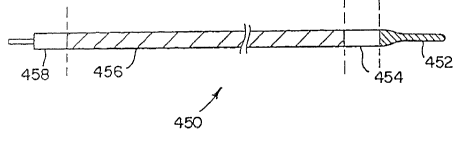
wire 450 is provided, as shown in Figure 1. The
imaging guide wire 450 includes a tip section 452, a
sensor section 454, a drive cable section 456, and a
proximal connector section 458. As mentioned above, an
essential requirement for the imaging guide wire is
that it possess an outer profile of a size that allows
it to fit through a guide wire lumen in conventional
interventional catheters. In catheters that use 0.018
inch guide wires, the guide wire lumen has a diameter
9
typically in a range between 0.020 and 0.022 inch. The
diameter of the proximal section 458 of the imaging
guide wire 450 may be as large as 0.020 inches but the
rest of the imaging guide wire should be not more than
approximately 0.018 inch. For use with catheters
designed with guide wire lumens of other sizes,
relative adjustments in dimension apply.
,.
2. Imaging Guide Wire Sensor
a. Image Resolution
The image resolution of the imaging guide
wire is limited by the optics of the aperture of the
ultrasonic sensor. For an unfocused transducer the
resolution can be approximated by using the maximum
between the angle of beam divergence and the aperture
width. The formula approximating the resolution from
angular beam spread is:
x = R * ~ / A
where,
x = resolution
R = range for sensor face
= wavelength of ultrasound
A = aperture width
For intravascular imaging, the depth of field
where the best resolution is desired is between 1 mm ,
and 3 mm from the face of the transducer. The outer
limit of useful information is out around 4 mm to 5 mm
from the transducer face. With these constraints, a
transducer should provide the best performance in this
range. For a flat, unfocused transducer, a preferred
transducer aperture width can be determined for a
selected operating frequency. The analysis in Table 1
is an approximation of actual performance since beyond
the near field the beam is uniform and approaches this
constant diffraction angle as distances increase. This
analysis is useful to get a coarse estimate of the
.._. - 10 -
expected resolution as a function of the independent
variables.
Table 1 shows the resolution for apertures of
0.5 mm, 0.4 mm, and 0.35 mm for operation at 30 MHz.
(A 0.5 mm aperture is disclosed in the first embodiment
described above in which the overall device profile is
on the order of 3 Fr). The data of Table 1 indicate
that for a system that will image out to approximately
5 mm radius (the range necessary for coronary arteries,
for example), the optics limit the aperture to about
0.35 mm (0.014 inch). It should be noted that the
resolution is improved out to the radius of 4 mm by up
to 30%.
A=0.35mm
A=0 . 5mm A=0 . 4mm
x (mm) x (mm) x (mm)
R=lmm 0.5 0.4 0.35
R=2mm 0.5 0.4 0.35
R=3mm 0.5 0.4 0.43
R=4mm 0.5 0.5 0.57
R=5mm 0.5 0.63 0.71
R=6mm 0.6 0.75 0.86
R=7mm 0.7 0.88 1.0
Table 1 Sensor at 30 MHz operation
By increasing the frequency up 40 MHz and
to
utilizing a method for reducing the signal scatter from
blood (as disclosed below), the resolution can be
further sensor face.
increased
in the
area close
to the
Moreover, the aperture size can be reduced. Table 2
shows the resolution for apertures of 0.5
mm, 0.4 mm,
and 0.35
mm for
operation
at 40 MHz.
A=0.5mm A=0.4mm A=0.35m
A=0 . 3mm
x (mm) x (mm) x (mm) x (mm)
R=lmm 0.5 0.4 0.35 0.3
R=2mm 0.5 0.4 0.35 0.3
R=3mm 0.5 0.4 0.35 0.36
R=4mm 0.5 0.4 0.41 0.48
R=5mm 0.5 0.47 0.51 0.6
4 R=6mm 0.5 0.56 0.61 0.71
0
R=7mm 0.5 0.66 0.71 0.83
- 11 - 2~9~U~~
Table 2 Sensor at 40 MHz operation
Table 2 shows that for a system that will
image to approximately a 5 mm radius, the optics limit
the aperture to about 0.3 mm (0.012 inch). It should
be noted that, compared to the 0.5 mm aperture, the
resolution is improved out to the radius of 4 mm by up
to 40s. The embodiments of the present invention for
imaging guide~wires relate in scale to this size.
The significance of a 0.3 mm (0.012 inch)
transducer aperture size is that this allows the
imaging guide wire to possess an 0.014 inch overall
device profile. This allows an imaging guide wire to
be used with conventional over-the-wire type catheters
that use a conventional 0.014 inch guide wire.
There are two significant factors to be
considered in providing an 0.014 imaging guide wire.
These factors relate to signal scatter from blood and
transducer design.
b. Imaging Guide Wire Transducer Design
It is essential to consider the design and
performance of the transducer sensor as the wavelength
width to length ratio is established in the range
consistent with the optics requirements set forth
above. With a transducer of the size required for an
imaging guide wire, it can be difficult to properly
match the impedance of the transducer sensor to the
drive cable with available materials and at the
required frequencies.
There are two well known methods to model
transducer performance. The model used for thickness
mode vibration is known as the KLM model (Krimholtz,
Leedom, Matthaei). This model is useful for modeling
thickness mode transducers that are substantially
clamped in the other dimensions. With a transducer of
a size that can be used in an imaging guide wire, the
- 12 -
width mode of oscillation and excitation is
significant. This diminishes the accuracy of the KLM
model when applied to a sensor used in an imaging
guide. This also makes the operation of a sensor with
this construction more difficult to work with. A
rectangular sensor can be made so that only its width
is a consideration, however, with a circular aperture
all directions should be considered.
r
Along with width oscillation being a
consideration, the energy coupling coefficient (kt2)
decreases significantly as the clamped construction is
compromised. The coupling coefficient effects the
signal level and ringdown performance so it is
advantageous to provide a material or mechanical
configuration that will give as high a kt2 value as
possible. This consideration must be reconciled with
the contrary considerations for aperture size.
The other model and method of constructing
transducers is based upon "phased array" considera-..
tions. This model can be similar to the model above
used with clamped thickness mode if a complex loading
impedance is used. With phased arrays, the width to
thickness ratio (G=W/T) of each phase element is
preferably within a range where G=0.1 to 2.0 for
reasonable performance. A maximum value for kt2 is
obtained within the range of G=0.5 to 0.8. Accord-
ingly, a sensor comprised of several separate elements,
similar to a phased array, can be advantageously
utilized in an imaging guide wire.
Such a sensor 500 is illustrated in Figures 2
and 2A. The sensor 500 is sliced parallel to the
longitudinal axis of the drive cable 352 (shown in
Figure 1) thereby forming discreet transducer elements
502. To minimize the width resonance of the sensor
500, the impedance between the elements 502 should be
kept as low as possible.
- 13 -
209~fl6~
The electrical excitation for the sliced
sensor 502 is similar to that of the sensor 42,
described above, and unlike conventional phased array
type transducers. In conventional phased array
excitation devices, each phased array element is
excited (and read) separately from the other elements
and separate electrical leads are required for each
element. A disadvantage of such conventional phased
.
array sensors is that the number of separate, discreet
leads for each element occupies a significant area
thereby limiting the size to which the device can be
reduced.
As shown in Figure 3, in one embodiment, the
elements or slices 502 are excited across the thiclaness
direction of the elements. Alternatively, the elements
can be excited across the width of each element. In
this embodiment, the thickness of the transducer 500 is
limited and constrained. However, through use of the
sliced transducer face, the effective width of the
transducer can be increased for the capacitance
calculation. This allows the transducer to be made
with the overall physical dimensions required for an
imaging guide wire but with an impedance matched
properly to the other system components, e.g. the drive
cable.
Alternative embodiments of the sliced
transducer sensor are shown in Figures 4, 5 and 6 in
which the sliced sensor 500 is adapted with a circu-ar
aperture. With a circular aperture, the slices can be
formed as straight lines thus forming rectangular
elements (Figure 4), circular concentric slices forr-.ing
circular elements (Figure 5), or spiral slices form_ng
spiral elements (Figure 6). A circular aperture ca~
also be formed on a rectangular substrate by
metallizing the areas required to make the circle a-ea
active. Piezoelectric material that is not metallized
- 14 -
on both sides and electrically connected to the signal
cable would not be active and would not effectively
form part of the acoustic aperture. In any of the
alternative embodiment geometries, the use of a sliced
transducer provides the capability to properly match
the impedance of the transducer to the rest of the
system components. Thus, transducer size is not a
limiting constraint at these dimensions.
,.
c. Additional Matching and Backing Layer
Embodiments
For a solid sensor in a larger imaging guide
wire, it is preferred that the transducer is air backed
and have double matching layers. Double matching
layers allow more energy to be transferred out the
front of the transducer and less out of the back. By
correct selection of the impedance and thickness of the
matching layers, an air backed sensor can produce a
near ideal pulse. It is desirable to keep the energy
out of the metal or composite sensor holder. This
feature is obtained by reducing the contact area
between the two surfaces.
For an embodiment in which a fluid is trapped
or otherwise exposed to the sensor, it is preferred to
keep any fluid or material out of the space between the
surfaces of the sensor and the mount. These surfaces
can be treated to increase the surface tension between
the surrounding fluid and bottom surfaces of the sensor
and the mount.
Another alternative embodiment for
accomplishing this is illustrated in Figures 7 and 7a.
In Figure 7, mounting tabs 508 are located over the
transducer 356 to aid in mounting the transducer 356 in
place. A protective sheath 510 is included to provide
a non-traumatic outer surface. By having a smooth end
section, a gap 511 is formed between the sensor face
512 and the outer sheath 510. This gap 511 is prefer-
- 15 -
ably filled with water. Alternatively, there are a
number of materials that have the acoustic impedance of
water and could be used as substitute alternatives for
filling of this gap, such as silicon oil, castor oil,
and many other fluids. It is preferred that this
material be biocompatible should a rupture occur.
Alternatively, the space 511 can be filled with a solid
material that has nearly the same acoustic properties
r
as water. Preferable materials include TPX, low
density PE and silicon rubbers. Even though silicon
rubbers have high attenuation they may be suitable.
A further alternative embodiment is illus-
trated in Figures 8 and 8a. Instead of a single, solid
material over the sensor 356, an exponential matching
layer 516 is provided and shaped into the circular form
of the holder 354. The exponential matching layer 516
is preferably formed of a series of layers in which the
impedance follows an exponential manner from one layer
to another. This type of matching layer is capable of
providing as near to ideal matching as can be realisti-
cally achievable.
A further alternative embodiment is illus-
trated in Figures 9 and 9a. Less ideal but still
suitable matching can be provided by forming the sheath
510 in a shape having a surface 520 that fully or
partially fills the space in front of the transducer
256 thereby additionally providing the function of the
matching layer of the previous embodiment. This sheath
510 with the formed surface 520 may be shrunk down over
the transducer section 356 providing for a non-filled
sensor face. The active area of the transducer 356
should be limited to the flat area provided by the
formed sheath 520. A formed sheath provides matching
nearly as good as that of the exponential matching
layers of the previous embodiment and may be easier to
construct.
- 16 - ~~90069
Energy that enters the sensor mount 354
should be minimized. By reducing the amount of energy
that is coupled into the backing, there is a corre-
sponding reduction in the amount of energy that can
reflect back into the sensor. The contact area between
the backing and the backing support is therefore kept
to a minimum. The energy can further be reduced by
limiting the energy that enters the backing support.
,.
As mentioned above, one method is to use a composite
metal and rubber or epoxy. This metal is preferably
sintered or powder in epoxy. Another way to attenuate
the energy that enters the backing layer or support is
to add a quarter wavelength spline structure 524 around
the backing layer support as shown in Figures 10 and
10a.
d. Wedge Transducer
Another alternative embodiment for the
transducer design in an imaging guide wire is shown in
Figure 11. In this embodiment, the transducer is a
wedge geometry transducer 530. Wedge transducers have
been used in many industries to provide a broadband
signal while coupling to a low acoustic impedance
medium. The wedge transducer material has a high
acoustic impedance so that the acoustic energy is more
easily coupled into the material. A very good material
for the PZT sensor and water interface is brass. This
provides for a broadband pulse with a short ringdown.
The angle between the wedge material and the transducer
face material 531 causes two waves to be formed. One
wave travels out of the wedge through the transducer
face material 531 to the blood and artery being imaged.
The other wave reflects off the face, stays inside the
wedge and is attenuated so the sensor can see the
return reflections from the forward wave. This attenu-
ation can be obtained using a number of different
- 17 -
techniques depending on the level of attenuation
needed.
One readily provided and effective method to
provide the necessary attenuation is to make the
backing material 532 off that side the same impedance
as the wedge material. A tungsten and rubber epoxy
mixture can be used for this purpose. The mixture
requires a large percentage of tungsten by weight to
get the impedance high enough to match the wedge
material. This mixture is highly attenuative and a
thickness of only a few mills of material is sufficient
for the needed attenuation. To add to the attenuation
at the wedge backing B interface, a quarter wavelength
grating surface 534 can be machined or etched into the
wedge material. This grating surface 534 reduces
reflection back into the wedge 530 sufficiently without
the use of a backing material at that location. Any
additional artifacts can be reduced or eliminated
through the use of the calibrated waveform pulser,
described above, or by canceling out the repetitive
return signal electrically.
The wedge transducer geometry allows for
making a transducer in a size necessary for use in an
imaging guide wire or even smaller. The minimum size
of a transducer formed with the wedge geometry would be
limited by the optics of the aperture, as described
above. The wedge geometry allows the use of nearly all
the cross section diameter for acoustic aperture
because the beam is bent to a small angle from
perpendicular to the wedge front face. Another
advantage of the wedge design is that it provides a
mechanical structure needed to support the sensor and
the guide wire tip. In this case this structure is an
integral acoustical part of the transducer.
- 18 -
e. Techniques For the Reduction of the
Scatter From Blood
As mentioned above, with an imaging guide
wire the frequency of operation can be approximately 40
MHz. At the short wavelengths corresponding to this
operating frequency, it is preferred to provide a means
to account for scattering due to structures (e. g.
particles) found in blood. In ultrasound imaging at
such frequencies, such scattering can obscure the
difference between the blood and artery or disease. A
Rayleigh scattering analysis that assumes spherical
bodies fairly portrays the observed phenomena.
One means for addressing this concern is to
use a vector averaging circuit to filter out the fast
moving blood scattering return signal. The spatial
frequency of the artery information is limited to twice
the angle of the beam. In practice, it works best to
sample about 25% faster than the maximum frequency.
Faster sampling rates do not provide any more useful
information about the artery. Multiple fast sampling
in the 30 microsecond to 100 microsecond range provides
information that can be used to average out noise,
random pulses, and fast-changing information from the
blood scattering. For broad band noise, the signal-to-
noise ratio is increased by the square root of the
sample number. For random pulses and the type of
signal received from blood scattering, the reduction is
proportional to the sample number. This would be less
than proportional for very dense return signals since
some may overlap.
Another way to reduce the scattering signal
from blood is to use a double frequency transducer.
Figure 12 illustrates an embodiment utilizing such a
transducer. The transducer 356 includes a first sensor
540 and a second sensor 542 located one over the other.
An additional layer may be needed between these two
- 19 - ~~~~~~9
sensors to separate and isolate them into two
relatively narrow bandwidth sensors. With this
construction, both sensors 540 and 542 are pulsed at
the same time and the return signals are frequency
multiplexed into different frequency bands. These
frequency bands are separated with analog or digital
filtering. It is preferred to acquire both signals
together and process them with a digital fourier
,.
analysis. This requires a significant processing
apparatus for real time imaging. Alternatively, analog
filtering to separate the frequency bands and dual
channel data acquisition may also be used for real time
implementation. The data acquired is processed by dual
data pipeline data acquisition front ends, as described
above. The resulting information is then combined by a
data pipeline function that would process the low
frequency information to find the blood/artery boundary
and then switch to the high frequency attenuation and
signal intensity for determining the material composi-
tion.
This technique effectively provides the
output of both a low frequency sensor and a high
frequency sensor at the same time. The low frequency
information is useful for blood-to-artery separation
and the high frequency information is useful for high
resolution artery imaging. The lower frequency is
preferably between 20 MHz and 30 MHz. This gives a
good low blood scattering signal and good resolution of
the artery edge. The upper frequency is preferably
about twice the lower frequency, but this ratio may be
from 1.5 to 3.
An alternative embodiment also incorporating
two sensors in an imaging guide wire enables both side-
looking and forward-looking imaging. In this alterna-
tive embodiment, one frequency sensor is pointed
sideways and the other frequency sensor is pointed
20 = z~~~~~9
forward. This is readily incorporated in a 3 French
imager, as described above, because the larger size
device can possess an open end housing formed of a hypo
tube in which the sensor is mounted. A forward-looking
imager has the ability to provide information about
where the imager is being pushed.
Multiple sensor devices may include more than
two sensors and two frequencies. As long as the
sensors do not overlap in frequency bandwidth
significantly, more than two sensors could be used.
This would be useful for 3D imaging where narrower
bandwidth sensors are used and more of them are
available for close, cross-sectional views.
3. Guide Wire Tip
Figure 13 shows an embodiment of the imaging
guide wire in which the tip portion 452 incorporates
features so that the imaging guide wire 450 can be used
for both imaging and for positioning. In Figure 46,
the tip section 452 of the imaging guide wire 450
includes a floppy tip 554 with a strain relief 556
section connecting the floppy tip 554 to the sensor
section 454. The strain relief section 556 provides
for a variable bending force between the floppy tip 554 ,
and the relatively stiff sensor section 454. This can
be provided by a gradually increasing core wire
diameter or by gradually increasing the size or the
diameter of the coil over the core wire.
In an imaging guide wire possessing
positioning functions combined with imaging functions,
one of the potential concerns relates to the inclusion
of a long, floppy tip conventionally used with guide
wires for steering in an artery. The concern is that
the floppy tip may twist off or scuff up the inside c-
the artery during rotation of the wire during imagine
An embodiment feature that addresses this concern is
_ 21 -
illustrated in Figure 14. In this embodiment, a
mechanism 558 is incorporated into the imaging guide
wire 450 that allows the tip 452 to stay stationary
with respect to the artery when the imaging guide wire
is being rotated for imaging but locks the tip 452 to
the wire when it is being used for steering during wire
placement. This mechanism 558 includes a means for
providing a fluid pressure on a hydraulic piston 560 in
the guide wire. When the piston 560 is pressurized,
the guide wire tip 452 is locked to the body of the
imaging guide wire. When the pressure is balanced
across the piston 560, the tip 452 will rotate freely.
4. Ima_~g~ Guide Wire Drive Cable
The drive cable section 456 is specifically
adapted to address the mechanical and electrical
requirements of the imaging guide wire. Mechanically,
the drive cable 456 preferably possesses very good
torque response in~order to be used for intravascular
positioning and good longitudinal stiffness for
pushability. The drive cable 456 should also exhibit
low angular whipping during rotation. Further, the
drive cable section 456 should be very straight.
Electrically, the drive cable 456 is preferably capable
of sending a signal from one end to the other with
minimum loss. In order to properly match the sensor
impedance, high impedance in the drive cable 456 is
preferred. The electrical impedance of the drive cable
456 is preferably in the range of 20 to 100 ohms.
An embodiment of the drive cable 456 is
illustrated in Figure 15. The drive cable 456 includes
a core wire 564, an insulation layer 566, a shield
layer 568, and a coil layer 570. The core wire 564 may
possess several alternative constructions. In one
embodiment, the core wire 564 is formed of a solid
wire. Alternatively, the core wire may be formed of
2090069
- 22 -
multi-strand copper or silver-plated copper wires. The
latter embodiment provides good electrical character-
istics and allows the drive cable 456 to be relatively
floppy. However, a multi-strand construction may not
provide sufficient longitudinal stiffness. Therefore,
the core wire may preferably be formed of a material
having a high modulus of elasticity thereby increasing
the longitudinal stiffness. Materials like stainless
steel, tungsten, and~beryllium copper are preferred.
Of these, tungsten is most preferred since it has the
highest yield strength and the highest conductivity.
To provide for low electrical loss in the
core wire 564, a high conductivity material is applied
to the outer surface of the core wire . Preferred
materials for applying to the outer surface of the core
wire ~ ;include silver or copper. Silver is most
preferred since it has the highest conductivity. These
materials are easily plated to a thickness suitable for
good electrical transmission. At high frequencies,
electrical current stays close to the surface of a
conductor and therefore a 0.001 inch of conductor
plating over the core wire is sufficient. In a
preferred embodiment, taking into account both
mechanical and electrical requirements, the ideal
thickness of the coating is less than 0.001 inch.
The insulation layer 566 in the imaging guide
wire separates the conductive core layer 564 from the
conductive shield layer 568. For electrical purposes,
this layer 566 is nonconductive and preferably has as
low of an dielectric constant as possible. If a solid
wire is used for the core wire 564, it is preferred
that a means be incorporated into the insulative layer
566 to restrict longitudinal motion between the core
wire 564 and the outer coil 568. If the insulative
layer 554 is made of Teflon* a direct bond may be
difficult to make between the layers. In this case,
* a trade-mark
A
- 23 - z~~~~~~
movement between the core wire 564 and the outer layers
can be restricted at the joint between the drive cable
456 and the sensor housing 354. This is preferably
accomplished by using a nonconductive sleeve to bond
between the core 564 and outer layers that will be
connected to the sensor housing 354. This sleeve is
made out of glass ceramic or other hard, nonconducting
material. To bond between the layers along the length
r
of the drive cable, holes are formed in the Teflon at
various patterns to allow glue or other bonding
material to be used to connect the layers together.
A material other than Teflon can be used for
the insulation layer 566. Such other materials include
glass strands or a solid extrusion of glass, kynar
strands, or a ceramic extrusion. The extrusions would
form a solid, uniform layer over the core wire out to a
given diameter. The strands would then be epoxied to
form a composite layer much like a fiber glass or other
composite structure that uses fiber and binder to
generate a unique high strength material.
The shield layer 568 is located over the
insulating layer 566 to make up the outer layer of a
coaxial signal cable. The shield 568 can be made from
a braid of wires or a coil of wires. In a preferred .
embodiment, these wires are rectangular silver-plated
copper wires. A single layer of coils may be used to
provide the smallest diameter drive cable. A low
resistance shield layer provides for RF emission
shielding and susceptibility. Cable loss is a function
of the core and shield total resistance, and accord-
ingly, it is desirable to provide the shield with as
low resistance as possible. For this reason, it is
preferred that a braid or double coil is used for the
shield layer.
The outer coil layers 570 are needed for good
torque transmission for performing the functions of
90
- 24 -
both the drive cable and guide wire. The outer coil
layers 570 are formed of copper or alternatively other
metals like stainless steel. In a proximal section of
the outer coil layer 570, a binder is used to bind all
the layers together over a length thereof so as to make
that portion of the imaging guide wire straight and
stiff. This proximal section is from the proximal
connector of the imaging guide wire to a location
corresponding the end of the guide catheter with which
the imaging guide wire would be used. This distance is
typically 130 cm. This allows the distal section of
the imaging guide wire to be relatively more flexible
where it needs to go through tight bends.
Another alternative way to provide additional
stiffness in a proximal section of the imaging guide
wire drive cable 456 is to provide another layer of
material over the metal coil outer layer 570 along a
proximal section. This additional layer may be formed
of other-than-metal strands of glass, kevlar or other
high strength materials. The strands would be used in
a coil or braid layer over the core cable 570. The
strands could then be epoxied to form a composite layer
much like a fiber glass or other composite structure
that uses fiber and binder thereby resulting in a ,
unique, high-strength material. As described above,
this can be a dual section composite in which one
section is made out of one fiber and binder and the
other section the same or different fiber and binder or
a combination thereof.
5. Imaging Guide Wire Proximal Section
A proximal
section 458 of the imaging guide wire provides several
functions. These functions include a connection for
electrical contacts for signal transmission, torque
transmission during imaging, torque and longitudinal
- 25 -
motion during guide wire placement and a connection to
an extension wire. Figures 16, 17, and 18 show
alternative embodiments for the proximal section 458 of
the imaging guide wire. In each of the three
embodiments, the proximal section 458 has approximately
the same diameter as the shaft portion 456 although the
proximal section 458 could range in size from a little
larger than the shaft portion 456 to much smaller. In
each of these embodiments, the proximal section 458
provides for electrical connection. The electrical
connection may be a static contact or a dynamic slip
surface in which case there is a slip ring. Torque
drive from the extension wire is accomplished by
fitting over a smaller (round square or other shape)
wire 571 as in Figure 16, or by fitting the wire inside
a (round, square, or other shape) hole 572 as in
Figures 17 and 18. The electrical contact connection
573 may be in an axially displaced configuration as
shown in Figures 16 and 17. Alternatively, the
contacts may be one inside the other as shown in
Figure 18.
With these proximal contact configurations
described above and shown in Figures 16 to 18, an
extension wire 574 is plugged into the end of the ,
imaging guide wire. The overall profile of the
extension wire 574 and the imaging guide wire 450
maintains a small diameter as illustrated in Figure 19.
The extension wire 574 is used for at least two
purposes. First, the extension wire 574 allows an
interventional catheter to be pushed into place over
the imaging guide wire after the imaging guide wire has
been positioned at the desired arterial location.
Second, the extension wire 574 is used to clap on to
for steering and pushing the imaging guide wire into
place initially. Alternatively, a short tool could
also be used that plugs into the end of the imaging
- 26 - y
guide wire that would allow steering and pushing the
imaging guide wire to the desired location in the
arteries.
When used for imaging, the proximal end of
the imaging guide wire plugs into an interface drive
device 576 that provides torque drive and an interface
to transfer the electrical signals from the imaging
guide wire electrical contacts. This connection is
illustrated in Figure 20. This section 576 includes a
proximal slip ring section 578 that would separate the
rotating mechanical drive from the electrical signals
on stationary hardware. This drive interface may
incorporate any of the three slip ring alternatives,
contacting, capacitive, and magnetic, as described
above.
Embodiments of the proximal interface 576 are
illustrated in Figures 21 and 21a. An external motion
restrictor 580 is preferably used when rotating the
imaging guide wire 450 to reduce any tendency for the
wire to whip around in the radial direction. This
restrictor 580 allows movement between the proximal
assembly and the catheter through which the imaging
guide wire extends. The restrictor 580 preferably
allows for the placement of the catheter as well as
movement of sensor within the catheter by pulling the
imaging guide wire back and forward while maintaining
the catheter stationary. Figures 21a and 21b illus-
trate two alternative embodiments for achieving this
motion restriction. Figure 21a shows an embodiment in
which a bellows type device 582 surrounds a portion of
the proximal end of the drive cable 456. Figure 21b
shows an embodiment in which a non-rotating close-
fitting tube 584 is fitted inside the catheter. The
use of a sterile contact plug 585 and a sterile sheath
586 allow for a convenient setup procedure, using
inexpensive, disposable or easily sterilizable parts.
.. - 27 -
6. Imaging Guide Wire Ancillary Equipment
An imaging guide wire is a relatively fragile
device and accordingly it is desirable to provide a
means to maintain the wire's straightness. For
example, during installation of any guide wire intra-
vascularly, there are numerous possible occasions for
the wire to be inadvertently bent and damaged. For
this reason, an imaging guide wire holder and delivery
device 590 may be used, as illustrated in Figure 22.
For installing the imaging guide wire in a guide
catheter 592 or any other catheter or sheath, the
fittings 594 are connected between the catheter 592 and
holder 590. An extension wire 574 (not shown) may be
connected to the imaging guide wire 450 and then pushed
to move the imaging guide wire 450 into place just
before exiting the catheter 592. At this point, the
fitting 594 is released and the extension wire 574 is
pushed through the holder 590 while holding the imaging
guide wire 450 steady. At this point, a clamp can be
placed on the extension wire 574 to push and steer the
imaging guide wire into place. An interventional
catheter may also be put in place at this time. The
extension wire 574 is then removed and the proximal
imaging assembly is snapped into place. Imaging would ,
begin by rotating the wire 450 and moving it in and
out.
B. Imaging Guide Wire Methods Of Use:
1. Method 1
In a first embodiment of operation, the
imaging guide wire 450 can be used as the primary guide
wire, i.e. to both position and to image. The imaging
guide wire 450, described above, can be used in this
manner. According to this method, the imaging guide
wire is routed into place as a conventional guide wire.
At this point, prediagnosis imaging may be performed if
- 28 -
considered appropriate by the physician. Imaging may
be done at this time by rotating the imaging guide wire
in place. In this embodiment, the imaging guide wire
possesses a nontraumatic tip and a smooth covering over
the wire to reduce the possibility that the wire may
damage the artery or the blood. In an alternative
method of operation, a sheath may be routed over the
imaging guide wire before the wire is rotated for
imaging. This would~also reduce or prevent any trauma
in the artery at the location of the sheath. The tip
of the imaging guide wire may extend distally outside
this sheath or the imaging guide wire tip could be
drawn back into the sheath before rotating. It should
be noted that imaging can be performed in real time,
and therefore to minimize problems with the rotation of
a bare wire, the speed of wire rotation can be low,
e.g. a fraction of a Hz or even done manually. If the
imaging guide wire were rotated manually, a constant
display can, at a minimum, provide information
concerning distances.
Once the imaging guide wire is in place and
an appropriate therapy is determined, a therapeutic
catheter, e.g. a balloon dilation catheter, can be
routed over the imaging guide wire to the desired ,
arterial location. At this stage, the imaging guide
wire can again be used to image by rotating the wire.
Images obtained at this stage show the arterial cross
section where the sensor is located. The imaging guide
wire can then be moved and operated at the location
where the treatment is performed. After the treatment,
the imaging guide wire can be left in place while the
catheter is removed and a second catheter is put in its
place for yet another treatment if considered appro-
priate. Otherwise, the imaging guide wire can be moved
to other locations to repeat the procedure, if neces-
sary.
- 29 --
In a further alternative method of operation,
once the catheter is in place over the imaging guide
wire, the catheter and the imaging guide wire can be
moved together to another arterial location. Here, the
tip can extend distally outside the catheter and the
image guide wire can be rotated and pushed to
facilitate advancing and positioning the wire and
catheter to the desired site. When used in this
manner, the imaging guide wire can be used as a
conventional guide wire. This has the potential to
save time since the catheter would not have to be
pulled back and replaced immediately for imaging and
treatment.
2. Method 2
The above described method of operation is
directed to the use of the imaging guide wire in
conjunction with a conventional over-the-wire catheter
in which the guide wire lumen extends the length of the
catheter. Other types of catheter designs are
available, such as the type of catheter in which the
catheter has a short guide wire lumen (SGWL) at the
distal end of the catheter and in which the guide wire
occupies a location adjacent to the catheter proximal ,
of a proximal entrance to the short guide wire lumen.
If the imaging guide wire is used with a catheter of
this type, somewhat different steps of operation may
apply. With a short guide wire lumen catheter, the
imaging guide wire may be first put in place in the
artery. The imaging guide wire may be provided in a
somewhat longer length, or a short extension wire can
be connected to the device used in method 1. The short
guide wire lumen catheter is then advanced over the
imaging guide wire and pushed into the desired arterial
location while holding the proximal end of the imaging
guide wire. A concern is that the imaging guide wire
- 30 - ~_ 209 40 69
is adjacent the short guide wire lumen catheter within
the guide catheter over a considerable portion of its
length. It may be desirable to reinforce the imaging
guide wire shaft along this portion of its length so
that it can be rotated without whipping. This may be
done by applying a stiffening composite layer, for
example. Alternatively, a reinforcing sheath may be
positioned over the imaging guide wire up to the
proximal guide wire lumen entrance.
With these
additional considerations accounted for, the imaging
guide wire may be operated in a manner similar to those
set forth above.
3. Method 3
According to this method, a conventional
guide wire is first used with a conventional
intravascular catheter. The guide wire and catheter
are positioned in a conventional manner. The
conventional guide wire is then withdrawn and the
imaging guide wire is put in its place via the guide
wire lumen of the conventional catheter. In this ,
method, the imaging guide wire can be constructed
somewhat differently from the imaging guide wire
described above. If used with a separate, conventional
guide wire for positioning, the imaging guide wire used
in this embodiment need not possess a distal steering
tip. Instead, all that would be required would be a
smooth end sensor section. A non-traumatic short soft
tip may be included at the end of the sensor section
for extending beyond the end of the catheter into the
artery. Further, in this embodiment the proximal end
of the imaging guide wire does not have to pass through
the catheter, and accordingly, there is no size
A
-,
_..,,
_ 31 _ Og~06g
~2
restriction on how large it is. A proximal connection
could be used very similar to what is described for use
with the 3 Fr imaging device, described above.
4. Method 4
In this method, a conventional guide wire is
used with a dual lumen catheter.
According to this
method, a conventional guide wire is advanced into the
desired arterial location. The dual lumen catheter is
routed over the conventional guide wire using one of
the lumens. This lumen can be the full length of the
dual lumen catheter or can be merged into the first
lumen at any point proximal from the distal tip. For
the dual lumen catheter, the conventional guide wire is
then partially pulled back to allow the imaging guide
wire to image through that section and extend beyond
the end of the dual lumen catheter. In this method,
any of the imaging guide wire embodiments may be used.
If an imaging guide wire, as described above in method
1 is used, the imaging guide wire could be left in ,
place and the catheter could be moved or exchanged over
it.
C. CCD Data Capture and Sensor Configurations
Among the major obstacles associated with
ultrasonic imaging configurations are the matching of
impedances between the sensor and the signal cable,
transmitting the signal down the cable with minimal
loss, and maintaining a high signal to noise ratio.
For phased array sensors, described below, and two
dimensional sensor arrays, there are additional prob-
- 32 _
lems related to parallel signals, such as crosstalk and
multiplexing limits.
In a further embodiment of the present
invention, an imaging transducer sensor is provided
having a charge coupled device, (CCD), associated
therewith. The CCD is an integrated circuit that could
be used to capture the high frequency waveform of the
sensor and send it back to the proximal end of the
device preferably both amplified and at a lower fre-
quency. The charge coupled device (CCD), as referred
to herein, may be one of a family of charge transfer
devices which may also include charge injection
devices . _._ _ _. _..____
Referring to Figure 28, there is depicted a
distal end of a imaging device 360 including a CCD 362,
a PZT transducer 364, a matching layer 366, a backing
material 368 all mounted in a holder 370. The signal
from the transducer 368 is input to the CCD 362. The
electrical connections 372 between the CCD 362 and the
signal and power wires 374 would be made using standard
IC wire bonding techniques. The input impedance of the
cell can vary widely based on the cell capacitance and
input resistance. This input impedance would be
designed to give the best pulse ringdown. The pulse is
generated by the CCD IC or alternatively the pulse may
come from a proximal pulser, as in the embodiment de-
scribed above. After the pulse, the CCD would be
clocked to store the input waveform from the sensor
364. After the waveform is acquired, further clocking
of the CCD array at a slower frequency will allow the
"reading" of the stored value and transmitting this to
the proximal electronics for further processing and
display.
The device 360 provides numerous advantages
for intravascular ultrasound imaging. It allows nearly
perfect impedance matching independent of the sensor.
- 33 - z~~~~~~
It allows the reduction in frequency of the transmis-
sion of the signal to the proximal end at very low
noise susceptibility or emission. This would allow the
reduction of the current coaxial wire design to a
single wire signal design. As few as two wires would
be necessary if the pulsing is remote and communica-
tions are done over the power lines.
In the embodiment shown in Figure 28, the CCD
362 and the sensor 364 are next to each other. By
using a PZT sensor with PVDF matching layer with an
overhang tab for top contact connection, the connection
between the sensor and the CCD is made by having a
large metal pad on the IC to contact the PVDF con-
ductive layer.
In an alternative embodiment 375 shown in
Figure 29, a transducer 376 located over a CCD 378.
This embodiment uses a copolymer material for the
transducer 378 and mounts it over the CCD 378. This
provides an electrical sensor plane as part of the CCD
378 by using a large area top conductive layer.
In further related embodiments, a CCD array
can be used in sensor devices having more than one
sensing area, e.g. phased arrays and linear arrays such
as described in the specification below, for sequential
sensors mounted along the axis of the device for 3-D
imaging. Such embodiments utilize the same type of
circuit for the CCD array as described above but use
parallel paths. Such a configuration is similar to
that currently being used in cameras. The CCD imaging
catheter functions as follows. Photons excite the
electrons that are stored into a 2-D CCD shift array.
Once the values are loaded into the shift array, they
are then shifted to one edge of the IC one row at a
time where they are shifted in the other dimension to a
circuit that measures, amplifies and sends out the
information one pixel at a time. A device very similar
- 34 -
to this could be used in phase arrays, where like the
single sensor CCD, the signal is read and input into
the CCD at one end of the shift register and it comes
out the other end. This would allow the simultaneous
acquisition of all of the sensor array elements and
allow the transfer of the total information to the
proximal circuitry with very little loss or distortion
from noise or crosstalk. Here, as in the single sensor
design, the sensor material could be located over the
CCD or next to it.
This concept could be extended further in an
embodiment of a CCD acoustical sound beam imager. This
device would be similar to that of CCD arrays used in
cameras, however, instead of having a cell area
designed to generate electrons from a light source, the
charge could come from a small area of piezoelectric
material. The piezoelectric material could be placed
over the CCD surface areas would be defined on the top
metallization layer of the IC that would capture and
transfer the piezoelectric charge into the input cell
of the 2-D shift register array. Once the data are
loaded into the shift array, they are then shifted to
one edge of the IC one row at a time where they are
shifted in the other dimension to a circuit that
measures, amplifies and sends the information out one
pixel at a time. This device would be able to take a
snapshot of all the acoustical 2-D wave front one point
in time.
This concept could be even further extended
to provide for a shift register for each of the
acoustical pixels. This would allow for capturing all
of the 2-D waveforms in time. Such a device would be
very useful for 3-D imaging in a non-moving device. A
forward-looking configuration could be constructed in
which the device is placed at the end of the catheter
or is placed behind an acoustical lens in the focal
.._ ~fl9flfl~9
- 35 -
plane. This would allow the acquisition and direct
display of the image within the focal region of the
device. Acoustical excitation could be generated by a
single pulse from a dispersive acoustical generator.
This generator could be a piezoelectric layer over the
CCD.
D. Sequential Sensor Mounting for 3-D
Three dimensional (3-D) images would be very
useful to visualize the extent of certain diseases
present in vessels. 3-D imaging allows for slicing,
rotating and displaying the information so that volume
and cross sections can be visualized. A 3-D recon-
struction requires information from a number of 2-D
cross sections as well as information about their
corresponding position along with the vessel. Acquir-
ing information for 3-D reconstruction can be obtained
by starting at one position in the artery and moving
the sensor past the area to be reconstructed. There
are drawbacks associated this technique, however, such
as the fact that in coronary arteries the rotational
axis of the artery is hard to define in time since this
axis is moving. Also, obtaining good distance measure-
ments along the length of the artery can be difficult ,
because of the stretching of the drive shaft or the
sheath especially if the whole catheter has to be
moved. This stretching can present a problem since the
displacement distance may be measured proximally with a
distance transducer. For example, as the catheter or
the drive shaft is pushed in from a proximal end, fric-
tion could prevent the sensor from moving at all. This
would produce a significant distortion in the 3-D
reconstruction. Also, the duration of time needed to
acquire all the information required for a 3-D recon-
struction could be a drawback by limiting the capabil-
ity for rapid update of the 3-D image.
- 36 -
Referring to Figure 30, there is depicted a
distal end of an ultrasonic imaging device 390 that
provides for 3-D imaging. This device contains
multiple sensors 392 along its axis. The multiple
sensors 392 are located and mounted in a mounting
holder 394 which is mounted on a distal end of a drive
cable 396. The holder 394 would be connected to the
drive cable 396, and driven thereby, in a manner
similar to that used~for mounting a single sensor
holder. The multiple sensors 392 may include an
arbitrary number of sensors depending on the number of
cross sections required. Each sensor would be located
at a constant, known spacing in the mounting holder
394. The sensor holder 394 may have flexible sections
398 between each of the sensors so that each of the
sections can flex as it is being rotated. This could
also facilitate delivery and use of this device. Each
of the multiple sensors 392 would be operated to scan
the cross section where it is positioned.
There are alternative transmission schemes
for transmitting the information signals from each
sensor section to the proximal end of the device. For
example, the signals from all the sensor sections could
be transmitted in parallel, or alternatively, signals ,
from each individual sensor section could be transmit-
ted one at a time by multiplexing, or a combination of
these two methods could be used. A multiplexes would
select which sensor section is currently active and
send its signal down the cable. There may be some
advantages in transmitting one signal at a time using a
multiplexes at the proximal end of the sensor array,
such as a reduction in crosstalk between channels and
the elimination of multiple high frequency signal
wires.
The conditioning hardware for reconstructing
a 3-D image in a reasonable amount of time may include
.w_ 2~~~~69
- 37 _
parallel processing units each working on a section of
the image. Each one of these could require a powerful
processor. Economies may be provided by using a net-
work of Intel I860 type processors. The data acquisi-
tion and data pipelining would be very similar to that
described elsewhere in this specification. The 3-D
processing might be best implemented in the raw data
pipeline. Alternatively, it could be implemented as a
parallel data path into a graphics pipeline allowing
simultaneous display of one of the sensor's cross
section being displayed in real time along with a 3-D
image of the total region.
In a further embodiment, these multiple
sensors could be used in a phased sensor operation in
which the beam is swept and pointed along the axis of
the device. This may be a desirable configuration
since it would allow some "forward-looking" along with
3-D acquisition. If this were implemented, it would be
preferable that the sensor elements be constructed
having a smaller dimension in the direction along the
device axis.
For a sensor array configuration, the sensor
sections would not have to be rotated to obtain an
image. By holding the device motionless, an image
would be obtained of a cross section of the wall of the
artery facing the sensors. This would for most appli-
cations be very useful information. 3-D information
could still be obtained by rotating the whole device.
E. Acoustical indexing for 3-D
An alternative approach to 3-D imaging is
shown in Figures 31 and 32. This alternative approach
would use a longitudinal indexing pattern 400 on a
sheath 402 for 3-D imaging. The indexing pattern could
be made to vary along the length of the sheath 402.
The pattern 400 would be used to determine the location
- 38 -
along the length of the sheath 402 at which the trans-
ducer (which would be insider the sheath as in the
previously described embodiments) is located. This
information could be used for acquiring 3-D information
of the artery as the transducer was moved with respect
to the sheath. The pattern 400 could possess a binary
pattern, a gray scale pattern, or other patterns to
indicate a change in position between the sheath and
the transducer. The~pattern could be applied over just
the distal length on the sheath or over the entire
length.
The pattern 400 may be encoded for incre-
mental or absolute registration. For incremental
registration, only one bit of information would be
required. In such a case, external direction informa-
tion would typically be generated. For absolute regis-
tration, two bits of information would be provided and
used in quadrature, thereby allowing the direction to
be determined. For absolute position information, gray
scale encoding may be preferable. Gray scale coding
has the property that only one bit changes in going
from one state to the next. This prevents errors
compared to binary scale for example, since there is no
way of ensuring in binary scaling that all bits will
change simultaneously at the boundary between two
encoded values for binary or other codes.
Patterns for both radial acoustic indexing
and 3-D lateral indexing may coexist on the sheath.
Both patterns could be formed of the sheath material or
could be formed of different materials. One pattern
could be formed on the inner side of the sheath while
the other on the outer side. Also, these patterns
could be formed on the same surface.
- 39 -
Data Graphics Pipeline Architecture
In ultrasonic intravascular imaging, a large
amount of data needs to be processed between the
transducer being pulsed and the image being displayed
and various means can be used for this processing. For
example, processing can range from all analog to all
digital. In most digital systems, the conditioned
signal is acquired through data acquisition, processed
by a computer, and displayed through some graphics
hardware. This can be accomplished over a computer
buss as long as there is a limited amount of trans-
ferring being done. Current systems are very basic in
the digital conditioning and image processing, and can
utilize this approach.
It would be preferred to use digital condi-
tioning functions to enhance the ultrasonic image or to
provide for feature extraction. This would likely
require a different data flow architecture to provide
for additional data transfers needed to produce the
image reasonably quicklx. Figure 33 depicts a pipeline
structure that provides this architecture. This
architecture includes a dual pipeline: one for raw data
and another for graphics data. The analog input from
the sensor/conditioning is acquired from a high speed ,
data acquisition circuit. This circuit synchronizes
the raw data pipeline and transfers the data down the
pipeline at a lower speed. The data is passed from one
function to the next in real time or near real time
speeds. This pipeline basically processes polar data.
Since there would be much less data in the polar
domain, it would be preferable to process this data as
much as possible. These processing functions may
include deconvolutions, fourier transform processing,
neurocomputing processing or other techniques to
enhance the raw data and do feature extraction.
- 40 -
Some of these pipeline functions include the
provision for recording and playback of raw data.
Also, a function may be provided for the buffering of
raw data as the catheter is advanced or withdrawn
through the guide wire lumen of the interventional
catheter. This would provide the physician with
information about the entire artery from the incision
location to the coronaries. This data would likely not
need to be viewed at the time it is obtained, however,
it would be available for analysis off line after the
procedure. The raw data can be stored on an optical
disk, e.g. a WORM, that can store up to 1 Gbyte of
data. It is estimated that during the relatively short
period of time that the imaging guide wire is being
advanced or withdrawn through the guide wire lumen of
the catheter, the data is being generated at a rate of
approximately 100 Nmytes/minute . ~ - ___
A small variation on this architecture would
include the addition of parallel pipelines. This could
be done for example by taking the raw data acquisition
output, branching off to a second LUT, and combining
the two at the initial graphics pipeline function.
This would allow two displays of the same raw data at
the same time in different locations on the screen. ,
This would be desirable if a real time enhanced display
is desired while at the same time showing a slower 3-D
reconstruction or enhanced feature detection.
The data pipeline and graphics pipeline
architecture, as described above, are advantageously
integrated into a system environment. Figure 23 shows
the pipeline structure integrated into one type of
system environment. Figure 23 shows how the communi-
cation portion of the architecture can be implemented
to allow the central system CPU to handle pipeline
setup and configuration. This allows user input to
effect changes in overlays, images and signal condi-
2~9~~r
- 41 -
tinning of data. Not every pipeline function may
require a direct interface to a common buss. An
alternative to common bussing is daisy-chained
communications. Here, the common processor would be
able to perform the setup and configuration tasks using
a serial or parallel communication link. An external
controller may be provided in the overall system
configuration. This controller may issue commands to
M
the system or may be directly memory-mapped to the
functions on the system. This intersystem communica-
tion may employ techniques known and accepted to those
of skill in the art. In the first method, the external
controller may be connected in a serial or parallel
manner and communicate with the system CPU. As with
the keyboard, these commands can be queued and
processed, or a handshaking can occur for synchronized
command execution and communication. The memory-mapped
external system control is performed by having the
external system take control of the system common buss
and accessing the hardware and memory directly.
Non-contactincr slip rings
With mechanical rotating imaging transducers,
one of the major concerns relates to making a good ,
electrical contact between the rotating drive shaft and
the proximal electronics from the proximal end of the
imaging device. In a previous embodiment in a 3 Fr
size imager, the transmission of the electrical signal
from the imager elongate shaft to the proximal
electronics is provided by a mechanical contacting slip
ring assembly. Although the slip ring assembly, as
described above, provides excellent transmission, in
alternative embodiments, it would be advantageous, and
potentially a simplification of the interface, if a
non-contacting means were employed to couple electrical
signal between the rotating and non-rotating parts.
- 42 -
Two alternative means for providing this transmission
link are capacitive coupling and magnetic coupling.
A first embodiment of the signal coupling
assembly is shown in Figure 24. This embodiment
employs capacitive coupling. Capacitive coupling can
be used when the capacitance is large enough between
the rotating and non-rotating contact rings. The
capacitance is a function of the surface area, the gap
distance and the effective dielectric constant. For a
30 Mhz signal, 100 pF would be more than enough
capacitance to provide suitable coupling. A values
greater or less than this would work also.
Capacitive contact rings 600 are.shown to be
longitudinally spaced, although alternatively, the
rings 600 could be positioned radially. If positioned
radially, one ring would be placed on the inner
diameter and the other contact ring on the outer
diameter of the assembly.
With either capacitive or magnetic non-
contacting slip rings, the mechanical energy is
transferred by a keyed configuration or a friction fit.
There are other means that could be used to transfer
the mechanical energy, for example by a magnetic drive.
By making the rotating contact rings out of a magnetic
material or by placing a permanent magnetic in the
assembly, the slip and drive shaft could be rotated
without physical contact. A similar principle is used
in stepper motors. There are several well known ways
of generating an appropriate rotating magnetic field
that the rotating contact rings would follow or of
generating a stepping multi-phase magnetic field that
would drive the center through the rotation phases that
following the stepper rotation.
Figure 25 shows an embodiment of a magnetic
non-contacting slip ring assembly 604. This
alternative embodiment includes a rotating and a non-
- 43 -
rotating transformer coil 608 and 610. The energy is
transferred by magnetic fields through the magnetic
circuit. A consideration with this embodiment is air
gaps reducing the coupling between the two coils. For
this reason, the gap area 612 is enlarged to minimize
this problem.
K. EEPROM Catheter Information Storage
,
In present preferred and alternative
embodiments of ultrasound imaging catheters, there are
numerous parameters that are device dependant.
Currently, all imaging device dependant information is
entered manually or by shunting contact pins to provide
some device type information. These parameters may be
as simple as device type, frequency, device serial
number, and production information. Other imaging
parameters that are sensor dependant include those that
would be used for a calibrated waveform pulser or
coefficients that describe the acoustic waveform that
would be used in image enhancement routines, as
described above. This information must be entered into
the system before imaging begins. However, it is not
very user-friendly to force the user to enter the
information manually into the system. ,
A feature that can be incorporated into any
of the embodiments discussed herein provides for
automatic imager information entry. An embodiment
incorporating this feature is shown in Figure 26. The
device dependant information is stored in a non-
volatile storage medium 614. Such a storage medium is
an EEPROM. In this embodiment, the information is
available when the imaging catheter or imaging guide
wire is plugged into the driving apparatus and control
system. The means for connection could be direct
wiring or an isolated reading means could be used. A
minimum of two wires are typically needed to transfer
- 44 -
information. Common serial EEPROM devices are avail-
able that operate off three wires and have a wide range
of storage capacity. Also potentially available but
not as desirable is parallel access non-volatile
storage.
Another easy method of entering this informa-
tion is to provide a separate data card or disk. This
can be plugged into the system and the computer control
can read the information before imaging begins.
L. Cath Lab System Integration
To use the imaging catheter or guide wire,
drive and electrical connections must be made. A setup
for achieving and facilitating this type of activity is
illustrated in Figure 27. Figure 27 shows a motor box
620 attached to the edge of a patient table 622. A
gooseneck device 624 extends the catheter connector
over the table 622 and holds the imaging catheter or
guide wire in place. It is important to keep the
imaging catheter or guide wire straight while imaging.
This gooseneck type device 624 allows movement back and
forward easily to follow to doctor as the imaging is
performed. Before and after imaging, the gooseneck
device 624 and imaging catheter can be pushed back out ,
of the way to eliminate some of the clutter on the
patient table 622 as well as to protect the imaging
drive shaft from getting bent. This gooseneck device
624 could have cables internal or external to its
supporting structure. The goose neck device 624
preferably possesses a physical configuration and
structure that can support a weight at a distance and
be moved between two three-dimensional points.
Ultrasound imaging in catheter labs is
currently performed by wheeling an ultrasound imaging
system into the cath lab, setting up the system and
catheter and then imaging. There are other methods of
1
- 45 - f 20900fig
system integration that depend on the catheter lab
setup. In prior cath lab setups, a direct connection
is made between the motor 630 and conditioning unit
(MCU) . The motor is typically in a cabinet on a
cart and the MCU is mounted on the table. In this
configuration, the proximal drive cable is laying
across the floor and can be tripped on if the system is
not next to the doctor. When the system is not next to
the doctor, the MCU should have a connector on the
floor, the table or hanging from the ceiling.
According to~a preferred setup, a connector
634 is mounted to the table 622 so the MCU 632 cable
follows the table 622 when it is moved. The system
also has a plug 636 and could be unplugged for portable
configurations. In this configuration, there is also a
connector for video input from the fluoroscope and
video outputs for displaying on the doctors' overhead
monitor.
Other system configurations include a rack
mount system integrated into existing or modified
catheter lab control hardware. In this configuration,
the system is already on-line and when the doctor needs
to perform an imaging procedure, the MCU 620 is mounted
to the table 622 and plugged in. At this time, imaging
could begin. The external controller could issue the
system commands and the video outputs are multiplexed
and displayed at the doctors overhead screen.
Another alternative configuration provides
for the system to be located within the MCU 620. This
could be provided if the system electronics were small
enough to fit within a reasonable sized box to place on
the table rack. Here, there is a manual interface on
the unit and that can be operated remotely from an
external controller. Also, a small monitor can be
provided internally, but the preferred method of
viewing would be externally on the overhead monitor.
A
- 46 - ~~9~~69
In this configuration, there is a plug for
communications, video signals and power.
It is intended that the foregoing detailed
description be regarded as illustrative rather than
limiting and that it is understood that the following
claims including all equivalents are intended to define
the scope of the invention.