Note: Descriptions are shown in the official language in which they were submitted.
2p~~f Z1U
INTERSTITIAL X-RAY 1VEEDLE
BACKGROUND OF THE INVENTION
The present invenUon generally pertains to X-ray apparatus and is par-
ticularly directed to an interstitial X-ray needle.
An X-ray apparatus is used for radiation therapy of cancer patients. One
such apparatus, as described in United States Patent No. 2,748.293 to
Reiniger,
includes an elongated X-ray tube with a converter element being disposed at a
tip
of the tube for converting emitted electrons into X-rays; and an elongated
outer
casing enclosing the tube and deeming a coolant flow chamber through which
coolant may flow to transfer heat from the tip of the tube. The tube is
inserted
into a cancer patient's body through a body cavity to position the converter
ele-
ment so that the X-rays can be concentrated at the tumor and thereby minimize
radiation damage to ad)acent undiseased tissue. However, the size of such an X-
ray apparatus is too large for insertion of the tube through the skin, whereby
the
applicability of X-ray therapy for treatment of cancerous internal body parts
has
been limited to only those body parts that can be accessed through body
cavities.
SUMMARY OP' THE INVENTION
The present invention provides an interstitial X-ray needle, comprising
an elongated X-ray tube coupled to an electron emitter at one end of the tube,
with a converter element being disposed at a tip of the other end of the tube
for
converting emitted electrons into X-rays: a solenoid coil wound around the
tube
for providing a magnetic field that confines the emitted electrons within a
narrow
beam; an elongated outer casing enclosing the tube and coil; and means within
the casing defining coolant flow chambers for directing coolant to and from
the tip
of the tube.
-i-
CA 02090210 2002-03-05
72046-53
The intersti~:ial X-ray needle of the present
invention may be of suwr~ small diameter that a portion of
the casing extending at least approximately five centimeters
from the tip of the tu:oe has a maximum outside diameter of
approximately two millimeters. An X-ray needle of such
small diameter may be :~rcserted in a patient's body without
significant damage to tissue between the skin and the tumor
site, thereby significantly increasing the applicability of
X-ray therapy for treatment of cancerous internal body
parts.
To prevent electron loss and stray X-radiation,
the solenoid coil is wound around the beam-transport tube in
order to provide a magnetic field that tightly confines the
emitted electrons.
In accordance with the present invention, there is
provided an interstitia:~ X-ray needle, comprising an
elongated X-ray tube coupled t:o an electron emitter at one
end of the tube, with a converter element being disposed at
a tip of the other ena of the tube for converting emitted
electrons into X-rays; a solenoid coil wound around the tube
for providing a magnetir_ field that confines the emitted
electrons within a narrow beam; an elongated outer casing
enclosing the tube and. coil; and means within the casing
defining coolant flow czambers for directing coolant to and
from the tip of the tube; wherein the needle is of such
small diameter that tree needle may be inserted in a
patient's body without: significant damage to tissue between
the skin and a tumor :>i t.e .
In accordance with the present invention, there is
further provided an i~-Lterstitial X-ray needle, comprising an
elongated X-ray tube coupled to an ei~ectron emitter at one
-2-
CA 02090210 2002-03-05
72046-53
end of the tube, with a converter element being disposed at
a tip of the other end of the tube for converting emitted
electrons into X-rays; a solenoid coil wound around the tube
for providing a magnet:i.c field that: confines the emitted
electrons within a narrow beam; an elongated outer casing
enclosing the tube and coil, wherein a portion of the casing
extending at least appa-aximately five centimeters from the
tip of the tube has a maximum outside diameter of
approximately two millimeters; and means within the casing
defining coolant flow chambers for directing coolant to and
from the tip of the tube.
In one aspect of the present invention, the
coolant-flow-chamber-defining means comprises a pipe
coaxially disposed between the casing and the tube for
defining an inner annu:rar flow charnber between the tip of
the tube and a first opening in the casing and an outer
annular flow chamber between the tip of the tube and a
second opening in the easing.
In another aspect: of the present invention, the
coolant-flow-chamber-defining means comprises a plurality of
pipes disposed between the casing and the tube wherein each
pipe defines an input low chamber between the tip of the
tube and at least one :inlet opening in the casing and
wherein the space between the tube and the casing not
occupied by the pipes defines an output flow chamber between
the tip of the tube and an outlet opening in the casing.
Additional features of the present invention are
described in relation to the description of the preferred
embodiment.
-2a-
2(~90~~ ~
BRIIw:F' DESCRIPTION OF' THE DRAWING
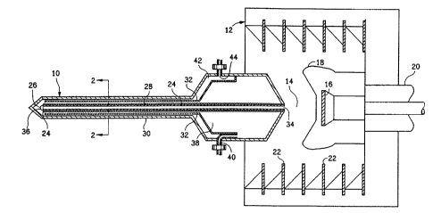
Figure 1 is a diagram of an X-ray apparatus including a preferred em-
bodirnent of the interstitial X-ray needle of the present invention.
Figure 2 is a sectional view of the needle of Figure 1 taken along lines
z-2.
Figure 3 is a diagram of a portion of the needle illustrating an alternative
preferred embodiment of the flow-chamber defining means.
Figure 4 is a sectional view of the needle of Figure 3 taken along lines
4-4.
DESCRIPTION OP' THE PREFERRED EMBODIMENTS
Referring to Figures 1 and 2, an X-ray apparatus containing a preferred
embodiment of the Interstitial X-ray needle of the present invention includes
the
needle 10 and a diode housing 12 for receiving the needle. The diode housing
12
includes a vacuum chamber 14 containing an electron emitter 16 and a control
grid 18. The electron emitter 16 is connected to a high voltage cable 20,
which is
connected to a high voltage source (not shown). Insulators 22 are stacked be-
tween the electron emitter 16 and the diode housing 12.
The needle 10 includes an elongated X-ray tube 24, a converter element
26, a solenoid coil 28, an elongated outer casing 30 and a pipe 32.
The X-ray tube 24 has an open end 34 which opens into the vacuum
chamber 14 to couple the X-ray tube 24 to the electron emitter 16.
-3-
2~90~~~J
The converter element 26 is disposed at a tip 36 of the other end of the
tube 24 for converting electrons emitted from the electron emitter 16 into X-
rays.
The solenoid coil 28 is wound around the tube 24 for providing a mag-
netic field that confines the emitted electrons within a narrow beam. The
electron
beam can be confined to a diameter of approximately 0.4 millimeter when the
solenoid coil 28 provides a magnetic field of approximately 20 gauss. For a
coil 28
having 13 ohms resistance and wound at 20 turns-per-centimeter, the required
current in the winding is only 0.8 amperes and the required voltage across the
coil
is only O.1 volts, whereby the power expended in the winding is only 0.08
watts.
The outer casing 30 encloses the tube 24 and coil 28.
The pipe 32 is coaxially disposed between the outer casing 30 and the
tube 24 for defining an inner annular flow channel 38 between the tip 36 of
the
tube 24 and a coolant inlet 40 in the casing 30, and an outer annular flow
cham-
ber 42 between the tip 36 of the tube 24 and a coolant outlet 44 in the casing
30.
For a needle 10 of the embodirnent of Figures 1 and 2, including a ten-
centimeter long tube 24 having an inside diameter of 0.64 millimeter and an
out-
side diameter of 0.81 millimeter wound with a single layer of #33 magnetic
wire of
0.22 millimeter diameter at approximately 40 turns-per-centimeter, an outer
casing 30 having an outside diameter of 2.8 millimeters and an inside diameter
of
2.16 millimeters, and a pipe of 1.52 millimeters inside diameter and 1.83 mil-
limeters outside diameter, a water flow rate of 87 milliliters-per-minute is
obtained
at an inlet pressure of 20 pounds-per-square-inch, whereby for a 20 watt heat
rate
at the tip 36 of the needle 10, the water temperature rise over ten minutes is
less
than 5 degrees Celsius.
-4-
Referring to Figures 3 and 4, in an alternative preferred embodiment, the
needle l0A includes an elongated X-ray tube 24, a com~erter element 26, a
solenoid coil 28, an elongated outer casing 30 and a plurality of pipes 46.
'The
pipes 46 are disposed between the casing 30 and the tube 24. Each pipe defines
an input flow chamber 48 between the tip 36 of the tube 24 and at Ieast one
inlet
opening (not shown) in the casing 30: and the space 50 beriveen the tube 24
and
tile casing 30 not occupied by Lhe pipes 46 defines an output now chamber be-
tween the tip 36 of the tube 24 and an outlet opening (not shown) in the
casing
30.
For a needle lOA of the embodiment of Figures 3 and 4, including a ten-
centimeter long tube 24 having an inside diameter of 0.64 millimeter and an
out-
side diameter of 0.81 millimeter wound with a single layer of #33 magnetic
wire of
0.22 millimeter diameter at approximately 40 turns-per-centimeter, an outer
casing 30 having an outside diameter of 2.8 millimeters and an inside diameter
of
2.16 millimeters, and four pipes each having outlet orifice )ets 52 of 0.15
mil-
limeter directed at the tip 36 of the needle 10A, a w2ter flow rate of 10
milliliters-
per-minute is obtained at an inlet pressure of 50 pounds-per-square-inch,
Wll(:1'Cl)y fUl' a 20 wall heat rate at the tip 36 of the needle 10A, the
water tempera-
ture rise over ten minutes is approximately 28 degrees Celsius.
The tube 24, casing 30 and pipe 32 or pipes 46 typically are rigid and
straight, but also may be made of flexible materials or may be curved rather
than
straight so as to enable insertion of the tip of the needle to portions of the
body
that are not directly accessible through soft tissue.
The X-ray apparatus described herein may be operated at a relatively low
power level of 14 watts when delivering a radiation dose of approximately 100
Gray over' ten minutes duration to tissue located one centimeter from the con-
verter element 26 by operating with an electron emater voltage of 200
kilovolts
and a beam current of 0.07 milliamperes.
-5-
In addition to providing benefits incident to its size, the miWature inter-
stitial X-ray needle of the present invention also may generate controlled
hyper-
thermic temperatures for application to the treated tumor, which combined with
the radiation treatment may provide a synergistic healing effect.
-6-
