Note: Descriptions are shown in the official language in which they were submitted.
20so3ss
TITLE OF THE INVENTION
An Apparatus for Medical Diagnosis Utilizing Masking
of Fixation Point
BACKGROUND OF THE INVENTION
Field of the Invention
The present invention relates to an apparatus for
medical diagnosis utilizing masking of fixation point.
More specifically, the present invention relates to an
apparatus for medical diagnosis utilizing masking of
fixation point in which eye fixation of a subject is
detected to enable diagnosis of diseases related to brain
function such as Alzheimer's disease.
Description of the Background Art
The number of patients suffering from Alzheimer's
disease is estimated to be four million in the United
States and about a million in Japan. Compared with senile
dementia such as cerebrovascular disease popular among
Japanese, the cause of Alzheimer's disease is not known,
and much effort has made to find the cause so as to enable
early diagnosis and early medical treatment. However, it
is difficult to discriminate Alzheimer's disease from
cerebrovascular disease when there is no typical symptoms.
There has been a strong demand of accurate method of
discrimination, since development of disease,
pharmaceutical treatment and so on are different for these
~ 2 0 9 0 3 5 9
diseases.
Hachinski's ischemic score has been proposed as a
method of discriminating these two diseases. According to
this ischemic score, a point is given dependent on whether
or not the patient has an anamnesis of apoplexy, cerebral
infraction or the like and if the points exceeds a
prescribed number, it is determined as the cerebrovascular
disease, and otherwise it is determined to be Alzheimer's
disease. However, discrimination is still difficult by
this method if the patient has no such anamnesis.
It has been known that neuropsychological symptom
which is considered to be an impairment of '~tool
disfunction" such as visual cognitive disfunction appears
from relatively early period of Alzheimer's disease. In
view of this fact, Fujii et al. has reported the following
analysis carried out by utilizing eye movement. More
specifically, a problem of copying a cube on the right
side while watching an original of the cube on the left
side is presented. Even a patient who is in the initial
stage I of Alzheimer's disease and does not show apparent
constructional apraxia is reported to show characteristic
symptom similar to a so called Balint syndrome; that is,
the patient cannot stare at one point, or more
specifically, abnormal distribution of gazing point
appears, saccade deviated from both the presented cube and
- 2090359
the depicted drawing by the patient is generated, or the
point of gazing is fixed at the same point for a long
period of time. In Alzheimer's disease, it is supposed
from MRI (nuclear magnetic periorbital inspection) that
there is caused disfunction of parietal lobe which is
related to spatial vision. Accordingly, constructional
disfunction derived from degradation in function of the
rear association areas with the parietal lobe being the
center, degradation of function of positional recognition
of a target point or recognition of depth derived from
disfunction of external spatial vision such as disfunction
of eye movement, disfunction of coordinate transformation
system between the coordinate of eye movement system and
the coordinate of the center of one's body axis, or
visual-motor disfunction, is supposed to be a possible
cause of the aforementioned symptoms. As for the
extension of fixation time, relation with one's memory has
been studied.
SUMMARY OF THE INVENTION
Therefore, an object of the present invention is to
provide an apparatus for medical diagnosis utilizing
masking of fixation point in which the fixation point is
masked for a prescribed time period by a prescribed
pattern of a desired size after a lapse of a desired time
period, and based on particular movement of the line-of-
20 9 0359
sight of the patient, on the percentage of correct answer
with respect to a prescribed task and so on, objective
diagnosis of symptoms related to brain function can be
obtained which is not influenced by subjective
determination such as caused by interviews, and which does
not incur unpresent pain of injection or the like.
Briefly stated, in the present invention, an image
which is a task for diagnosis is presented to a subject,
the movement of the eyeball of the subject is detected,
and based on the movement of the eyeball, the actual
movement of the line-of-sight of the subject is
calculated. When there is generated a point of fixation
in the calculated movement of the line-of-sight, then,
after a prescribed period, a desired masking image of a
desired size is inserted to the presented image for a
prescribed time period. Whether or not the disease is
related to brain function is determined in accordance with
the movement of the line-of-sight calculated at that time.
Therefore, the present invention enables diagnosis of
diseases related to the brain function based not on
objective determination such as obtained through interview
but on subjective determination, without imposing pain of
injection or the like on the patient.
In accordance with a preferred embodiment of the
present invention, movement of the head portion of a
2090359
subject is detected, and the line-of-sight is calculated
in accordance with the movement of the head portion and
the movement of the eyeball while the subject is gazing at
the presented image.
In accordance with a more preferred embodiment,
fixation duration distribution, eye movement velocity
distribution and movement of the line-of-sight are
calculated in accordance with the calculated movement of
the line-of-sight, so as tG determine whether or not the
disease is related to the brain function.
Further, according to a more preferred embodiment,
determination as to whether the disease is related to the
brain function is given on the basis of the time period in
which the masking image is inserted in the presented image
and on the percent of correct answer with respect to the
task.
The foregoing and other objects, features, aspects
and advantages of the present invention will become more
apparent from the following detailed description of the
present invention when taken in conjunction with the
accompanylng drawings.
BRIEF DESCRIPTION OF THE DRAWINGS
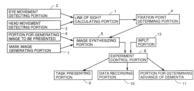
Fig. l is a block diagram showing one embodiment of
the present invention.
Fig. 2 shows an example in which the eye movement
2090359
detecting portion and the head movement detecting portion
shown in Fig. 1 are attached to goggles.
Fig. 3 shows a specific example of the head movement
detecting portion.
Fig. 4 shows a specific example of the eye movement
detecting portion.
Fig. 5 shows the principal of the head coordinate
system with the subject being the center.
Fig. 6 is a flow chart showing specific operation of
one embodiment of the present invention.
Fig. 7 shows an example of an image in which the
fixation point of the line-of-sight of the subject is
masked.
Fig. 8 shows a relation between the eye movement and
the masking process.
Fig. 9 shows locus of the line-of-sight when a
subject suffering from Alzheimer's disease is counting the
circles with the fixation point masked for 66msec at 4.
Fig. 10 shows locus of the line-of-sight when a
healthy person is counting the circles with the fixation
point masked for 66msec at 4.
Fig. 11 shows a relation between the percentage of
correct counting and the onset of masking.
DESCRIPTION OF THE PREFERRED EMBODIMENTS
2S Fig. 1 is a block diagram of one embodiment of the
--6--
2U90359
present invention. Referring to Fig. 1, the line-of-sight
calculating portion 1 calculates the position of the line-
of-sight of the subject. An eye movement detecting
portion 2 detects eye movement of the subject and applies
the detected output to the line-of-sight calculating
portion 1. A head movement detecting portion 3 detects
the movement of the head portion of the subject and
applies the detected output to the line-of-sight
calculating portion l. When the position of the line-of-
sight of the subject is calculated, the line-of-sight
calculating portion 1 applies the result of calculation to
a fixation point determining portion 4. The fixation
point determining portion 4 determines the fixation point
of the subject based on the position of the line-of-sight.
A portion 6 for generating an image to be presented
generates an image which is to be presented to the subject
as a task for diagnosis, and applies the image signal to
an image synthesizing portion. A masking image generating
portion 7 generates an image signal for masking, for a
prescribed time period, the fixation point, at which the
subject gazes, of the image presented to the subject with
a prescribed masking pattern of a desired size after the
lapse of a desired time period. The image signal is
applied to the image synthesizing portion.
An experiment control portion 8 presents the image
2090359
synthesized in the image synthesizing portion 5 at a task
presenting portion 9, records data at the data recording
portion 10 and determines the advance of dementia by the
degree of advance of dementia determining portion 11. A
CRT display, for example, is used as the task presenting
portion 9 which displays the image synthesized in the
image synthesizing portion 5. The data recording portion
10 records data such as the onset of masking, the
percentage of correct counting and so on. The degree of
advance determining portion 11 determines the possibility
of Alzheimer's disease based on the percentage of correct
counting. Further, an input portion 13 is connected to
the experiment control portion 8. The input portion 13 is
for inputting, when the subject counts the image as the
task, the counted value.
Fig. 2 shows an example in which the eye movement
detecting portion and the head movement detecting portion
shown in Fig. 1 are attached to goggles, Fig. 3 shows a
specific example of the head movement detecting portion,
and Fig. 4 shows a specific example of the eye movement
detecting portion.
A subject wears the goggles 12 shown in Fig. 2, which
has, at a lower portion on one side, the eye movement
detecting portion 2 attached. The eye movement detecting
portion 2 includes a light emitting diode 21 provided at
20903S9
the center and photodiodes 22 and 23 provided on both
sides thereof as shown in Fig. 4(a). A light emitting
diode radiating infrared rays having relatively wide
directivity of about +21 is used as the light emitting
diode 21, while ones having acute directivity of about
+10 are used as the photodiodes 22 and 23. The light
beam emitted from the light emitting diode 21 to the eye
ball 26 is reflected from the iris of the eye 27 and from
the white of the eye 28 with different reflectivity as
shown in Fig. 4 (b) and (c), and the difference in
reflectivity is amplified by an operational amplifier 25.
If the difference is calculated, a horizontal output (left
and right) is obtained as shown in Fig. 4(b), and if the
sum is calculated by an operation amplifier 24, a vertical
(up and down) output is obtained as shown in Fig. 4(c).
The head movement detecting portion 3 is formed of a
magnetic sensor as shown in Fig. 3. More specifically,
the head movement detecting portion 3 includes a
orthogonal coil serving as a source 31 and an orthogonal
coil serving as a sensor 32. In accordance with an
instruction from a control portion 33, a driving circuit
34 drives the orthogonal coil of the source 31 to generate
a magnetic field. When the subject wearing the head
movement detecting portion 3 moves, a voltage is induced
in the sensor 32, which voltage is detected by the
2090359
detecting circuit 35, the detected output therefrom is
calculated by the control portion 33, and thus data
corresponding to the movement of the head is output.
Fig. 5 is an illustration showing the principle of
the head coordinate system with the subject being the
center. Referring to Fig. 5, the head coordinate system
detected by the head movement detecting portion 3 will be
described. The head coordinate system includes two
systems, that is, XY coordinate system realized by the
translational movement of the subject with respect to the
object of monitoring such as shown in Fig. 5(a), and a
polar coordinate system based on the rotation movement of
the head such as shown in Fig. 5(b). The amount of head
movement in respective coordinate systems are defined as
(Hx, Hy, Hz), (H~, H~, H~). In this embodiment, the
direction toward the object of monitoring is represented
by the Y axis, the horizontal movement is represented by
the X axis and the vertical movement is represented by the
Z axis, as an example. H~ represents the rotation of the
X axis, that is, the movement of one's neck upward or
downward. H~ represents the rotation of the Y axis, that
is, the movement of inclining ones neck once from the left
shoulder to the right shoulder. H~ represents rotation in
the Z axis, that is, rotation of one's neck in the left or
right direction.
-10-
-- 2090359
The line-of-sight changes by the horizontal movement
of the head (Hx, Hy, Hz), and when this movement is
changed in the equivalent of rotation angle of the eye
ball (Ex, Ey), the following equations (1) and (2) are
obtained.
Ex = 180/~ tan Hx / (D+Hy) ... (1)
Ey = 180/~ tan Hz / (D+Hy) ... (2)
where D: distance from the subject to the point of
gazing.
When the neck is inclined by H~ to the left shoulder
or to the right shoulder, the coordinate of the eye
movement system rotates. Therefore, the eye movement
coordinate system (Xe, Ye) inclined by H~ must be changed
to the coordinate system (Xe', Ye') which is orthogonal to
5 the original object of monitoring.
Xe' = Xe cosH~ + Ye sinH~ ... (3)
Ye' = -Xe sinH~ + Ye cosH~ ... (4)
The movement of the line-of-sight (Xh, Yh) realized
by the head movement is represented by the following
equations (5) and (6) derived from the equations (1) and
(2).
Xh = Ex + H~ ... (5)
Yh = Ey + H~ ... (6
Therefore, the movement of the line-of-sight (Vx, Vy)
5 taking the head movement into account is represented by
~- 2090359
the following equations (7) and (8), from equations (3) to
(6).
Vx = Xe' + Xh (7)
Vy = Ye' + Yh (8)
By employing the equations (7) and (8) above, the
ordinary movement of one's line-of-sight effected by
combining head movement and eye movement can be
reproduced.
Fig. 6 is a flow chart showing specific operation of
one embodiment of the present invention, Fig. 7 shows an
example of the image with the fixation point of the line-
of-sight of the subject being masked, and Fig. 8 shows a
relation between the time of masking and the time of eye
fixation.
A specific operation of one embodiment of the present
invention will be described with reference to Figs. 1 to
8. In step SP1 (simply referred to as SP in the
drawings), the amount of head movement (Hx, Hy, Hz), (H~,
H~, H~) described with respect to Fig. 5 is applied as the
data of head movement from the head movement detecting
portion 3 to the calculating portion 1. In step SP2, the
eye ball coordinate system (Xe, Ye) is applied as the data
of eye movement from the eye movement detecting portion 2
to the calculating portion 1. In step SP3, the
calculating portion 1 carries out calculations of the
-12-
-
2090359
above mentioned equations (1) to (8) in each of the
sampling periods i, i+l, i+2 .... Consequently, the
values of H~i, H~i, H~i, Hxi, Hyi, Hzi, X ei, Y ei, Vxi and
VYi of each sampling period are calculated. The fixation
point determining portion 4 calculates the fixation point
based on the line-of-sight calculated by the line-of-sight
calculating portion 1, in step SP4. In this embodiment,
when the next sample is within the scope Dth of fixation
point determined by the threshold velocity Vth and the
sampling period Ts from the fixation point at present,
then this sample is regarded as the same fixation point as
the present one. S/sec is determined as an exemplary
threshold velocity Vth in accordance with the nature of
pursuit eye movement. When the sampling period is lOmsec,
then Dth = 5/sec x O.Olsec = 0.03 = 3~
The portion 6 for generating the image to be
presented generates an imas2 as a task in step SP5. In
this embodiment, a task is assigned to the subject that
the subject should count the number of O in a pattern
including 0, ~ and 3. The portion 6 for generating the
image to be presented generates image signals of the
pattern including circles 0, triangles ~ and squares ~.
The image signals thus generated are applied to the image
synthesizing portion 5, and the experiment control portion
8 applies the image signals to the image presenting
-13-
2090359
portion 9 so that the pattern is displayed. The masking
image generating portion 7 generates mask image signals
for masking a portion of the task pattern displayed and
applies the generated signals to the image synthesizing
portion 5, as shown in Fig. 7. The image synthesizing
portion 5 inserts the mask image at the fixation point in
the task image determined by the fixation point
determining portion 4, in step SP6. The experiment
control portion 8 applies the image signals with the mask
image inserted to the task image to the image presenting
portion 9 to be displayed. The timing for masking
described above is as shown in Fig. 8. As is apparent
from Fig. 8, the pattern image including circles,
triangles and squares is presented, after the lapse of
time period Ts after the subject gazes the image, the task
image is masked by the mask pattern for a prescribed time
period Tr, and then the original image is resumed.
The portion for determining degree of advance of
dementia 11 receives information related to masking and
data of position of the line-of-sight from the experiment
control portion 8, calculates the fixation duration
distribution in step SP7, calculates the locus of the
line-of-sight in step SP8 and calculates eye movement
velocity distribution in step SP9, using the onset of
masking, the size of the masking, the type of masking and
A
- 2090359
duration of masking as parameters.
Fig. 9 shows the line-of-sight when the subject
suffering from Alzheimer's disease counts the number of
circles, and Fig. 10 shows the locus of the line-of-sight
when a healthy person counts the number of circles. The
aforementioned fixation time distribution represents the
distribution of time while the subject is gazing at
respective points (the points of circles shown in Figs. 9
and 10), the locus of the line-of-sight represents a line
connecting one point of fixation and the next point of
fixation, and the eye movement velocity distribution
represents the velocity when the line-of-sight moves from
one fixation point to another fixation point. Generally,
when a masking pattern of the task image is displayed, the
eyeball moves in a quick and leaping movement called
saccade in order to avoid masking, and therefore the
distribution of the fixation time reduces at this point.
However, a patient suffering from Alzheimer's disease
having damage on visual function related to spatial vision
is incapable of changing the strategy (visual searching)
of moving eyeballs as a masking trigger. Therefore, the
change in the fixation time distribution such as observed
in the case of healthy persons cannot be found, and
therefore the degree of advance determining portion 11
determines the possibility and degree of advance of
20903S9
dementia in accordance with the degree of such change. As
is apparent from the comparison between Figs. 9 and 10, in
the case of the patient suffering from Alzheimer's
disease, there is not a correspondence between the
fixation point and the circles in the pattern, as compared
- with a healthy person. Similar change is observed in the
eye movement velocity distribution, and the degree of
advance determining portion 11 determines, in step SP10,
possibility and the degree of advance of dementia in
accordance with the degree of such change.
Then, the degree of advance determining portion ll
determines the possibility and the degree of advance of
dementia based on the onset of masking and the percentage
of correct answer with respect to the task and on the size
of masking in step SP11. Fig. 11 shows a relation between
the onset of masking and the percentage of correct
answers. Experiments with no mask, (normal) and with mask
with the onset of masking changed to 200msec, 134msec and
66msec were carried out in order, the eye movement at the
experiments were monitored, and the numbers of counted
circles in respective experiment were reported by the
subject. When the numbers of circles were input through
the input portion 13, and the degree of advance
determining portion 11 calculated the percentage of
correct answers. The experiment control portion 8 records
-16-
2090359
the data in the manner shown in Fig. 11 by the data
recording portion 10 in accordance with the result of
calculation.
As is apparent from ~ig. 11, the percentage of
correct answers is quite high in case of healthy persons,
even if the masking is started at 66msec from the start of
gazing. The percentage slightly decreases when the
subject is suffering from cerebrovascular disease which is
called MID: multi-infarct dementia, which is popular among
Japanese. The percentage decreases as the time of
presentation becomes shorter when the subject is suffering
from Alzheimer's disease. Based on the masking start time
at which the percentage falls and on the percentage of
correct answers, the degree of advance of the Alzheimer's
disease can be determined, and the Alzheimer's disease can
be distinguished from other dementia such as MID.
As described above, according to the embodiment of
the present invention, eye movement of the subject is
detected to calculate the actual movement of the line-of-
sight, an image as a task for diagnosis is presented tothe subject, a mask image is inserted to the presented
image when fixation point is generated in the movement of
the line-of-sight and whether or not the disease is
related to the brain function is determined in accordance
with the output of the line-of-sight. Therefore,
2090359
diagnosis of diseases related to brain function can be
given based on subjective determination, not on objective
determination such as obtained by interview, without
imposing pain of injection or the like on the subject.
Although the present invention has been described and
illustrated in detail, it is clearly understood that the
same is by way of illustration and example only and is not
to be taken by way of limitation, the spirit and scope of
the present invention being limited only by the terms of
the appended claims.
-18-