Note: Descriptions are shown in the official language in which they were submitted.
Edward Duane Ping ~on, Paul Guy Thomson 6-8 (~ 5050~
2091271
r
PERCUTANEOUSLY INSERTABLE, NEEDL~-SIZED
TISSUE RETRACTOR AN~ SYSTEM
Technical Field
This invention relates to suxgical instruments and
particularly to a needle-sized tissue retractor and system
that are percutaneously inserted into a cavity of a subject.
'
Backaround of the Invention
Minimally invasive endoscopic surgery is performed using
two or more trocar sheaths for gaining access to a cavity of
a patient. These trocar sheaths provide access to the
surgical site for various medical instruments such as
forceps, scissors, and scopes. The advantages of endoscopic
surgery over traditional, open surgery include decreased
trauma for the patient, smaller wounds and less scarring,
faster healing and recovery time, and decreased risk of
infection.
A prohlem presented by endoscopic surgical procedures is
that a surgeon has no direct visualization of the surgical
site. Instead, visualization of the surgical site is by a
video camera and monitor. The surgeon uses medical devices
inserted through access sheaths to manipulate tissue
adjacent the surgical site for lifting tissue out of the
line of sight. A solution to this problem is the use of an
EXPRESS MAIL CERTIFICATE
"Express Mailing" label number ~B ~3~ ~c/s
Date of Deposit ~lay ~, /qq~
I hereby certify that this paper is being deposited l~ith the United States Postal Service
"Express Mail Post Office to Addressee" service under 37 CFR 1.10 on the date indicated above
and is addressed to the Commissioner of Patents and Trademarks, Washington, D.C. 20231.
~ ~ .J ~f s ~
(Typed or printed name of person mailing paper or fee
~Signature of per n mailing paper or fee)
_
: ,., ;' ' ': ' i -
Edward Duane Pingl on, Paul Guy Thomson 6-8 (~ 5050~ 0 9 ~ 2 71
additional access sheath for inserting forceps or a grasper
to the surgical site for retracting tissue. A limitation of
using these sheaths, however, is that the trocar sheaths
normally range in size from 3 to 10 mm and leave deep,
cylindrical wounds where a significant bulk of tissue is
removed. As a result, surgeons tend to use as few access
sheaths as possible.
In endoscopic surgical procedures, the surgeon does not
directly manipulate tissue as in traditional, open surgery.
To perform the task of moving tissue aside and away from the
surgical site, the surgeon or surgical assistants must
insert forceps or a grasper through an access sheath, grasp
the desired tissue, move the tissue aside, and then manually
maintain the position of the grasper and tissue for the
duration of a surgical procedure. A problem with manually
stabilizing medical devices such as a grasper is that the
surgeon or surgical assistants lack control over the grasper
and tissue due to muscle fatiyue or inadvertent movement
over the duration of the procedure. ~nother problem with
the use of these known medical devices for repositioning
tissue is that one or both hands of each surgeon or surgical
assistant are occupied. When the surgeon or surgical
assistant must release a device prematurely to perform a
more urgent task, as often happens during surgery, the
tissue returns to its original position. As a result, the
surgeon's line of sight or access to the surgical site is
obstructed, and operative time is extended.
Summarv of the Invention
The foregoing problems are solved and a technical
advance is achieved in an illustrative tissue retractor that
is percutaneously inserted into a cavity of a patient
through a needle-sized trocar sheath. The retractor
comprises an elongated member and a tissue grasper
positioned about the distal end of the elongated member.
The grasper has open and closed positions for engaging and
" ::
Edward Duane Pingl ~on, Paul Guy Thomson 6-8 (1 5050)
20~1~71
releasin~ tissue. The grasper in the closed position and
the elongated member are sized for insertion through a
needle-sized trocar sheath having an outside diameter in a
range of 10 to 22 gauge. The needle-sized trocar sheath and
retractor advantageously minimize trauma to the patient and
the bulk associated with commonly used trocar sheaths
ranging in size from 3 to 10 mm. The retractor also
includes an easily operated handle that is positioned about
the proximal end of the elongated member for operating the
grasper between the open and closed positions.
The elongated member of the retractor includes an outer
cannula and an inner rod extending through the passage of
the outer cannula. The grasper includes pivotedly
interconnected jaws operable between the open and closed
positions with the first jaw connected to the distal end of
the outer cannula and the second jaw conn~cted to the distal
end of the inner rod. The handle includes an enclosure
having a cavity therein and a piston slidably positioned in
the cavity for operating the grasper between the open and
closed positions. The proximal end of the outer cannula is
connected to the enclosure, whereas the piston is connected
to the proximal end of the inner rod. Retaining tubes are
positioned on the outer cannula to advantageously fixedly
position the cannula with respect to the enclosure. The
handle further includes a spring positioned in the cavity
- that engages both the enclosure and the piston for
maintaining the grasper in one of the open and closed
position. The elongated member of the retractor further
includes an outer sheath of, for example, nylon. The outer
cannula and inner rod are inserted in the passage of the
outer sheath. The outer sheath is connected to the handle
for easy percutaneous insertion of the elongated retractor
member into the cavity of a patient.
The retractor system includes the tissue retractor and
the needle-sized trocar sheath through which the tissue
retractor is extended therethrough. The retractor system
'
- . . .
:
Edward Duane Pingl~_on, Paul Guy Thomson 6-8 (~. 5050)
209~2~1
further comprises a trocar stylet insertable through the
passaye of the trocar sheath and stylet into the cavity of
the patient. The trocar stylet includes a pointed distal
end that extends from the distal end of the sheath when the
stylet is inserted through the passage of the sheath. The
sheath further includes a retention cap positioned about the
proximal end of the sheath. The cap includes a second
passage extending therethrough that communicates with the
passage of the sheath. The cap also includes a seal
positioned about the passage of the cap for fixedly
positioning the retractor when inserted through the passage
of the cap and sheath. This advantageously allows the
physician to percutaneously grasp and retract tissue and
fixedly position the retractor and tissue in a desirable
position so as not to interfere with the line of sight
during a minimally invasive endoscopic procedure. The
retractor system further comprises a retention flange that
is positioned around the outside surface of the sheath and
is movable and fixedly positionable therealong. The
retention flange is utilized to advantageously fixedly
position the sheath and retractor with respect to the
surface tissue of the patient.
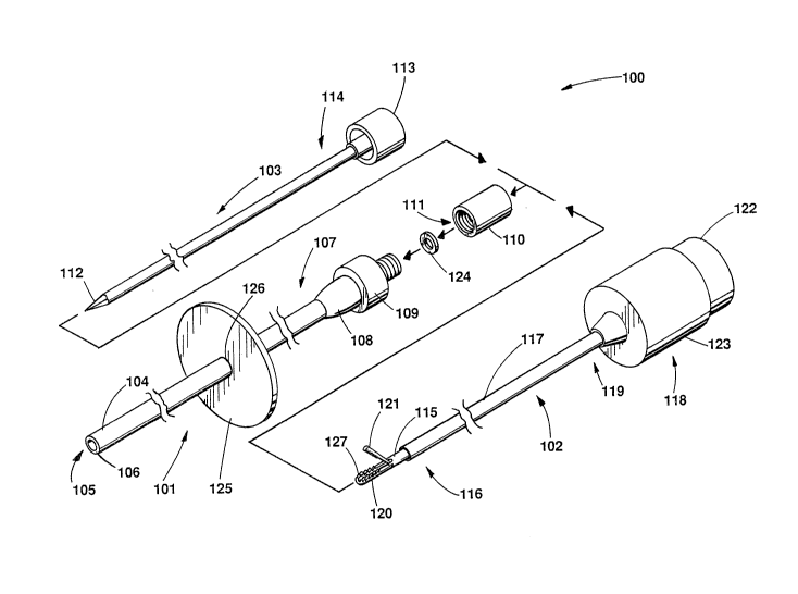
Brief DescriPtion of the Drawinq
FIG. 1 depicts an illustrative percutaneously
- insertable, needle-sized retractor and system of the present
invention; and FIG. 2 depicts an enlarge~, partially
sectioned, side view of the retractor o~ FIG. 1.
Detailed Description
Depicted in FIG. 1 is an illustrative percutaneous
retractor system 100 comprising needle-sized trocar sheath
101, tissue retractor 102, and trocar stylet 103 for
percutaneous insertion into the cavity of a patient. The
trocar sheath comprises a polytetrafluoroethylene radiopaque
material tube 104 having passage 105 extending
, ~ ,
:., :: . ::, :;
Edward Duane Pingl_~on, Paul Guy Thomson 6-8 (~ -5050)
2091271
longitudinally therethrough. Tube 104 has an outside
diameter in a range of 10 to 22 gauge (.028" to .134") and
is approximately 13 cm in length. Preferably, tube 104 has
an 8 French (.105") outside diameter and a wall 106
thickness of approximately .016". Fixedly attached using,
for example, medical grade adhesive about proximal end 107
of the tube are well-known male and female Luer lock
connector adapters 108 and 109. Also positioned about the
proximal end of the tube is retention cap llO threadably
engaging female connector adapter 109. Passage 111 extends
through the retention cap and connector adapters to
communicate with passage 105 of the polytetrafluoroethylene
tube. Trocar stylet 103 is inserted through passages 105
and 111 of the tube and retention cap. When the trocar
stylet is fully inserted in tube 104, pointed distal end 112
of the stylet extends distally from passage 105 of the tube.
The trocar sheath with the pointed distal end of the stylet
extending therefrom is then percutaneously inserted into the
cavity of a patient through the skin and subtending tissue
and organs. The trocar stylet stiffens the sheath for
advancement through the skin and subtending tissues and
organs. A well-known connector cap 113 is fixedly
positioned about a proximal end 114 of the stylet with, for
example, medical grade adhesive for positioning and
manipulating the stylet with respect to the trocar sheath.
The trocar stylet preferably comprises a stainless steel
material rod approximately 17 cm in length ttith an outer
diameter of approximately .068". The distal end of the rod
is ground to a point for easy insertion into the cavity of
a patient.
When the trocar stylet and sheath are inserted into the
cavity of a patient, the stylet is removed from the passage
of the sheath. Tissue retractor 102 is then inserted
through the passage of the trocar sheath and into the cavity
of the patient. The retractor includes grasper 115
positioned at distal end 116 of elongated member 117.
s
Edward Duane Pingi_~on, P~ul Guy Thomson 6-8 ~ 5050~
2091271
Tissue retractor 102 also comprises push-button handle 118
attached about proximal end 119 of elongated member 117.
Handle 118 is grasped by the middle and inde~ fingers along
with the thumb of the physician to operate pivotedly
interconnected jaws 120 and 121 of grasper 115 between open
and closed positions. Jaws 120 and 121 have well-known
alligator teeth for grasping tissue or, alternatively, a
well-known hounds tooth configuration. Normally, the
grasper is maintained in the closed position with jaws 120
and 121 engaging one another. The jaws of the grasper are
operated to the open position by the physician pressing
push-button piston 122 of the handle which slidably moves in
enclosure 123 of the handle. Retaining cap 110 of the
trocar sheath includes seal 124 positioned about passageway
15 111, which is compressed to engage elongated member 117 of
the retractor. This is accomplished by the physician
turning retention cap 110, compressing the seal, and fixedly
positioning the retractor in passages 105 and 111 of the
trocar sheath and retention cap.
The retractor system further comprises a well-known
retention flanye 125, such as the Molnar retention disk
commercially available from Cook Urological, Incorporated,
Spencer, Indiana. The disk includes passage 126 of which
trocar sheath tube 104 is inserted therethrough. Tha disk
is moved along the length of the tube to engage the surface
of the skin and fixedly position the trocar sheath with
respect to the cavity. With the retention cap fixedly
positioning the grasper, and the retention flange fixedly
positioning the trocar sheath with respect to the surfa~e
tissue of the patient, the grasper can effectively grasp,
retract, and fixedly position tissue during a minimally
invasive endoscopic procedure.
Depicted in FIG. 2 is an enlarged, partially sectioned,
side view of tissue retractor 102. Tissue retractor 102
includes needle-sized elongated member 117 with tissue
grasper 115 positioned about distal end 116 and handle 118
:.-: '"' ` :: -
:
Edward Duane Pin(~l _on, Paul Guy Thomson 6-8 (~ 5050)
2091271
positioned about distal end 119 thereof. Jaws 120 and 121
of the grasper are depicted in this figure in the open
position with alligator teeth 127 positioned on opposing
faces of the jaws. Elongated member 117 comprises an outer
5 cannula lZ8, inner rod 129, and outer sheath 130. Outer
cannula 128 comprises a 12.125" length of an 18.5 gauge
regular wall tube with an outside diameter of approximately
.046" and passage 131 extending longitudinally therethrough
with an inside diameter of .030". Distal end 116 of the
10 outer cannula is connected to stationary jaw 121. Proximal
end 119 of the outer cannula is connected to handle
enclosure 123 through passage 134 with proximal and distal
retaining tubes 132 and 133 on opposite sides of enclosure
wall 135, as shown.
Inner rod 129 comprises a 22 gauge thin-wall stainless
steel tube with an outside diameter of approximately .028"
and an inside diameter of approximately .014". A mandrel of
stainless steel wire is inserted through the passage of the
thin-wall tube and soldered about the proximal and distal
20 ends thereof. Distal end 116 of the inner rod is connected
to movable jaw 120 via interconnecting link 136 in a well-
known manner. The proximal end of the inner rod is
connected to push button piston 122 via passage 137 thereof
and fixed thsreto with a well-known set screw 138, as shown.
Handle 118 comprises machined, vinyl material enclosure
123 with cavity 139 formed therein, as shown. The handle
further comprises machined, vinyl material push button
piston 122 slidably positioned in cavity 139. Push button
piston 122 also includes cavity 140 communicating with
30 enclosure cavity 139. Positioned in cavity 139 of the
enclosure and cavity 140 of the piston around outer cannula
128 and inner rod 129 is spring 141 engaging opposite facing
walls 135 and 14Z and providing expansion tension thereon.
Spring 141 is commercially available as model number LC-
35 0380-8 from Lee Spring, Inc., Des Plaines, Illinois.
" :. : : '
,
.: . . : . ~
. ., .;
.. : ~ . : .
Edward Duane PingL ,on, Paul Guy Thomson 6-8 (~ -5050)
~; 2091271
H~ndle enclosure 123 also includes outer shoulder 143
extending distally therefrom about proximal end 119 of
elongated member 119. Passage 134 extends therethrough of
which proximal and distal retaining tubes 132 and 133 are
friction welded on outer cannula 128 on opposite sides of
enclosure wall 13 5, as shown.
Elongated member 117 also includes outer sheath 130
comprising, for example, a nylon material tube having an
outside dimension of .065" and an inside dimension of . 050" .
Proximal end 119 of the nylon material outer sheath is glued
to shoulder 143 of the handle enclosure next to distal
retaining tube 133.
Handle enclosure 123 comprises a machined, vinyl
material cylindrical cup with a length of approximately
.750" and an overall length with outer shoulder 143 of
approximately 1.000". The depth of enclosure cavity 139 is
approximately .650". The outside diameter of the handle
enclosure is approximately . 750" . The diameter of the
cavity is approximately .650". Shoulder 143 has an
outermost diameter of .250" with 15 degree tapered sides.
Passage 134 is approximately . 052" in diameter. The distal
end of the shoulder is countersunk to accept 17 gauge thin-
wall, stainless steel, distal retaining tube 133, which is
approximately .04" in length. Proximal retaining tube 132
25 is a 17 gauge thin-wall, stainless steel tube approximately
.300" in length. Outer sheath nylon tube 130 is
approximately 5 French (.066") in diameter.
Handle 11~ assumes operated and relaxed states
corxesponding to the open and closed positions of grasper
115. With spring 141 engagincl the push button and handle
enclosure, the handle assumes the relaxed state with the
grasper in the closed position. To operate the handle to
the operated state, the physician grasps handle enclosure
123 with the middle and index fingers and depresses push
button 122 with the thumb. As a result, spring 141 is
compressed and tissue grasper 115 and jaws 120 and 121 are
... ~ . :
': ' ~, ~ '`'' ,."'; '''""`
' ` ` ~: , ,,
: .
~ Edward Duane Pingl ~on, Paul Guy Thomson 6-8 (~ 5050)
-' -, 2091271
operated to the open position. This is accomplished by
inner rod 121. To operate the retractor to the closed
position, the physician simply releases the push button.
spring 141 separates the push button and handle enclosure,
causing jaws 120 and 121 to close. It is to be
understood that the above-described percutaneously
insertable needle-sized retractor and system is merely an
illustrative embodiment of the principles of this invention
and that other needle-sized retractors and systems may be
devised by those skilled in the art without departing from
the spirit and scope of this invention. It is contemplated
that the tissue grasper at the distal end of the retractor
may comprise other configurations such as a biopsy punch,
normally opened rather than closed jaws, cutting blades, and
the like. It is further contemplated that the handle may
take on other shapes of which to operate the tissue grasper
between open and closed positions. It is also contemplated
that the materials of the grasper may comprise other well-
known and commercially available materials.
.:
''`
., 9
~''
.
.:, : ;: , . :,,. :- . ;, :: . :. . : -
: . . , . , :. ~ :: . .~. . . . . .:
: : . ; . . . .. :,: . :. :~: ., , :. : : .. .: