Note: Descriptions are shown in the official language in which they were submitted.
P~-5066-CIP, CIP2, CIP3 ~ ~ 2 ~ ~ 7
VASCULAR STENT
Technical Field
The present invention generally relates to vascular
stents.
Backaround of the Invention
A stent, generally speaking, is a device that can be
placed within the lumen or interior space, of a tubular
structure for supporting and assuring patency of a
contracted, but otherwise intact, lumen. (Patency the state
of being freely open, is particularly important in the field
of angioplasty, which is concerned with the reconstruction
of blood vessels.) Stents are used, for example, for
holding blood vessels open or for back tacking intimal flaps
inside vessels after angioplasty. More generally, however,
stents can be used inside the lumina of any physiological
conduit including arteries, veins, vessels, the biliary
tree, the urinary tract, the alimentary tract, the
tracheobronchial tree, the genitourinary system, and the
cerebral aqueduct. Furthermore, stents can be used inside
;lumina of animals other than humans.
In the field of angioplaety, the most common angioplasty
surgical procedure is percutaneous transluminal coronary
angioplasty, or ''PTCA'I, which is employed for enlarging
; 30 narrowed arteries near the heart. In a PTCA procedure, a
balloon-tip catheter is maneuvered into position in a
narrowed artery where the balloon is expanded in order to
dilate this area of narrowing. After the arterial lumen is
dilated, the balloon at the catheter tip is deflated and the
~35 catheter is removed from the enlarged artery. A vascular
;~ stent can be used to dilate an artery after a suboptimal
PTCA dilation.
In practice, the above-described conventional PTCA
procedure has several shortcomings. One drawback is that
'
'''
:,.
~ . . . . :,. : ..
.-,: ~
P7~-50~;6-CIP, CIP2, CIP3
approximately one-third of all PTCA patients suffer from
restenosis, a chronic regrowth of obstructive tissue that
narrows the lumen. Typically, restenosis occurs within six
months following an angioplasty procedure. Since a majority
of these restenosis patients also display symptoms of
deteriorating cardiac status, they frequently must undergo
additional PTCA procedures or more risky coronary artery
bypass graft surgery. Unfortunately, those patients who
undergo repeated PTCA procedures tend to restenose at an
even higher rate than first-time PTCA patients.
A second, and sometimes fatal, complication of coronary
angioplasty is the abrupt re-closure of a previously dilated
section of a vessel. There are many different factors that
are thought to contribute to abrupt re-closure after PTCA
including obstructive flaps of disrupted wall tissue, vessel
wall spasms with luminal contraction, and thrombus formation
at the site of dilation. Vascular stents can be used like
a scaffold to mechanically bridge areas of narrowing (flaps
or thrombus) and oppose spasms, and therefore, maintain
artery patency.
Many of the factors responsible for abrupt closure (post
balloon inflation) may also influence the development of
restenosis, and therefore, long term patency. In this
regard, vascular stents, by virtue of their ability to limit
elastic recoil of the vessel wall and to eliminate the
negative physical consequences of PTCA (including
obstructing intimal flaps and dissection) may be useful in
limiting restenosis.
Therefore, there are two potential benefits of vascular
stents in the treatment of vascular disease: 1) prevention
of abrupt arterial closure, and 2) prevention of restenosis.
~r Summar~ of the Invention
,~,.~,,
Generally speaking, the present invention provides a
vascular stent for reducing hemodynamic disturbances caused
by angioplasty and the stent itself. In a preferred
~: .
~ 2
....
: , ., . : -
:
.... . . , . -: .. :
- : - . .:
PA-5066-CIP, CIP2, CIP3
3~
embodiment, the stent is formed from a single filament of
low memory bio-compatible material having a series of U-
shaped bends. The filament is wrapped around a mandril in
a circular fashion in order to align opposing curved
5 portions of each bend which are then connected. The stent
therefore provides a maximum amount of structural support
for the lumen while minimizing the level of hemodynamic
disturbance inside the lumen.
The tubular stent shown in the embodiments of the
invention is a co-planar structure as opposed to a woven or
knitted structure.
The present inventor has found that vascular stents
require substantial flexibility in their unexpanded state in
order to allow them to bend and conform to the tortuous
shape of the vessel through which they are inserted. This
need for flexibility during insertion is especially
important for older patients since their blo~d vessels tend
to be more tortuous and less flexible than those of younger
patients. The present inventor has also found that,
vascular stent6 should be rigid and have a high hoop
strength in their expanded state. Although the reasons for
the success of rigid stents are not entirely clear, it has
been suggested that rigid stents are less likely to pulsate
inside vessels, and therefore, they are less likely to rub
against the veæsel intima once they are in place.
~:
~E~ef Descri~tion of the Drawinq
Figure 1 shows a filament shaped into a compressed
planar wave used to make the nearly sinusoidal waveform of
Figure 2;
; Figure 2 shows the planar wave of Figure 1 expanded
along its longitudinal centerline to form a nearly
sinusoidal waveform used in making a stent;
; 35 Figure 3 shows an alternative waveform that can also be
used in making a stent;
Figure 4 shows another alternative waveform that can be
.
. . - . . .
.. . . . .
: . , . - . . .
P~-5066-CIP, CIP2, CIP3 2 ~ ~ 2 ~ 37
used in making a stent;
Figure 5 shows the waveform of Figure 3 spirally wrapped
around a round mandril;
Figure 6 shows a connection for the end of the filament
after the waveform of Figure 3 is completely wrapped around
the mandril;
Figure 7 shows a preferred alternative waveform that can
be used in making a stent;
Figure 8 shows the relative positions of the U-shaped
bends in each component section of the preferred alternative
waveform of Figure 12:
Figure 9 shows the preferred alternative waveform of
Figure 7 being wrapped around a cylindrical mandril;
Figure 10 shows in an expanded state a side elevation of
a stent formed from the preferred alternative waveform of
Figure 7 by wrapping it around a mandril in a circular
fashion in order to align the curved portion of each bend;
Figure 11 shows an opposite side elevation of the stent
in Figure 10;
Figure 12 shows an end view of the stents in Figures 10
and 11;
Figure 13 shows a stent mounted on a balloon-tip
catheter ready for insertion into a lumen;
. Figure 14 shows a stent being used with a graft to
repair a pseudo-aneurysm in the common femoral artery;
~ Figure 15 shows two stents being used with a graft to
''! bypass an occlusion in the femoral-popliteal artery;
Figure 16 shows a stent being used with a graft to
repair an aorto-iliac aneurysm;
Figure 17 is a schematic illustration of a planar
~` waveform which is used to form the stent;
:~ Figure 18 illustrates the waveform of Figure 17 being
wrapped around a mandril;
` Figure 19 illustrates an alternative embodiment of the
`; 35 waveform of Figure 17 being wrapped around a mandril;
~; Figure 20 shows the arrangement of the waves around the
'
-:: - -
~, . : ,, .. . :
: - . . . :
:~ -.:. . ..
- :. ::
. . .: .- .: :
P.~-5066-CIP, CIP2, CIP3 ,~ ~ 2 3 ~ ~
circumference of the mandril when the stent is formed in its
unexpanded state as in Figure 18;
Figure 21 shows the arrangement of the waves around the
circumference of the mandril when the stent is formed in its
unexpanded state as in Figure 19;
Figure 22 is an enlargement of one of the cells in the
stent of Figures 20 and 21 when the stent is in an expanded
state:
Figure 23 shows a stent being used with a graft to
repair a pseudo-aneurysm in a common femoral artery;
Figure 24 is a side-elevational view of a stent
according to the other preferred embodiment in compressed
condition;
Figure 25 is a side elevational view of the stent of
Figure 24 in expanded condition;
Figure 26 is an end view of the stent of Figure 25;
Figure 27 is a cross-sectional view which is taken along
the plane of the line 4-4 in Figure 25 for viewing in the
direction of the arrows;
20Figure 28 is an enlarged cross-sectional detail, taken
along the plane of the line 5-5 in Figure 26 for viewing in
. the direction of the arrows;
~ Figures 29A and 29B are views that correspond in
:- orientation to Figure 4 and which schematicall~ show the
stent of Figure 1 embedded in the lumen of a blood vessel;
:. Figure 30 is a schematic illustration of a planar
waveform of a continuous wire which is used to form the
stent of Figures 24 and 25; and
Figures 31A and 31B are illustration of the continuous
.; 30 waveform of Figure 27 wrapped around the circumference of a
` mandril for forming the stent in its compressed condition.
;`:
Detailed Descri~tion
35 The stent is preferably formed from a continuous wire.
The term "wire", as used here, should not be construed as
limited to just metallic materials. In fact, the stent may
': . ' ,. . . ;~' .
~: '
-
P.~-5066-CIP, CIP2, CIP3
be formed from any type of filament. The stent may also be
made from groups of filaments or fibers which are wound or
braided together in order to form a continuous filament.
Also, several distinct filaments may also be attached
together by any conventional means such as butt-welding. It
is also possible to mold the stent in its unexpanded state.
To prevent the stent from recoiling to its unexpanded
state after it has been implanted, the stent is preferably
made from a "low memory" material that does not try to
resume its original shape after it is deformed.
Alternatively, the size of the wire can be chosen so that
when the stent is expanded, the wire is stressed beyond its
plastic yield point but not beyond the ultimate stress at
which the material cracks or breaks. Both the unformed wire
and the unexpanded stent may be annealed in order to reduce
the stresses which are created in the wire during the stent
formation process.
The stent material is preferably radio-opaque so that
the location of the stent can be verified through
,,
fluoroscopic examination. The stent should also be made
from a biocompatible (e.g. stainless steel) and/or
bioabsorbable (e.g. Vicryl) material with a smooth surface
for minimizing the stent's effect on surrounding tissue and
bodily fluids such as blood. The stent may also be coated
with antithrombolytic or anticoagulatory agents such as
-,Dextran, Heparin, t-PA, polytetrafluoroethylene, or ultra
low-temperature isotropic carbon.
~; Figure 1 shows a filament 11 formed in a compressed
planar waveform. Preferably, the filament 11 is made from
0.013 to 0.05 cms (0.005 - 0.020 inch) diameter stainless
,, ~
steel wire; however, it can be made from materials such as
titanium, tantalum, gold, copper and copper alloys,
combinations of these materials, or any other biologically-
compatible materials with a low shape-memory level. (In the
present context, a low shape-memory level implies that the
stent will not contract to its compressed shape after it is
:
:
. - .,:: : , :
:. :. .: . ,~. .,.: , :
- - ,: :~ - ,
:, ~ :,
PA-5066-CIP, CIP2, CIP3
~ `2~7
inserted and internally expanded in a lumen.) The filament
ll can also be formed from several separate strands which
are wrapped or woven together.
The compressed waveform pattern in Figure 1 is
5 preferably formed generally in the shape of a compressed
sinusoid, but can have any wave-like pattern. In the
drawing, it should be noted that the waveforms at the ends
l9 and 21 of the wire having smaller amplitudes than the
waveforms 15 in the middle of the wire. The drawing shows,
for example, four reduced amplitude peaks 17 at each of the
ends 19 and 21, respectively. Preferably, the heights of
the reduced amplitude waveforms are one-half to two-thirds
of the heights of the larger waveforms.
In Figure 2, the aompressed waveforms of Figure 1 are
~15 expanded along their longitudinal centerline into a nearly
- sinusoidal waveform by stretching the compressed waveforms
`from their ends. (The broken line shows the longitudinal
centerline of the expanded waveforms.) At both ends 19 and
21, the longitudinal centerline of the smaller waveforms is
displaced from the longitudinal centerline of the waveforms
~;near the middle of the wire. At one end 19, for instance,
the centerline of the smaller waveforms 17 is displaced
below the broken line; at the end 21, by way of contrast,
the centerline of the smaller waveforms is dieplaced above
the broken line.
In practice the above-described expanded waveforms
preferably have a period of about eight millimeters. The
larger waveforms 15 preferably have a peak~to-peak amplitude
of eight millimeters while the smaller waveforms 17 are one-
half to two-thirds the height of the larger waveforms.
;However, other sizes may be used. Although all of the
waveforms normally have the same period, they are not
necessarily sinusoidal, regular, repeating, or continuous.
Figures 3 and 4 show the expanded state of two
alternative waveforms that can be used to form the above-
described stent. The period of each waveform in the
,
,
P~-506~-CIP, CIP2, CIP3
4~ 7
waveform of Figure 3 is preferably one-half of the peak to
peak amplitude of the waveform. In Figure 3, the
longitudinal centerlines of the small waveforms 17a at the
ends of the device are approximately parallel to each other,
but the centerline of the large waveforms 15a is inclined
relative to the longitudinal centerlines of the smaller
waveforms, preferably at an inclination angle of
approximately 45. In Figure 4, the waveform is similar to
that of Figure 3 except that the centerline of the larger
waveforms 15b is perpendicular to the centerline of the
smaller waveforms 17b; in other words, the inclination angle
of the larger waveforms is approximately 90.
Figure 5 shows the expanded waveform of Figures 3 formed
into a stent by wrapping it, in a spiral, around a mandril
21. Similar waveforms could also be used. For instance, if
:
the waveform of Figure 4 were used, the longitudinal
centerline of the large waveforms would remain parallel to
the centerline of the mandril and the peaks of the waveforms
would be wrapped around the mandril, perpendicular to the
centerline of the mandril.
As shown in Figure 5, the centerline of the large
waveforms 15a is arranged to spiral along the length of the
mandril 31. One side of each of the larger waveforms 15a is
arranged approximately parallel to the longitudinal axis of
the mandril 31, and the remaining sections of each of the
waveforms are arranged at a small angle to the longitudinal
axis of the mandril. (In the drawing, the "small" angle has
been greatly exaggerated for purposes of illustration.) It
will be appreciated that the shown arrangement allows the
` 30 stent to be wound in a very tight spiral.
By forming the above-described stent as a tight spiral
on a mandril, the stent expands primarily in the radial
direction, with relatively slight movement at the ends, as
it is expanded internally in a lumen. Even greater radial
expansion might be achieved by the wrapping the waveform as
a circle around the mandril. However, such as radially-
:'' ' : -` ~ . - ,
P~-5066-CIP, CIP2, CIP3
3~7
wrapped configuration would use an excessive amount of
filament per unit surface area to support the lumen,
especially where the filaments were allowed to overlap.
In Figure 6, each of the last three smaller waveforms
17a (from Figure 5) at the end of the stent is wrapped with
its longitudinal centerline around the circumference of the
~ mandril. It should be noted that the peaks of the last
- three smaller waveforms (indicated in Figure 6 by the
letters "a", "b" and "c" respectively) are approximately the
- 10 same distance from the edge of the mandril, and the fourth
peak "d" is slightly further away from the end of the
mandril. Also, the end of the stent near peak "a" is
connected to the apex of peak "d", the result of this
~- connection is that peaks "a", "b" and "c" are substantially
equally spaced around the circumference of the mandril and
~ are all at the approximately same distance from the end of
`~ the mandril.
In practice, the connection between the loop and the
- filament is slidable along the filament 11, thereby allowing
for radial expansion. Although this connection can be
easily made using a loop as shown, it can also be made by,
r~( for example, using a bracket. The connector could also be
made by brazing, welding or gluing the end to the filament.
When the above-described stent is wound around a mandril
in the shape of a tight spiral, the non-expanded ~orm of the
.
stent provides a profile that is lower than conventional
stents, and the "tines" of the non-expanded stent are almost
~; parallel and packed closely together. This is important
because such stent can be accommodated through a smaller
incision and, therefore, reduces blood loss during surgery.
Furthermore, such a stent can provide an expansion ratio of
about 10:1, enabling it to be used in large arteries.
As shown in Figure 12, the connections at the ends of
i the filament 11 create a circular hoop near each end of the
stent with no sharp edges, or point, protruding from the
perimeter to project into a lumen or to catch on the balloon
., - ~ :
.,.:
.. . . . . .
P~-5066-CIP, CIP2, CIP3
or plaque inside of a vessel. Also, because the centerline
of the smaller waveforms is arranged along the circumference
of the stent, the end hoops allow the stent to fit snugly
inside the lumen and prevent migration. In other words, in
this arrangement, the hoops expand radially to lock the
expanded stent in place in a lumen while permitting only
limited longitudinal expansion.
Figure 7 shows a preferred alternative waveform which
can be used in making a stent. The waveform of Figure 7 is
formed from a series of U-shaped bends having substantially
straight legs on each side of the curved portion of each
U". The legs are preferably parallel; but they may also be
formed at angles to each other. The curved portions are
preferably semi-circular; however, other shapes of curves
can be used to connect the straight legs in each bend. The
curved portions may have the same or different sizes. It is
also preferred that the curved portions are connected to the
straight portions at the tangent of each curve in order to
prevent any discontinuities in the length of the filament.
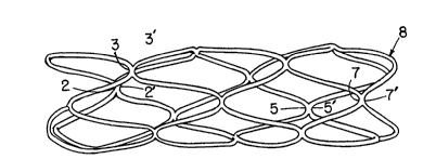
Figure 8 shows the relative positions of the U-shaped
bends for each component section A, B, C of the preferred
alternative waveform of Figure 7. Sections A and C of the
waveform are upside down mirror images of each other. The
broken lines in Figure 8 are reference lines which are
preferably equally spaced and parallel. However, it is also
possible to form the stent so that the top and bottom
reference lines are parallel to each other but not equally
; spaced from or parallel to the other reference lines.
Defining the distance between the reference lines as one
unit of measurement, then each of the U-shaped bends in end
sections A and C each have a different length. For example,
U-shaped bend 1 is one unit long while U-shaped bend 3 is
three units long. In contrast, each of the waveforms in
section B has one long leg which is four unit long and one
short leg which is three units long. For example, the left
leg of U-shaped bend 5 i6 four units long while the right
. . .. , :,
. ; . . ~
: . - , ~ :
: . . - : :
PR-5066-CIP, CIP2, CIP3
~2:~37
leg is three units long as measured between the reference
lines. Each of the curved portions, except for the ends of
the filament, are preferably semicircular with a diameter of
- one unit. The curved portion at each end of the filament is r
5 preferably one half of the semicircular arc. However, other
shapes and proportions may also be used to appropriately
size the stent.
As shown in figure 9, the stent is formed by wrapping
the waveform of Figure 7 around a mandril which is
10 preferably cylindrical. However, mandrils with other shapes
could also be used. The waveform is preferably wrapped
around the mandril so that the legs of each U-shaped bend
are parallel to the axis of the mandril. In this
configuration, a single wire may be formed into an extremely
15 rigid tubular structure with very little material to disturb
flow inside the lumen. However, the waveform might also be
wrapped around the mandril in a slightly spiral manner.
Once the waveform is wrapped around the mandril, the outside
^ edge of curves on the same reference line will be arranged
back-to-back adjacent to (or overlapping with) each other.
For example, the outside edge of curve 1 will be back-
to=back with the outside edge of curve 1'. Similarly the
outside edge of curve 7 will be adjacent to curve 7'. The
outside edges of these U-shaped bends can then be fastened
25 together by any conventional means such as weldinq, brazing,
soldering, or gluing.
Figures 10, 11, and 12 illustrate the stent which is
formed by wrapping the waveform of Figure 8 around a
circular mandril with the reference lines arranged on the
30 circumference of the mandril. It will be apparent that each
of the labeled U-shaped bends on parallel reference lines in
Figure 8 have been connected in Figures 10 and 11. For
- example, U-shaped bend 7' is shown to be connected to U-
shaped bend 7 at the top of Figure 10. Although it is
35 preferred that the U-shaped bends are welded, it is also
possible to form the connecting portions of the filament
11
. , ~ ,
.. .. .. . : ~,
P~-5066-CIP, CIP2, CIP3
2~33 ~
from a single piece of material in order to eliminate the
need for connecting each of the appropriate U-shaped bends.
The ends of the filament are also connected back to the
filament and trimmed in order to remove any excess filament
precluding from the free end.
The rigidity of the structure may be controlled by
welding less than all of the adjacent curved portions. For
- example, a stent with only half the U-shaped portions welded
together would be approximately half as rigid as a stent
with all the tangent points welded together. Of course, the
stent can also be used without any connections between
adjacent curved portions.
, The lowest possible profile (i.e. diameter) is provided
;~ by arranging the long leg of each U-shaped bend parallel to
the axis of the catheter before it is inserted into a lumen.
This arrangement increases the diameter to which the stent
can be expanded without, at the same time, decreasing the
~` end-to-end length of the stent. By increasing or decreasing
the length of the long leg of each U-shaped bend, one can
alter the expansion ratio without altering the profile.
Consequently, a nearly unlimited circumferential expansion
ratio may be created without contracting the stent along its
longitudinal axis. The expansion ratio is therefore nearly
independent of this profile.
When expanded, each of the U-shaped portion~ in the
stent may assume a rhomboidal pattern where the legs of each
U-shaped bend are no longer parallel. The expansion ratio
of the stent may therefore exceed lO to 1 in terms of the
expanded diameter versus the unexpanded diameter of the
stent. Consequently, the outside surface of the stent
touching the vessel is small while the effective support
area is very large. This feature dramatically reduces the
possibility of causing any hemodynamic disturbances inside
the vein or artery because of the stent. The large
expansion ratio also allows the stent to be used with
smaller incisions. Moreover, this configuration allows the
~-5066-CIP, CIP2, CIP3
stent to be flexible in the radial direction in order to
accommodate the pulsation of an artery.
The stent may also be coated with anti-thrombolytic
agents in order to limit the thrombotic formation which
often aGcompanies angioplasty.
Figures 13-16 illustrate a typical stent of which could
represent any one of the embodiments described above.
Figure 13 shows a typical stent mounted on a 4/5 F balloon
(4-lOmm) with a 6/7 F sheath. The apparatus of Figure 13 is
preferably used with a .078-.091 guide sheath. Figure 14
shows the stent inside a graft, being used to repair a
pseudo-aneurysm in a common femoral artery. The stent 8 is
: placed inside graft 9 which blocks off pseudo-aneurysm 13.
Although the stent is shown to be completely inside graft 9,
it may also extend outside the edges of the graft in order
to provide additional support for the incisions at the end
of the graft.
Figure 15 shows two stents being used at each end of a
graft to bypass an occlusion 23 in, for example, the
femoral-popliteal artery. Figure 16 illustrates how three
stents can be used with a branched graft to repair an aorto-
iliac aneurysm 33. The graft 9 is placed inside the
aneurysm and secured at one end to the aorta 35. The other
ends of the graft are similarly stented to iliac branches
37.
Figure 16 also illustrates how the catheter of Figure 17
can be used to insert the stent 8 inside a lumen.
Typically, a short incision is made in the lumen (for
example, a vein or artery) and the stent, which is mounted
on the balloon, is then slipped into the incision. When the
stent is in place, the balloon is expanded in order to
expand the stent against the inside walls of the lumen.
Once the stent is in place, the balloon is deflated and
removed through the inside of the stent and the incision in
order to leave the stent in place.
Various advantages of the present invention can now be
13
- , - . ., .- . . .. . .. . ... . . ...
PA-5066-CIP, CIP2, CIP3
2 ~
understood. For example, the above-described stent uses
substantially less material than conventional stents
(especially knitted ones with overlapping wires) and,
therefore, introduces a substantially lesser quantity of
5 foreign material into a lumen. The stent also provides a
maximum amount of structural support with a minimum amount
of material. As another example, the above-described stent
connects its filament ends back onto the filament to prevent
thrombosis in blood vessels or damage to any type of a lumen
wall such as is caused by stents that have loose wire ends
that protrude into a lumen.
Another advantage of the above-described stent is that
it provides substantial radial expansion with only limited
~longitudinal migration and, therefore, reduces the problem
-; 15 of migration inside a lumen. More particularly, the hoops
and end component sections at each end of the above-
described stent reduce migration by securing the stent
inside of a lumen. In the preferred embodiment, the hoops,
end component sections, as well as the spiral shape of the
stent itself are oriented to inhibit longitudinal growth of
the stent during radial expansion.
Yet another advantage of the above-described stent is
that it provides sufficient flexibility to allow
implantation in tortuous lumens and in applications where
lumen bending i8 required. This overcomes the pxoblem with
conventional stents that are so stiff that they are
difficult to negotiate through a tortuous vessel during
implantation. Furthermore, a stiff stent can cause damage
to certain vessels, such as those around joints, that
require flexibility.
The stent is formed from a continuous wire shaped into
the planar pattern or waveform illustrated in Figure 17.
The pattern in Figure 17 includes a series of alternating U-
shaped waves having a period p with peaks 10 and valleys 12
interconnected by substantially straight sections 14. The
straight sections 14 are substantially parallel to each
14
' ~
PA-5066-CIP, CIP2, CIP3
2 ~ 7
other in Figures 17, 18 and 20 and are therefore depicted as
straight vertical lines in those figures. However, the term
"substantially parallel" also refers to the configuration of
the straight portions 14 illustrated in the compressed
planar (and generally sinusoidal) waveforms of Figures 19
and 21. The peaks 10 and valleys 12 are preferably
semicircular and arranged to intersect straight portions 14
at the tangent of each curved peak or valley so that there
are no discontinuities in the wire. ~owever, other curved
or linear shapes may also be used to form the peaks 10 and
valleys 12. Each U-shape wave includes an ascending side
14a and a descending side 14b.
The outermost portions of the peaks 10 and valleys 12 in
the middle ~ection of the waveform are aligned along
parallel axes 16 and 18, respectively. The axes 16 and 18
form an acute angle a with respect to the straight portions
14. The angle ~ is preferably 45 so that if distance
between each straight section is one unit, then each U-
shaped wave in the middle section has one leg that is three
units long while the other leg is four units long as
illustrated by the parallel horizontal reference lines in
Figure 17. Other relative dimensions and angles, however,
can be used. A curved stent can also be formed by, for
example, slightly increasing the length of every third wave
and decreasing the length of a corresponding wave in order
to form an arched configuration where one side of the
tubular body is slightly longer than another side.
There are two waves 20 of different amplitudes at each
end of the stent which each have two sides of the same
length. The end sections of the waveform include peaks 10a,
10b, and 10c at one end of the stent and valleys 12a, 12b,
and 12c at the other end. The outer edges or apexes, of
valleys 12a, 12b, and 12c are aligned along axis 28 which is
substantially perpendicular to the straight portions 14
(i.e. horizontal in Figure 17). Similarly, the apexes of
peaks 10a, 10b, 10c are aligned with an axis 30 which is
` ' ~ ' ~' I
.
PA-5066-CIP, CIP2, CIP3
also perpendicular to the straight portions 14 of the waves
22 but displaced from axis 30. The ends of the wire 2~, 26
are preferably formed into half a valley 12 at one end and
half of a peak 10 at the other end. The ends 26 may also
include a small, straight portion (not shown) which may be
parallel or perpendicular to the straight portions 14.
Referring to Figure 18, the stent is formed by wrapping
~ the waveform of Figure 17 around a mandril 32. The peak 10
;~ of one wave coincides with the valley 12 of another wave
when the waveform of Figure 17 is wrapped around mandril 32
with straight portions 14 aligned with the longitudinal, or
central, axis of the mandril 32. Figure 18 illustrates the
end 24 of the waveform wrapped around the mandril 32 so that
the end 24 is tangent to point 24'. Similarly, end 26 will
be tangent to point 26' when the wave is completely wrapped
around the mandril 32. The ends 24, 26 of some or all of
the junctions are then bonded to one another over relatively
short lengths to form bonded cells by spot welding, spot
brazing, soldering, tying looping, adhesive bonding, or
other suitable means to the points 24' and 26' respectively,
so that the ends of the wire are not exposed where they
could snag or otherwise interfere with the placement of the
stent in the vessel.
In practice, electric resistance welding has been found
to offer the most secure metal to metal bond by minimizing
the amount of oxidation that occurs during bonding process.
As the wire is wrapped on the mandril, some or all of the
successive junctions between the peaks 10 and valleys 12 may
be bonded in a similar manner until the stent is complete.
The flexibility of the stent can be controlled by bonding
fewer than all of the peaks 10 to corresponding valleys 11.
The stent may then be compressed on consecutively small
diameter mandrils so that the straight sections 14 in Figure
17 are no longer exactly parallel, but still "substantially
parallel", i.e. less than 10 from being parallel, to the
longitudinal axis of the mandril so the wave pattern takes
16
:~ : : ' ,.,,, :~ .:-
P~-5066-CIP, CIP2, CIP3 2~3~ 7
on a generally sinusoidal shape such as the one illustrated
in Figures 19 and 21. The planar waveform of Figure 17 may
also be compressed perpendicular to straight sections 14 in
order to form the nearly sinusoidal pattern illustrated in
5 Figure 19 before being wrapped around the mandril 3~. The
stent is then removed from the smallest mandril and the
stent is arranged on the balloon catheter.
The structure of the stent of the present invention is
capable of expanding radially when subjected to the internal
pressure of an expanding catheter balloon. The peaks 10 and
valleys 12 between the waves operate like flexible junctions
or hinges to allow the straight portions 14 to swing
outwardly, oblique to the central axis of the body of the
stent. Unlike hinges, however, after the stent is expanded,
the junctions resist displacement of the straight sections
in the opposite direction (for example, due to the
compressive force of the lumen) which would tend to reduce
the diameter of the expanded stent. The resistance of these
junctions to compression (i.e. hoop strength) is caused by
placing a stress on the material at the junction which
exceeds the elastic limit of the material so that the
material near the junction is plastically deformed and this
resists any tendency for the stent to collapse inside a
lumen. The wire and the bonding material should therefore
be a low memory material.
Figures 17 and 18 illustrate a waveform where the period
(or wavelength) of each wave p is roughly one-fourth of the
mandril circumference c. This configuration has been found
to minimize the number of waves, the number of bonds between
waves, and amount of wire required to adequately support the
lumen. For the embodiments illustrated in Figures 17 and
18, the end of the stent will have three peaks lOa, lOb and
lOc, and three valleys 12a, 12b and 12c exposed on the end
of the expanded stent. The apex of peaks lOa, lOb and lOc
and valleys 12a, 12b and 12c are equally spaced at 120, 240
and 360 degrees, respectively, around the end face of the
:~'
: , ~ ` ' .: , :' : : " . ,
P~-5066-CIP, CIP2, CIP3
2~233~
stent. This preferred configuration provides the maximum
lumen support and minimum profile (i.e. diameter) in the
unexpanded state using the least possible amount of foreign
material inside the body. Conventional stents have been
found to use more than three peaks or valleys around the end
circumference of the body which increases their unexpanded
profile and uses more material than is necessary. When the
stent is properly expanded, each apex of peaks lOa-lOc and
valleys 12a-12c moves only in the radial direction away from
the longitudinal axis of the tubular body of the stent.
Consequently, the present stent will not migrate inside a
lumen during expansion.
Figures 20 and 21 illustrate the arrangement of the
waves (from the waveforms of Figures 18 and 19,
respectively) around the circumference of the mandril 32 or
body of the stent when the stent is in its unexpanded state.
In both Figures 20 and 21, the straight portions 14 are
"substantially parallel" to longitudinal axis of the tubular
body of the stent which is illustrated by the centerline in
each of the Figures.
Figure 22 shows an enlargement of one of the cells 39
formed from the wave pattern of Figures 20 or 21 when the
stent is in an expanded state. The cell 39 can also be
described as a rhombic shape having four sides 34, 36, 38
and 40 where sides 34 and 36 are formed from one straight
portion 14 and sides 38 and 40 are formed from another
straight portion 14 which i8 adjacent to the other straight
portion. The wire is preferably bonded at the point of
tangency between adjacent sides 34, 36 and 38, 40 of cell
39. It is clear from Figure 10, 11 and 22 that the straight
portions will extend oblique to the central axis of the
tubular body (shown by the centerline in the figures) when
the stent is expanded to form a rhombic shaped cell.
The ultimate degree of expansion or expansion ratio of
the stent can be adjusted by changing the height of the
waves defined by the distance between axis 18 and axis 20.
18
- P~-5066-CIP, CIP2, CIP3 2 ~ 9 2 ~ 3 7
Increasing the length of straight sections 14 increases the
ultimate expansion ratio of the stent without affecting its
compressed or unexpanded diameter or profile. Consequently,
the ultimate expanded diameter of the stent is independent
of its unexpanded diameter so that one size stent can be
used with almost any size lumen. Moreover, even large
lumens can be supported with a stent that has a small
unexpanded profile so that bleeding and vessel damage is
minimized during implantation. In practice, the stent has
been found to work well with expansion ratios of between 1:1
and lO:1; however, larger expansion ratios are also
possible. The ultimate expansion ratio can also be
increased by decreasing the period of the waves p and/or the
distance between straight sections 14 so that more waves are
created around the circumference of the stent.
Figure 23 shows the stent, inside a graft, being used to
repair a pseudo-aneurysm in a common femoral artery. The
stent 8 is placed inside graft 41 which blocks off pseudo-
aneurysm 42. Although the stent is shown to be completely
inside graft 41, it may also extend outside the edges of the
graft in order to provide additional support for the
incisions at the end of the graft.
In another preferred embodiment, a vascular prosthesis
stent according to the present invention is constructed from
a continuous wire that is half-round (i.e. semi-circular) in
transverse cross-section. In other words, in transverse
cross-section, the wire has a semi-circular side and a
substantially planar side. In a completed stent, the semi-
circular wire profiles are all on the exterior of the stent
body while the planar portions of the wire are all on the
interior. As a result, the interior of the stent --
comprised of the cross-sectional diameters of the wires --
provides a generally smooth surface that minimizes blood
flow turbulence along the interior of the stent.
As compared to full-round wire stents, the stent of this
embodiment provides less topography or elevation of the
19
;. ' , : , :. :: , ,
:- :~- :
: ~ ~ ~ : .. ,. . - .
Pi~-5066-CIP, CIP2, CIP3
2~23$7
stent in a vessel. This is important because the stent is
a foreign body relative to the vessel and will elicit a
tissue reaction that covers the stent and incorporates it
into the vessel wall. In comparison to full-round wire
stents, the stent of this embodiment reduces the thickness
of foreign material which projects into the lumen and is in
contact with flowing blood. Because the stent is generally
flush with the vessel wall, it will incite a less exuberant,
thinner layer of healing tissue to cover the prosthesis.
This results in less compromise of the vessel lumen.
Therefore, in comparison to full-round wire stents of the
other embodiments, the stent of this embodiment will allow
a larger luminal diameter than full-round wire stents and,
therefore, provides a relatively larger internal flow
diameter of blood flow through a vessel.
In this preferred embodiment, the vascular prosthesis
stent has a sufficiently low profile (i.e. external
diameter) in its compressed state that the stent can be
inserted through a relatively small aperture in a blood
vessel wall, thereby minimizing bleeding and damage to the
vessel. Also, the low profile allows the stent to be easily
moved through narrow vessels.
Further, the vascular prosthesis stent has a compressed
profile which is independent of its expansion ratio. In
other words, the ultimate expanded diameter of the stent is
not a function of its compressed profile and, therefore, one
size stent can be used for lumens of a wide range of
diameters.
Still further in this preferred embodiment, the vascular
prosthesis stent has substantial flexibility in its
compressed state while being generally rigid and having a
high hoop strength in its expanded state. The flexibility
of the compressed stent is important, as mentioned above,
for inserting the stent through tortuous lumens. The hoop
strength is important for resisting the radial forces from
the artery after the stent is in place. Also, with the
PA-5066--CIP, CIP2, CIP3 2 ~ 9 2 3 ~ 7
stent being substantially rigid after it is expanded inside
a vessel, movement of the stent against the vessel intima is
reduced after the stent is implanted. The reduction in
movement is important for reducing trauma and for promoting
healing of the vessel
Even further still, the vascular prosthesis stent of
this embodiment, has a tubularly-shaped body comprised of a
plurality of oblong, open cells which are staggered around
the circumference of the body such that when the stent is in
its compressed condition, the long sides of each oblong cell
are substantially parallel to the stent's longitudinal axis.
The adjoining cells normally are bonded together at a point
between adjacent parallel sides on a cell so that, when the
stent is expanded, the adjacent sides of each cell extend at
an obligue angle to the longitudinal axis of the stent.
A vascular prosthesis stent, as shown in Figures 24 to
26, has a tubularly-shaped body 22 formed from a continuous
wire or the like. The tubularly-shaped body preferably is
comprised of a plurality of cells that are formed from the
continuous wire, with each of the cells having a plurality
of sides. The cell sides extend substantially parallel to
the longitudinal axis of the tubularly-shaped body when it
is compressed (Figure 24), but extend obliquely to the
longitudinal axis of the tubularly-shaped body when it is
expanded (Figure 25). The construction of the 0tent i5 as
described above except that, as can be seen in Figure 28,
the continuous wire that forms the tubularly-shaped stent
body is half-round (i.e., semi-circular) in transverse
cross-section. In other words, in transverse cross-section
the wire has a semi-circular side 25 and a substantially
planar side 27. The substantially planar side 27 generally
corresponds to the diameter of the wire. In practice, the
planar side is smooth and has a polished appearance.
From the following, it can be understood that it is
important for the stent wire to have a substantially planar
side, but it is not necessary that the remainder of the
21
`:
:- ;. .:: - .
. . ., . ,. ~ . : ~
: , : ~ ., . : :., : ~ . .
PA-5066-CIP, CIP2, CIP3
2~ 3~
periphery of the wire be semi-circular. Indeed, the
remainder of the periphery of the wire can have a variety of
arcuate and non-arcuate shapes.
As can be seen in Figure 27, the continuous wire is
wound such that the semi-circular wire profiles 25 are all
on the exterior of the tubularly-shaped stent body while the
planar portions 27 are all on the interior of the stent. As
compared to a full-round wire design, the orientation of the
half-round wire is important so that the interior of the
stent -- comprised of the cross-sectional diameters of the
wires -- provides a generally smooth surface that minimizes
blood flow turbulence along the interior of the stent and
reduces the thickness of reactive tissue required to cover
the prosthesis and incorporate it into vessel wall.
In use of the above-described stent, the stent is
maneuvered along a blood vessel until it reaches desired
location, whereat the stent is expanded by a balloon
catheter for lodging inside of a lumen. When so expanded,
the semi-circular profiles of the wires on the exterior of
the stent press into the vessel wall. In fact, as suggested
by Figure 29A, the stent may expand sufficiently that all of
the semicircular profiles on the exterior of the stent are
embedded in a vessel wall 29 to the extent that the planar
portions of the wire are substantially flush with the vessel
wall. As a result, the interior of the lumen i~ generally
smooth without impedance from the embedded stent.
There are several benefits to the stent to the
configuration shown in Figure 24. One benefit, as mentioned
above, is that the stent offers a generally smooth surface
that reduces turbulence on blood flowing along the lumina
supported by the stent and encourages blood platelet
; aggregation. As a result, this configuration minimizes the
traumatic effect of the stent on vessels and blood cells.
Further, this configuration promotes healing of the vessel.
As compared to full-round wire stents, the stent of this
embodiment provides less topography, or elevation, of the
22
,: ~ - - - . -
: - . . .: . .
:. :: ..... : ,, : :
. ::' -: . .:
P~-5066-CIP, CIP2, CIP3
stent in the vessel. This is important because the stent
configuration allows its planar surface to be embedded in a
manner substantially flush with the inner surface of the
vessel wall. Consequently, the normal healing reaction of
the vessel wall in response to the stent insertion is
relatively thin and less exuberant than that required to
incorporate a full-round wire design which projects further
into the lumen from the vessel wall. As an example, Figure
29B shows the vessel of Figure 29A with tissue healed over
the stent; typically, the tissue layer (intimal hyperplasia)
is about 100 angstroms thick.
Also in comparison to full-round wire stents, the stent
of this embodiment requires less reactive tissue to
incorporate the stent into the vessel wall. Again this is
important because the neointimal layer will be completed
faster when the reaction requires less reactive tissue.
Finally in comparison to full-round wire stents, because the
stent of this embodiment elicits a thinner circumferential
layer of tissue healing, it can yield a larger luminal
diameter than full-round wire stents and, therefore,
provides a larger internal flow diameter for blood flow.
Figures 30 and 31A show patterns or waveforms of the
wire that forms the stent similar to that described in
connection with Figures 17 and 18.
As further shown in Figure 30, the peaks 10 ~nd valleys
12 are interconnected by substantially straight sections 14.
The straight sections 14 are substantially parallel to each
other and, for that reason, are depicted as straight
vertical lines in the drawings. (The term "substantially
parallel" is intended to encompass the configuration of the
straight portions 14 in the compressed and expanded stent.)
In practice, the peaks and valleys are generally
; semicircular in shape and arranged to intersect the straight
portions 14 at the tangent of each curved peak or valley,
with the result that there are no discontinuities in the
wire.
23
.: - .
: ~ : . . ; ~; . :
.. .: , . .. . ...
PA-5066-CIP, CIP2, CIP3
20~37
The formation of the stent about the mandril in the
other preferred embodiment can be summarized by observing
that the continuous wire is formed into an asymmetric
undulating wave pattern around the circumference of the
tubularly-shaped stent body with each wave having a long
ascending side and a short descending side, with a peak
between the long ascending side and the short descending
side valley between the short descending side and a long
ascending side of an adjacent wave, and the ascending and
descending sides of each wave being arranged substantially
parallel to the longitudinal axis of the body when the body
is in compressed condition. Further, as the continuous wire
is wound around the cylindrical mandril, the wire
configuration is adjusted so that the nth peak comes into
tangency with the valley immediately following peak nth+3,
and so forth so that all peaks and valleys are in tangency.
Then pairs of the tangent points are fixed together over
relatively short lengths by means of for example a spot weld
to form a plurality of cells arranged substantially parallel
to the long axis of the mandril. Preferably, the long side
of the wave and the short side of the wave are in a ratio of
about 4:3. Also, as mentioned above, at least some of the
peaks and valley of the waves are bonded together to form a
plurality of cells.
It should be partiaularly noted the waveform is wrapped
around the mandril 32 so that the planar face of the half-
round wire is in contact with the mandril. That is, the
mandril surface is tangent to the substantially planar face
of the half-round wire and the semi-circular surface of the
half-round wire faces outward from the mandril. Thus, when
the tubularly-shaped stent is removed from the mandril, it
is in the compressed condition shown in Figure 24.
It should also be noted that the end 24 of the waveform
is wrapped around the mandril 32 so it is tangent to point
24'. Similarly, end 26 is tangent to point 26' when the
wave is completely wrapped around the mandril 32. In
24
PA-5066-CIP, CIP2, CIP3
6~ 3 7
practice, the ends 24, 26 are bonded ~as by welding,
brazing, soldering, tying, looping, adhesive, bonding, or
other suitable means) so that the ends of the wire are not
exposed to snag or otherwise interfere with the placement of
the stent in the vessel.
The planar waveform is compressed perpendicular to
straight sections 14 to form an undulating pattern before
being wrapped around the mandril 32. In these conditions,
the straight portions 14 are substantially parallel to
longitudinal axis of the tubular shaped stent body.
Referring again to Figure 24, it can be seen that the
side profile of the stent in its expanded state is defined
by cells that have generally rhombic shapes with four sides.
As mentioned above, the wire is bonded at the tangent points
between adjacent sides to form bonded cells. The above-
discussed straight portions 14 extend obliquely to the
; central axis of the tubularly-shaped body when the stent is
expanded as shown in Figure 25.
In operation, the compressed stent is mounted on a
catheter for insertion into a lumen. Then, during
implantation, the compressed stent 22 and a catheter balloon
are withdrawn inside the sheath onto the catheter while the
sheath is slid inside a vessel lumen. Then, after the
compressed stent 22 i5 moved to lts appropriate position,
the sheath is partially withdrawn so that the compressed
stent 22 and the balloon are exposed inside the lumen. The
balloon is then inflated and the stent 22 is expanded inside
the lumen. Finally, the balloon is deflated and the
catheter is removed from the lumen without the stent.
The stent material preferably has "low memory," which is
to say that it does not try to resume its original shape
after it is deformed. This is important for preventing the
stent from recoiling to its compressed condition after
implantation. In one preferred embodiment, the stent is
formed from about 0.006 to 0.020 inch diameter annealed
tantalum wire. The stent material may also be radio-opaque
,: ~
,,, . :: : : -
-: , .. ..
: ~ - , ,, :
PA-5066-CIP, CIP2, CIP3 2 ~ ~ 2 ~ ~ ~
to allow its location in a vessel to be verified through
fluoroscopic examination. Preferably, the stent is made
from a biocompatible material (such as stainless steel) or
a bio-absorbable material (such as Vicryl). The stent may
5 also be coated with anti-thrombolytic or anti-coagulant
agents such as Dextral, Heparin, t-PA,
polytetrafluoroethylene, or ultra low-temperature isotropic
carbon.
It is important for the stent wire to have a
substantially planar side, but the remainder of the
periphery of the wire can have a variety of arcuate and non-
arcuate shapes.
It has been found by the present inventor that an ideal
vascular prosthesis should include several features. The
stent should be formed from as little material as possible
with a low profile (i.e. diameter) in its unexpanded state
so that it can be inserted through the smallest possible
hole in the vessel wall in order to control bleeding and
damage to the vessel. A low profile also allows the stent
to be more easily moved through narrow vessels.
Furthermore, it is preferable that the unexpanded profile of
the stent be independent of its expansion ratio. In other
words, besides needing the smallest possible profile during
insertion, there is also a need to be able to change the
ultimate expansion ratio of the 6tent without affecting its
unexpanded profile so that one size stent can be used with
almost any size lumen.
The stent should also have high flexibility in its
unexpanded state and excellent hoop strength in its expanded
state. In practice, it has been found to be difficult to
design a stent with both of these characteristics.
Flexibility is needed to insert the stent through tortuous
lumens while hoop strength is needed to resist the radial
i forces from the artery once the stent is in place. The
stent should also be rigid once it is expanded inside a
vessel in order to minimize its movement against the vessel
26
- : : , ;. . ~.~ :
PA-506~>-CIP, CIP2, CIP3
~233~
intima after it is in place and to promote healing of the
vessel after placement. Furthermore, the flexibility of the
design should be adjustable without changing the size or
configuration of the stent.
The stent should be atraumatic to vessels and blood
cells. It should therefore be formed from as little
biocompatible material as possible. The stent should not
have any exterior tines or sharp edges which could damage
the wall of the vessel. It should also not have any
interior tines which could damage the catheter balloon or
cause hemodynamic disturbances which might interfere with
the flow of blood through the stent. The material from
- which the stent is formed is preferably a low memory, radio-opaque material. In other words, the stent should maintain
its shape without recoil once it is expanded inside the
vessel and should be visible during fluoroscopy in order to
be able to verify that the stent has not migrated from its
intended position.
In the preferred embodiments, the vascular stent
includes a continuous wire which is formed into a
substantially tubular body. The wire forms a plurality of
oblong, open cells which are staggered around the
circumference of the tube. When arranged substantially
parallel to the longitudinal or axis of the tubular body.
Adjoining cells may then be bonded together at a point
between adjacent parallel sides on a cell. When the body is
expanded, the adjacent sides of each cell extend oblique to
the longitudinal axis of the body.
, :, : ~ -
:- , ::~, :, - ~- , :
: . : , ~ ' .' , : ' ~ ' !
~'' ' , . ' , . . .,, ',,: :, . , ., :'
,:' ` : ' , '... : ' ' '. ' ': ~',`