Note: Descriptions are shown in the official language in which they were submitted.
2092~2~
W09t/~52 PCT/US~/~976
INCONTINENCE BLADDER CONTROL METHOD AND APPARATUS
BACKGROUND OF THE INVENTION
The present invention pertains generally to the
treatment of urinary incontinence and more particularly
to an incontinent bladder control method and apparatus
incorporating a prosthesis for selectively restricting
urine flow in a urethra.
Both males and females have an external sphincter
formed about the urethra which, when functioning
normally, constricts the urethra and prevents flow of
urine from the bladder except when the bladder is
voided during normal urination.
Urinary incontinence may result from several
causes. For example, in females stretching or
lengthening of the pelvic attachments to the bladder
and urethra (termed cystocele or urethrocele) may
occur, such as following a normal ~aginal parturition,
thereby allowing the bladder to descend from a normal
position (FIG. 1) into a lower position (FIG. 2) thus
functionally shortening the urethra. This form of
incontinence may be surgically corrected by re-securing
the bladder and urethra into a normal or near-normal
position in the pelvis (FIG. 3), thereby regaining
normal or additional urethral length. In this type of
incontinence, the essential elements of the sphincter
are intact.
A more difficult form of urinary incontinence
relates to iatrogenic injury to the urethral sphincter.
Such injury is common in the male following certain
types of prostate surgery (e.g., for prostate
malignancy and sometimes for benign prostatic
hypertrophy) and produces incontinence as result of
d-m~ge to or lcss ~f thc ~xter..al urcthra' sph nc~er.
This form of incontinence is treated by repair or
augmentation of the sphincter, or by substitution of
its function by implantation of a prosthetic sphincter.
~ - , . . . . .
.
. .
., . ; , . .
. : . . . : .: . : .
, :: ,, '.,, .~ ' ' : . : .~ . .,
. .
.. . . - ,
.
209~2~ --
W092/~52 PCT/U~901~ ~6 :~
It is not treatable by repositioning surgery, as in the
case of female urethrocele/cystocele, because that
surgery requires an intact sphincter.
There are numerous prior art prosthetic sphincters
5 for selectively closing and opening the urethra to
prevent incontinence. These devices typically
incorporate an inflatable cuff which surrounds the
urethra or encloses it on two sides, and which is
inflated to restrict urine flow in the urethra.
1~ Examples of such prosthetic sphincters are seen in U.S.
Pat. No. 4,571,749 to Fischell, U.S. Pat. No. 4,222,377
to Burton and in other prior patents referenced in the
accompanying information disclosure statement.
Implementing this approach can encounter surgical
difficulties and using it involves problems of control,
both with potentially serious complications. Surgery
in the female requires a difficult dissection behind
the bladder neck and urethra, ris~ing perforation of
the adjacent vaginal wall. In males, dissection in
this area encounters the prostate and rectum, risking
rectal injury/~istula.
After implantation, control and maintenance of
pressure in the cuff has been found to be difficult.
Inadequate pressure (inflation) applied by such prior
art devices may fail to occlude the urethra and thus
permit continued incontinence. When sufficient
pressure is applied, incontinence can be initially
prevented but then may recur as result of partial
tissue 108g or necrosis of the urethra due to excessive
localized pressure applied to the urethra by the
prosthetic sphincter.
Another drawback associated with the prior art
prozthetic aphin-ters, -~hl-h are activated ~y ~ransfer
of an incompressible fluid, relates to the complex
control systems used for inflating and deflating the
sphincter. Examples of such prior art systems are seen
.:
W092/~52 2 ~ 9 2 9 2 a PCT/US90/05976
in U.S. Pat. No. 4,571,749 to Fischell and in U.S. Pat. -
No. 3,744,063, which includes a fluidic control system
for inflating and deflating an artificial sphincter
that includes four check valves. Other examples are
disclosed in the accompanying information disclosure
statement. Accordingly, a need remains for a better
way to treat urinary incontinence, particularly in
males and in cases of iatrogenic injury to the external
sphincter.
SUMMARY OF INVENTION
It is an object of the present invention to
provide a method and apparatus for treatment of
incontinent bladder function which overcomes the above
enumerated disadvantages which are inherent in prior
art devices and methods.
A further object of the invention is to provide a
prosthesis which is simply constructed and which may be
easily used by a patient to selectively restrict or
permit urine flow in the urethra.
Another object is to provide such a urinary
incontinence treatment method and apparatus capable of
restricting urine flow without compressing the urethra
to the extent that tissue 108s or necrosis occurs.
Yet another object of the invention as aforesaid
25 is to enable treatment of incontinence in both males
and females in the same way and with similar
effectiveness.
The apparatus of the invention comprises a
re~ervoir containing fluid and an inflatable
30 compression means positionable between the bone of a
human pelvis and the urinary bladder and in fluid
communication with the reservoir. A releasable one-way
valve means is included between thr reservoir and
compression means for controlling and maintaining
35 inflation of the compression means. The compression
means is designed to fit between the posterior
- ~,: , ' . ' ,
,, , . . .
,.. . , .... , , . : . . : ~ .
. . . . .
: - . . , . . - ~ , :
2092~2a
WO 92/06652 PCr/US90/0597~ .
. ~: 4
symphysis of the patient's pubis and anterior side of
the patient's urethra. So positioned, inflation of the
compression means compresses the urethra along one side
and over an extended area to occlude the urethral
lumen. Means for directing inflation of the
compression means can be provided to direct expansion
of the compression means preferentially in an inferior-
posterior direction, i.e., parallel to the posterior
symphysis pubis, to impinge upon the anterior aspect of
the urethra.
The method of the instant invention comprises the
steps of (a) elevating the patient's bladder, (b)
elongating the urethra and (c) compressing a lengthwise
extent of the urethra. This is preferably done by
surgically implanting the inflatable compression means
at the neck of the elevated bladder between the pubis
and ventral side of the urethra and releasably
inflating the compression means. In~lation of the
compression means can be directionally channelled for
urging the same against the urethra 6ubstantially along
its length.
Placement and operational effectiveness of the
compression means are aided by elevating the bladder.
This functionally lengthens the urethra and reduces
lumen size so that it can be occluded more easily by
inflating the compression means. Inflation of the
compression means on only one side of the urethra and
over an extended area of its length minimizes risk of
necrosis of urethral tissue. Additionally, because
compression of the urethra is on one side and against
lower abdominal contents, control will be at least
partially responsive to intraabdominal pressure
variat~ons, e.g., d~a to b add2r filling, -cugh~ng, SG
as to help maintain continence.
Further objects, features and advantages obtained
by the instant invention will become more fully
.
,, , . .. .. . , . , , . . : , .
: . . :. ..
.. , . . , . ~ .
.. . . . . . ~
.- ~
, ' ' - ', ' '' :- :
~-~ W092/~2 2n92~25 PCT/US90/05976
apparent when the following detailed description of a
preferred embodiment is read in view of the
accompanying drawing.
BRIEF DESCRIPTION OF DRAWINGS
FIG. 1 is a simplified diagram showing a lateral
sectional view of a normal bladder and pubis of a human
female in standing position.
FIG. 2 is a view similar to FIG. 1 showing a
cystocele and urethrocele condition.
FIG. 3 illustrates a conventional surgical
correction of the condition shown in FIG. 2.
FIG. 4 is a view similar to FIG. 1 showing a
simplified diagram illustrating implementation of the
; present invention to correct urinary incontinence in
either male or female.
FIG. 5 is a more detailed lateral view of the
'~ device shown implanted in FIG. 4.
FIG. SA is a frontal elevation view taken along
line 5A-5A in FIG. 5.
' 20 FIG. 5B is a longitudinal section view taken along
line 5B-5B in FIG. 5.
FIG. 5C is a cross-sectional view taken along line
5C-SC in FIG. SB.
FIG. 6 is a sectional view taken along lines 6-6
, 2S in FIG. SA showing interior details and operation of
the first embodiment with the reservoir being inflated
and compre6sion balloon deflated in solid-lines and the
reservoir deflated and compression balloon inflated in
daehed lines.
FIG. 7 is a view similar to FIG. 6 of a second
embodiment of the invention, showing operation of the
releasable check valve to equilibrate the reservoir and
comprsssion balloon to p~r~it v^iding.
FIG. 8 is a more detailed female anatomic diagram
similar to the view of FIG. 4 showing a device
.
: - , :
~.,., . ~ . :-
- , ~ ,
,. ~ ,
, ,
: .
. .
. - . : - .
WO92/~s2 2 0 9 2 9 2 ~ PCT/US90/~97~ ;
constructed in accordance with the invention in
deflated condition t~:përmit voiding.
FIG. g is a view like FIG. 8 showing the
compression balloon in a filled condition for occluding
the urethra.
FIG. 10 is a male anatomic diagram similar to the
view of FIG. 8, showing the compression balloon
deflated in solid lines and inflated in dashed lines.
DETAILED DESCRIPTION
Turning now to the drawings, and more particularly
to FIG. 1, indicated generally at 10 is a simplified
diagram of a portion of normal anatomy of a female in
standing position. Included in this lateral view is a
bladder 11, bladder neck 12 and a urethra 14. Also
included is the pubis bone 16. The distal end 13 of
the urethra is a relatively fixed position by virtue of
attachments 17 to the inferior pubic arch and anterior-
superior vaginal tissues. A sphincter surrounding the
urethra normally maintains the lumen or central opening
through urethra 14 in a closed condition thereby
preventing urine from traversing the urethra. Relaxing
the sphincter opens the lumen to permit voiding o~
urine from bladder 11.
Turning now to FIG. 2, indicated generally at 18
i8 a view similar to FIG. 1 illustrating an anatomic
defect which may occur in females and which is referred
to as cystocele and/or urethrocele, producing "stress"
incontinence. Structures corresponding to those
previously illustrated and described in FIG. 1 bear the
same numbers in FIG. 2.
The cystocele/urethrocele condition is defined as
a downward migration of bladder 11, bladder neck 12 and
ur:thra 14 from the normal poEition EhOk~ ir. FIC. 1 to
the position shown in FIG. 2. Such migration is
typically a result of a structurally inadequate
muscular floor of the anterior pelvis. It is thought
.. , . , ................ - . , . ., :: . . . ::
- . - . . . .. . . . ..
W092/~2 2 0 ~ 2 ~ 2 5 PCTtUS90/05976
to be a normal aging process accelerated by preg~ancy
and vaginal delivery of the fetus. Stretching or
lengthening of the pelvic attachments to the bladder
and urethra permits bladder 11 and uret~ra 14 to
descend into the lower position shown in FIG. 2.
When in the lowered position of FIG. 2 with its
distal end 13 attached as indicated at 17, urethra 14
is effectively shortened and thus the lumen assumes a
larger effective diameter. With the lumen diameter so
enlarged, the sphincter itself may be distended, lack
sufficient range and/or strength to fully occlude the
lumen, thereby resulting in urinary stress
incontinence.
Urinary incontinence of the cystocele/urethrocele
type illustrated in FIG. 2 can typically be
successfully corrected by various conventional surgical
procedures. FIG. 3 illustrates the anatomy after
successful surgical correction of the
cystocele/urethrocele condition of FIG. 2. The
procedure consists of elevating the bladder and fixing
it by sutures 15 to the posterior surface of the
superior pubic arch. This functionally lengthens the
posterior surface of the urethra 14 by bringing bladder
neck 12 and urethra back into a more superior and
anterior position from the location shown in FIG. 2
while the attachments 17 retain the distal end 13 in
place below pubis 16.
A more difficult form of urinary incontinence
relates to iatrogenic injury which is common in the
male following surgery for prostate malignancy, and, in
some instances, surgery for benign prostatic
hypertrophy. This form of incontinence is secondary to
damagc to or los~ of tha muscla and/cr r.2rve al2m2nts
of the external sphincter mechanism. Prosthetic
surgery has been necessary to correct this type of
defect since normal muscle and/or nerve supply is
, : '' ' . '' . :
. .~
.- , ~ . - . ... .
W092/~2 2 0 9 2 9 2 ~ P~T/US90/05971 -
irreparably lost; Ihus,~a substitute sphincter must be
utilized. As dis~ussed above, the prior art methods
using artificial sphincters have various drawbacks that
my invention avoids as next described.
Turning now to FIG. 4, indicated generally at 20
is a prosthetic device constructed in accordance with
the invention implanted in a female. Anatomy
corresponding to that previously identified in FIGS. l-
3 is identified with the same numbers in FIG. 4.
Generally speaking, device 20 includes a
compressible reservoir balloon 22, an inflatable
balloon 24, which functions as an inflatable
compression means, and a tube 26 providing fluid
communication between balloons 22, 24. A lateral
attachment tab 28 fixed to the superior pubis 16
secures tube 26 and thus balloons 22, 24 in position as
shown. A quantity of a suitable liquid is contained
within tube 26 and balloons 22, 24. As more fully
explained, a transfer of liquid into balloon 24 is
used to selectively compress urethra 14 in an a~terior-
posterior direction in an area ~ust above the location
of the natural sphincter. The compression platform
against which the urethra is compressed is ultimately
the sacrum and coccyx with intervening rectum and
pelvic viscera providing a buffer ~see FIGS. 8-lO).
The total volume of fluid in the device 20 is
controlled by adding fluid to or subtracting fluid from
the device by inserting a noncoring needle through a
self-sealing diaphragm 23 in the anterior wall of
reservoir 22.
An important part of the surgical procedure of
implanting device 20 includes ~ixing the anterior-
superior bladder to ~hai pos~eirior r~ us fdscia abo~e
the level of the superior pubic rami. This is
accomplished by sewing a l cm. by 2-3 cm. felt matrix
or mesh patch 30 to the anterior bladder wall. The
.. . .
W092/~52 2 0 9 2 ~ 2 ~ - PCT/~S90/~976
patch in turn is then sewn to the posterior aspect of
the rectus fascia (not shown) via suture 32.
Additional detail concerning the implantation of device
20 and the anchoring of the bladder via suture 32 is
provided hereinafter. Attention is now directed to
FIGS. 5, 5A, 5B, 5C, 6 and 7 for more detailed
consideration of the structure of device 20.
Device 20, except for attachment tab 28 and a pair
of staples 34, 36 which are used to anchor the lateral
attachment tabs 29 of attachment 28 as shown in FIG. 4,
is constructed of or encased by conventional plastic
implantable material.
Reservoir balloon 22 is shaped to contain a
relatively large volume of fluid while maintaining a
relatively small anterior-posterior width as viewed in
FIG. 4. The relatively wide lateral dimension of
balloon 22, as viewed in FIG. 5A, overlies the broad
expanse of anterior pubic bone 16 when implanted.
Compression balloon 24 similarly has a somewhat
flattened shape with an oval cross section best seen in
FIGS. 5B and 5C. This shape helps locate and maintain
the compression balloon ~n position between the
concavity of the pubic symphysis and the anterior
urethra.
In the first embodiment shown in FIG. 6, balloon
24 includes a restraining means or skirt-like cup 38
fixedly attached to tubing 26 as shown in FIG. 5B . As
fluid moves from balloon 22 to balloon 24 via tube 26,
in a manner which is hereinafter more fully described,
cup 38 restricts expansion of balloon 24 to a direction
substantially downwardly along an axis 40 in FIG. 5B.
The dashed line outline 24' in FIG. 5B illustrates the
confi~ratinn o, thê lower port'or o, balloo.l 24 when
the same is further inflated from the solid-line view
of FIG. 5B. The dashed line configuration is obtained
'
.. .
:
W092/~52 2 0 9 2 9 2 ~ PCT/US90/~97~-~
because of the restraining action of cup 38 on the
expansion of upper portion of balloon 24.
FIGS. 6 and 7 are more~dètailed sectional views of
device 20 and an alternate embodiment 44 respectively,
constructed in accordance with the invention. Both
embodiments of the invention as disclosed in FIG. 6 and
FIG. 7 incorporate the same structure in balloon 22 up
to and including the attachment of the same to tube 26.
Thus, in the views of FIGS. 6 and 7 all structure to
the left of the break-line in tube 26 is substantially
identical in each embodiment and thus contain the same
reference numerals in the various figures.
Referring to FIG. 6, a check valve 46 is
incorporated into balloon 22 at the entrance to tube
26. In the example shown valve 46 includes a resilient
cylindrical valve body 48 having an axial bore 50. One
end of bore 50 communicates with balloon 22 along a
substantially planar side 52 of valve body 48 while the
other end of bore 50 communicates with tube 26 along an
opposite convex or dome-shaped side 54 of the valve
body. A resilient circular membrane 56 is attached
about the circumference of side 54 to the inside of the
wall of balloon 22 and, in the view of FIG. 6, is
flushly sealed against side 54. Membrane 56 has pair of
openings 57, 59 spaced radially apart from the center
of the membrane and from axial bore 50. In the closed
position shown in FIG. 6, a greater pressure on the
right side of membrane 56 seals the membrane against
dome 54 to block openings 57, 59 and prevent fluid flow
from tube 26 to bore 50.
The embodiment of FIG. 7 illustrates check valve
46 in its open condition. Compression of valve body 48
vi~ a pati~nt's thum~ 7~ and fore,ng6r 74 d~fnr~s ~he
valve body 48 and lifts membrane 56. This action opens
holes 57, 59, allowing fluid to flow via bore 50 into
reservoir 22. Additional det~ils concerning the
- . . ..
.. : , . -. ~ , . . .. . . ..
~ .
. . . . .. .. : , . -
: . - . :
W092/~2 2 0 9 2 9 2 5 P~T/US90/05976
. ~
opening of check valve 46 are provided hereinafter in
connection with the description of operation of the
various embodiments of the invention.
At its juncture with tube 26, balloon 22 forms a
resilient connecting member 58. Member 58, as seen in
FIGS. 6 and 7, includes thickened walls which
resiliently maintains member 58 in a domed shape spaced
axially from the valve body as shown in the drawing.
The structure of valYe 46 is believed to be known and,
by itself, is not my invention.
When balloon 22 is compressed, as indicated by
dashed lines 22', fluid is discharged through bore 50
and openings 57, 59 and tube 26 into the compression
balloon, distending the compression balloon as
indicated by dashed lines 24', 66'. When valve 46 is
squeezed between the patient's fingers, sufficient
fluid in the distended compression balloon flows back
to the reservoir balloon to equilibrate the pressures
in both balloons. The compression balloon thus
contracts.
In the embodiment of FIG. 6, the inflatable
balloon 24 has an entrance connected to tube 26 and has
a wall of uniform thickness. Cup 38 is also shown
fixedly connected to tube 26 with an upper portion
including the entrance to balloon 24 retained inside
cup 38. Cup 38 is made of an implantable material
which is stiffer or thicker than the wall of balloon 24
and, as later described in more detail, does not deform
when balloon 24 is inflated. Cup 38 functions to
direct balloon expansion upon inflation so that the
greatest expansion occurs along the central axis 64 of
tube 26, as indicated by dashed lines 24'.
In the e~bodi~ar.t C4 FIC. 7, an -.f'~table balloGn
66 is fixedly attached to tube 26. The embodiment of
FIG. 7 does not include a discrete retaining member,
like cup 38 in FIG. 5B and FIG. 6, but functions in
,
'' ::: ' . i .
~ .. -
,
W092/06652 2 0 9 2 !1 2 5 12 1~US90/0597 :.~. f
essentially the same manner. Unlike the wall of
balloon 24 in FIG. 5B, the wall of balloon 66 varies
axially in thickness. As can~be seen, the thickest
portion of the balloon o ~ rs adjacent its attachment
to tube 26. The wall tapers uniformly about the
circumference of the balloon, down to minimum thickness
at a latitude midway between the end of the balloon
attached to tube 26 and the outermost balloon end.
Thereafter, balloon 66 is formed of a substantially
uniform thickness wall. With balloon 66 so formed, the
outermost end of the balloon tends to expand more
rapidly in response to balloon inflation than the
remainder of the balloon. ~his causes maximum
expansion of balloon 66 along the axis 70 of tu~e 26,
lS as indicated by a dashed line 66' in FIG. 7.
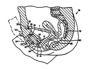
Turning now to FIG. 8, device 20 is shown after
being implanted in a female patient indicated generally
at 76. Structure previously identified herein is
identified with the same number in FIG. 8. Additional
anatomical structure includes the coccyx 78, such
comprising the lowermost portion of the spine. Also
illustrated are the rectum 80 and vagina 82. In the
vlew of FIG. 8, balloon 24 is shown in a substantially
deflated or contracted condition.
In FIG. 9, balloon 24 is illustrated in an
inflated condition such that urethra 14 is compressed
in an anterior-posterior direction between balloon 24
and the tissue posterior to urethra 14, thereby
occluding the urethral lumen as illustrated. A portion
of the patient's forefinger 84 is shown in dashed lines
compressing the reservoir balloon 22 against the pubis
16. This action forces fluid from reservoir balloon 22
through tuba ~6 to inflatab~e ballocn 2~. The balloon
24 expands axially, preferentially compressing a
lengthwise portion of the urethra.
W092/~52 2 0 9 2 ~ 2 ~ PCT/~S90/05976
In FIG. 10, device 20 is illustrated implanted in
a male patient indicated generally at 86. Included in
male patient 86 is a coccyx 88, a rectum 90 and a
prostate gland 92, shown in dashed lines, encircling
- 5 urethra 94. The urethra depends from bladder 96, there
being a bladder neck 98 formed between the bladder and
urethra 94. Device 20 is mounted via attachment tab 28
to pubis bone lOO.
Surgical access for the implantation of the
proposed incontinence device is via a standard lower
vertical mid-line abdominal or horizontal
(Pfannenstiel's) incision, with separation of the
rectus muscles to gain access to the retropubic
(anterior pelvic) space and to the superior pubic rami.
Each of devices 20, 44 are attached to the anterior-
superior aspect of the anterior pubic rami on either
side of the symphysis pubis by staples 34, 36 driven
into pubis 16 ~pubis lOO in FIG. lO) through lateral
attachment tabs 29. Reservoir balloon 22 is implanted
in a subcutaneous pocket overlying the anterior pubic
rami and symphysis in an area accessible to the patient
~or manual actuation (compression of the reservoir
balloon). The underlying bone serves as a platform
against which the reservoir is compressed.
Inflatable balloon 24 ~or 66 in FIG. 7) is
connected to reservoir balloon 22 over the superior
aspect of the symphysis pubis via tubing 26.
; Attachment tab 28 is integrated with tube 26 and serves
as the only point of fixation of the device to bone or
! 30 adjacent structures. Balloon 24 is implanted behind
the pubic symphysis and above the pubic arch within the
retropubic space of the pelvis. Balloon 24 is
positionsd so th t tha bladder neck and urethra ~re
compressed by it before the urethra passes through the
pelvic diaphragm (not shown) under the pubic arch.
.
.
,
.
. : .
W092/06652 2 0 9 2 9 2 ~ pcr/us9o/o597~--
14
A~ter separation of the rectus muscles via the
earlier described standard surgical approaches, only '
blunt dissection may be necessary to gain access within
the retropubic space for implanti;ng the balloon 24 in a
fixed relationship wi~h the a*ja~cent superior urethra
and bladder nec~. No dissect'ion posteriorly or
laterally of the urethra and bladder neck is necessary.
This greatly simplifiés the surgical procedure and
avoids the possibility of rectal or vaginal injury.
Venous structures in this area, particularly in the
mid-line retropubic space, are numerous and large.
Little or no dissection of these veins is required.
Ligation, if necessary, or compression of these veins
by the device 20, should not produce venous stasis
since ample collateral veins are present laterally.
Balloons 24, 66 are narrow in the anterior-
posterior dimension and wider in the lateral dimension,
and oval in cross section to conform to the concavity
of the posterior pubic symphysis. This shape
stabilizes the compression device between the anterior
bladder, bladder neck, and superior urethra
posteriorly, and the concave posterior aspect of the
pubic symphysis, anteriorly. The bladder as well as
' the pelvic contents hold the inflatable balloon in
position behind the pubic symphysis at or near the mid-
line. No additional fixation of device 20 to the ,''
posterior pubic bone or pelvic structure is necessary,
as the shape of the device allows for stable
positioning in this location within the concavity of
the pelvis (between the diverging'arms of the inferiorpu~ic rami, anteriorly).
An integral and important part of the surgical
proccdus2 includes 3 ~3ns to ~ thc ~nt2rivr-
superior bladder to the posterior rectus fascia above
the level of the superior pubic rami. Tube 26 is
routed through the mid-line fascia throu~h the incision
~ . ~ , ~... . . . .
W092/~52 ` 2 ~ 9 2 S 2 ~ PCT/US90/05976
between the rectus muscle bodies at or near their
insertion on the superior aspect of the anterior pubic
rami. The fascial incision is closed in standard
fashion with interrupted sutures. The most inferior
sutures bracket the interconnecting tubing as it exits
the pelvis, thus securing fascial tissue around it and
preventing herniation. Before fascial closure, a felt
matrix or mesh patch 30 (in FIG. 4) of a biologically
inert material, such as Dacron~, is sewn to the
anterior bladder wall over a distance of 2-3 cms.
transversely. This, in turn, is sewn via suture 32, to
the posterior aspect of the rectus fascia prior to the
fascial closure, well above the rectus insertions and
device 20. Tissue incorporation into the felt occurs
both from the bladder aspect and the fascial aspect,
effecting a secure union. This fixes the bladder to
anterior structures (abdominal wall) thus stabilizing
the bladder and urethra and preventing inferior
migration of the bladder with expansion of balloon 24
as might otherwise occur. This concept is an extension
of existing surgical principles with regard to stress
urinary incontinence correction in the female.
In conjunction with this portion of the surgery,
electrodes can be incorporated within the bladder wall,
or affixed to it, to record the status of bladder
filling via a strain gauge or similar instrument. This
sensor, in turn, is linked to a warning device, for the
patient who has deficient sensory enervation, or to
nursing sta~f for the incompetent or incapacitated
patient, to signal the need for voiding. The potential
bene~it of such a bladder warning system is great for
institutionalized patients who are incapable of normal
control (pat'2nts with ~'zh~..2r's D s~3s2, atc.~.
This requires an attentive nursing staff but would be a
vast improvement over the incontinence that is often
. ,...:
:, :
:; . ' '. - . ' . . .
.
W092t~2 2 0 9 2 9 2 ~ PCT/US90/0597F-,
encountered in nursing home and convalescent center
environments.
Because of the bladder fixation to be employed in
this surgery, and the attendant temporary bladder
dysfunction that is frequently sëen with similar
surgical procedures (e.g., for correction of stress
incontinence in the female), it is likely that a
temporary form of urinary drainage will be necessary in
conjunction with the above described surgery and
placement of a device constructed in accordance with
the invention. A bladder catheter is placed in the
mid-line through the fascial closure at a level higher
than the placement of device 20 (again incorporated
between fascial interrupted sutures). A Foley catheter
or similar retention device is utilized for this
purpose and is positioned adjacent the fixation felt 30
to Aid in bringing the bladder into close opposition to
the anterior abdominal wall via traction on the
catheter during the post-operative period. This
catheter is removed when voiding function is re-
established and the patient is accomplished in the
operation in the device and its voiding valve. At that
time, the wound should be well-healed and the bladder
well-fixed and stabilized anteriorly.
Reservoir balloon volume is carefully monitored at
the time of surgery to ensure that adeguate bladder
emptying is possible when inflatable balloon 24 (in
devise 20) is deflated, or at equal pressure with the
reservoir. A portion of the reservoir balloon that is
accessible from the anterior-superior aspect of this
prosthesis component is designed with a self-sealing
diaphragm 23 to allow perforation by a non-coring
neadla introduced thrcugh adjacent skin to add or
subtract fluid volume.
With reference to FIGS. 8 and 9, after the device
is implanted and the patient wishes to close the
.. . ~ : . . . .
W092/~52 2 0 9 2 ~ 2 5 PCT/US90/05976
17
urethra to prevent bladder voiding, forefinger or
fingers 84 is used to compress balloon 22 against pubis
16. When such compression occurs, as shown in FIGS. 6
and 7, fluid in balloon 22 is forced through bore 50.
The increased pressure distends membrane 56 away from
side 54 of valve body 48 thereby allowing fluid flow
from bore 50 through holes 57, 59, and into tube 26
thereby inflating compression balloon 24, 66 and
ultimately compressing the urethra between the balloon
and the tissue posterior to the urethra. When the
patient removes his or her finger(s), back pressure of
the fluid in the compression balloon seals membrane 56
against the bore 50, blocking back flow of fluid.
When the patient desires to void his or her
bladder, the patient can compress valve body 48 between
his or her thumb 72 and forefinger 74 as shown in FIG.
7. Such compression lifts membrane 56 away from side
54 of the valve body thereby permitting flow from
compression balloon 24 through tube 26 and holes 57, 59
in membrane 56. The fluid passes through bore 50 and
back into reservoir balloon 22, thus allowing the
device to resume the configuration shown ~n FIG. 8.
Wlth the balloon no longer inflated, the urethra opens,
permitting voiding. After voiding, the patient again
compresses the reservoir balloon with his or her
forefinger to inflate balloon 24 thereby occluding the
urethra lumen, as illustrated in FIG. 9, to prevent
incontinence.
Since the inflatable balloon in each embodiment
expands primarily along the longitudinal axis of tube
26, increasing expansion is directed in an inferior
direction perpendicular to the pelvic diaphragm (not
sh~ .). Compressic.. cf th2 supsr~or ursthr3 r2sults
from expansion of the inflatable balloon over a broad
surface area. The risk of tissue necrosis is minimal
since the urethra is compressed only in the inferior-
'~ ,
-
~ .
' . ~., ~ '.
.. . .
W092/~52 2 0 9 2 9 2 ~ PCT/US90/~976~-j
18
posterior direction and only with sufficient fluid
transfer to effect continence. The compression is
directed only upon the anterior wall of the urethra,
ultimately compressing the uréthra against the sacrum
and coccyx posteriorly, with intervening rectum and
pelvic contents providLng a buffer.
With balloon 24 inflated, the urethra, already
elongated and stabilized, is compressed and further
lengthened as it is urged posteriorly by the expanding
balloon. As the urethra is lengthened, the diameter of
the lumen therein decreases, thus requiring less force
to occlude the same. The area of compression of the
urethra exceeds the anatomic size of the external
sphincter in males. The locus of compression is
immediately above the urogenital diaphragm (above the
external sphincter) in the male. Since the inflatable
balloon 24 is secured only by attachment tab 28, it is
somewhat mobile. This mobility permits the balloon to
be forced into a more inferior position with sudden
increased abdominal pressure (such as with coughing,
sneezing, etc.) or as directed by the patient (via
voluntary Val Salva maneuver) to effect increased
urethral compression. This voluntary patient maneuver
can be ut~lized in the competent patient having intact
bladder sensation in circumstances such as sudden
bladder contraction.
It can be seen that the invention provides a
bladder incontinent control method and apparatus which
is easily operated and controlled by the patient. The
patient controls both the degree of urethral
compression, via incremental transfer of fluid from the
patient-accessible reservoir balloon, and voiding
.u,.ction. ~h2 lattar is effected by tha patiant or
nursing personnel by a ~ingle manipulation which
effects rapid urethral decompression. Another
advantage of this invention is the ease of surgical
. ' ~ , .
' '
- '
- Wos2/~s2 ~0 9 ~ ~ 2 ~ PCT/US90/~976
19
access via standard anterior lower abdominal
approaches, avoiding lateral and posterior dissection
around the urethra and bladder neck. The concept
utilizes urethral compression over a broad area at the
highest level feasible, i.e., at the bladder neck and
superior urethra. This allows the use of the proposed
device in patients who have failed inflata~le cuff
applications or other surgical treatments at a lower
level.
Having illustrated and described the principles of
my invention in two alternative embodiments, in both
males and females, it should be appreciated that
additions and modifications may be made without
departing from such principles. I claim all variations
and modifications within the spirit and scope of the
following claims.
:~
.