Note: Descriptions are shown in the official language in which they were submitted.
2093~97
The present invention concerns a correction lens which is
implantable on to the front side of the natural crystalline lens of an
eye.
An implantable correction lens which can be used for the
treatment of myopia, hypermetropia and astigmatism is fitted by
implantation on to the front side of the nautral crystalline lens of
the eye requiring correction. The correction lens has an optical lens
portion which is normally of a circular configuration, a positioning
portion and a support portion adjoining same. When the correction lens
is in the implanted condition, the positioning portion and the support
portion are between the iris and the front surface of the natural lens
of the eye.
The implanted lens thus serves as a substitute for
conventional spectacle lenses, contact lenses which are fitted on to
the cornea or other correction procedures such as the removal of
layers of the cornea.
According to the present invention there is provided a
correction lens which is implantable at the front of a natural lens of
an eye, comprising an optical lens portion and a haptic at least
partially surrounding the optical lens portion, wherein the haptic is
subdivided in a radial direction into an inner haptic portion around
the optical lens portion and an outer haptic portion, the haptic
portions having outer boundary lines which lie at least partly on
circular arcs, wherein the outer haptic portion at its rear side
which in the implanted condition of the correction lens is towards the
natural lens has a geometrical configuration which differs from the
configuration of the surface geometry of the rear faces of the optical
lens portion and the inner haptic portion, and wherein beginning at
the outer boundary line of the inner haptic portion and extending
towards the outer edge of the outer haptic portion , the rear side of
the outer haptic portion extends perpendicularly to the optical axis
of the optical lens portion.
2093~97
As will be seen in greater detail hereinafter, in the
implantable correction lens according to the invention, the haptic
which comprises an inner haptic portion at least partially surrounding
the optical lens portion and an outer haptic portion or support
portion which forms the outward edge region of the correction lens is
of such a configuration in the outer haptic portion that the rear side
thereof extends at least substantially perpendicularly to the optical
axis of the optical lens portion. The outer haptic portion or support
portion has, at its front and rear sides, flat surfaces which extend
at least substantially straight and parallel to each other. In the
implanted condition of the lens, that geometrical configuration means
that, in the outer haptic portion or outward edge region, the haptic
doe s not curve round to follow the curvature of the natural
crystalline lens of the eye but lifts away from the surface thereof.
As a result, even in a situation involving capillary adhesion between
the implanted correction lens and the front surface of the natural
lens of the eye, the haptic, in the outward portion thereof, has a
tendency to lift progressively away from the surface of the natural
lens. That considerably reduces the chafing effect on the zonule
fibers, when the natural lens of the eye performs its natural
movements. The overall diameter of the correction lens is preferably
such that the outward haptic portion lies in the region of the zonule
fibers which extend between the natural lens of the eye and the
ciliary muscle. The fact that the rear side of the outer haptic
portion extends substantially per,oe ndicularly relative to the lens
axis, with the rear side thereof being substantially matched to the
radial configuration of the zonule fibers ensures reliable contact
such as properly to define the position of the correction lens, while
however the area of contact with the natural lens of the eye is as
small as possible in order not to impair the metabolic procedure
involved.
2 0 ~ 7
Preferably the lens has a circular optical lens portion while
the haptic p~rtion may be made up of a plurality of parts. mus, to
provide a reduction in lens surface area, the haptic may have lateral
boundary edges which depart frcm an arcuate shape, at the nine o'clock
and three o'clock sides. The lateral boundary edges extend at least
substantially parallel to each other. The upper and lower boundary
edges at the twelve o'clock side and the six o'clock side extend in a
continuous arcuate shape between the respective ends of the lateral
boundary edges which extend in at least substantially straight line.
The two arcuate lines may have a common center point which coincides
with the optical axis of the lens. They are arcs which extend
concentrically around the circular optical lens portion, the center
point of which is also on the optical axis of the lens.
As indicated above, extending between the outer edge portion
or outer haptic portion, which forms the support portion thereof, and
the optical lens portion, is the inner haptic portion which
constitutes a positioning portion for the lens. The front side of the
positioning portion or inner haptic portion is of such a configuration
as to ensure, in conjunction with the iris lying thereagainst, that
the correction lens is positioned in the desired position, relative to
the natural lens of the eye. In that condition the optical axes of the
natural lens and the correction lens should at least approximately
coincide. The front side of the inner or positioning portion of the
haptic forms a sliding surface for movement of the iris thereover.
That front side of the inner haptic portion may be of a concave, flat
or convex configuration. Preferably the inner haptic portion is such
that, beginning from the circular junction between the inner haptic
portion and the optical lens portion, the inner haptic portion
decreases in thickness, for example tapers, towards the outer edge of
the correction lens.
The junction between the inner haptic portion and the outer
haptic portion or support portion also extends along two arcs which
2093097
are concentric relative to the circular edge of the optical lens
portion and the two upper and lower arcuate boundary edges of the
outer haptic portion. The tw~ arcs defining the junctions between the
inner haptic portion and the outer haptic portion also extend
continuously between the boundary edges at the tw~ sides of the lens.
The overall geometrical configuration of the correction lens,
in the implanted condition, ensures that the surface of the natural
lens of the eye remains accessible for the metabolism procedure which
occurs at that location. The haptic may additionally be provided with
openings or holes so that the area of the natural lens, which is
covered by the correction lens, is still further reduced. All boundary
edges of the lens can be rounded off so that the lens does not have
any sharp edge configurations.
In order to provide that the implanted lens is fitted in
position in such a way as to give the best possible floating effect,
it is of a specific weight which is approximately equal to that of the
eye chamber fluid, namely about 1.1.
So that the correction lens is prevented from turning after
being implanted in the eye, the respective peripheral portion of the
haptic, which is in the region of the ciliary sulcus of the eye,may be
of a non-uniform or variable curved configuration which however only
insignificantly departs from the circular shape of the ciliary sulcus.
That further provides for gentle engagement between the ~espective
peripheral portion of the haptic of the correction lens and the
adjoining eye tissue. The variable curved configuration at the
respective peripheral portions of the haptic may be polygonal or may
involve convex and/or concave configurations. The respective
peripheral portion of the haptic may be fonmed alternately by planar,
convex or concave and circular contour segments. The depth or the
height of the respective convex or concave configurations, relative to
the adjacent peripheral portions, are such that they are only a
~093~97
fraction of their longitudinal dimension in the peripheral direction.
That ensures that, upon implantation of the ~, the concave or convex
portions which thus constitute projections on or recesses in the
haptic do not lose their desired shape and form a reliable means for
preventing the implanted lens from turning in the eye in which it is
implanted. The periphery of the lens retains a configuration which
approximates to an arcuate shape.
Preferably, the correction lens according to the invention
has tw~ peripheral haptic parts which are arranged diametrally
opposite relative to the axis of the lens and which, after
implantation of the correction lens, are disposed in the region of the
ciliary sulcus of the eye. The boundary lines of the contour segments
at the peripheral parts of the haptic may be straight or arcuate. At
the transitions between contour segments of different configurations,
for example between contour segments which are of a straight and an
arcuate configuration respectively, that arrangement provides
anchorage points for the adjacent tissue of the eye without the latter
beccming irritated and inflamed. The above-mentioned transitions or
intersections further contribute to ensuring that the correction lens
is prevented frcm turning in the eye, in the desired manner.
Preferably, the above-mentioned concave or convex portions at
the respective peripheral parts of the haptic may be of a depth or
height respectively, relative to the adjacent peripheral regions,
which corresponds to between about one sixth and one third of their
longitudinal extent in the peripheral direction. Thus, the depth or
height thereof respectively may be between about 0.2 and 0.4 mm while
the longitudinal dimension may be between about 0.8 and 1.4 mm in the
peripheral direction.
Embodiments of the corrections lens according to the present
invention will now be described by way of example with reference to
the accompanying drawings in which:
2093~97
Figure 1 is a view in section through a first embodiment of a
correction lens according to the invention,
Figure 2 is a plan view of the lens shown in Figure 1,
Figure 3 is a plan view of a second embodiment of a lens
according to the invention,
Figure 4 is a plan view of a third embodiment of a lens
according to the invention,
Figure 5 is a plan view of a fourth embodiment of a lens
according to the invention,
Figure 6 is a plan view of a fifth embodiment of a lens
according to the invention,
Figure 7 is a plan view of a sixth embodiment of a lens
according to the invention,
Figure 8 is a plan view of a seventh embodiment of a lens
according to the invention,
Figure 9 is a plan view of an eighth embodiment of a lens
according to the invention, and
Figure 10 is a plan view of a ninth embodiment of a lens
according to the invention.
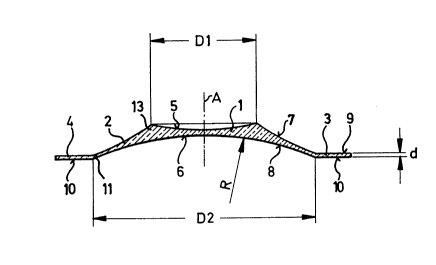
Referring firstly to Figures 1 and 2, as shcwn therein the
lens body of the illustrated correction lens according to the invention
comprises an optical lens portion generally indicated at
and a haptic which is disposed at least in part around the optical
lens portion 1. The haptic illustrated camprises first and second
portions, namely an inner haptic portion or positioning portion 2
which adjoins and at least partially surrounds the optical lens
portion 1, and an outer haptic portion or support portion which
adjoins the positioning portion 2 and which is made up of first and
second parts 3 and 4. In the illustrated embodiment the optical lens
20~3097
portion 1 is circular and its optical axis, which is the axis of the
correction lens, is identified by the line A. The optical lens portion
1 fonms the actual correction portion of the lens and is of a
biconcave shape having a front side 5 and a rear side 6. The rear side
6 has a radius of curvature which is matched to the outside of the
natural crystalline lens of the eye to be corrected by implantation of
the correction lens. The radius of curvature R of the rear side 6 of
the optical lens portion 1 is for ex.ample about 10 mm + 1 mml while the
diameter of the optical lens portion 1, as indicated at Dl, is for
example about 4 mm + 1 mm.
At a circular annular connecting location as indicated at 13
in Figures 1 and 2, the optical lens portion 1 passes into the haptic,
more specifically the inner haptic portion or positioning portion 2.
Starting fram the connecting location 13, the cross-section of the
inner haptic portion 2 decreases in a tapering configuration in an
outward direction, as can be seen in particular fram Figure 1. In the
illustrated embodiment the inner haptic portion 2 is of a biconcave
configuration. It is also possible however for the inner haptic
portion 2 to be of a plane-concave configuration, in which case the
rear side 8 of the inner haptic portion 2 is of the same radius of
curvature R as the rear side 6 of the optical lens portion 1. The
front side 7 of the inner haptic portion 2 can be of a concave or
planar configuration. It is also possible however for the front side 7
to be convex. The inner haptic portion 2 fonms a sliding surface for
iris movement, in particular for the pupil. The configuration of the
front side 7 of the inner haptic portion, namely concave, planar or
convex, depends on the thickness of the optical lens portion 1.
Adjoining the inner haptic portion 2 which surrounds the
optical lens portion 1 in the form of a one-piece surface, in a
radially outward direction, is the outer haptic portion defined by
first and second edge regions or support parts as indicated at 3 and
209~097
4. As can be seen in particular from Figure 1, the rear face 10 of the
support parts 3 and 4 differs considerably from the configuration of
the rear faces 6 and 8 of the optical lens portion 1 and the inner
haptic portion 2. The rear faces 10 are of a flat configuration and
S extend at least substantially perpendicularly to the axis A of the
lens. In the illustrated embodiment, the front sides 9 of the su~port
parts 3 and 4 also extend at least substantially perpendicularly to
the optical axis A and are also of a flat configuration. The thickness
d of the support parts 3 and 4 is for exdl~le about 0.1 mm. The
junctions, as indicated at 11, between the inner haptic portion 2 and
the support parts 3 and 4, are of an arcuate configuration. The two
arcs defining the junctions 11 have a co~mon center point which lies
on the axis A of the lens. The arcuate junctions 11 terminate at
lateral boundary edges 14 and 15 which are at least substantially
straight edges. In the illustrated embodiment, the arcuate junctions
11 extend over an angular region as indicated at a in Figure 2, of
about 90. That angular region may also be for example between about
80 and about 100, depending on the width B of the lens, being
defined by the spacing between the two lateral boundary edges 14 and
lS of the lens. In the illustrated embodiment the width B of the lens
body is about 6 mm ~ 1 mm.
The two outer edge regions or support parts 3 and 4 defining
the outer haptic portion also have outer boundary edges as indicated
at 16 and 17, which are also of a continuous arcuate configuration and
which each terminate at the ends of the respective lateral boundary
edgs 14 and 15. The two arcs providing the boundary edges 16 and 17
may also have a common center point which lies on the axis A of the
lens.
The overall diameter of the lens body, as indicatedat D3 in
Figure 2, that is to say, the spacing between the two arcuate boundary
edges 16 and 17, is for example about 11 mm, in the illustrated
embcdiment. It will be appreciated however that the overall diameter
2093~97
of the lens body can be varied according to the eye into which the
correction lens is to be implanted. Such variations are generally in a
range of about t 1 mm.
In the illustrated embodiment, the diameter D2 illustrated in
Figure 1, being the spacing between the tw~ arcuate junctions 11, is
about 8 mm, that is to say approximately double the diameter Dl of the
optical lens portion 1. The diameter D2 can also vary, in dependence
on the diameter D3. The diameter D2 is such that it is less than three
quarters of the diameter D3.
Reference will now be made to Figures 3 and 4 showing another
embodiment of the lens according to the invention, in which the two
lateral boundary edges 19 and 20 of the lens body are of such a shape
as to extend arcuately, possibly in a circular configuration in an
inward direction so that they are concave. m e embodiment shown in
Figure 3 further has concave portions formed by recesses 21 and 22,
approximately in the middle of the tw~ edge regions or support parts 3
and 4 of the outer haptic portion. Both the concavely shaped lateral
boundary edges 19 and 20 and also the recesses 21 and 22 contribute to
improving the metabolism procedure at the outward face of the natural
crystalline lens cn which the correction lens according to the
invention is implanted, by virtue of the reduction in the surface area
of the correction lens.
Looking now at Figure 5, shown therein is an embodiment of
the correction lens which is of a generally star-shaped configuration.
Reference numeral 1 again denotes the optical lens portion and
reference numeral 2 shows the inner haptic portion. The outer haptic
portion which adjoins the inner haptic portion 2 is formed by the
projections 23, 24 and 25 which are at an angular spacing from each
other of about 120. It will be seen that the inner haptic portion 2
is formed by a circular ring.
The lens body consisting of the optical lens portion 1 and
2~93097
the haptic may be made in one piece. A transparent biocompatible
material can be used as the material for the lens, and it is
preferably in the form of a soft and/or resilient material. The lens
material is preferably also hydrophilic and/or gas-permeable, in
particular being permeable to oxygen. ~he lens mate~ial may be for
ex~l~le silicone rubber, polyhydroxy ethyl methacrylate, a copolymer
of silicone and methyl methacrylate, polyvinylpyrrolidone and other
materials which are compatible with the eye tissue.
It may be noted at this point that, in order further to
reduce the surface area of the correction lens and to improve
accessibility to the surface of the natural crystalline lens of the
eye, for example for the metabolism procedure, the lens body may also
be provided in the region of its haptic with openings as indicated at
18 in Figure 2, for example in the form of round holes or the like.
Referring now to Figures 6 through 10, the embodiments
illustrated therein again comprise the optical lens portion 1 which is
surrounded by the haptic formed by an inner haptic portion 2 and an
outer haptic p~rtion as indicated at 3. In the illustrated embodiments
the haptic has the substantially straight lateral boundary edges 14
and 15. It will be a~preciated however that it is also possible for
the lateral boundary edges to be of an inwardly curved or concave
configuration.
The outer haptic portion has tw~ peripheral parts, one
thereof being shown in each of Figures 6 through 10. In the implanted
condition of the correction lens, the peripheral part constituting a
support part 3 as illustrated in Figures 6 through 10 lies in the
region of the ciliary sulcus. The tw~ peripheral parts of the haptic
extend over an angular range as indicated at a, relative to the
optical axis A of the lens. That angle a may be of the order of
magnitude of between about 50 and 90.
20~3as7
In the illustrated embodiments, the haptic peripheral parts
have a non-uniform or variable curved configuration at their
peripheral edge.
Looking more specifically at the embodiments shown in Figures
6 and 7, the curved configuration of the haptic periphery is a
polygon, within the angular range a.
In the embodiment shown in Figure 7, it is a pure polygon
which comprises three sides indicated at 26, 27 and 28.
On the other hand, in the emb~diment shown in Figure 6,
concave recesses 29 and 30 are provided in the sides 26 and 28 of the
polygon, that is to say in the two outer sides of the polygon
illustrated therein, with the middle side 27 of the polygon being
straight. The concave recesses 29 and 30 are so arranged that their
inward ends are disposed at intersections 31 and 32 between the sides
26 and 28 respectively, and the side 27 disposed therebetween. However
the concave recesses 29 and 30 may also be arranged in the middle or
at another location along the sides 26 and 28 of the polygon.
In the embodiment shown in Figure 8 the haptic peripheral
part, within the angular range a, is of a substantially arcuate
configuration with convex projections 34 and 35 extending outwardly
therefrom.
In the Figure 9 embodiment, the haptic peripheral part is
once again of a substantially arcuate configuration, with concave
recesses 29 and 30 extending radially inwardly therefrom.
In the Figure 10 embodiment, the haptic peripheral part is of
a polygonal configuration made up of the sides 26, 27 and 28 which
intersect at the intersections indicated at 31 and 32. Provided in the
region of the sides26 and 28 of the polygon are convex portions or
raised portions 34 and 35, which thus project outwardly relative to
the polygon peripheral configuration of the haptic portion. The two
inner ends of the convex portions 34 and 35 æ e disposed at the
20s3as7
intersections 31 and 32 where the respective sides 26 and 28 of the
polygon, meet the side 27 which is therebetween.
The depth of the concave recesses 29 and 30 in the
e~bodiments shown in Figures 6 and 9, relative to the adjacent
peripheral region, is between about 0.2 and 0.4 mm. In the embodiment
shown in Figure 6, the depth is measured in relation to the extension,
as shown in broken lines, of the respective sides of the polygon, in
the region of the recesses 29 and 30.
In the embodiment shown in Figure 9, the depth is similarly
measured in relation to the extension shown in broken line of the
arcuate peripheral edge, in the region of the recesses 29 and 30.
In the embodiments shown in Figures 8 and 10, the height of
the convex portions or projections 34 and 35 is measured in relation
to the continuation, shown in broken lines, of the adjacent arcuate
segments 33, in the region of the projections 34 and 35 (Figure 8) or
in relation to the broken-line extension of the sides 26, 27 and 28 of
the polygon, in the region of the projections 34 and 35 (Figure 10).
In the embodiments of Figures 6, 8, 9 ar.d 10 the longitudinal
extent in the peripheral direction of the concave recesses 29 and 30
and the convex portions or projections 34 and 35 is between about 0.8
and 1.4 mm.
In the embodiments shown in Figures 6 through 10, the non-
uniform or varying curved configuration of the peripheral part of the
haptic still approximates to an arcuate shape. That arcuate shape is
of a diameter which is suited to the inside diameter of the ciliary
sulcus of the eye in which the lens is to be implanted. That diameter
is generally in the region of between about 11.0 and 13.5 mm. The
variations in contour which cause the curved configuration to be non-
uniform, namely the recesses 29 and 32, and the projections 34 and 35,
as well as the transitions between those recesses or projections and
the other parts of the peripheral configuration provided by the sides
12
20~0~7
26 through 28 of the polygon or the arcs 33, provide a holding means
which prevents the correction lens from turning in the eye, about the
optical axis A of the lens, when the lens is in the implanted
condition. That point also applies in regard to the embodiment shcwn
S in Figure 7, in which the peripheral contour is a pure polygon. It
will be appreciated that although the polygon in the illustrated
e~bcdiment in Figure 7 has three sides 26, 27 and 28, it is also
possible to use a different number of sides for the polygon and in
particular more sides. The intersections 31, 32 between the sides of
the polygon provide the necessary holding effect to prevent the
co,L~ction lens from turning.
Th.e embodiments shown in Figures 6, 7 and 10 in which the
basic contour of the periphery of the haptic portion is a polygon
further afford the advantage that the lens body can be cut to size
from a larger lens body blank, as a basic shape, to adapt it to the
diameter of the ciliary sulcus of the eye into which the correction
lens is to be implanted. In that way, it is possible exactly to adapt
the overall diameter of the diametrally oppositely disposed haptic
peripheral parts, to the corresponding dimension of the eye in which
the correction lens is to be implanted.
In the embodiments illustrated in the drawings the optical
lens portion 1 is generally of a diameter of about 4.0 mm while the
width of the haptic portion 2 from one side edge 14 to the other is
for example about 6.0 mm.
When the correction lens is a high-myopia lens with a
strength of about -50 dpt, the diameter of the optical lens portion is
about 0.2 nm.
The embodiments illustrated in Figures 6 through 10 may be of
a cross-section like that illustrated in Figure 1. It will be
appreciated that the embodiments shown in Figures 6 through 10 are
also suitable for myopia lenses with haptics of a different
2~3097
configuration, for example haptics which are not subdivided into tw~
regions. In that case also the illustrated edge shapes provide that the
correction lens is prevented from turning in the eye in ~hich it is
implanted.
S The correction lens according to the invention is preferably
used to deal with severe myopia, for which purpose the optical lens
p~rtion 1 is in the form of a negative lens. Instead of the biconcave
configuration of the optical lens portion it may also be of a concave-
convex or toric shape.
Upon implantation of the correction lens, the pupil of the eye
is enlarged so that the correction lens, with its diameter D3, can be
fitted between the iris and the outside surface of the natural lens of
the eye. The optical lens portion 1 can then be aligned with its lens
axis A on the optical axis of the natural lens of the eye. The edge
regions or support parts 3 and 4 forming the outer haptic portion are
then in the region of the zonule fibers. The natural movement of those
fibers is not or only slightly impeded, by virtue of the specific
configuration of the above-mentioned edge regions of the haptic
portion.
The implantable correction lens can ensure satisfactory
positioning and fixing thereof relative to the crystalline lens of the
eye requiring correction and for example can be suitably fixed in
position by capillary adhesion, while at least substantially reducing a
chafing effect on the zonule fibers extending between the natural lens
of the eye and the ciliary muscle upon natural lens movement.
It will be appreciated that the above-described embodiments of
the correction lens according to the invention have been set forth
solely by way of example and illustration of the principles of the
invention and that various other modifications and alterations may be
made therein without thereby departing from the spirit and scope of the
invention.